diagnosis of infectious laryngotracheitis by chick embryo inoculation
TRANSCRIPT

J. COMPo PATH. 1964. VOL. 74· II9
DIAGNOSIS OF INFECTIOUS LARYNGOTRACHEITIS BY CHICK EMBRYO INOCULATION
By
F. T. W. JORDAN
Department of Veterinary Preventive Medicine, University of Liverpool
INTRODUCTION
Ever since Burnet (1934) demonstrated that the virus of Infectious Laryngotracheitis (I.L.T.) would grow and produce pocks on the chorioallantois (CAM) of the ,developing chick embryo, egg inoculation has been used as a means of isolating the virus. One of the disadvantages of this method in the diagnosis of I.L.T. is that bacteria contaminating the inoculum may kill the embryo before virus growth has occurred. This often happens when the inoculum is prepared from tracheal exudate or lungs, especially when these have been removed from the carcase and despatched to the laboratory for diagnosis. In our experience despatch of the material in 50 per cent. glycerol broth or glycerol saline as advocated by Webster (1959) did not appear to reduce bacterial contamination. Even when whole carcases were received for diagnosis and inocula were prepared from tracheal swabs suspended in penicillin and streptomycin broth, much of the material killed the embryos and was, therefore, valueless' for diagnosis.
Another factor influencing the isolation of the virus is the method of virus extraction from such tissues as the trachea, lungs and CAMs. Grinding the material with powdered sterile glass in a Griffith tube is a common and satisfactory method, but it was thought worthwhile to compare it with the extraction of virus by fluorocarbon emulsification. This method depends on the separation of non-viral protein from the nucleoprotein of virus particles by fluorocarbon emulsification of homogenized material. Gessler, Bender and Parkinson (1956) obtained higher virus titres following the extraction of vaccinia and Rous sarcoma virus from CAMs by fluorocarbon emulsification than by their control method which was presumably maceration of the CAM.
Demonstration of the virus also depends on the technique of embryo inoculation. Apart from inoculation onto the dropped CAM, as used by Burnet (1934) and described by Beveridge and Burnet (1946), other methods have been advocated, and Fabricant (1957) described a method of embryo inoculation through the air-sac. In view of the simplicity of inoculation by this latter technique and the fact that Fabricant (1957) suggested that it could be used for the isolation of the viruses of infectious bronchitis, Newcastle disease, fowl pox and I.L.T., it was decided to compare it with the method of Burnet in the isolation of I.L.T.
In this paper comparisons were made of the following: (I) the ability of antibiotic solutions and glycerol broth to suppress bacterial growth when used as suspending media for tracheas removed from chickens which had died of I.L.T.: (2) the value of three different solutions of antibiotics (penicillin and streptomycin; penicillin, streptomycin and chlortetracycline; penicillin, streptomycin and chloramphenicol) in suppressing bacterial growth in inocula prec
120 DIAGNOSIS OF I.L.T. BY EMBRYO INOCULATION
pared from tracheal swabs: (3) virus extraction from tissues by grinding with sterile sand and by emulsification of the homogenized material with a fluorocarbon (Arcton 63) * using tracheas and lungs from the carcases of chickens which had died of I.L.T., and also CAMs with confluent lesions produced by the virus: and (4) the amounts of virus growth following the inoculation of embryos onto the dropped CAM and inoculation through the air-sac.
MATERIALS AND METHODS
Comparison of Antibiotic and Glycerol Broths in Suppressing Bacterial Growth in Tracheas
Suppression was assessed by embryo survival and virus isolation following the inoculation of tracheal exudate. The carcases of 27 chickens which had died of I.L.T. in IS field outbreaks were received at this laboratory. I.L.T. virus was isolated from all of them by examination of tracheal swabs and the carcases were stored at - 27°C. As opportunity arose each carcase was thawed at room temperature and the trachea, which included the larynx and syrinx, was removed aseptically. The trachea was opened, a swab taken and examined for I.L.T. virus, and the trachea then divided into two longitudinal halves, one part being placed in a one-ounce bottle containing IS rol. of antibiotic broth and the other in a bottle containing the same quantity of 50 per cent. glycerol in 10 per cent. broth in saline. The antibiotic broth consisted of IO per cent. broth containing 1,000 units of penicillin, I ftg. of streptomycin, 10 per cent. of a saturated solution of chloromycetin and 50 units of nystatin per ml.
The bottles containing the tracheas were kept in the dark at room temperatures varying between 17 and 26°C. to simulate postal conditions. At intervals of three days each half trachea was removed, placed on a sterile filter paper and a swab touched at several sites along its length; each half trachea was then returned to its bottle. Each swab together with 0·5 ml. of the suspending fluid from which the trachea was taken was placed in a separate sterile bottle and 2 ml. of antibiotic broth added; after allowing it to stand at room temperature for an hour it was inoculated in 0·1 ml. quantities onto the CAM of six I2-q-day-old chick embryos. These were incubated for three days at 37°C. and examined for I.L.T. virus. Examination of tracheas was discontinued when virus was not isolated on two consecutive occasions. All dead embryos were examined for evidence of bacterial contamination by smear from the CAM, and, if this was negative, by culture of the CAM in broth.
Value of Different Antibiotics in Suppressing Bacterial Growth in Inocula Prepared from Tracheal Swabs
Three antibiotic solutions were prepared in IO per cent. broth. Solution A contained 1,000 units of penicillin and I ftg. of streptomycin per ml. Solution B contained, in addition, I ftg. chlortetracycline per ml. Solution C contained 10 per cent. saturated chloramphenicol solution in addition to the antibiotics in A.
Three swabs were taken from each trachea of 163 chicken carcases received at this laboratory. The swabs from anyone trachea were taken
* Arcton 63 (Trifluorotrichloroethane) I.e.I.
F. T. W. JORDAN 121
from the same site but no attempt was made to swab fro~ precisely the same site in different tracheas. They were placed in separate bijou bottles containing 2 ml. of antibiotic solution A, Band C respectively. Mter standing for an hour at room temperature they were examined individually for LL.T. virus by inoculating 0'1 ml. onto the CAM of each of six 12-14-day-old embryos. The eggs were incubated for three days at 37°C., candled daily, and all dead embryos were examined for bacterial growth. If bacterial contaminants were isolated from any of the dead embryos then all three swabs prepared from that trachea were assumed to be contaminated.
Virus Extraction by Grinding with Sterile Powdered Glass Compared with Homogenization and Emulsification with Fluorocarbon (Arcton 63)
The two methods for the extraction of the virus were compared on tracheas and lungs from affected chickens and on confluent CAMs. Mter the virus had been extracted from the tissues by grinding or homogenization followed by emulsification, the extract in each case was suspended in equal volumes of diluent and the titre determined.
The tracheas and lungs of ten chickens which had died in different outbreaks of LL.T. were removed aseptically. Each trachea was.divided longitudinally into two halves. One half was placed in a Griffith tube and ground with sterile powdered glass after which 10 ml. of McIlvaine's buffer pH 7'2 containing 1,000 units penicillin, I fLg. streptomycin and 10 per cent. of a saturated solution of chloramphenicol was added and the suspension centrifuged at 3,000 r.p.m. for 10 minutes. The other half trachea was placed in a sterile one-ounce bottle, 6 ml. of McIlvaine's buffer added and emulsified for 10 minutes at approximately 10,000 r.p.m. in an M.S.E. homogenizer; then 4 ml. of Arcton 63 was added and the material centrifuged at 3,000 r.p.m. for 10 minutes. The titre (EID5o) of the virus in the supernatant was determined in each case.
The lungs from each carcase were opened along the primary bronchus and divided into medial and lateral portions. A medial portion of one lung together with the lateral portion from the other was placed in two Griffith tubes, ground, 15 ml. of McIlvaine's buffer was added and the material centrifuged as above. The remaining portions of each lung were emulsified together in 9 ml. of McIlvaine's buffer, 6 ml. of Arcton 63 was added and the mixture centrifuged at 3,000 r.p.m. for 10 minutes. In each case the titre of the supernatant was determined by egg inoculation.
Two confluent CAMs for each of three strains of LL.T. virus were prepared. The CAMs were weighed and one of each pair was ground in a Griffith tube. McIlvaine's buffer was added to give a suspension of 1/10 weight/volume which was centrifuged at 3,000 r.p.m. for 10 minutes. The other CAM was homogenized for 10 minutes in McIlvaine's buffer to give a 1/6 weight/volume and then Arcton 63 added to give a final weight/volume of CAM to diluent of 1/10. This mixture was centrifuged at 3,000 r. p.m. for 10 minutes. In each case the embryo infectivity of the supernatent suspensions was determined.
Comparison rif Virus Growth Following Inoculation onto the Dropped CAM and Inoculation through the Air Space and Shell Membrane
The comparison of these methods of egg inoculation was made using both field and laboratory strains of virus. From ten different field outbreaks of LL. T. a swab was taken from the trachea of one chicken which
122 DIAGNOSIS OF I.L.T. BY EMBRYO INOCULATION
had died of the disease. Each swab was placed in 10 per cent. broth containing penicillin, streptomycin and chloramphenicol as described previously, and also Mycostatin * 50 p.gfml. The broth was allowed to stand for one hour at room temperature and o' I ml. was then inoculated onto the dropped CAM of each often twelve-day-old chicken embryos by the method of Beveridge and Burnet (1946), referred to as the "standard" method. A further ten twelve-day-old embryos were each inoculated with the same quantity of inoculum by the method suggested by Fabricant (1957) and Gorham (1957), referred to as the "vertical" method. In this method a small hole is pierced through the shell and outer shell membrane immediately over the centre of the air sac and the egg placed vertically with the air sac uppermost. An inoculum of o· I ml. is deposited on the inner shell membrane by a fine needle which is then directed downwards to pierce the inner shell membrane and the underlying CAM. The needle is removed and the egg incubated in the vertical position. After incubation for three days at 37°C. the CAMs were removed and examined for pocks.
A similar procedure was used with ten strains of virus which had been passaged between five and eight times on CAMs in the laboratory. From each strain fresh confluent CAMs were prepared and a one-in-ten weightf volume dilution made. The titre of this suspension was determined by preparing ten-fold dilutions and inoculating o· I ml. of each dilution into each of six twelve-day-old embryos by each method. The inoculated eggs were incubated for three days at 37°C.
In order to determine the influence on virus growth of the volume of inoculum introduced by the two methods, varying amounts of a 10-3
dilution of one laboratory strain freshly prepared from confluent CAMs was used. This dilution was chosen as the one most likely to give countable pocks. Ten eggs were inoculated by the standard route and ten by the vertical, each with o· I ml. of inoculum. This was repeated for further groups of eggs with inocula of 0'2; 0'3; 0'4; 0'5; 0·6; and 1'0 ml. per egg. Mter incubation for three days the CAMs were examined for virus growth.
RESULTS
Antibiotic and Glycerol Broth as Suspending Media for Tracheas
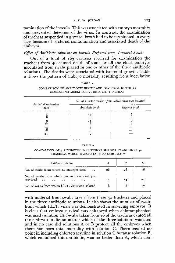
After storage of the carcases, I.L.T. virus was isolated from 25 of the 27 tracheas immediately before suspension in antibiotic or glycerol broth and only these 25 are considered for the purpose of this experiment. From one of the 25 tracheas it was not possible to isolate the virus after suspension. From all the remaining 24 tracheas virus could be isolated from the half trachea in antibiotic broth for a longer period than from the half suspended in glycerol broth. Table I shows that I.L.T. virus was recovered from all 24 half tracheas after suspension in antibiotic broth for three days and from two of them after as long as 2 I days. In contrast, when the tracheas were suspended in glycerol broth, the virus was isolated from only nine after three days and from none after 12 days. Only one of the 25 tracheas suspended in antibiotic broth showed bacterial con-
* Mycostatin-Squibbs.
F. T. W. JORDAN 123
tamination of the inocula. This was associated with embryo mortality and prevented detection of the virus. In contrast, the examination of tracheas suspended in glycerol brClth had to be terminated in every case because of bacterial contamination and associated death of the embryos.
Effect of Antibiotic Solutions on Inocula Prepared from Tracheal Swabs
Out of a total of 163 carcases received for examination the tracheas from 40 caused death of some or all the chick embryos inoculated from swabs placed in one or other of the three antibiotic solutions. The deaths were associated with bacterial growth. Table 2 shows the pattern of embryo mortality resulting from inoculation
TABLE I
COMPARISON OF ANTIBIOTIC BROTH AND GLYCEROL BROTH AS SUSPENDING MEDIA FOR 25 BISECTED TRACHEAS
No. of bisected tracheas from which virus was isolated Period of suspension
(days) Antibiotic broth Glycerol broth
3 24 9 6 18 6 9 11 2
12 8 0
15 4 -18 2 -21 2 -
TABLE 2
COMPARISON OF 3 ANTIBIOTIC SOLUTIONS USED FOR SWABS FROM 40
TRACHEAS WHICH CAUSED EMBRYO MORTALITY
Antibiotic solution A B
No. of swabs from which all embryos died .. 26 26
No. of swabs from which one or more embryos
C
16
survived .. .. . . . . . . . . 14 14 24
No. of swabs from which LL.T. virus was isolated 8 8 17
with material from swabs taken from these 40 tracheas and placed in the three antibiotic solutions. It also shows the number of swabs from which I.L.T. virus was demonstrated in surviving embryos. It is clear that embryo survival was enhanced when chloramphenicol was used (solution C). Swabs taken from 16 of the tracheas caused all the embryos to die no matter which of the three solutions was used and in no case did solutions A or B protect all the embryos when there had been total mortality with solution C. There seemed no point in including chlortetracycline in solution C because solution B, which contained this antibiotic, was no better than A, which con-
124 DIAGNOSIS OF I.L.T. BY EMBRYO INOCULATION
tained only penicillin and streptomycin. With solutions A and B all the embryos inoculated with material from 26 of the 40 swabs died.
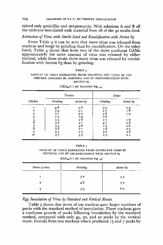
Extraction of Virus with Sterile Sand and Emulsification with Arctan 63 From Table 3 it can be seen that more virus was released from
tracheas and lungs by grinding than by emulsification. On the other hand, Table 4 shows that from two of the three confluent CAMs approximately the same amount of virus was released by either method, while from strain three more virus was released by emulsification with Arcton 63 than by grinding.
TABLE 3
AMOUNT OF VIRUS EXTRACTED FROM TRACHEAS AND LUNGS OF TEN CHICKEN CARCASES BY GRINDING AND BY EMULSIFICATION WITH
ARCTON 63
(EID.%ol ml. inoculum logo 10)
Trachea Lungs
Chicken Grinding Arcton 63 Grinding Arcton 63
I 4°8 3°6 3°9 2°g 2 3°9 3 ' 1 2°0 1 0 8 3 5"1 4 °0 3°8 log
4 2°5 log 0°8 -5 3°3 2 0 8 2°7 1 0 8 6 5°0 4° 1 4° 1 2 0 8 7 4°9 3°8 1°2 0°6 8 3°8 2°1 1 0 8 oog g 4°2 2°3 3° 2 2°6
10 4°7 3°5 2°g 2°1
TABLE 4
AMOUNT OF VIRUS EXTRACTED FROM CONFLUENT CAMS BY GRINDING AND BY EMULSIFICATION WITH ARCTON 63
(EID50/oo I mlo inoculum log. 10)
Strain of virus Grinding Arcton 63
5"2
2
3
Egg Inoculation of Virus by Standard and Vertical Routes Table 5 shows that seven of ten tracheas gave larger numbers of
pocks with the standard method of inoculation. Three tracheas gave a confluent growth of pocks following inoculation by the standard method, compared with only 45, 30, and 22 pocks by the vertical route. Inocula from two tracheas which produced 15 and 7 pocks by
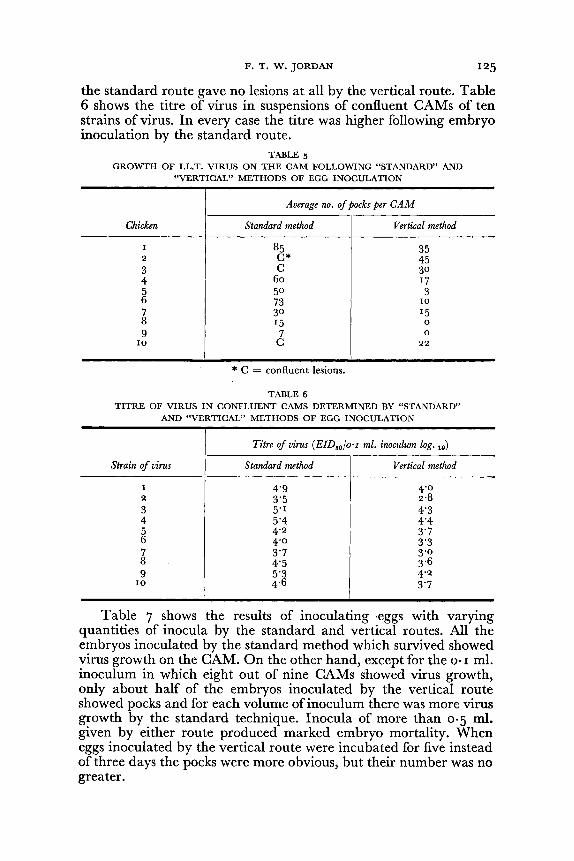
F, T, W, JORDAN 125
the standard route gave no lesions at all by the vertical route, Table 6 shows the titre of virus in suspensions of confluent CAMs of ten strains of virus, In every case the titre was higher following embryo inoculation by the standard route,
TABLE 5 GROWTH OF I.L,T, VIRUS ON THE CAM FOLLOWING "STANDARD" AND
"VERTICAL" METHODS OF EGG INOCULATION
Average no, of pocks per CAM
Chicken Standard method Vertical method
I 85 35 2 C* 45 3 C 30 4 60 17 5 50 3 6 73 10 7 30 15 8 15 0 9 7 0
10 C 22
* C = confluent lesions,
TABLE 6 TITRE OF VIRUS IN CONFLUENT CAMS DETERMINED BY "STANDARD"
AND "VERTICAL" METHODS OF EGG INOCULATION
Titre qf virus (E1D501o'I ml, inoculum log, 10)
Strain qf virus Standard method Vertical method
I 4'9 4'0 2 3'5 2,8 3 5'1 4'3 4 5'4 4'4 5 4'2 3'7 6 4'0 3'3 7 3'7 3'0 8 4'5 3,6 9 5'3 4'2
10 4,6 3'7
Table 7 shows the results of inoculating ,eggs with varying quantities of inocula by the standard and vertical routes, All the embryos inoculated by the standard method which survived showed virus growth on the CAM, On the other hand, except for the 0' I ml. inoculum in which eight out of nine CAMs showed virus growth, only about half of the embryos inoculated by the vertical route showed pocks and for each volume of inoculum there was more virus growth by the standard technique. Inocula of more than 0'5 ml. given by either route produced marked embryo mortality. When eggs inoculated by the vertical route were incubated for five instead of three days the pocks were more obvious, but their number was no greater,
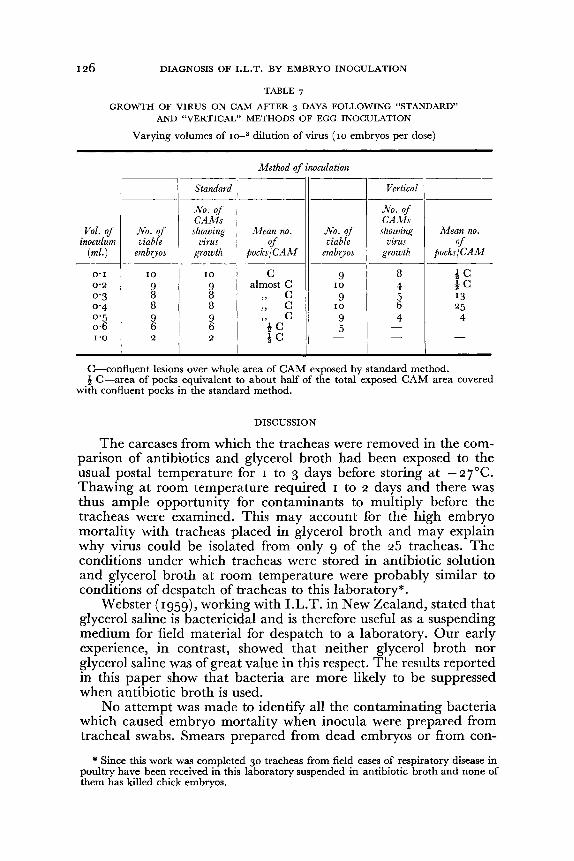
DIAGNOSIS OF I.L.T. BY EMBRYO INOCULATION
TABLE 7
GROWTH OF VIRUS ON CAM AFTER 3 DAYS FOLLOWING "STANDARD" AND "VERTICAL" METHODS OF EGG INOCULATION
Varying volumes of 10-3 dilution of virus (10 embryos per dose)
Method of inoculation
Standard Vertical --~---~-
No. of No. of CAMs CAAIs
Vol. of No. of showing Alean no. No. of showing Mean no. inoculum viable virus of viable virus of
(mi.) embryos growth pocks/CAM embryos growth pocks/CAM ---~-
0·1 10 10 C 9 8 iC 0·2 9 9 almost C 10 4 iC 0·3 8 8
" C 9 5 13
0·4 8 8 "
C 10 6 25 0·5 9 9 "
C 9 4 4 0·6 6 6 iC 5 - -1·0 2 2 iC - - -
C-confluent lesions over whole area of CAM exposed by standard method. i C-area of pocks equivalent to about half of the total exposed CAM area covered
with confluent pocks in the standard method.
DISCUSSION
The carcases from which the tracheas were removed in the comparison of antibiotics and glycerol broth had been exposed to the usual postal temperature for 1 to 3 days before storing at - 27°C. Thawing at room temperature required 1 to 2 days and there was thus ample opportunity for contaminants to multiply before the tracheas were examined. This may account for the high embryo mortality with tracheas placed in glycerol broth and may explain why virus could be isolated from only 9 of the 25 tracheas. The conditions under which tracheas were stored in antibiotic solution and glycerol broth at room temperature were probably similar to conditions of despatch of tracheas to this laboratory*.
Webster (1959), working with I.L.T. in New Zealand, stated that glycerol saline is bactericidal and is therefore useful as a suspending medium for field material for despatch to a laboratory. Our early experience, in contrast, showed that neither glycerol broth nor glycerol saline was of great value in this respect. The results reported in this paper show that bacteria are more likely to be suppressed when antibiotic broth is used.
No attempt was made to identify all the contaminating bacteria which caused embryo mortality when inocula were prepared from tracheal swabs. Smears prepared from dead embryos or from con-
* Since this work was completed 30 tracheas from field cases of respiratory disease in poultry have been received in this laboratory suspended in antibiotic broth and none of them has killed chick embryos.
F. T. W. JORDAN 127
taminated CAMs cultured in broth showed that the preponderance of organisms were gram negative rods. Proteus was frequently identified by cultural examination and Pseudomonas was also observed. Both these groups of bacteria are insensitive to chlortetracycline but are suppressed by chloramphenicol, which probably explains the greater protection afforded by the antibiotic solution containing this substance.
Gessler et at. (1956) found that emulsification of homogenized CAM with fluorocarbon (C2F3CI3) gave higher titres of vaccinia virus than other methods of virus extraction. Porter (1956), working with tobacco and cucumber mosaic viruses found that, while a higher titre of tobacco mosaic virus could be obtained following fluorocarbon emulsification than by other methods, this was not true for cucumber mosaic virus. No explanation was offered for this difference. Our results indicate very little difference between the two methods and since emulsification with fluorocarbon is much more time-consuming there seems no reason to use it.
Virus growth on the CAM is measured by examination of that area of the membrane that is affected. In the case of the CAM dropped by the standard method this area is bounded by the junction of the dropped CAM with the shell membrane. In contrast, the embryo inoculated through the air-sac may show lesions at any site on the CAM and it is necessary to examine the whole CAM for evidence of them. There were fewer pocks by the vertical method and they were smaller in size by this method of inoculation after three days' incubation. Our results with field material show that some positive cases of I.L. T. would have been missed if reliance had been placed solely on the vertical method of inoculation, whereas they were detected by the standard method. No cases positive by the vertical method were negative by the standard method. For diagnostic purposes, therefore, the vertical method of inoculation is not suitable.
In determining the titre of virus present in suspensions of confluent CAMs our results agree with those of Hitchner and White (1958), who also obtained higher titres by inoculating eggs onto the dropped CAM (standard method). Whereas they obtained differences of about 2 10g/10, our results showed a difference a little less than 1 log/1O" More recently Gentry (1963) has found no significant difference in the titre of I.L.T. virus between the two methods of inoculation, but this may be because he modified the vertical method by puncturing the inner shell membrane at six sites instead of only one, as used by Hitchner and White (1958) and ourselves. This may well increase the amount of inoculum reaching the CAM. Fabricant (1957) suggested that the vertical method of inoculation is more effective for the routine detection of a variety of respiratory viruses of the chicken but our results do not confirm this for I.L.T. virus. Gorham (1957) stated that the vertical method permits, in a single injection, a combined inoculation of the CAM, the allantoic cavity
DIAGNOSIS OF I.L.T. BY EMBRYO INOCULATION
and the yolk sac. This means that the I.L.T. virus in the inoculum would be dispersed throughout the embryo and that there would thus be less available for growth on the CAM. Since I.L.T. virus inoculated into the yolk sac would clearly be unavailable for growth on the CAM this may account for the relatively smaller amount of virus growth which results on the CAM when the vertical route of inoculation is used.
CONCLUSIONS
Ten per cent. broth containing antibiotics was preferable to 50
per cent. glycerol broth as a suspending medium for tracheas taken from chickens affected with I.L.T. in that embryo mortality was reduced and the diagnosis of I.L.T. was enhanced. The most satisfactory mixture of antibiotics for suppressing bacterial growth in swabs from field material was 1,000 units per ml. of penicillin, I fJ-g. per ml. of streptomycin and ten per cent. of a saturated solution of chloramphenicol.
I.L.T. virus was released in greater quantity from chicken tracheas and lungs by grinding with sterile sand or sterile glass than by homogenization followed by extraction with the fluorocarbon Arcton 63. In contrast, more virus was released from infected chick embryo chorio-allantois by emulsification with Arcton 63.
I.L.T. virus was more readily grown when inoculated onto the dropped CAM than when inoculated onto the shell membrane.
ACKNOWLEDGMENTS
I wish to thank Mrs. H. Evanson for technical help, Professor E. G. White for his interest and advice and the Agricultural Research Council for a grant towards the cost of the work.
REFERENCES
Beveridge, W. I. B., and Burnet, F. M. (1946). The Cultivation of Viruses and Rickettsiae in the Chick Embryo, M.R.C. Special Report, 256, H.M.S.O.; London.
Burnet, F. M. (1934). Brit. ]. expo Path., 15, 52. Fabricant, J. (1957). Avian Dis., 1, 62. Gentry, R. F. (1963). Ibid., 7,31. Gessler, A. E., Bender, C. E., and Parkinson, M. C. (1956). Trans. N.Y
Acad. Sci. Ser. 11,18, 701, 707. Gorham, J. R. (1957). Amer. ]. vet. Res., 18,691. Hitchner, S. B., and White, P. G. (1958). Poult. Sci., 37,684. Porter, C. A. (1956). Trans. N.Y. Acad. Sci. Ser. 11,18, 704. Webster, R. G. (1959). N.Z. vet. ].,7,67.
[Receivedfor publication, September 14th, 1963]