diagnosis & treatment of cough - the lung...
TRANSCRIPT

Diagnosis & Treatment
Of
Cough

a.Diagnosis :
- Details History
- Physical Examination
- Investigation
b. Treatment of cough

Detail history provides valuables clues for etiology of the cough
- Acute or chronic
- Ass. Symptoms s/o. of Respiratory infection
- Seasonal or ass. with wheezing
- Ass. with symptoms suggestive of post nasal drip or GER
- Ass with fever or sputum? If sputum what’s its character?
- Having any ass. Disease or risk factors
like – Cigarette Smoking, Environmental Exp.
- Patient taking an ACE inhibitor?
- Change of voice, weight loss & anorexia

Physical Examination-Examination of Nose
-DNS - Polyps
- Nasal Discharge
- Examination of oro-pharynx
-Erythema of mucosa
- Post Nasal Drip
-Examination of Chest
-Inspection
- Palpation
-Percussion
-Auscultation

Investigation
1. Evaluation of Infection
(a Microbiological Studies, (b Imaging Studies
(c Routine Blood Tests
2. Allergological & Physiological Examination
(a Physiological Studies (b Bronchoscopy
(c Sputum Examination (d Blood Tests
e) Chest CT (f Misc. Studies

1. Evaluation of Inflection


Microbiological Studiesi. Collection of Sample : Collect prior to starting Antimicrobial
t/t
Indirect Sample :- Sputum
Urine
Blood
Direct Sample :- Tracheal Aspiration
Bronchial washing
Bronchoalveolar lavage
Transbronchial lung Biopsy

ii. Rapid Diagnostic Test : Useful in t/t decisions & Outpatient settings.
Sputum Gram Stain :- - Useful in Rapid Identification of Bacteria
- Based on Morphology & staining
Special Stains :- - Pathogens don’t stain with
gram stain
- T.B. & Non T.B. Mycobacterium –
Z.N. stain
- Fungi – PAS Stain
- Legionella – Gimenez Stain

Immuno fluorscence :- Ag - Ab. Complex illuminated under UV light
Emitted fluorescence examine with
a fluorescence microscope e.g.
Legionella, Pnemocystitis
Antigen Tests :- - Detection of Ag in Samples
- Kits are available Commercially for e.g. Pnemoccocal Urinary Antigen, Influenza, RSV
Genetic Tests :- - DNA probes & PCR

iii. Culture & Identification Test :- Sample collect prior to Antimicrobial t/t
- Not suitable for Rapid clinical diagnosis
- Perform to Isolate pathogen and do drugs sensitivity
Bacteria :- - 106 – 107 CFU / ml in Sputum Culture -> +
- 103 - 104 CFU / ml in BAL, Bronchial washing -> +
Medium for -> Mycobacterium -> ogawa medium
Legionella -> BCYE alfa medium
Fungus -> Sabourouds medium

Imaging Studies- Typical findings on CXR can help confirm a diagnosis of
infection.
a. Lung field Infiltration :- Lobar or Brochopneumonia
Infectious Pneumonia Pneumonia Like infiltrates
- CHF
- Eosinophilic Pneumonia
b. Nodules (with or without cavitations) – T.B.
- Non T.B.
Mycobacteriosis

Routine Blood Test
- Peripheral Leukocyte count
- CRP
- ESR
- 3 parameter helps in knowing degree & course of inflammation & t/t effects.
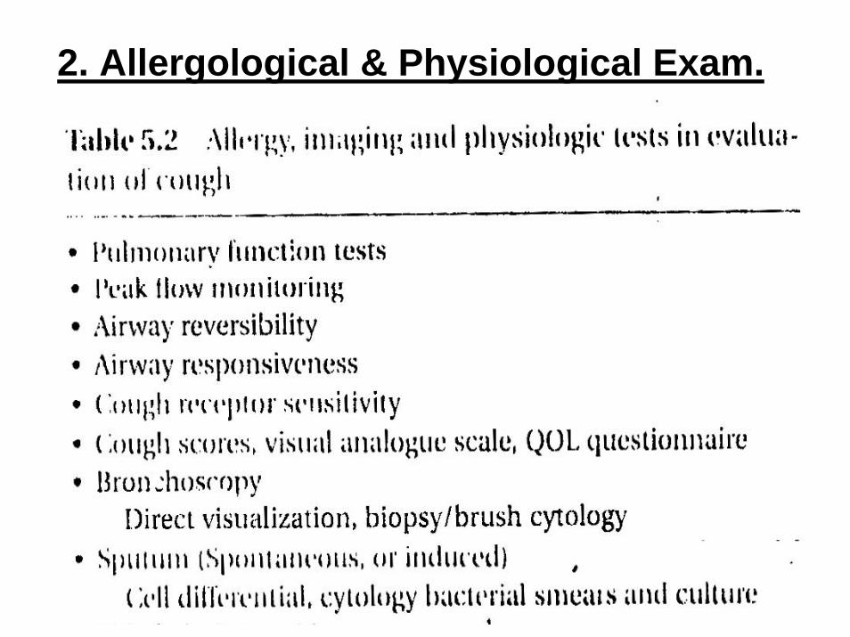
2. Allergological & Physiological Exam.

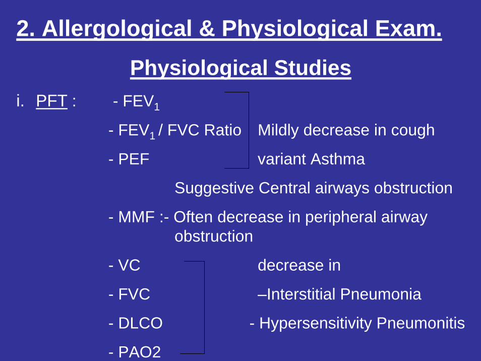
2. Allergological & Physiological Exam. Physiological Studies
i. PFT : - FEV1
- FEV1 / FVC Ratio Mildly decrease in cough
- PEF variant Asthma
Suggestive Central airways obstruction
- MMF :- Often decrease in peripheral airway obstruction
- VC decrease in
- FVC –Interstitial Pneumonia
- DLCO - Hypersensitivity Pneumonitis
- PAO2

ii. Airway reversibility Test : FEV1 is measure before and 15-30 minutes after inhalation of B2 agonist
- Cough variant asthma :- Pre FEV1 > then Pre FEV1 of classic asthma
- Improvement of coughing after inhalation of B2 agonist is diagnosis

iii. Airway responsiveness test : Evaluate threshold of Bronchial contractility by bronchoconstrictor like
{Methacholine, histamine}
given by inhalation stating at low dose to higher dose
- Not always specific for diagnosis of cough variant asthma
a. Air sensitivity :- Assess by the threshold when constriction first occur
b. Airway Reactivity :- Assess, magnitude of subsequent constriction from the slope of dose –
response curve

Bronchoscopy
Visual Examination of - Central Lung tumors
- Endobronchial T.B.
- F.Bodies
If any abnormality found - Do Endo bronchial Biopsies
- Brush cytology
- Bronchial wash cytology
- Bacteriology Tests

Blood Test
ECP (Eosinophilic cationic protein) – Marker of airway Inflammation
- Increase in atropicasthma& cough variant asthma
- Blood Eosinophilcounts are also use but less specific then ECP

CHEST CT
Central Airway Lesion – Tumors- Endobronchial T.B
Parenchymal Lung disorder – Interstitial pneumonia- Hyper sensitivity pneumonitis
OTHERS-Sinus X-Ray
--24 Hours esophageal ph monitoring

TREATMENT
According to – Duration of Cough - Type of Cough
a. Duration of Cough – Acute - Sub acute- Chronic
b. Type of Cough – Dry - Productive

Duration of Cough Guidline for Treating the common causes of Acute Cough


Guidelines for Treating the most common causes of Subacute Cough


Guidelines for Treating the most common causes of Chronic Cough


Type of Cough
a. Dry Cough – Antihistamine, - Decongestant
b. Productive Cough – Expectorant - Mucolytics- Decongestant- Antihistamine with Low Atropine like activity

Cough Suppresant Expectorants Mucolytics
Dextromethrophan Ammonium Salts Acetyl Cysteine
Guaiapate Guacitisal Ambroxol HCL
Noscapine Guaiacol Bromohexine
Pholcodine Guaiphensin Carbocistine
Piperidion Ipecacuanha Telmesteine
Sodium Dibunate

Complication of Cougha. Non Specific Complaints – Fatigue
- Anorexia
- Nausea & Vomiting
b. Musculoskeletal Complaints – Ribs Fracture- Stretching & Pulling of
Intercostals muscles- Frank rupture of musclese.g. Rectus Abdominal muscles
c. Severe Coughing - Pneumothorax- Pneumomediastinum- Cough Syncope

All the best………