diagnostic accuracy of post-mortem mri for thoracic...
TRANSCRIPT
PEDIATRIC
Diagnostic accuracy of post-mortem MRI for thoracicabnormalities in fetuses and children
Owen J. Arthurs & Sudhin Thayyil & Oystein E. Olsen & Shea Addison &
Angie Wade & Rod Jones & Wendy Norman & Rosemary J. Scott & Nicola J. Robertson &
Andrew M. Taylor & Lyn S. Chitty & Neil J. Sebire & Catherine M. Owens &
for the Magnetic Resonance Imaging Autopsy Study (MaRIAS) Collaborative Group
# The Author(s) 2014. This article is published with open access at Springerlink.com
AbstractObjectives To compare the diagnostic accuracy of post-mortem magnetic resonance imaging (PMMR) specificallyfor non-cardiac thoracic pathology in fetuses and children,compared with conventional autopsy.Methods Institutional ethics approval and parental consentwas obtained. A total of 400 unselected fetuses and childrenunderwent PMMR before conventional autopsy, reportedblinded to the other dataset.Results Of 400 non-cardiac thoracic abnormalities, 113(28 %) were found at autopsy. Overall sensitivity and speci-ficity (95 % confidence interval) of PMMR for any thoracicpathology was poor at 39.6 % (31.0, 48.9) and 85.5 % (80.7,89.2) respectively, with positive predictive value (PPV)
53.7 % (42.9, 64.0) and negative predictive value (NPV)77.0 % (71.8, 81.4). Overall agreement was 71.8 % (67.1,76.2). PMMR was most sensitive at detecting anatomicalabnormalities, including pleural effusions and lung or thoracichypoplasia, but particularly poor at detecting infection.Conclusions PMMR currently has relatively poor diagnosticdetection rates for the commonest intra-thoracic pathologiesidentified at autopsy in fetuses and children, including respi-ratory tract infection and diffuse alveolar haemorrhage. Thereasonable NPV suggests that normal thoracic appearancesat PMMR exclude the majority of important thoraciclesions at autopsy, and so could be useful in the contextof minimally invasive autopsy for detecting non-cardiacthoracic abnormalities.
O. J. Arthurs :O. E. Olsen :C. M. OwensDepartment of Radiology, Great Ormond Street Hospital for ChildrenNHS Foundation Trust, London, UK
O. J. Arthurs (*) :N. J. SebireUCL Institute of Child Health, London, UKe-mail: [email protected]
S. Thayyil : S. AddisonPerinatal Neurology and Neonatology, Imperial College London,London, UK
A. WadePaediatric Epidemiology and Biostatistics Unit, UCL Institute ofChild Health, London, UK
R. Jones :W. Norman :A. M. Taylor :C. M. OwensCentre for Cardiovascular Imaging, UCL Institute of CardiovascularScience, London, UK
R. Jones :W. Norman :A. M. Taylor :C. M. OwensCardiorespiratory Division, Great Ormond Street Hospital forChildren NHS Foundation Trust, London, UK
R. J. ScottDepartment of Histopathology, University College London HospitalNHS Trust, London, UK
N. J. RobertsonAcademic Neonatology, UCL Institute forWomen’s Health, London,UK
L. S. ChittyGenetics and Genomic Medicine, UCL Institute of Child Health,Great Ormond Street, London, UK
L. S. ChittyUCLH NHS Foundation Trusts, London, UK
N. J. SebireDepartment of Histopathology, Great Ormond Street Hospital forChildren NHS Foundation Trust, London, UK
DOI 10.1007/s00330-014-3313-8
Received: 14 February 2014 /Revised: 7 May 2014 /Accepted: 3 July 2014 /Published online: 31 August 2014
Eur Radiol (2014) 24:2876–2884
Key Points• PMMR has relatively poor diagnostic detection rates forcommon intrathoracic pathology
• The moderate NPV suggests that normal PMMR appear-ances exclude most important abnormalities
• Lung sampling at autopsy remains the “gold standard” forpulmonary pathology
Keywords MRI . Autopsy . Pathology . Fetuses . Children
Introduction
Paediatric and perinatal autopsy rates have declined overrecent decades, leading to post-mortem cross-sectional imag-ing being proposed as a possible alternative or adjunctiveapproach [1, 2]. Post-mortem magnetic resonance imaging(PMMR), when used in conjunction with other ancillary in-vestigations (collectively known as minimally invasive autop-sy [MIA]), has recently been shown to have a high diagnosticaccuracy rate for cause of death or main diagnosis comparedwith traditional autopsy in fetuses, stillbirths and children [3].Whilst cardiac and neurological diagnoses account for themajority of causes of death in these age groups (12 and28 % respectively [3]), less is known about how well thoracicpathology can be diagnosed across a range of likely diagnoses,gestational age and childhood age ranges.
A systematic review of post-mortem imaging [4] iden-tified several previous studies [5–8] which had attemptedto investigate the accuracy of non-cardiac thoracic diag-nosis, and reported a relatively good estimated pooledsensitivity of 82 % (95 % confidence intervals 49,95 %) and specificity of 86 % (58, 96 %) for lungpathology. However, these studies were not fully blindedbetween reporting radiologists and pathologists, includedhighly selected small cohort groups (range, 6–37 sub-jects), with pooled data from only 73 fetuses and nochildren, giving wide confidence intervals. Breeze et al.[9] independently reported a 2.5 % sensitivity and 87 %specificity for eight lung lesions in 30 fetuses, whenassessed using PMMR, with a reported diagnostic accu-racy of 83 %.
The aim of theMARIAS study was specifically to establishthe diagnostic utility of less invasive autopsy, using PMMR,compared with traditional autopsy, in an unselected popula-tion for the major diagnosis or cause of death. These resultshave already been published [3]. Here, we use the detailedMARIAS dataset for sub-analysis of the PMMR findingsspecifically for non-cardiac thoracic abnormalities, irrespec-tive of whether the abnormalities contributed to the maindiagnosis or cause of death.
Materials and methods
Study participants
The study design has previously been described in detail [10].Briefly, we performed pre-autopsy PMMR in an unselectedsequential cohort of fetuses and children (up to 16 years),referred for conventional autopsy between March 2007 andSeptember 2011, to Great Ormond Street Hospital for Chil-dren and University College Hospital, London. The study hadinstitutional approval (reference 04/Q0508/41), and parentalconsent was obtained as stipulated by the ethics committee[3]. All fetuses and children (≤16 years) undergoing conven-tional autopsy were eligible for recruitment. PMMR wasperformed as soon as practically possible. Cases were exclud-ed if consent was not available or if PMMR could not beperformed before autopsy. The bodies were stored in a mor-tuary at 4 °C.
Imaging technique
Post-mortem magnetic resonance imagings (MRIs) weremostly scheduled to be done out of hours (6 pm to 8 am),causing least disturbance to clinical services. All examinationswere performed on a 1.5-T MRI scanner (Avanto; SiemensMedical Solutions, Erlangen, Germany) with a conventionalphased-array body coil, using whole-body three-dimensional(3D) T2-weighted turbo spin echo (TSE, TR 3,500 ms, TE276 ms, voxel size 0.8×0.8×0.8 mm, 2 averages), 3D T1-weighted volumetric interpolated breath-hold examination(VIBE; TR 5.9 ms, TE 2.4 ms, flip angle 25°, voxel size0.8×0.8×0.8 mm, 8 averages) and 3D constructive interfer-ence in the steady state (CISS) sequence (TR 9.2 ms, TE4.6 ms, flip angle 70°, voxel size 0.6×0.6×0.6 mm, 4 aver-ages), as published previously [10]. The first 20 cases (total423) were used for MR sequence optimisation and for imagertraining.
Reporting PMMR images
PMMR images were reported by one of two specialist paedi-atric radiologists (O.O. and C.M.O.), with 12 and 18 years’experience, using the OsiriX platform (OsiriX Foundation,Geneva, Switzerland). The radiologist was given the age andbasic clinical details of the case, but was blinded to results ofancillary investigations and autopsy findings.
Reporting of autopsy data
Conventional autopsies were performed by one of severalexperienced specialist perinatal/paediatric pathologists (atleast 8 years experience) in accordance with national guide-lines, and were reported blinded to the MRI findings [3, 11].
2877Eur Radiol (2014) 24:2876–2884
Data and statistical analysis
Data were collected into a specific Microsoft Access database(version 2003; Microsoft, Redmond, WA, USA), which wasdeveloped as part of a study examining paediatric autopsies[12]. The radiologists and pathologists could only access theirrespective sections of the database until all data were reported.Prior to data unmasking, a pathologist (N.J.S.), radiologist(A.M.T.) and neonatologist (S.T.) reviewed all the PMMRand pathology reports, to define and categorise the findingsaccording to predefined criteria [10]. The results presentedhere are those as recorded at the time of initial PMMRreporting, not following subsequent specialist unblinded re-view, so as to reflect how PMMR would be used in clinicalpractice.
In order to clarify the nature of the discrepancies, we thenretrospectively reviewed the imaging and pathology findings.We excluded cardiac abnormalities, as they have been report-ed elsewhere [13] and divided specific thoracic diagnoses intoone of seven categories, which were: respiratory tract infec-tion (lung parenchymal consolidation at PMMR, pneumonia,pneumonitis and aspiration at autopsy), pulmonary haemor-rhage, lung or thoracic hypoplasia/agenesis, pulmonarycongestion/oedema, isolated pleural effusion, trachea-oesophageal fistula, and “other” categories.
Primary outcomes were sensitivity, specificity, positive-predictive valve (PPV) and negative predictive value (NPV),where PMMR (the index test) defined the diagnosis comparedwith conventional autopsy (the “gold standard”), with 95 %confidence intervals (CI). Concordance was defined as thesum of true positives and true negatives divided by all cases.Exact methods were used to calculate confidence intervals[14]. SPSS (version 19 for Macintosh; SPSS IBM, New York,USA) was used for data analysis. Sub-group analysis wasperformed based on age (fetuses <24 weeks gestation, fetuses≥24 gestation, and children including all newborns, infantsand children ≤16 years of age).
Results
Demographic data
PMMR was performed on 403 cases, although autopsy datacould not be retrieved in 3 cases. Of the remaining 400 cases,277/400 (69 %) were fetuses (consisting of 185 at ≤24 weeks’gestation and 92 at >24 weeks’ gestation) and 123/400 (31 %)were children (consisting of 42 newborns aged <1 month, 53infants >1 month to ≤12 months, 28 children >12 months to≤16 years). Hospital-requested autopsies (fetal medicine unitor in-patients) accounted for 292 cases, with HM Coroner(Medical Examiner)-requested autopsies accounting for the
remainder: 6 (2.2 %) fetuses and 102 (82.9 %) children. Mean(SD) time between death/delivery and PMMR was 4.5 (2.5)days.
Autopsy findings
Summary details of autopsy pathology in this dataset havebeen briefly reported previously [3]. Conventional autopsystudies revealed a total of 113/400 thoracic (28 %) abnormal-ities across all groups. At autopsy, in fetuses <24 weeks (n=185), the thorax was normal in 146/185 (78.9 %) cases,abnormal in 35/185 (19 %) and non-diagnostic in 4/185(2.1 %). Of the 113 abnormalities, the most common abnor-malities reported were infection in 15/113 (13.3 %) and pul-monary hypoplasia in 11/113 (6 %) cases. In fetuses>24 weeks (n=92), the thorax was abnormal in 16/92(17 %), with infection being the commonest abnormalityreported in 9/92 (10 %) cases. In newborns and children (n=123), the thorax was abnormal in 62/123 (50.5 %) cases, withthe commonest abnormalities being infection in 24/123(19.5 %) and aspiration/haemorrhage in 12/123 (9.7 %) cases.
Non-diagnostic cases
There were 27/400 (6.7 %) non-diagnostic thoracic PMMRscans; all of which occurred in fetuses ≤24 weeks. There weretwo thoracic abnormalities (lung hypoplasia) in this group.Subsequent analysis of the data has been performed on theremaining 373 cases, which had 111/373 (29.8 %) thoracicabnormalities identified by conventional autopsy.
Comparison of PMMR to conventional autopsy
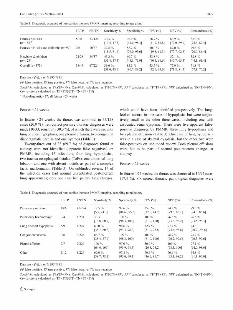
The overall sensitivity, specificity, PPV, NPV, and concor-dance across the three subgroups with 95 % confidence inter-vals are given in Table 1.
Thoracic PMMR had poor sensitivity and specificity acrossall groups, with slightly higher sensitivity in children thansmaller fetuses (45 vs 30 %), but poorer specificity (61 vs97 %), and poor overall concordance (71.8 %). Overall sensi-tivity was poor at 39.6 %, although specificity was higher at85.5 %.
PMMR was most sensitive at detecting anatomical abnor-malities, including pleural effusion (100 %) and lung or tho-racic hypoplasia (60 %; Table 2). PMMR was particularlypoor at detecting infection with sensitivity of 12.5 % andPPV of 25.0 %. Although overall concordance, specificityand NPV were high across different pathologies, this waslargely due to the majority of normal cases in this study.
Examples of concordance between PMMR and conven-tional autopsy findings are shown in Figs. 1, 2, and 3; exam-ples of a lack of concordance are shown in Figs. 4 and 5.
2878 Eur Radiol (2014) 24:2876–2884
Fetuses <24 weeks
In fetuses <24 weeks, the thorax was abnormal in 33/158cases (20.9 %). Ten correct positive thoracic diagnoses weremade (10/33; sensitivity 30.3 %), of which there were six withlung or chest hypoplasia, one pleural effusion, two congenitaldiaphragmatic hernias and one hydrops (Table 3).
Twenty-three out of 33 (69.7 %) of diagnoses found atautopsy were not identified (apparent false negatives) onPMMR, including 15 infections, four lung hypoplasias,two trachea-oesophageal fistulae (ToFs), one abnormal lunglobation and one with absent nostrils as part of a complexfacial malformation (Table 3). On unblinded review, 14 ofthe infection cases had normal unventilated post-mortemlung appearances; only one case had patchy lung changes,
which could have been identified prospectively. The lungslooked normal in one case of hypoplasia, but were subjec-tively small in the other three cases, including one withassociated renal dysplasia. There were five apparent false-positive diagnoses by PMMR: three lung hypoplasias andtwo pleural effusions (Table 3). One case of lung hypoplasiawas in a case of skeletal dysplasia, but the other two werefalse-positives on unblinded review. Both pleural effusionswere felt to be part of normal post-mortem changes atautopsy.
Fetuses >24 weeks
In fetuses >24 weeks, the thorax was abnormal in 16/92 cases(17.4 %). Six correct thoracic pathological diagnoses were
Table 1 Diagnostic accuracy of non-cardiac thoracic PMMR imaging, according to age group
FP/TP FN/TN Sensitivity % Specificity % PPV (%) NPV (%) Concordance (%)
Fetuses <24 wks(n=158)a
5/10 23/120 30.3 %[17.4, 47.3]
96.0 %[91.0, 98.3]
66.7 %[41.7, 84.8]
83.9 %[77.0, 89.0]
82.3 %[75.6, 87.4]
Fetuses >24 wks and stillbirths (n=92) 9/6 10/67 37.5 %[18.5, 61.4]
88.2 %[79.0, 93.6]
40.0 %[19.8, 64.3]
87.0 %[77.7, 92.8]
79.3 %[70.0, 86.4]
Newborn & children(n=123)
24/28 34/37 45.2 %[33.4, 57.5]
60.7 %[48.1, 71.9]
53.8 %[40.5, 66.6]
52.1 %[40.7, 63.3]
52.8 %[44.1, 61.4]
Overall (n=373) 38/44 67/224 39.6 %[31.0, 48.9]
85.5 %[80.7, 89.2]
53.7 %[42.9, 64.0]
77.0 %[71.8, 81.4]
71.8 %[67.1, 76.2]
Data are n (%), n or % [95 % CI]
FP false positive, TP true positive, FN false negative, TN true negative
Sensitivity calculated as TP/(TP+FN); Specificity calculated as TN/(TN+FP); PPV calculated as TP/(TP+FP); NPV calculated as TN/(TN+FN);Concordance calculated as (TP+TN)/(TP+TN+FP+FN)a Non-diagnostic=27, all fetuses <24 weeks
Table 2 Diagnostic accuracy of non-cardiac thoracic PMMR imaging, according to pathology
FP/TP FN/TN Sensitivity % Specificity % PPV (%) NPV (%) Concordance (%)
Pulmonary infection 18/6 42/224 12.5 %[5.9, 24.7]
92.6 %[88.6 , 95.2]
25.0 %[12.0, 44.9]
84.2 %[79.3, 88.1]
79.3 %[74.3, 83.6]
Pulmonary haemorrhage 0/4 8/224 33.3[13.8, 60.9]
100 %[98.3, 100]
100 %[51.0, 100]
96.6 %[93.3, 98.2]
96.6 %[93.5, 98.3]
Lung or chest hypoplasia 8/9 6/224 60.0 %[35.7, 80.2]
96.6 %[93.3, 98.2]
52.9 %[31.0, 73.8]
97.4 %[94.4, 98.8]
94.3[90.7 , 96.6]
Congestion/oedema 0/6 3/224 66.7 %[35.4, 87.9]
100 %[98.3, 100]
100 %[61.0, 100]
98.7 %[96.2, 99.5]
98.7 %[96.3, 99.6]
Pleural effusion 7/7 0/224 100 %[64.6, 100]
97.0 %[93.9, 98.5]
50.0 %[26.8, 73.2]
100 %[98.3, 100]
97.1 %[94.0, 98.6]
Other 5/12 8/224 60.0 %[38.7, 78.1]
97.8 %[95.0, 99.1]
70.6 %[46.9, 86.7]
96.6 %[93.3, 98.2]
94.8 %[91.3, 96.9]
Data are n (%), n or % [95 % CI]
FP false positive, TP true positive, FN false negative, TN true negative
Sensitivity calculated as TP/(TP+FN); Specificity calculated as TN/(TN+FP); PPV calculated as TP/(TP+FP); NPV calculated as TN/(TN+FN);Concordance calculated as (TP+TN)/(TP+TN+FP+FN)
2879Eur Radiol (2014) 24:2876–2884
made (6/16; sensitivity 37.5 %; Table 3). There were tenapparent false negatives (10/16; 62.5 %), including nine in-fections and one lung hypoplasia (Table 3). On unblindedreview, the lungs had normal unventilated post mortem ap-pearances in all nine infections. The lung hypoplasia wasmissed in a severely macerated 24-week-gestation fetus.
There were nine apparent false positives by PMMR: fourpleural effusions, three lung hypoplasias and two focal lunglesions (Table 3). On unblinded review, all four pleural effu-sions were present but interpreted as normal post-mortemchange at autopsy. In one of the lung hypoplasias, the lungslooked subjectively small on PMMR in a case of massivesacrococcygeal teratoma, but were histologically normal sizeand weight at autopsy. Two other lung hypoplasias were over-calls. In one “lung lesion”, the lungs looked patchy on PMMRbut were normal at autopsy, including histological examination,and in the other the lungs looked normal on PMMR but weredistorted by large amounts of intra-pleural gas.
Newborns and children
In newborns, infants and children, the thorax was abnormal in62/123 cases (50.4 %). Twenty-eight correct pathologicaldiagnoses were made (28/62; sensitivity 45.2 %; Table 3).Thirty-four thoracic diagnoses were not correctly identified(34/62; 54.8 %), including 18 infections, 8 with pulmonaryhaemorrhage, 2 trachea-oesophageal fistulae, 3 pulmonarycongestion/oedema and 1 case each of lung hypoplasia, pul-monary hypertension and hyalinemembrane disease (Table 3).On unblinded review, there were patchy lung changes, whichcould have been interpreted as abnormal in only six of thecases in which infection was diagnosed at autopsy; in the other12 cases, the lungs looked normal on PMMR. Equally, therewere patchy lung changes in five of the cases of pulmonaryhaemorrhage and two cases of pulmonary congestion andoedema, but the lungs looked normal in the other three andone case respectively. The lungs looked normal on PMMR in
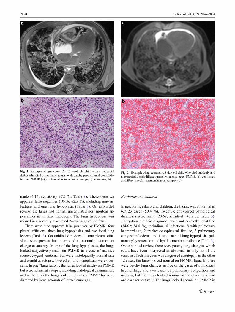
Fig. 1 Example of agreement. An 11-week-old child with atrial-septaldefect who died of systemic sepsis, with patchy parenchymal consolida-tion on PMMR (a), confirmed as infection at autopsy (pneumonia; b)
Fig. 2 Example of agreement. A 3-day-old child who died suddenly andunexpectedly with diffuse parenchymal change on PMMR (a), confirmedas diffuse alveolar haemorrhage at autopsy (b)
2880 Eur Radiol (2014) 24:2876–2884
the cases of HMD, pulmonary hypertension and hypoplasia.The trachea-oesophageal fistulae were not identified.
There were 24 apparent false positives, including 18 infec-tions, 2 lung hypoplasias, 2 focal lung lesions, 1 pleuraleffusion and 1 dilated oesophagus (Table 3). On unblindedreview, there were patchy lung changes in 17 out of 18 casesof infection, and both cases of focal lung lesions, althoughnone of these could be confirmed as abnormal at autopsy,including histology. In one case of lung hypoplasia the childhad a skeletal dysplasia (osteogenesis imperfecta type IIa)with a small, dysmorphic ribcage, although the lungs werehistologically normal and of normal weight at autopsy, but theother was an overcall. The case of pleural effusion wasinterpreted as normal post-mortem change at autopsy. Theoesophagus was dilated in one case on PMMR but no abnor-mality was detected at autopsy.
Across all groups, the commonest abnormality was infec-tion (consolidation on PMMR, pneumonia or pneumonitis atautopsy), accounting for 43% of all diagnoses made, althoughonly six correct diagnoses were made on PMMR, with 42apparent false negatives and 18 false positives made. Thisgives a sensitivity of 12.5 % (5.9, 24.7) and specificity of92.6 % (88.6, 95.2), and concordance of 79.3 % (74.3, 83.6)for infection alone (Table 2).
Discussion
In this large prospective study, PMMR demonstrated poordiagnostic utility for detection of thoracic pathology in fe-tuses, newborns and children. Whilst the specificity was highat 86 %, due to the large number of normal cases, the sensi-tivity and positive predictive value were poor at 40 and 54 %
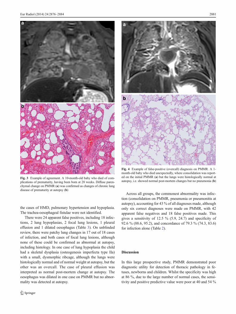
Fig. 3 Example of agreement. A 10-month-old baby who died of com-plications of prematurity, having been born at 28 weeks. Diffuse paren-chymal change on PMMR (a) was confirmed as changes of chronic lungdisease of prematurity at autopsy (b)
Fig. 4 Example of false-positive (overcall) diagnosis on PMMR. A 1-month-old baby who died unexpectedly, where consolidation was report-ed on the initial PMMR (a) but the lungs were histologically normal atautopsy, i.e. showed normal post-mortem changes but no pneumonia (b)
2881Eur Radiol (2014) 24:2876–2884
respectively. Overall diagnostic accuracy was reasonable at72 %. PMMR was most sensitive at detecting anatomicalabnormalities such as lung or chest hypoplasia and post-mortem pleural effusions, but poor at detecting infection andpulmonary haemorrhage.
PMMR showed a 9.5 % apparent false-positive rate forthoracic imaging, which is in keeping with results from otherbody systems [3]. Apparent false positives do not necessarilyrepresent a significant failure of the minimally invasive au-topsy model, since the vast majority (75 %) were overcalls ofpossible infection (consolidation/pneumonia) in children,which would lead to additional investigations and organ sam-pling for histopathological confirmation. There were severalfalse positives where normal post-mortem accumulation offluid in bodily cavities was identified but mis-interpreted as
pathological pleural fluid, and most of the correct interpreta-tions of lung hypoplasia were recorded in the context ofskeletal dysplasia.
Our accuracy data in this large unselected population islower than those previously published [5–8], with lower sen-sitivity and overall concordance, but similar specificity. This islikely to be due to the difficulty in diagnosing infection,particularly in children, where such cases were not includedin the several previous studies. Breeze et al. [9] reported areported a 62.5 % sensitivity (95 % CI=29.0, 96.0) and 87 %specificity (95%CI=78.9, 100), for eight lung lesions over 30fetuses. Our findings are similar, in that fetal lung hypoplasiawas usually easily detected, but was also overcalled. Theirseries only had two false-positive lung diagnoses (2/30=6.7 %), and their PPV was 71.4 % (35.9, 91.8), NPV 87.0 %(67.9, 95.5), and overall concordance 83 % (66.4, 92.7 %), allgreater than our data but with much wider confidence inter-vals. We postulate that lung PMMR is more difficult across alarger cohort of children when reported in a blinded fashion.
Infection (pneumonia or lung parenchymal consolidation)was by far both the commonest abnormality in our study, andalso the most difficult to achieve correctly. There are severaldiagnostic difficulties highlighted by our study, and a smallstudy of 44 children has previously identified similar difficul-ties in detecting lung parenchymal changes using whole-bodypost-mortem CT [15]. In unventilated fetal lungs, infectionwas missed in the majority of cases, and infection was equallyovercalled and missed in the paediatric population, with orwithout patchy signal changes in the lungs at post-mortem.We conclude that infection or pneumonia is currently difficultto identify at fetal PMMR, and that lung sampling should beperformed in all cases where sepsis is suspected or possible. Inolder children, misinterpreting consolidation as normal post-mortem change, and vice versa, represents a real challenge inPMMR, recognised by other authors [16]. Several featureswithin the lungs may be interpreted as consolidation (as theywould indicate such in life), whereas at autopsy the unequallydistributed fluid accumulation within the lung parenchyma isa variant of normal post-mortem changes. We also failed todiagnose eight out of 12 cases of pulmonary haemorrhage,which were interpreted as ‘normal’ post-mortem changes.However, there are variable histological degrees of pulmonarycongestion and intra-alveolar fluid in the majority of cases ofinfant death, regardless of cause. The high apparent false-negative rate (16.2 %; apparent pathologies present at autopsywhich were not reported on PMMR) in this study also mainlyrelates to patchy but diffuse lung parenchymal change, i.e.pneumonia or haemorrhage. The majority of thoracic missesand overcalls in this study were in newborns and children, andmostly related to both overcalling and missing pneumonia.
A normal thoracic scan in fetuses predicts normal pathol-ogy in over 80 % of cases, probably because lung aerationdoes not present a confounding problem in differentiating
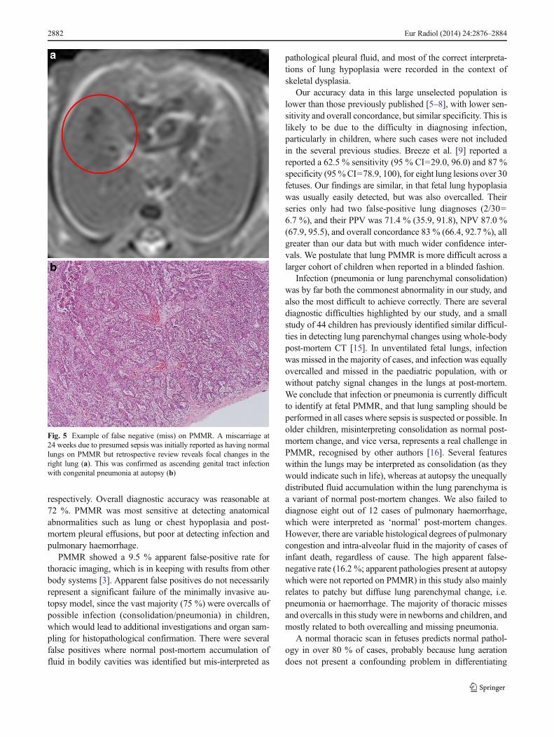
Fig. 5 Example of false negative (miss) on PMMR. A miscarriage at24 weeks due to presumed sepsis was initially reported as having normallungs on PMMR but retrospective review reveals focal changes in theright lung (a). This was confirmed as ascending genital tract infectionwith congenital pneumonia at autopsy (b)
2882 Eur Radiol (2014) 24:2876–2884
between other causes of death, such as CDH or pulmonaryhypoplasia, but this does not hold true for children. Ourcurrent practice is that unless a focal lung lesion is identifiedon PMMR, then standard lung histopathology should be per-formed in all childhood cases. It may be possible that in futurethis could be performed by percutaneous or endoscopic routes[17], but the accuracy of this approach to detect patchy lungpathology, compared with open sampling, remainsundetermined.
Lung abnormalities are notoriously difficult to detect usingother types of post-mortem imaging. For instance, lung opac-ities on post-mortem radiographs have little correlation withhistologically diagnostic pneumonia [18]. Lung PMMR isunlikely to have higher diagnostic accuracy than thoracicMRI in life, and high-resolution computed tomography (CT)remains the mainstay of diagnostic imaging for parenchymalchanges, including interstitial lung disease and more subtlepathology. Whilst infant and childhood lung MRI is improv-ing, it is notoriously sequence-dependent and prone to respi-ratory and cardiac motion artefacts [19]. The post-mortemstate negates several of these issues, but clearly brings newdiagnostic challenges.
The main strength of this study is the double-blinded studydesign and large prospective data collection of unselectedcases, which allows categorical reporting of both conventionalautopsy and PMMR data, in an independent way. By identi-fying small pleural effusions, and lung parenchymal changesmore accurately, it is likely that correct interpretation ofPMMR images will be easier in the future.
A limitation of this study was that these cases were allinterpreted by both specialist paediatric radiologists in a
tertiary centre and, as such, is unlikely to represent the situa-tion in many other centres. It also highlights both the difficultyin interpreting cases correctly, as these are predominantlyinterpretation rather than observational errors, and thereforeemphasises the need for specialist education in this field inorder to maximise the diagnostic yield from both imaging andautopsy. We did not perform post-mortem CT in our cohort,the diagnostic accuracy of which in this setting remains to beevaluated. We also did not measure temperature duringPMMR in this study, which may have had an impact on thereporting radiologists’ ability to correctly interpret PMMRimages [20]. We also did not evaluate more advanced MRtechniques, such as T2 relaxometry of the lungs or diffusion-weighted imaging. Semi-quantitative measures of water redis-tribution may be able to differentiate pathological from non-pathological changes in the lungs, but such approaches requirefurther investigation.
PMMR currently has relatively poor diagnostic detectionrates for the commonest intra-thoracic pathologies identifiedat autopsy in fetuses and children, including infection andhaemorrhage. The relatively high negative predictive valuesuggests that normal thoracic appearances at PMMR excludesthe majority of important thoracic lesions at autopsy, and socould be useful in the context of minimally invasive autopsyfor excluding non-cardiac thoracic abnormalities.
Acknowledgments The scientific guarantor of this publication isAndrew M. Taylor. The authors of this manuscript declare no relation-ships with any companies, whose products or services may be related tothe subject matter of the article. This study has received funding by thePolicy Research Programme in the Department of Health (0550004). Theviews expressed are not necessarily those of the Department. The study
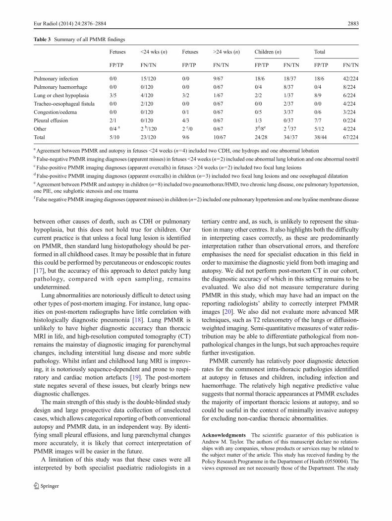
Table 3 Summary of all PMMR findings
Fetuses <24 wks (n) Fetuses >24 wks (n) Children (n) Total
FP/TP FN/TN FP/TP FN/TN FP/TP FN/TN FP/TP FN/TN
Pulmonary infection 0/0 15/120 0/0 9/67 18/6 18/37 18/6 42/224
Pulmonary haemorrhage 0/0 0/120 0/0 0/67 0/4 8/37 0/4 8/224
Lung or chest hypoplasia 3/5 4/120 3/2 1/67 2/2 1/37 8/9 6/224
Tracheo-oesophageal fistula 0/0 2/120 0/0 0/67 0/0 2/37 0/0 4/224
Congestion/oedema 0/0 0/120 0/1 0/67 0/5 3/37 0/6 3/224
Pleural effusion 2/1 0/120 4/3 0/67 1/3 0/37 7/7 0/224
Other 0/4 a 2 b/120 2 c/0 0/67 3d/8e 2 f/37 5/12 4/224
Total 5/10 23/120 9/6 10/67 24/28 34//37 38/44 67/224
aAgreement between PMMR and autopsy in fetuses <24 weeks (n=4) included two CDH, one hydrops and one abnormal lobationb False-negative PMMR imaging diagnoses (apparent misses) in fetuses <24 weeks (n=2) included one abnormal lung lobation and one abnormal nostrilc False-positive PMMR imaging diagnoses (apparent overcalls) in fetuses >24 weeks (n=2) included two focal lung lesionsd False-positive PMMR imaging diagnoses (apparent overcalls) in children (n=3) included two focal lung lesions and one oesophageal dilatatione Agreement between PMMR and autopsy in children (n=8) included two pneumothorax/HMD, two chronic lung disease, one pulmonary hypertension,one PIE, one subglottic stenosis and one traumaf False negative PMMR imaging diagnoses (apparent misses) in children (n=2) included one pulmonary hypertension and one hyaline membrane disease
2883Eur Radiol (2014) 24:2876–2884
was also supported by grants from the British Heart Foundation (CI/05/010). TheMR facility at the UCL Centre for Cardiovascular Imaging wasfunded by the British Heart Foundation (CI/05/010). None of the fundingbodies had any role in analysis of data, results or conclusions of the study.A.M.T. is supported by an NIHR Senior Research fellow award, NJS issupported by an NIHR Senior Investigator award, and O.A. and S.T. aresupported by NIHRClinician Scientist awards. L.S.C., N.J.S. and A.M.T.receive funding from the Great Ormond Street Hospital Children’s Char-ity. This work was undertaken at GOSH/ICH, UCLH/UCL includingsupport from the NIHR GOSH Biomedical Research Centre. One of theauthors has significant statistical expertise. Institutional Review Boardapproval was obtained. Written informed consent was obtained from allsubjects (patients) in this study.
Some study subjects or cohorts have been previously reported byThayyil et al. [3]. Methodology: prospective, diagnostic study,multicentre study.
Open Access This article is distributed under the terms of the CreativeCommons Attribution Noncommercial License which permits any non-commercial use, distribution, and reproduction in any medium, providedthe original author(s) and the source are credited.
References
1. Shojania KG, Burton EC (2008) The vanishing nonforensic autopsy.N Engl J Med 358:873–875
2. Sieswerda-Hoogendoorn T, van Rijn RR (2010) Current techniquesin postmortem imaging with specific attention to paediatric applica-tions. Pediatr Radiol 40:141–152
3. Thayyil S, Sebire NJ, Chitty LS, Wade A, Chong WK, MARIASCollaborative Group et al (2013) Post-mortem MRI versus conven-tional autopsy in fetuses and children: a prospective validation study.Lancet 382:223–333
4. Thayyil S, Chandrasekaran M, Chitty LS, Wade A, Skordis-WorrallJ, Bennett-Britton I, Cohen M, Withby E, Sebire NJ, Robertson NJ,Taylor AM (2010) Diagnostic accuracy of post-mortem magneticresonance imaging in fetuses, children and adults: a systematic re-view. Eur J Radiol 75:e142–e148
5. Huisman TA,Wisser J, Stallmach T, Krestin GP, Huch R, Kubik-HuchRAC (2002) MR autopsy in fetuses. Fetal Diagn Ther 17:58–64
6. Alderliesten ME, Peringa J, van der Hulst VP, Blaauwgeers HL, vanLith JMC (2003) Perinatal mortality: clinical value of postmortemmagnetic resonance imaging compared with autopsy in routine ob-stetric practice. BJOG 110:378–382
7. Roberts IS, Benbow EW, Bisset R et al (2003) Accuracy of magneticresonance imaging in determining cause of sudden death in adults:comparison with conventional autopsy. Histopathology 42:424–430
8. Patriquin L, Kassarjian A, Barish M et al (2001) Postmortem whole-body magnetic resonance imaging as an adjunct to autopsy: prelim-inary clinical experience. J Magn Reson Imaging 13:277–287
9. Breeze AC, Cross JJ, Hackett GA, Jessop FA, Joubert I, Lomas DJ,Set PA, Whitehead AL, Lees CC (2006) Use of a confidence scale inreporting postmortem fetal magnetic resonance imaging. UltrasoundObstet Gynecol 28:918–924
10. Thayyil S, Sebire NJ, Chitty LS, Wade A, Olsen O, Gunny RS,Offiah A, Saunders DE, Owens CM, Chong WK, Robertson NJ,Taylor AM (2011) Post mortem magnetic resonance imaging in thefetus, infant and child: a comparative study with conventional autop-sy (MARIAS protocol). BMC Pediatr 11:120
11. Royal College of Pathologists Working Party on the Autopsy (2006)Guidelines on Autopsy Practice: Scenario 9: Stillborn infant(singleton). June 2006. http://www.rcpath.org/Resources/RCPath/Migrated%20Resources/Documents/G/G001Autopsy-Stillbirths-Jun06.pdf. Accessed 01 Nov 2013
12. Weber MA, Klein NJ, Hartley JC, Lock PE, Malone M, Sebire NJ(2008) Infection and sudden unexpected death in infancy: a system-atic retrospective case review. Lancet 371:1848–1853
13. Taylor AM, Sebire NJ, Ashworth MT, Schievano S, Scott RS, ChittyLS, Robertson N, Thayyil S, Magnetic Resonance Imaging AutopsyStudy Collaborative Group et al (2014) Post-mortem cardiovascularmagnetic resonance imaging in fetuses and children—a masked com-parison study with conventional autopsy. Circulation 129:1937–1944
14. Wilson EB (1927) Probable inference, the law of succession, andstatistical inference. J Am Stat Assoc 22:209–212
15. Proisy M, Marchand AJ, Loget P, Bouvet R, Roussey M, Pelé F,Rozel C, Treguier C, Darnault P, Bruneau B (2013) Whole-bodypost-mortem computed tomography compared with autopsy in theinvestigation of unexpected death in infants and children. Eur Radiol23:1711–1719
16. Germerott T, Preiss US, Ebert LC, Ruder TD, Ross S, Flach PM,Ampanozi G, Filograna L, Thali MJ (2010) A new approach invirtopsy: postmortem ventilation inmultislice computed tomography.Legal Med (Tokyo) 12:276–279
17. Sebire NJ, Weber MA, Thayyil S, Mushtaq I, Taylor A, Chitty LS(2012) Minimally invasive perinatal autopsies using magnetic reso-nance imaging and endoscopic postmortem examination (“keyholeautopsy”): feasibility and initial experience. J Matern Fetal NeonatalMed 25:513–518
18. Olsen OE (2006) Radiography following perinatal death: a review.Acta Radiol 47:91–99
19. HirschW, Sorge I, Krohmer S,Weber D,Meier K, Till H (2008)MRIof the lungs in children. Eur J Radiol 68:278–288
20. Ruder TD, Hatch GM, Siegenthaler L, Ampanozi G,Mathier S, ThaliMJ, Weber OM (2012) The influence of body temperature on imagecontrast in post mortem MRI. Eur J Radiol 81:1366–1370
MaRIAS (Magnetic Resonance Imaging Autopsy Study)Collaborative group
Ms. Shea Addison (Research Assistant, UCL), Dr. Michael Ashworth(Consultant in Paediatric Pathology, GOSH) Dr. Alan Bainbridge (MRPhysicist, UCL), Dr. Jocelyn Brookes (Consultant in Interventional Ra-diology, UCH), Prof. Lyn Chitty (Consultant in Fetal Medicine andGenetics, UCL), Dr. WK ‘Kling’ Chong (Consultant in Paediatric Neu-roradiology, GOSH), Dr. Andrew Cook (Senior Lecturer in CardiacMorphology, UCL), Dr. Enrico de Vita (MR Physicist, UCL), Dr. RoxanaGunny (Consultant in Paediatric Neuroradiology, GOSH), Dr. BrianHarding (Consultant in Paediatric Neuropathology, GOSH), Dr. TomJacques (Consultant in Paediatric Neuropathology, GOSH), Mr. RodJones (Research MR radiographer, UCL), Dr. Mark Lythgoe (Director,Centre for Advanced Biomedical Imaging, UCL), Dr. Marion Malone(Consultant in Paediatric pathology, GOSH), Wendy Norman (ResearchMR radiographer, UCL), Dr. Oystein Olsen (Consultant in PaediatricChest and Abdomen Imaging, GOSH), Dr. Cathy Owens (Consultant inPaediatric Chest and Abdomen Imaging, GOSH), Dr. Amaka C. Offiah(Consultant in Paediatric Musculoskeletal Imaging, previously GOSH,currently Sheffield Children’s Hospital), Dr. Nikki Robertson (Readerand Consultant in Neonatology, UCH), Dr. Tony Risdon (Consultant inPaediatric Forensic Pathology, GOSH), Prof. Neil Sebire (Professor ofPerinatal and Paediatric Developmental Pathology, GOSH), Dr. Rose-mary Scott (Consultant in Perinatal pathology, UCH), Dr. Dawn Saunders(Consultant in Paediatric Neuroradiology, GOSH), Dr. Silvia Schievano(Senior Research Fellow in Medical Engineering, UCL), Ms. AngieScales (Family liaison sister, GOSH), Prof. Andrew Taylor (Chief Inves-tigator; Professor of Cardiovascular Imaging, UCL), Sudhin Thayyil(Senior Clinical Lecturer and Honorary Consultant in Neonatology,UCL), Angie Wade (Senior Lecturer in Medical Statistics, UCL).
2884 Eur Radiol (2014) 24:2876–2884