diagnostic accuracy of small volume cone beam …...diagnostic accuracy of small volume cone beam...
TRANSCRIPT

Diagnostic accuracy of small volume cone beamcomputed tomography and intraoral periapicalradiography for the detection of simulated externalinflammatory root resorption
C. Durack1, S. Patel1,2, J. Davies3, R. Wilson4 & F. Mannocci11Department of Conservative Dentistry, Dental Institute, King’s College London, London, UK; 2Specialist Practice, 45 WimpoleStreet, London; 3Department of Dental Radiology, Dental Institute, King’s College London, London, UK; 4Department of RestorativeDentistry, Dental Institute, King’s College London, London, UK
Abstract
Durack C, Patel S, Davies J, Wilson R, Mannocci F.
Diagnostic accuracy of small volume cone beam computed
tomography and intraoral periapical radiography for the detec-
tion of simulated external inflammatory root resorption. Inter-
national Endodontic Journal, 44, 136–147, 2011.
Aim To compare in an ex vivo model the ability of
digital intraoral radiography and cone beam computed
tomography (CBCT) to detect simulated external
inflammatory root resorption lesions, and to investigate
the effect of altering the degree of rotation of the CBCT
scanners X-ray source and imaging detector on the
ability to detect the same lesions.
Methodology Small and large simulated external
inflammatory resorption (EIR) lesions were created on
the roots of 10 mandibular incisor teeth from three
human mandibles. Small volume CBCT scans with
180! and 360! of X-ray source rotation and periapical
radiographs, using a digital photostimulable phosphor
plate system, were taken prior to and after the creation
of the EIR lesions. The teeth were relocated in their
original sockets during imaging. Receiver operator
characteristic (ROC) analysis and kappa tests of the
reproducibility of the imaging techniques were carried
out and sensitivity, specificity, positive and negative
predictive values (PPV and NPV) were also determined
for each technique.
Results The overall area under the ROC curve (Az
value) for intraoral radiographywas 0.665, compared to
Az values of 0.984 and 0.990 for 180! and 360! CBCT,respectively (P < 0.001). The sensitivity and specificity
of 180! and 360! CBCT were significantly better than
intraoral radiography (P < 0.001). CBCT, regardless of
the degree of rotation, had superiorNPVs (P < 0.01) and
PPVs (P < 0.001) to periapical radiography. The intra-
and inter-examiner agreement was significantly better
for CBCT than it was for intraoral radiography
(P < 0.001). The ability of small volume CBCT to detect
simulated EIR was the same regardless of whether 180!or 360! scans were taken. Examiners were significantly
better able to identify the exact location of the artificial
resorption lesions with CBCT than they were with
periapical radiographs (P < 0.001).
Conclusion CBCT is a reliable and valid method of
detecting simulated EIR and performs significantly
better than intraoral periapical radiography. Small
volume CBCT operating with 360! of rotation of the X-
ray source and detector is no better at detecting small,
artificially created EIR cavities than the same device
operating with 180! of rotation.
Keywords: cone beam computed tomography, diag-
nosis, external inflammatory resorption, periapical
radiography.
Received 8 July 2010; accepted 8 October 2010
Introduction
Root resorption is the loss of dental hard tissue, namely
cementum and dentine, as a result of odontoclastic
cell action (Hammarstrom & Lindskog 1985). Root
Correspondence: Conor Durack, 70 Milligan Street, Lime-house, London, E14 8AU, UK (e-mail: [email protected]).
doi:10.1111/j.1365-2591.2010.01819.x
International Endodontic Journal, 44, 136–147, 2011 ª 2010 International Endodontic Journal136

resorption may be simply classified by its location in
relation to the surface of the root. Internal resorption
occurs on the root canal wall whilst external resorption
affects the roots outer surface (Tronstad 1988, Trope
2002). External root resorption is a common compli-
cation after severe dental luxation (Andreasen &
Vestergaard Pedersen 1985) and avulsion (Andreasen
& Hjørting-Hansen 1966a, b) injuries. Three main
types of external resorption associated with dental
traumatic injuries have been described: surface
resorption, inflammatory resorption and replacement
resorption (Andreasen & Hjørting-Hansen 1966a, b).
The prevalence of external inflammatory resorption
(EIR) following all types of luxation injury is approxi-
mately 4.9%. However, the frequency of EIR
following intrusive luxation injuries alone is 38%
(Andreasen & Vestergaard Pedersen 1985). EIR occurs
in 30% of replanted, avulsed teeth (Andreasen et al.
1995a).
EIR is characterized radiographically by bowl shaped
radiolucent lesions along the surface of the root, with
radiolucencies in the adjacent alveolar bone (Andrea-
sen & Hjørting-Hansen 1966a). EIR is dependent on the
presence of infected necrotic tissue within the root
canal space of the affected tooth for its development
and progression (Andreasen & Hjørting-Hansen 1966b,
Andreasen 1981). The process begins as surface
resorption. In some instances, this normally transient
event will expose underlying dentinal tubules, allowing
bacterial toxins from within the root canal to penetrate
through to the periodontal tissues. The toxins stimulate
and intensify the resorption process ensuring its pro-
gression and the resorption proceeds towards the root
canal space (Andreasen & Hjørting-Hansen 1966b,
Andreasen 1981).
EIR may advance rapidly, such that an entire root
may be resorbed within a few months (Andreasen &
Andreasen 2007). Clinical treatment of EIR is based on
the effective removal of the causal agent, i.e. the
infected necrotic pulpal tissue in the root canal space.
This will arrest the resorptive process, and create an
environment conducive to hard tissue repair of the
damaged root surface (Cvek 1973, 1992, Dumsha &
Hovland 1995). It is therefore essential that root canal
treatment is initiated as soon as radiographic signs of
EIR are identified (Cvek 2007). The earlier the resorp-
tion is diagnosed and treated the better the prognosis is
for the affected tooth. Failure to diagnose and treat the
condition may result in tooth loss.
The diagnosis of EIR in clinical situations is usually
based solely on the radiographic demonstration of the
process (Andreasen et al. 1987). The initial radio-
graphic signs of EIR can potentially be visualized as
early as two weeks after replantation of avulsed teeth
(Andreasen & Hjørting-Hansen 1966a). However, the
limitations of conventional radiographic imaging in
dentistry have been well reported on. The diagnostic
yield of radiographs are reduced by adjacent anatom-
ical interferences (Bender & Seltzer 1961, Revesz et al.
1974, Kundel & Revesz 1976, Grondahl & Huumonen
2004), geometric distortion (Grondahl & Huumonen
2004) and by the compression of three dimensional
structures on to a two dimensional shadow-graph
(Webber & Messura 1999, Cohenca et al. 2007, Patel
et al. 2009a). These limitations may result in late
diagnosis of EIR. Several studies have demonstrated
that conventional intraoral radiography is not a
reliable technique for detecting external root resorption
in its early stages (Andreasen et al. 1987, Chapnick
1989, Goldberg et al. 1998). Computed tomography
(CT) (Silveira et al. 2007) and cone beam computed
tomography (CBCT) (Liedke et al. 2009) have demon-
strated good diagnostic ability in the identification of
simulated external resorption. Currently there is little
published research directly comparing the diagnostic
ability of CBCT and conventional intraoral radiography
in this area.
The purpose of this study was to compare in an
ex vivo model, the ability of digital intraoral radiogra-
phy and CBCT to detect simulated external inflamma-
tory root resorption lesions, and to investigate the effect
of altering the degree of rotation of the CBCT scanners
X-ray source and detector on the ability to detect the
same lesions.
Materials and methods
Twelve mandibular incisor teeth in three partially
dentate, intact, dry, human mandibles obtained from
the anatomy department of King’s College Dental
Institute were used in this study. Prior to use, each
mandible was soaked for 90 min in warm soapy water
(Fairy Liquid Original, Procter and Gamble, Weybridge,
UK). The mixture served to reduce the surface tension
of the mandibles and increase their wetness and
resilience preceding tooth extraction and radiological
imaging. Intraoral periapical radiographs (Digora"Optime; Soredex, Tuusula, Finland) and CBCT (3D
Accuitomo 80; J Morita, Kyoto, Japan) scans of the
mandibular incisor teeth were then taken to identify
any existing resorption or periapical disease associated
with the teeth. A putty matrix (polyvinyl-siloxane
Durack et al. Diagnosis of external root resorption using CBCT and intraoral radiography
ª 2010 International Endodontic Journal International Endodontic Journal, 44, 136–147, 2011 137

impression material, President, Coltene AG, Alstatten,
Switzerland), encasing the incisal edges and the lingual
aspects of the crowns of the incisor teeth, and extend-
ing to a level below the alveolar crest, was created for
each mandible. This was used as a template to ensure
complete reseating of the extracted teeth prior to
further imaging. Each experimental tooth was then
atraumatically extracted and examined under a dental
operating microscope (3 step entree Dental Microscope;
Global, St Louis, MO, USA) to confirm the roots were
intact and unaffected by any resorptive process. Two
teeth on one mandible had damaged root surfaces and
were eliminated from the study, reducing the subject
material to 10 teeth.
The 10 teeth were randomly allocated to two groups,
A and B. The five teeth in group A had simulated
resorption cavities created on their buccal surfaces and
the five teeth in group B had lesions created on a
proximal surface (Fig. 1). The resorption cavities were
machined equidistant between the crestal bone and the
root apex, on the assigned root surface. Steel rose head
burs (Henry Schein, Gillingham, UK) with diameters of
0.5 and 1.0 mm were selected to create simulated
resorption cavities. Metal stops were laser welded
around the circumference of the burs to create hemi-
spherical cavities of 0.5 mm · 0.25 mm and
1 mm · 0.5 mm respectively. The lesions were created
using an endodontic motor (X-SMART DUAL#, Dents-
ply, Addlestone, UK) set at 1000 rpm. A dental
operating microscope was used to ensure accurate
location of the cavities. Following the creation of ‘small’
lesions (0.5 mm · 0.25 mm), the teeth were relocated
in the mandibles and digital periapical radiographs and
CBCT scans of the lower anterior teeth were taken in an
identical manner to the base line images (see Radio-
graphic technique section) (Figs 2–4). The teeth were
subsequently removed from their sockets again and the
lesion dimensions were increased to the size of the
‘large’ cavities (1 mm · 0.5 mm). A final set of CBCT
scans and periapical radiographs was then taken
(Figs 5–7).
Radiographic technique
A digital photostimulable phosphor plate (PSP) system
(Soredex) was used to capture the intraoral radio-
graphic images. Intraoral radiographs were exposed
with an X-ray unit (Heliodent, Sirona, Bensheim,
Germany) operating at 65 kV and 7 mA. The exposure
time was 0.16 s. Two specifically designed jigs were
created to allow standardized reproducible radiographs
and CBCT scans of the subject teeth to be taken. The jig
used for the intraoral radiographs (Fig. 8) allowed the
mandible and their teeth to be set at distance of
36.5 cm from the X-ray source. The mandibles could
be removed and relocated on their own individual putty
(Coltene AG) templates on top of a wooden disc. The
thickness and distribution of the putty ensured that the
teeth were perpendicular to the X-ray beam, producing
parallel images. The disc could move on runners so that
each tooth could be centred on the X-ray tube in turn.
The disc rotated to allow a change in angulation of the
teeth in relation to the X-ray source for parallax
images. A mathematical protractor measured the angle
between the teeth and the X-ray source. For parallel
radiographs the teeth were aligned with the 90º line on
the protractor, which was centred on the X-ray tube.
For parallax images the disc was rotated, left and right
so that 20º horizontal angles were created between the
X-ray source and the tooth. The tube head was fixed in
position during imaging by the wooden jig. A sheet
of acrylic (Plexiglas"; Evonik Industries AG, Essen,
Germany) 1 cm thick was placed between the X-ray
tube and the mandible, prior to taking each radiograph,
in order to recreate the soft tissue attenuation occur-
ring clinically.
For the CBCT images a small volume scanner (J
Morita) was used. A purpose built jig was constructed
to position the mandibles correctly for the scans. The jig
Figure 1 Flow chart demonstrating the allocation of the
experimental teeth to their groups.
Diagnosis of external root resorption using CBCT and intraoral radiography Durack et al.
International Endodontic Journal, 44, 136–147, 2011 ª 2010 International Endodontic Journal138
consisted of a plywood platform with a 2 cm length of
dowel rod attached to the centre of its undersurface.
When positioning the jig the scanners chin rest was
removed and the jig was located in the vacated slot by
means of the dowel attachment. The entire inferior
border of each mandible was embedded in separate
putty (Coltene AG) matrices, placed and allowed to set
on specifically created grooves on the top of the
platform. The thickness and distribution of the impres-
sion material in each matrix was such that the lower
anterior teeth of each mandible were inclined with their
long axes perpendicular to their supporting platform.
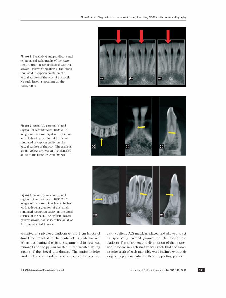
(a) (b) (c)
Figure 3 Axial (a), coronal (b) and
sagittal (c) reconstructed 180º CBCT
images of the lower right central incisor
tooth following creation of the ‘small’
simulated resorption cavity on the
buccal surface of the root. The artificial
lesion (yellow arrows) can be identified
on all of the reconstructed images.
(a) (b) (c)
Figure 2 Parallel (b) and parallax (a and
c), periapical radiographs of the lower
right central incisor (indicated with red
arrows), following creation of the ‘small’
simulated resorption cavity on the
buccal surface of the root of the tooth.
No such lesion is apparent on the
radiographs.
(a) (b) (c)
Figure 4 Axial (a), coronal (b) and
sagittal (c) reconstructed 180º CBCT
images of the lower right lateral incisor
tooth following creation of the ‘small’
simulated resorption cavity on the distal
surface of the root. The artificial lesion
(yellow arrows) can be identified on all of
the reconstructed images.
Durack et al. Diagnosis of external root resorption using CBCT and intraoral radiography
ª 2010 International Endodontic Journal International Endodontic Journal, 44, 136–147, 2011 139

This reduced the amount of up-righting of the data
which would be required using the software. Once the
impression material had set the matrices could be
removed and replaced on the platform ensuring repro-
ducible and accurate relocation of each mandible on
the jig for every scan. Prior to each scan, the acrylic
sheet (Evonik) was secured over the X-ray source using
sellotape (Henkel Consumer Adhesives, Winsford, UK)
in order to attenuate the beam and replicate the soft
tissue scatter encountered in a clinical setting. Expo-
sure parameters of 90 kV, 3.0 mA and a 17.5 s scan
were used for the CBCT scanner when capturing 360º
(a) (b) (c)
Figure 5 Parallel (b) and parallax (a and
c) digital, periapical radiographs of the
lower right central incisor (indicated
with red arrows), following creation of
the ‘large’ simulated resorption cavity on
the buccal surface of the root of the
tooth. The lesion cannot be identified on
the radiographs.
(a) (b) (c)
Figure 7 Axial (a), coronal (b) and sag-
ittal (c) reconstructed 180º CBCT images
of the lower right lateral incisor tooth
following creation of the ‘large’ simu-
lated resorption cavity on the distal
surface of the root. The artificial lesion
(yellow arrows) can be identified on all of
the reconstructed images.
(a) (b) (c)
Figure 6 Axial (a), coronal (b) and
sagittal (c) reconstructed, 180º CBCT
images of the lower right central incisor
tooth following creation of the ‘large’
simulated resorption lesion on the buccal
surface of the root. The artificial lesion
(yellow arrows) can be clearly identified
on all of the reconstructed images.
Diagnosis of external root resorption using CBCT and intraoral radiography Durack et al.
International Endodontic Journal, 44, 136–147, 2011 ª 2010 International Endodontic Journal140
images. The scans exposure time was reduced to 9 s for
the 180º scans. All CBCT data were resliced to produce
0.16 mm slice intervals and 1.2 mm slice thickness.
The brightness and contrast of all images were
optimized to allow the best possible visualization of
the lesions.
Radiological assessment
Eight examiners, comprising three endodontists (n = 3)
and five endodontic postgraduates (n = 5) assessed the
radiographs and CBCT scans in the following sequence
over four sessions: session 1 – radiograph examination,
session 2 – CBCT 180º and 360º examination, session 3
– repeat radiographs and session 4 – repeat CBCT scans.
The repeat sessions (sessions 3 and 4) were carried out
to assess intra-examiner agreement. In sessions 3 and 4,
half of the original images were re-randomized and
shown to the examiners again. There was at least one
week between each session. The radiographs, the 180º
CBCT scans and the 360º CBCT scans were viewed in
separate pre-randomized orders in each session. All
examination sessions were carried out in a quiet,
darkened room. Prior to Sessions 1 and 2 the examiners
were calibrated. The examiners were shown examples of
CBCT images and radiographs of simulated resorption
defects, of various sizes, created on teeth used in pilot
studies. They were trained how to use the CBCT
software and how to adjust the contrast of the digital
periapical radiographs. Each investigator demonstrated
an ability to assess the calibration images proficiently,
prior to examining the experimental images.
The radiographs were viewed on a laptop computer
(Dell Inspiron 1720; Dell, Round Rock, TX, USA) as a
PowerPoint presentation (Microsoft, Seattle, WA, USA).
A parallel radiograph and two parallax views were
provided for each tooth under examination. The tooth
to be examined was arrowed to avoid confusion.
Examiners were asked to note down whether or not
they thought there was an EIR lesion present on
the root of the arrowed tooth. Confidence in their
observation was recorded using a five point scale as
follows: 1 – external resorption lesion definitely not
present, 2 – probably not present, 3 – unsure, 4 –
probably present and 5 – definitely present. The
examiners were also asked to note, using the following
5 point scale, on which surface of the root the visualized
lesion was located: 1 – mesially, 2 – distally, 3 –
buccally, 4 – lingually and 5 – unsure. Finally, the
examiners were asked to indicate the lesion they
visualized, using the PowerPoints ‘ink annotations’
function located in ‘pointer options’. The pointer
created a red dot at a point on the slide of the examiners
discretion. The examiner was asked to directly ‘dot’ any
resorption lesion they could see on the arrowed tooth.
For the CBCT scans the examinerswere presentedwith
the primary reconstructed CBCT data. Prior to examin-
ing the scans, the examiner was tutored on how to
upright the teeth on the scans using the One Volume
Viewer software (JMorita). The examinerwas then asked
to scroll through the axial, coronal and sagittal slices for
each tooth. Again, they were asked to note down
whether or not a lesion was present, how confident they
were about the presence or absence of the lesion, and on
which root surface the lesionwas located, using the same
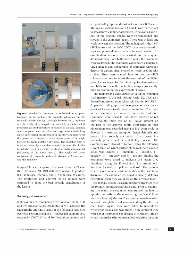
(a)
(b)
a
bc d
f
f
g
h h
d
g
a
bh
che
ji
j
ie
Figure 8. Mandibular specimen (a) embedded in its putty
template (b) to facilitate its accurate relocation on the
rotatable wooden disc (c). The angle between the X-ray beam
and the tooth being imaged is measured by a mathematical
protractor (d) fixed in position in relation to the disc. Stainless
steel wire pointers (e) centred on and perpendicular to the long
axis of each incisor are embedded in the putty and hover over
the protractor to ensure accurate measurement of the angle
between the teeth and the X-ray beam. The phosphor plate (f)
is set in position by a standard anterior intra-oral film holder
(g) which is fixed in a wooden jig (h) designed to ensure exact
positioning of the X-ray tube (i). The acrylic soft tissue
equivalent (j) is securely positioned between the X-ray source
and the mandible.
Durack et al. Diagnosis of external root resorption using CBCT and intraoral radiography
ª 2010 International Endodontic Journal International Endodontic Journal, 44, 136–147, 2011 141

scales and methods as described for the radiograph
examination. When the examiner identified a resorption
lesion on the CBCT scan they were asked to notify the
author and a static image, containing slices in all the
orthogonal planes, which the examiner felt most clearly
demonstrated the resorption,was copied to aPowerPoint
presentation by the author. The examiner was then
asked to mark the resorption lesion as before, in as many
of the planes as s/he could, using the ‘ink annotation’
function. The dotted images were then saved.
Data analysis
The raw data were analysed using Stata# software
(Stata 10, College Station, TX, USA). The sample size of
10 teeth and the use of eight of examiners was
predetermined by reference to previous work (Patel
et al. 2009a). Power calculation using data from this
study indicated that 10 teeth and 8 observers would be
sufficient for a 90% chance of showing a difference of
0.2 in the receiver operator characteristic (ROC) area
under the curve. The diagnostic accuracy of each
imaging system and examiner in the detection of
simulated EIR was assessed using receiver operating
characteristic (ROC) curve analysis to estimate the area
under the ROC curve (Az value). Sensitivity, specificity
and positive and negative predicitive values (PPV) and
(NPV) were determined. Kappa analysis was used to
compare the repeatability of ROC scores for the three
radiological techniques. A further Kappa analysis was
carried out to compare the accuracy of the imaging
techniques in allowing the examiners to identify the
surface on which the resorption lesion was created.
Results
The ROC analysis revealed a lower Az value for digital
periapical radiography (0.584) in the detection of small
EIR lesions when compared with 180º (0.969) and
360º (0.979) CBCT (P < 0.001). The Az value for
intraoral radiography in the detection of larger lesions
was 0.733 and was less than that for 180º (1.000) and
360º (1.000) CBCT (P < 0.001). Regardless of the
lesion size, the overall Az value for intraoral radiogra-
phy was lower (0.665), compared to Az values for 180º
(0.984) and 360º (0.990) CBCT (P < 0.001) (Table 1).
Regardless of lesion size the sensitivity of intraoral
radiography was lower (86.9) compared to 180º (100)
and 360º (100) CBCT (P < 0.001). The specificity of
180º and 360º CBCT was 92.3 and 95.5 respectively.
These results were, again, regardless of lesion size and
were better than the specificity of periapical radiogra-
phy, which was 43.1 (P < 0.001).When comparing ‘no
lesion’ with ‘any lesion’ the PPV for radiographs was
43.1 compared to values of 95.6 for 180º CBCT and
97.5 for 360º CBCT (P < 0.001). CBCT, regardless of the
degree of rotation had NPV of 100 compared to NPVs of
85 for intraoral radiography (P < 0.01) (Table 2).
The kappa values for intra-examiner agreement were
0.292 for intraoral radiography, 0.853 for 180º CBCT
and 0.895 for 360º CBCT (P < 0.001) (Table 3). The
kappa scores for inter-examiner reproducibility were
0.122 for radiographs and 0.639 and 0.672 for 180º
and 360º CBCT respectively (P < 0.001) (Table 4).
Table 1 Comparison of agreement with gold standard
diagnosis [mean (SD) area under the curve from ROC
analysis] for the three radiological techniques, n = 8
Radiograph 180! CBCT 360! CBCT
No lesion vs. both
sizes of lesion
0.665 (0.057) 0.984* (0.013) 0.990* (0.014)
No lesion vs.
small lesion
0.584 (0.051) 0.969* (0.026) 0.979* (0.029)
No lesion vs.
large lesion
0.733 (0.098) 1.000* (0.000) 1.000* (0.000)
*significantly different from radiographs (P < 0.001).
Table 2 Mean (SD) percentage sensitivity, specificity, PPV and
NPV for each radiological technique comparing ‘no lesion’
with ‘any lesion’
Sensitivity Specificity PPV NPV
Radiograph 86.9 (9.3)* 43.1 (4.6)* 43.1 (13.3)* 85 (12.0)!
180! CBCT 100 (0.0) 92.3 (5.5) 95.6 (3.2) 100 (0.0)
360! CBCT 100 (0.0) 95.5 (4.8) 97.5 (2.7) 100 (0.0)
*Significantly different from 180º and 360º CBCT (P < 0.001),!P < 0.01.
Table 3 Comparison of repeatability of ROC scores (mean (SD)
Kappa values) for the three radiological techniques, n = 8
Radiograph 180º CBCT 360º CBCT
Original vs
repeated score
0.292 (0.148) 0.853* (0.151) 0.895* (0.065)
*Significantly different from radiographs (P < 0.001).
Table 4 Kappa values for inter-examiner agreement in read-
ing radiographs and CBCT images
Radiograph 180º CBCT 360º CBCT
Inter-examiner
kappa score
0.122* 0.639 0.672
*Significantly different from 180º and 360º CBCT (P < 0.001).
Diagnosis of external root resorption using CBCT and intraoral radiography Durack et al.
International Endodontic Journal, 44, 136–147, 2011 ª 2010 International Endodontic Journal142

Examiners were also significantly better able to
identify the exact location of the resorption lesions
with CBCT than they were with radiographs
(P < 0.001) (Table 5). There was no significant differ-
ence in the ability of 180º and 360º CBCT to detect
simulated EIR.
Discussion
In this study, simulated EIR cavities were created on
mandibular incisor teeth in dry human mandibles.
Under ideal circumstances human cadavers would
have been used in order to accurately replicate the soft
tissue attenuation and scatter from the X-ray source.
Appropriate specimens, however, were not available for
this study. The X-ray beam for the intraoral radio-
graphs and CBCT scans were attenuated by placing a
1 cm thick acrylic plate (Evonik), as used by Noujeim
et al. (2009), between the X-ray source and the
specimen. It may be argued that a thickness of acrylic
greater than 1 cm would be necessary to reproduce the
absorbtion and scatter created during clinical CBCT
imaging as the X-ray beam encounters the cervical
spine and other areas of the mandible. A pilot study
demonstrated that the images captured using the 1 cm
thick acrylic as a soft tissue equivalent closely mim-
icked corresponding clinical images on patients. How-
ever, this is a subjective assessment.
The intraoral radiographs were captured using a
digital system. This produced a dynamic image allow-
ing the user to alter brightness and contrast easily.
Borg et al. (1998) have shown that there is no
difference between digital [PSP and charged couple
device (CCD)] and film-based radiographs in the detec-
tion of artificially simulated external resorption lesions.
Another study, however, reported that conventional
film and CCD sensors were better at detecting external
resorption than PSP sensors (Kamburoglu et al. 2008).
The authors conceded that the reason for the difference
may have been due to limitations of the specific PSP
system used by them in their study (Orex Digident,
Yokneam, Israel). The PSP system used in this study,
on the other hand, was the latest generation of the one
used by Borg et al. (1998).
The periapical radiographs were viewed as Power-
Point slides. Theoretically transferring digital images to
PowerPoint may affect the diagnostic yield of the
radiographs, but with care is unlikely. Steps were taken
during the original process of saving the images to
minimize any reduction in image quality during the
preparation of the slides. On close inspection, no
subjective difference in image quality was noticed
between the original images and the same images
viewed as PowerPoint slides.
A final CBCT data set is based on a series of combined
individual basis images. Increasing the degree of
rotation of the CBCT scanners’ X-ray source and
detector means an increased number of basis images
(and the acquisition of more data), with a potential
improvement in diagnostic yield. However, with higher
degrees of rotation comes increased radiation exposure
to the patient. Two sets of CBCT scans were taken in
this study, differing only in the degree of rotation of the
X-ray source around the experimental specimen –
either 180º or 360º. This allowed us to assess any
difference in diagnostic yield resulting from the
increased radiation associated with the higher degree
of rotation. The placement of the specimens in specially
created jigs during imaging ensured that all images
were standardized and reproducible.
It would have been desirable to use maxillary
anterior teeth in this experiment. These are the teeth
most frequently subject to luxation (Andreasen &
Vestergaard Pedersen 1985) and exarticulation injuries
(Andreasen et al. 1995a,b,c,d). However, intact human
maxillae with sufficient and appropriate teeth to carry
out the experiment proved impossible to source. Con-
sequently, the resorption was simulated on incisors in
human mandibles. After maxillary incisors these are
the teeth most commonly subject to dental traumatic
injuries (Lenstrup & Skieller 1959, Andreasen &
Hjørting-Hansen 1966a, Andreasen & Vestergaard
Pedersen 1985). The simulated EIR lesions were
machined in the roots using steel rose head burs.
Every effort was made to ensure standardization and
reproducibility of the cavity sizes, using similar tech-
niques to other authors (Andreasen et al. 1987, Hintze
et al. 1992). The simulated external inflammatory root
resorption lesions created in this study were hemi-
spherical. This clearly does not reflect the clinical
reality in which root resorption lesions are irregular in
shape. Future studies involving the use of standardized
simulated resorption lesions of irregular shape are
Table 5 Comparison of accuracy in identifying the affected
surface (mean (SD) kappa values) for the three radiological
techniques, n = 8
Radiograph 180º CBCT 360º CBCT
Gold standard vs
surface score
0.248 (0.092) 0.946* (0.031) 0.964* (0.022)
*Significantly different from radiographs (P < 0.001).
Durack et al. Diagnosis of external root resorption using CBCT and intraoral radiography
ª 2010 International Endodontic Journal International Endodontic Journal, 44, 136–147, 2011 143

necessary. As stated previously, EIR lesions are associ-
ated with destruction of the adjacent bone, represented
radiographically as radiolucencies. Under ideal circum-
stances this boney destruction would have been repli-
cated in the experiment. However, in order to create
access to prepare the boney defects accurately at a
point directly adjacent to the simulated resorption
defects, the intact mandibles would have to be split in a
coronal plane through their sockets. The different
segments would then have to be repositioned prior to
imaging. This would be highly destructive of valuable
human samples, and would inevitably produce radio-
graphic representations of the fracture lines on the
images to be examined. Examination of the root
surfaces would most likely be impaired by this process
and so it was decided not recreate these boney defects.
The results of this study are in agreement with
earlier studies which indicate that intraoral radiogra-
phy is an inadequate method for detecting resorption
defects at an early stage (Andreasen et al. 1987,
Chapnick 1989, Goldberg et al. 1998). Two cavity
sizes were used in this experiment – small (0.5 mm
diameter and 0.25 mm depth) and large (1 mm diam-
eter and 0.5 mm depth). The dimensions of these
cavities were similar to the ‘small’ and ‘medium’ cavity
dimensions, respectively, used in previous studies
(Andreasen et al. 1987, Chapnik et al. 1989, Goldberg
et al. 1998). In the study by Andreasen et al. (1987) no
small resorption defects could be detected using film
based intraoral radiographs despite the fact that the
pre-operative radiograph and multiple angled radio-
graphs of the subject teeth were available to the
examiners. In a similar study by Goldberg et al. (1998)
examiners also failed to diagnose any small resorption
defects using conventional radiography when the pre-
operative radiographs were not available to them for
comparison purposes. In an attempt to overcome the
shortcomings of conventional radiography, the ability
of other imaging systems to detect external root
resorption have been investigated. Digital subtraction
radiology (Kravitz et al. 1990, Hintze et al. 1992) and
tune apertured computed tomography (Nance et al.
2000) have been shown to be better capable of
identifying simulated external root resorption cavities
than conventional intraoral radiography. In addition,
flat panel volume computerized tomography has
potential applications in the detection of external root
resorption at an early stage (Hahn et al. 2009).
Case reports in the literature outlining the usefulness
of CBCT and CT as diagnostic and treatment planning
tools in the diagnosis and management of external
resorption have been restricted to cases of external
cervical resorption (Kim et al. 2003, Cohenca et al.
2007, Patel & Dawood 2007). Clinical studies directly
comparing the ability of intraoral radiographs and
CBCT to detect and diagnose EIR are limited. However,
one clinical study did report that CBCT is superior to
conventional radiography in diagnosing and determin-
ing the extension of non-specific inflammatory resorp-
tion on root surfaces (Estrela et al. 2009). Patel et al.
(2009b), in a further clinical study, compared the
accuracy of conventional intraoral radiography and
CBCT in the diagnosis and management of external
cervical and internal resorption lesions. The authors
reported CBCT to be 100% accurate in the diagnosis of
the presence and type of the root resorption and the
overall sensitivity of intraoral radiographs was lower
than CBCT. Critically, however, the nature of these
clinical studies does not preclude the occurrence of false
negative results. That is, as the root surfaces cannot be
visually examined, it is impossible to rule out the
presence of resorption lesions which may not have been
detected by the imaging systems. For this reason,
ex vivo studies similar to the current one are necessary
to accurately measure and compare the sensitivity of
imaging devices in the detection of EIR.
One ex vivo study (Alqerban et al. 2009) compared
the ability of two CBCT systems and conventional
panoramic radiography to detect simulated external
surface resorption lesions, of varying sizes, associated
with canine impaction. The authors reported that small
and medium field of view CBCT systems were superior
to conventional panoramic radiography in the detec-
tion of simulated external resorption cavities, regardless
of the cavity size. There was no statistical difference
between the diagnostic ability of the CBCT systems.
However, conventional panoramic radiography is
rarely utilized in endodontic specific investigations.
Intraoral radiography is currently the imaging tech-
nique of choice when assessing traumatically injured
permanent teeth which may develop EIR (Flores et al.
2007a,b). The current study directly compares the
ability of CBCT and digital periapical radiography to
detect early EIR lesions under experimental conditions.
In that regard, to our knowledge, this study is unique.
The selection of teeth with pre-experimental intact
surfaces ensured that the experimental resorption
cavities were the only ones on the root surfaces.
In this study some small lesions were identified on
periapical radiographs. The radiographs had a poor Az
value of 0.584 for the detection of small cavities in the
ROC analysis. This contrasts starkly with the Az values
Diagnosis of external root resorption using CBCT and intraoral radiography Durack et al.
International Endodontic Journal, 44, 136–147, 2011 ª 2010 International Endodontic Journal144
approaching 1.000 for CBCT. The specificity of peri-
apical radiography was poor. Part of the reason for this
may have been due to the possible ‘expectation’ of some
examiners that they should visualize simulated resorp-
tion defects as they were aware that this is what they
were looking for. A contributory factor may also have
been the difference in the patterns of boney trabeculae
between mandibles. In one mandible in particular the
superimposition of boney trabeculae on the roots of
some teeth seemed to mimic the experimental defects.
In a clinical setting the examiners may have been more
confident that what they were viewing was a normal
trabecular pattern as they would not necessarily have
been expecting to see resorptive defects. Nevertheless
the same ‘expectations’ would have applied when the
examiners viewed the CBCT images. The results clearly
demonstrate that CBCT has excellent sensitivity and
specificity in the detection of simulated external
resorption lesions, and that CBCT performs significantly
better than digital intraoral radiography in this regard.
This is due to the fact that CBCT provides information
in the third dimension, coupled with the fact that
images can be reconstructed such that overlying noise
is eliminated.
The intra-examiner agreement for periapical radio-
graphs in this study was poor. This suggests that
intraoral periapical radiography is an unreliable meth-
od of detecting simulated early EIR. It is likely that the
anatomical noise and the compression of three-dimen-
sional anatomical structures associated with the peri-
apical radiographs played a significant part. The noise
created by the trabecular patterns was, again, likely to
have confused examiners leading to uncertainty when
attempting to detect simulated resorption defects. The
intra-examiner kappa scores for CBCT, however, were
very good and undoubtedly were due to the fact that
this imaging technique overcomes many of the short-
comings of conventional radiographic imaging.
The inter-examiner agreement was significantly
better with CBCT compared to digital radiography.
When taken together the inter and intra examiner
kappa scores obtained here provide evidence of the
validity and reliability of CBCT for detecting EIR defects.
The reliability and validity of intraoral radiography is
poor, in comparison.
The effective radiation dose to patients when using
CBCT is higher than digital radiography and any
benefit to the patient of the CBCT scan should outweigh
any potential risks of the procedure in order to be
justified (Farman & Farman 2005). The radiation
should be as low as reasonably achievable (ALARA).
This study does demonstrate that when all other
exposure parameters are the same, limited volume
CBCT scans with 360º of rotation are no better at
detecting EIR than the same scans taken with 180º of
rotation, and as such cannot be justified in a clinical
setting.
Given the financial and emotional impact that
premature tooth loss can have on individuals and
given the evidence presented here and in other similar
literature, a re-thinking on how traumatized teeth are
radiographically assessed and followed up is warranted.
Nevertheless, radiological imaging is only one of a
number of diagnostic aids used in dentistry. In clinical
situations in which a definitive decision can be made
about the pulpal status of a tooth based on sensibility
testing, clinical signs and symptoms and conventional
radiographic imaging, endodontic treatment will be
indicated without the need for further CBCT imaging.
Regardless of the type of ERR, CBCT imaging can only
be justified clinically in situations where the possible
information it provides will impact directly on treat-
ment.
Conclusion
The results of this study highlight the limitations of
intraoral periapical radiography in the detection of
simulated EIR. CBCT overcomes these shortcomings
and provides a reliable and valid method of detecting
artificially created EIR defects. Earlier detection of the
resorption process in ‘at risk’ individuals may lead to
more prompt and appropriate treatment, ultimately
resulting in improved prognoses for affected teeth.
References
Alqerban A, Jacobs R, Souza PC, Willems G (2009) In-vitro
comparison of 2 cone-beam computed tomography systems
and panoramic imaging for detecting simulated canine
impaction-induced external root resorption in maxillary
lateral incisors. American Journal of Orthodontics and Dento-
facial Orthopedics 136, 764e1–e11.
Andreasen JO (1981) Relationship between surface and
inflammatory root resorption and changes in the pulp after
replantation of permanent incisors in monkeys. Journal of
Endodontics 7, 294–301.
Andreasen JO, Andreasen FM (2007) Avulsions. In: Andrea-
sen JO, Andreasen FM, Andersson L, eds. Textbook and Colour
Atlas of Traumatic Injuries to the Teeth, 4th edn. Chapter 17.
Oxford: Blackwell Munksgaard, pp. 444–88.
Andreasen JO, Hjørting-Hansen E (1966a) Replantation of
teeth. I. Radiographic and clinical study of 110 human teeth
Durack et al. Diagnosis of external root resorption using CBCT and intraoral radiography
ª 2010 International Endodontic Journal International Endodontic Journal, 44, 136–147, 2011 145

replanted after accidental loss. Acta Odontoligica Scandinavica
24, 263–86.
Andreasen JO, Hjørting-Hansen E (1966b) Replantation of
teeth. II. Histological study of 22 replanted anterior teeth in
humans. Acta Odontoligica Scandinavica 24, 287–306.
Andreasen FM, Vestergaard Pedersen B (1985) Prognosis of
luxated permanent teeth – the development of pulp necrosis.
Endodontics and Dental Traumatology 1, 207–20.
Andreasen FM, Sewerin I, Mandel U, Andreasen JO (1987)
Radiographic assessment of simulated root resorption cav-
ities. Endodontics and Dental Traumatology 3, 21–7.
Andreasen JO, Borum MK, Jacobsen HL, Andreasen FM
(1995a) Replantation of 400 avulsed permanent incisors.
4. Factors related to periodontal ligament healing. Endodon-
tics and Dental Traumatology 11, 76–89.
Andreasen JO, Borum MK, Jacobsen HL, Andreasen FM
(1995b) Replantation of 400 avulsed permanent incisors.
1. Diagnosis of healing complications. Endodontics and Dental
Traumatology 11, 51–8.
Andreasen JO, Borum MK, Jacobsen HL, Andreasen FM
(1995c) Replantation of 400 avulsed permanent incisors.
2. Factors related to pulpal healing. Endodontics and Dental
Traumatology 11, 59–68.
Andreasen JO, Borum MK, Andreasen FM (1995d) Replanta-
tion of 400 avulsed permanent incisors. 3. Factors related to
root growth. Endodontics and Dental Traumatology 11, 69–
75.
Bender IB, Seltzer S (1961) Roentgenographic and direct
observation of experimental lesions in bone: I. Journal of the
American Dental Association 62, 152–60.
Borg E, Kallqvist A, Grondahl K, Grondahl H-G (1998) Film
and digital radiography for detection of simulated root
resorption cavities. Oral Surgery, Oral Medicine, Oral Pathol-
ogy, Oral Radiolology and Endododontology 86, 110–4.
Chapnick L (1989) External root resorption: an experimental
radiographic evaluation. Oral Surgery Oral Medicine Oral
Patholology 67, 578–82.
Cohenca N, Simon JH, Mathur A, Malfaz JM (2007) Clinical
indications for digital imaging in dento-alveolar trauma.
Part 2: root resorption. Dental Traumatology 23, 105–13.
Cvek M (1973) Treatment of non-vital permanent incisors
with calcium hydroxide. II. Effect on external root resorption
in luxated teeth compared with effect of root filling with
gutta percha. Odontologisk Revy 24, 343–54.
Cvek M (1992) Prognosis of luxated non-vital maxillary
incisors treated with calcium hydroxide and filled with gutta
percha. Endodontics and Dental Traumatology 8, 45–55.
Cvek M (2007) Endodontic management and the use of
calcium hydroxide in traumatized permanent teeth. In:
Andreasen JO, Andreasen FM, Andersson L, eds. Textbook
and Colour Atlas of Traumatic Injuries to the Teeth, 4th edn.
Chapter 22. Oxford: Blackwell Munksgaard, pp. 598–657.
Dumsha T, Hovland EJ (1995) Evaluation of long-term
calcium hydroxide treatment in avulsed teeth – an in vivo
study. International Endodontic Journal 28, 7–11.
Estrela C, Reis Bueno M, Alencar AHG et al. (2009) Method to
evaluate inflammatory root resorption by using cone beam
computed tomography. Journal of Endodontics 35, 1491–7.
Farman AG, Farman TT (2005) A comparison of 18 different
X-ray detectors currently used in dentistry. Oral Surgery,
Oral Medicine, Oral Pathology, Oral Radiology and Endodon-
tology 99, 485–9.
Flores MT, Andersson L, Andreasen JO et al. (2007a) Guide-
lines for the management of traumatic dental injuries. I.
Fractures and luxations of permanent teeth. Dental Trauma-
tology 23, 66–71.
Flores MT, Andersson L, Andreasen JO et al. (2007b) Guide-
lines for the management of traumatic dental injuries. I.
Avulsion of permanent teeth. Dental Traumatology 23, 130–
6.
Goldberg F, De Silvio A, Dreyer C (1998) Radiographic
assessment of simulated external root resorption cavities in
maxillary incisors. Endodontics and Dental Traumatology 14,
133–6.
Grondahl H-G, Huumonen S (2004) Radiographic manifesta-
tions of periapical inflammatory lesions. Endodontic Topics 8,
55–67.
Hahn W, Fricke-Zech S, Fricke J et al. (2009) Detection and
size differentiation of simulated tooth root defects using flat-
panel volume computerized tomography (fpVCT). Oral
Surgery, Oral Medicine, Oral Pathology, Oral Radiology and
Endodontology 107, 272–8.
Hammarstrom L, Lindskog S (1985) General morphological
aspects of resorption of teeth and alveolar bone. International
Endodontic Journal 18, 93–108.
Hintze H, Wenzel A, Andreasen FM, Sewerin I (1992) Digital
subtraction radiography for assessment of simulated root
resorption cavities. Performance of conventional and
reverse contrast modes. Endodontics and Dental Traumatology
8, 149–54.
Kamburoglu K, Tsesis I, Kfir A, Kaffe I (2008) Diagnosis of
artificially induced external root resorption using conven-
tional intraoral film radiography, CCD, and PSP: an ex vivo
study. Oral Surgery, Oral Medicine, Oral Pathology, Oral
Radiology & Endodontology 106, 885–91.
Kim E, Kim K-D, Roh B-D, Cho Y-S, Lee S-J (2003) Computed
tomography as a diagnostic aid for extracanal invasive
resorption. Journal of Endodontics 29, 463–5.
Kravitz LH, Tyndall DA, Bagnell CP, Dove SB (1990)
Assessment of external root resorption using digital
subtraction radiography. Journal of Endodontics 18, 275–
84.
Kundel HL, Revesz G (1976) Lesion conspicuity, structured
noise, and film reader error. American Journal of Roentgen-
ology 126, 1233–8.
Lenstrup K, Skieller V (1959) A follow up study of teeth
replanted after accidental loss. Acta Odontologica Scandinavia
17, 503–9.
Liedke GS, Silveira HED, Silveira HLD, Dutra V, Figueiredo JAP
(2009) Influence of voxel size in the diagnostic ability of
Diagnosis of external root resorption using CBCT and intraoral radiography Durack et al.
International Endodontic Journal, 44, 136–147, 2011 ª 2010 International Endodontic Journal146

cone beam tomography to evaluate simulated external root
resorption. Journal of Endodontics 35, 233–5.
Nance RS, Tyndall D, Levin LG, Trope M (2000) Diagnosis of
external root resorption using TACT (tuned-aperture com-
puted tomography). Endodontics and Dental Traumatology 16,
24–8.
Noujeim M, Prihoda TJ, Langlais R, Nummikoski P (2009)
Evaluation of high-resolution cone beam computed tomog-
raphy in the detection of simulated interradicular bone
lesions. Dentomaxillofacial Radiology 38, 156–62.
Patel S, Dawood A (2007) The use of cone beam computed
tomography in the management of external cervical
resorption lesions. International Endodontic Journal 40,
730–7.
Patel S, Dawood A, Mannocci F, Wilson R, Pitt Ford T (2009a)
Detection of periapical bone defects in human jaws using
cone beam computed tomography and intraoral radiogra-
phy. International Endodontic Journal 42, 507–15.
Patel S, Dawood A, Wilson R, Horner K, Mannocci F (2009b)
The detection and management of root resorption lesions
using intraoral radiography and cone beam computed
tomography – an in vivo investigation. International End-
odontic Journal 42, 831–8.
Revesz G, Kundel HL, Graber MA (1974) The influence of
structured noise on the detection of radiologic abnormali-
ties. Investigative Radiology 6, 479–86.
Silveira HL, Silveira HE, Liedke GS, Lermen CA, Santos RB,
Figueiredo JAP (2007) Diagnostic ability of computed
tomography to evaluate external root resorption in vitro.
Dentomaxillofacial Radiology 36, 393–6.
Tronstad L (1988) Root resorption – etiology, terminology and
clinical manifestations. Endodontics and Dental Traumatology
4, 241–52.
Trope M (2002) Root resorption due to dental trauma.
Endodontic Topics 1, 79–100.
Webber RL, Messura JK (1999) An in vivo comparison of
diagnostic information obtained from tuned-aperture com-
puted tomography and conventional dental radiographic
imaging modalities. Oral Surgery, Oral Medicine, Oral Pathol-
ogy, Oral Radiology and Endodontology 88, 239–47.
Durack et al. Diagnosis of external root resorption using CBCT and intraoral radiography
ª 2010 International Endodontic Journal International Endodontic Journal, 44, 136–147, 2011 147