diagnostic tests and laboratory values - study with … tests and laboratory values ... tilt-table...
TRANSCRIPT
Diagnostic Tests and
Laboratory Values
CLPNA Self-Study Course
2017
CLPNA Diagnostic Tests & Laboratory Values – P a g e | i
Acknowledgements
he development of this resource guide is an initiative of the College of Licensed Practical Nurses of
Alberta (CLPNA). Production of this professional development initiative has been made possible through
a grant from Alberta Labour, Foreign Qualification Recognition branch.
Content and Review
Dr. John Collins has attained broad experience in the fields of nursing and education. He has worked as an
RN and an RPN in clinical practice, administration, research, and education. As an individual who has
completed undergraduate, graduate, and postgraduate studies, John values the principle of lifelong learning
and encourages others to follow this path with a view to providing excellence in client care.
Jason Richmond is an advanced-care paramedic with experience in health care, education, and curriculum
development. He is currently pursuing a master of education in distance education. Jason was the founding
chairperson of the Continuing Education Centre for Emergency Services, which continues to provide free
continuing education. Further, he is a strong advocate for integrated practice between health professions
and open education.
Editing of this module was done by Heather Buzila, who has broad editorial experience that includes
educational materials and fiction and nonfiction manuscripts.
Programming and Production
The design and programming of this course was done by Russell Sawchuk of Steppingstones Partnership,
Inc., and Learning Nurse Resources Network.
Legal and Copyright
This self-study course is intended to support the continuing education of Alberta’s Licensed Practical Nurses.
This course is intended as a refresher and is not a substitute for proper accreditation or training. Always
follow your employer’s proper policies and procedures.
© College of Licensed Practical Nurses of Alberta, 2017
Published by the College of Licensed Practical Nurse of Alberta St. Albert Trail Place 13163 – 146 Street Edmonton, Alberta T5L 4S8 Tel: 780-484-8886 Website: www.clpna.com Email: [email protected]
T
CLPNA Diagnostic Tests & Laboratory Values – P a g e | ii
Diagnostic Tests and Laboratory Values
Table of Contents
Introduction to Diagnostic Tests and Laboratory Values ......................................................1
Module 1: Diagnostic Procedures ........................................................................................2
Introduction ........................................................................................................................................ 2
Angiography ........................................................................................................................................ 3
Arthroscopy ........................................................................................................................................ 4
Bone scan ............................................................................................................................................ 5
Bronchoscopy ..................................................................................................................................... 6
Computed Tomography (CT Scan or CAT Scan) .................................................................................. 7
Cystoscopy .......................................................................................................................................... 8
Doppler studies ................................................................................................................................... 9
Echocardiography ............................................................................................................................. 10
Electrocardiogram (ECG, EKG) .......................................................................................................... 11
Electroencephalogram (EEG) ............................................................................................................ 12
Electromyography (EMG) .................................................................................................................. 13
Endoscopy ......................................................................................................................................... 14
Magnetic Resonance Imaging (MRI) ................................................................................................. 15
Myelography .................................................................................................................................... 16
Pulmonary Function Tests (PFT) ....................................................................................................... 17
Radiography (X-Ray) Chest, Abdomen ............................................................................................. 18
Thallium Scan .................................................................................................................................... 20
Tilt-table Test .................................................................................................................................... 21
Transesophageal Echocardiogram (TEE) .......................................................................................... 22
Ultrasound (Sonography) ................................................................................................................. 23
Summary............................................................................................................................................ 24
Module 2: Application of Nursing Knowledge to Diagnostic Tests ....................................... 25
Introduction ...................................................................................................................................... 25
Scenarios ........................................................................................................................................... 25
Case Studies ...................................................................................................................................... 26
Answers ............................................................................................................................................ 27

CLPNA Diagnostic Tests & Laboratory Values – P a g e | iii
Table of Contents
Module 3. Overview of Laboratory Tests .......................................................................... 31
Introduction ...................................................................................................................................... 31
Common Lab Tests ........................................................................................................................... 32
CHEMISTRY TESTS .................................................................................................................. 33
Albumin (AL) ............................................................................................................................... 33
Ammonia (AMM, NH3) .............................................................................................................. 34
Amylase (AMY) ........................................................................................................................... 35
Aspartate Transaminase (AST) ................................................................................................... 36
Blood Urea Nitrogen (BUN) ........................................................................................................ 37
Chloride (Cl) ................................................................................................................................ 38
Creatine Kinase (CK) ................................................................................................................... 39
Creatinine (Cr) ............................................................................................................................. 40
D-dimer (DDIMER) ...................................................................................................................... 41
Glucose (GLU) ............................................................................................................................. 42
Ischemia-Modified Albumin (IMA) ............................................................................................. 43
Lactate Dehydrogenase (LDH) ................................................................................................... 44
Potassium (K) ............................................................................................................................. 46
Sodium (Na) ............................................................................................................................... 47
Troponins (cTnT & cTnI) ............................................................................................................. 48
Summary .................................................................................................................................... 48
HEMATOLOGY TESTS .............................................................................................................. 49
Complete Blood Count (CBC, Diff) .................................................................................................... 49
Components of Complete Blood Count (CBC) ........................................................................... 49
Hemoglobin (Hgb) ...................................................................................................................... 51
Hematocrit or Packed Cell Volume (PCV) ................................................................................... 51
Myoglobin (Mb) ......................................................................................................................... 52
Natriuretic Peptides: BNP/or NT-proBNT .................................................................................. 53
Platelet Count (Thrombocytes) .................................................................................................. 54
White Blood Cell Count (WBC) ................................................................................................... 55
White Blood Cell Types (WBC Differential) ................................................................................ 56
Summary .................................................................................................................................... 57
Coagulation Studies .......................................................................................................................... 58
Prothrombin Time (PT) .............................................................................................................. 58
Partial Thromboplastin Time (PTT) ............................................................................................ 59
International Normalized Ratio (INR) ........................................................................................ 60

CLPNA Diagnostic Tests & Laboratory Values – P a g e | iv
Table of Contents Diabetes Studies ............................................................................................................................... 61
Fasting Plasma Glucose Test (FBS) ............................................................................................. 61
Oral Glucose Tolerance Test (OGTT) .......................................................................................... 61
Hemoglobin A1C (HbA1c) .......................................................................................................... 62
URINE TESTS .......................................................................................................................... 63
Urinalysis (UA) ............................................................................................................................ 63
Urine C & S (Culture & Sensitivity) ............................................................................................. 64
Urine Toxicology ........................................................................................................................ 65
Summary ........................................................................................................................................... 65
Module 4. Nursing Implications and Applications of Lab Values ......................................... 66
Introduction ...................................................................................................................................... 66
Questions / Case Study ..................................................................................................................... 66
Self-Assessment ................................................................................................................................ 68
Summary ........................................................................................................................................... 68
Endnotes ........................................................................................................................................... 69
Comments
This resource is intended for Licensed Practical Nurses and adds to competency in nursing practice and
the safety of clients. It is recommended that the LPN refer to CLPNA’s Competency Profile for LPNs:
http://www.clpna.com/members/continuing-competency-program/competency-profile-for-lpns/ for
additional information regarding their scope of practice.
Additional Comments
The information found here is useful for the LPN’s role as a caregiver and educator (explaining various lab
tests, the collection of specimens, and preparing clients for diagnostic testing). Further reading or access
to additional resources for training purposes is recommended. Some resources are posted at the end of
this course.
CLPNA Diagnostic Tests & Laboratory Values – P a g e | 1
Introduction to Diagnostic Tests & Laboratory Values
icensed Practical Nurses (LPNs) are responsible for providing safe, competent, and quality care to clients.
It is an expectation of practice that they will critically appraise all assessment data to arrive at the best
nursing care for each client. This course provides information and learning activities related to diagnostic
tests and laboratory (lab) values, one source of client assessment data. An LPN should understand the
rationale for conducting specific diagnostic and laboratory tests when delivering care, as well as the
implications of the results of those tests. The course lists common diagnostic and lab tests and values in an
organized manner, with opportunities for participants to test their knowledge through short case studies
and interactive quizzes.
Purpose
According to Alberta’s LPN Competency Profile, an entry-level LPN meets the knowledge requirement
for obtaining, assessing, and monitoring diagnostic tests and lab values commonly used in health care.1 This
course is intended to review and extend an LPN’s knowledge pertaining to diagnostic tests and lab values so
that safe and timely care may be delivered to clients.
Course Outcomes
On completion of this course participants will
identify and describe commonly used diagnostic procedures and lab tests;
describe commonly ordered diagnostic procedures and lab values;
recognize principles of client teaching to the preparation and management of clients undergoing
diagnostic and lab testing;
differentiate normal from abnormal results; and
implement appropriate action as it relates to results to maintain a safe, competent, and ethical care
to clients.
Course Outline
This course consists of four modules:
Module 1: Introduction to and overview of diagnostic tests. This section gives the learner a brief
overview of commonly ordered diagnostic tests used in a variety of settings where an LPN provides
client care.
Module 2: Nursing implications for diagnostic tests. This module encourages the learner to apply
knowledge of diagnostic tests through responding to nursing case studies.
Module 3: Overview of lab values. The topics discussed in this section focus on values limited to the
most commonly ordered chemistry, hematology, and urine tests.
Module 4: Nursing implications for lab tests. This section provides an opportunity to integrate
information from Module 3 to check the LPN’s competence through interactive online quizzes.
L
CLPNA Diagnostic Tests & Laboratory Values – P a g e | 2
Module 1: Diagnostic Procedures
Introduction
iagnostic testing enables health care providers to diagnose, monitor, and treat conditions or anticipate
changes in the health statuses of individuals. LPNs are involved in both direct and indirect care of
clients where diagnostic procedures are anticipated (long-term care, maternity, pediatrics, community and
public health, medical and surgical units in hospitals). Regardless of the nursing environment, client teaching
is vital to encourage collaboration with procedures and facilitate obtaining the most accurate results. The
LPN is proactive in assisting clients undergoing diagnostic testing.
The following diagnostic tests will be discussed in this module:
Angiography
Arthroscopy
Bone scan
Bronchoscopy
Computed tomography (CT scan or CAT scan)
Cystoscopy
Doppler studies
Echocardiography
Electrocardiogram (ECG, EKG)
Electroencephalogram (EEG)
Electromyography (EMG)
Endoscopy
Magnetic resonance imaging (MRI)
Myelography
Pulmonary function tests (PFT)
Radiography (X-ray) chest, abdomen
Thallium scan
Tilt-table test
Transesophageal echocardiogram (TEE)
Ultrasound (Sonography)
Module Outcomes
Upon completion of this module, the participant will be able to
list commonly used diagnostic tests;
identify the indications for various diagnostic tests;
explain the procedure(s) for various diagnostic tests;
recognize interfering factors in diagnostic testing; and
state the nursing implications of diagnostic tests.
Nice to Know…
This module uses some terminology that you should familiarize yourself with. For example, normal
findings refer to diagnostic results that are within expected or typical ranges. Indications refer to the
reasons for obtaining diagnostic studies (e.g., establish a diagnosis, monitor therapy, screen for disease).
Procedure relates to the preparation of the client before the test and care of the client during and after
the test. Interfering factors refers to those factors that could influence or alter test results (e.g.,
medications). Nursing implications refer to those aspects of care that are within the role of the LPN,
including the need for client teaching.
D
CLPNA Diagnostic Tests & Laboratory Values – P a g e | 3
Angiography
Description2
n X-ray test that uses a special dye and camera (fluoroscopy) to take pictures of the blood flow in an
artery (e.g., aorta) or a vein (e.g., vena cava). Common angiograms can look at arteries close to the
heart, lungs, brain, head or neck, legs or arms, and the aorta.3
Indications
To find the cause of chest pain or pressure (e.g., from myocardial infarction, angina, or pericarditis).
Procedure
1. A thin tube (catheter) is placed into a blood vessel in the groin or wrist (femoral or radial artery or
vein) or just above the elbow (brachial artery or vein) and guided to the specific area.
2. An iodine dye is injected into the vessel to make the area show clearly on X-ray pictures.
Interfering Factors
Movement during the filming can distort the X-ray picture.
Nursing Implications
Make sure the consent form is signed.
Explain the procedure to the client and provide support.
Instruct the client to be NPO for 8–12 hours before the test (check local policies).
Discontinue anticoagulants before the test.
Monitor for excessive bleeding post-test.
Record vital signs pre- and post-test.
Adjust vascular closure devices.
A

CLPNA Diagnostic Tests & Laboratory Values – P a g e | 4
Arthroscopy
Description4, 5
This test is used to visually examine the interior of a joint with a specially designed fibre-optic endoscope.
Arthroscopy permits concurrent surgery or biopsy using a technique called triangulation, in which
instruments are passed through a separate cannula.
Indications
To reveal a torn meniscus, chondromalacia, dislocation, subluxation, fracture, and degenerative
articular cartilage.
To assess torn anterior cruciate or tibial collateral ligaments.
To diagnose Baker cysts and ganglion cysts, synovitis, and rheumatoid and degenerative arthritis.
To detect foreign bodies associated with gout, pseudogout, and osteochondromatosis.
Procedure
1. The surgeon anesthetizes the joint, makes a small incision, and passes a cannula through the incision
and positions it in the joint cavity.
2. He or she inserts the arthroscope through the cannula and examines the knee structure, taking
photographs for further study.
3. After the procedure, the arthroscope is removed, the joint is irrigated, and an adhesive strip and
compression bandage are applied to the site.
Interfering Factors
None reported.
Nursing Implications
Make sure the consent form is signed.
Instruct the client to fast after midnight before the procedure.
Shave the area 13 cm above and below the joint.
Watch for fever and swelling, increased pain, and localized inflammation at the incision site.
Administer analgesics as ordered.
Monitor client’s circulation and sensation in the leg.
Instruct the client to report fever, bleeding, drainage, or increased joint swelling or pain.
Tell the client that showering is permitted after 48 hours, but baths should be avoided until the postoperative visit.
Tell the client that he or she may resume his or her usual diet following the procedure.
CLPNA Diagnostic Tests & Laboratory Values – P a g e | 5
Bone Scan
Description6, 7
A bone scan permits imaging of the skeleton using a scanning camera after intravenous injection of a
radioactive tracer compound.
Indications
To help detect bone cancer.
To diagnose bone trauma associated with pathological fractures and infection.
To help stage cancer.
To monitor degenerative bone disorders.
Procedure
1. The client is injected with a radioactive tracer compound that collects in bone tissue in increased
concentrations at sites of abnormal metabolism.
2. When scanned, these sites appear as hot spots that are commonly detectable months before
radiography can reveal a lesion.
3. As the scanner head moves over the body, it detects low-level radiation emitted by the skeleton and
translates this into a chart to produce a two-dimensional picture of the scanned area.
Interfering Factors
A distended bladder may obscure pelvic detail.
Nursing Implications
Make sure the consent form is signed.
Advise the client to drink lots of fluids in the interval between injection and actual scanning (about
one to three hours).
Instruct the client to void immediately before the procedure.
The client may have to be repositioned several times to obtain adequate views.
Check injection site for redness and swelling.
After the procedure, instruct the client to drink lots of fluids and to empty bladder frequently for the
next 24–48 hours.
Provide analgesics, as needed, for pain resulting from positioning on the scanning table.

CLPNA Diagnostic Tests & Laboratory Values – P a g e | 6
Bronchoscopy
Description8
The direct inspection of the larynx, trachea, and bronchi through a flexible bronchoscope. The scope has a
lens with a light at its distal end.
Indications
To detect and remove foreign bodies and secretions.
To inspect the larynx, trachea, and bronchus for lesions.
Procedure
1. The test is conducted while the client is lying supine or in Semi-Fowler’s position with
head hyperextended.
2. The bronchoscope will be inserted through the client’s nose or mouth.
Interfering Factors
Improper labelling of specimens.
Nursing Implications
Make sure the consent form is signed.
Explain the procedure to the client, and provide support.
Check with the department regarding NPO status.
Administer pre-medications as ordered (e.g., atropine to dry the mouth).
Take vital signs pre- and post-test.
Advise the client that the procedure takes approximately one hour.
Recognize complications post-test: laryngeal edema, bronchospasm, pneumothorax, and possible
bleeding from the biopsy site.
Check for hemoptysis and the client’s gag reflex before giving food or liquids.

CLPNA Diagnostic Tests & Laboratory Values – P a g e | 7
Computed Tomography (CT Scan or CAT Scan)
Description9, 10
Pictures of the body are taken using radiation and computer-enhanced imaging.
Indications
To screen for coronary artery disease, head, liver, and renal lesions, tumours, edema, metastatic
disease, vascular diseases, and bone destruction.
Procedure
1. Consent form must be signed.
2. Medications can be taken up to two hours before the test (check with radiology department).
3. If contrast dye is used, usually NPO before test (eight hours before test if morning appointment). For
afternoon scheduling, NPO after full liquid breakfast (check with radiology department).
Interfering Factors
Presence of dentures, hairpins, jewellery for CT head.
Presence of barium (an enema may be ordered).
Nursing Implications
Explain the procedure to the client, and provide reassurance.
Ensure IV site is patent before the test.
Obtain a client history of allergies to contrast dye (or allergies to seafood or iodine).
Advise the client that if contrast dye is used, a warm, flushed sensation may be felt in the face or
body.
Inform the client that the test may take approximately 30 minutes to one hour to complete.

CLPNA Diagnostic Tests & Laboratory Values – P a g e | 8
Cystoscopy
Description11, 12
The direct visualization of the bladder wall and urethra using a cystoscope (lighted telescopic lens). Usually
performed by a urologist.
Indications
To detect renal calculi and renal tumours.
To remove renal stones.
To determine the cause of UTI, dysuria, and hematuria.
Procedure
1. The test is conducted under local anesthesia (inserted into the urethra) or general anesthesia.
2. Check with the specialist regarding NPO status before the test.
3. The client lays flat with legs and feet in stirrups.
4. The scope is entered through the urethra, and a sterile solution is slowly inserted to fill the bladder,
making it easier to visualize.
Interfering Factors
None reported.
Nursing Implications
Make sure the consent form is signed.
Explain the procedure to the client, and provide support.
Offer sedation up to an hour before the test.
Assess urinary patterns (amount, colour, odour).
Take vital signs pre-and post-test.
Inform the client that there may be some pressure or burning discomfort during and post-test.
CLPNA Diagnostic Tests & Laboratory Values – P a g e | 9
Doppler Studies
Description13, 14
Doppler ultrasonography evaluates blood flow in the major blood vessels of the arms and legs and within
the extracranial cerebrovascular system.
Indications
To measure systolic pressure, which helps detect the presence, location, and extent of peripheral
arterial occlusive disease.
To detect abnormal carotid blood flow.
To monitor clients after arterial reconstruction and bypass grafts.
Procedure
1. A handheld transducer directs high-frequency sound waves to the artery or vein being tested.
2. The sound waves strike moving red blood cells and are reflected back to the transducer at
frequencies that correspond to blood-flow velocity through the vessel.
3. The transducer then amplifies the sound waves to permit direct listening and graphic recording of
blood flow.
Interfering Factors
None reported.
Nursing Implications
Make sure the consent form is signed.
Explain the procedure to the client, and provide support.
Tell client that he or she will be asked to move arms to different positions and to perform breathing
exercises as measurements are taken.
Apply water-soluble conductive jelly to the tip of the transducer to provide coupling between the
skin and the transducer.
After the procedure, remove the conductive jelly from the skin.
Take vital signs pre-and post-test.
CLPNA Diagnostic Tests & Laboratory Values – P a g e | 10
Echocardiography
Description15, 16
Produces an audio-visual representation of the heart and its blood flow by recording sound waves that are
bounced off the organ.
Indications
To evaluate cardiac structures and function.
To measure cardiac output (volume of blood).
To identify the cause of abnormal heart sounds.
To assess the damage to muscle, dysfunction of valves, and abnormality of blood flow.
To evaluate myocardial disease.
Procedure
1. Performed with the client positioned on his or her left side.
2. Acoustic gel applied to the skin over the chest.
3. Transducer moved over chest and upper abdomen to obtain images.
Interfering Factors
Dressings and scarring of the chest may adversely affect results.
Nursing Implications
Explain the procedure to the client, and provide support.
Inform the client that the test usually takes 15 to 45 minutes to complete.

CLPNA Diagnostic Tests & Laboratory Values – P a g e | 11
Electrocardiogram (ECG, EKG)
Description17, 18
A test that checks for problems with electrical activity
of the heart. It shows this electrical activity as
tracings on paper/screen (called waves).
Indications
To find the cause of chest pain or pressure
(e.g., from myocardial infarction, angina, or
pericarditis).
To find the cause of symptoms related to
cardiac health.
To determine the effectiveness of medications and check on implanted devices (pacemaker).
Procedure
1. Performed with client lying in a supine position.
2. Certain areas on the arms, legs, and chest may be cleaned and shaved to improve electrode
adhesion.
3. Several electrodes are attached to the skin on each arm, leg, and chest; these electrodes are
attached to a machine that traces heart activity onto a paper.
Interfering Factors
Electrodes not securely adhered to the skin.
Moving and talking during the test.
Nursing Implications
Explain the procedure to the client, and provide support.
Assist in removing all jewellery from the neck, arms, and wrists.
Inform the client that tests usually take 5 to 10 minutes to complete.
CLPNA Diagnostic Tests & Laboratory Values – P a g e | 12
Electroencephalogram (EEG)
Description19
A test that measures and records the electrical activity of the brain.
Indications
To check for epilepsy.
To check for problems with loss of consciousness or dementia.
To watch brain activity and physical problems in the brain, spinal cord, or nervous system.
Procedure
1. May be performed while client is awake, drowsy, asleep, or a combination of these.
2. Special sensors called electrodes are attached to the head.
3. These leads extend onto wires to a computer.
4. Computer records the brain’s electrical activity.
Interfering Factors
Drugs (e.g., sedatives, barbiturates, anticonvulsants, and tranquilizers).
Alcohol.
Oily hair and hairspray.
Nursing Implications
Make sure the consent form is signed.
Explain procedure to the client, and provide support.
Advise client of NPO status up to 12 hours before the test.
Ensure hair is clean and avoid oils, creams, or lotions on client’s hair/head.
If EEG is conducted under little or no sleep, plan to have client get a ride home after the test.

CLPNA Diagnostic Tests & Laboratory Values – P a g e | 13
Electromyography (EMG)
Description20, 21
EMG is the recording of electrical activity of selected skeletal muscle groups at rest and during voluntary
contraction.
Indications
To diagnose neuromuscular disorders.
To assess spinal nerve disorders.
Procedure
1. A needle electrode is inserted percutaneously into a muscle.
2. The muscle’s electrical discharge (or motor unit potential) is then measure and displayed on an
oscilloscope screen.
Interfering Factors
Drugs that affect myoneural junctions, such as cholinergics, anticholinergics, and skeletal muscle
relaxants, will interfere with EMG results.
Nursing Implications
Make sure the consent form is signed.
Explain the procedure to the client, and provide support.
Restrict cigarettes, coffee, tea, and cola for two to three hours before the test.
Check for a history of medications that may interfere with test results.
If client experiences residual pain, apply warm compresses and administer analgesics as ordered.

CLPNA Diagnostic Tests & Laboratory Values – P a g e | 14
Endoscopy
Description22
A flexible fibre-optic scope is inserted directly into an organ or cavity of the body. This test is performed by a
gastroenterologist.
Indications
To visualize the internal structures of the esophagus, stomach, and duodenum.
To obtain a cytology specimen; biopsy forceps or a cytology brush can also be inserted through the
endoscope.
Procedure
1. This test is conducted under local anesthesia (of the throat) and IV sedation.
2. Check with specialist regarding NPO status before the test (varies but should be at least two hours
to clear the stomach).
Interfering Factors
Barium from previous GI testing can interfere with visualization of the mucosa.
Nursing Implications
Make sure the consent form is signed.
Explain the procedure to the client, and provide support.
Dentures, eyeglasses, jewellery, and clothing should be removed from the neck to the waist.
Encourage the client to void before the test.
Record pre-and post-test vital signs.
Check that gag reflex has returned post-test before offering food and fluids.
CLPNA Diagnostic Tests & Laboratory Values – P a g e | 15
Magnetic Resonance Imaging (MRI)
Description23, 24
MRI scanners use strong magnetic fields and radio waves to create images of the body.
Indications
To define the structure of internal organs and to detect edema, infarction, CNS problems, blood flow
or hemorrhage, injury, and tumours.
To stage disease in the spine, head, heart, bone/joints, and abdomen.
Procedure
1. The client must lie still on a narrow table with a cylinder-type scanner around the body area being
scanned.
2. The procedure takes approximately 45 minutes to an hour to perform.
Interfering Factors
Movement during the procedure can distort images.
Metal in the body could cause critical injury to the client.
Nursing Implications
Consent form must be signed.
Check facility policy and procedures for specific instructions for food and fluid restrictions.
Remove all jewellery, including watches, glasses, hairpins, and any metal objects.
Ask the client about any concerns related to claustrophobia; relaxation techniques or sedative might
be used.
Explain the procedure, and ascertain if the client has any metal prosthetics/shrapnel/wires left in
their body, as tissue damage may result.
CLPNA Diagnostic Tests & Laboratory Values – P a g e | 16
Myelography
Description25, 26
Myelography combines fluoroscopy and radiography to evaluate the subarachnoid space after injection of a
contrast medium. The contrast medium should flow freely through the subarachnoid space, showing no
obstruction or structural abnormalities.
Indications
To locate a spinal lesion, ruptured disk, spinal stenosis, or abscess.
To detect arachnoiditis, spinal nerve root injury, and skull tumours.
Procedure
1. Because the contrast medium is heavier than CSF, it will flow through the subarachnoid space to the
dependent area when the client, lying prone on a fluoroscopic table, is tilted up or down.
2. The fluoroscope allows visualization of the flow of the contrast medium and the outline of the
subarachnoid space.
3. X-rays are taken for a permanent record.
Interfering Factors
None reported.
Nursing Implications
Make sure the consent form is signed.
Instruct the client to restrict food and fluids for eight hours before the test.
Check for hypersensitivity to iodine and iodine-containing substances, contrast media, and drugs
associated with the procedure.
If the client received a water-based contrast medium, elevate the head of the bed 30 to 45 degrees
for six to eight hours.
Monitor vital signs, neurologic status, and intake and output.
Encourage the client to drink extra fluids.
If fever, back pain, or signs of meningeal irritation (headache, irritability, or neck stiffness) develop,
keep the room quiet and dark, and provide an analgesic or antipyretic as ordered.
Client may resume usual diet and activities the day after the test.
CLPNA Diagnostic Tests & Laboratory Values – P a g e | 17
Pulmonary Function Tests (PFT)
Description27
This test checks how well the lungs are functioning. In PFTs, several tests are conducted (e.g., spirometry, gas
diffusion, inhalation challenge test, and exercise stress test).
Indications
To investigate lung problems (COPD, asthma).
To measure the severity of lung disease.
To check to determine what impact the treatment for lung disease is having.
Procedures
Spirometry: The most common lung function test. It measures how much and how quickly air is
inhaled and exhaled from the lungs. The client breathes into a mouthpiece attached to a machine,
which displays these measures.
Gas diffusion test: Measures the amount of oxygen and other gases that move through the lungs’
air sacs (alveoli) per minute. This test indicates how well gases are being absorbed into the blood
from the lungs.
Inhalation challenge: Used to measure how the airways respond to substances that may be causing
asthma or wheezing. The client inhales increasing amounts of a substance through a nebulizer.
Spirometry readings are taken to evaluate lung function before, during, and after inhaling the
substance.
Exercise stress test: Looks at how exercise affects the lungs. Spirometry readings are
performed before, during, and after exercise and again at rest.
Interfering Factors
The use of bronchodilators before the PFT may return false results.
Lack of client cooperation or misunderstanding instructions.
Nursing Implications
Make sure the consent form is signed.
Explain the procedure to the client, and provide support
Advise the client not to smoke for at least four hours before the test.
Assess for signs and symptoms of respiratory distress.
Record vital signs pre-and post-test.
CLPNA Diagnostic Tests & Laboratory Values – P a g e | 18
Radiography (X-Ray) Chest, Abdomen
Description28, 29, 30
Radiography involves the use of X-rays to produce images related to bone structure and tissue in the body.
X-ray beams pass through the body and are absorbed in different amounts, depending on the density of the
material (e.g., bone has high density, causing light images or white structures to be produced). These
examinations are completed quickly, and the test itself does not cause the client pain.
X-Ray Chest
Indications
To evaluate pulmonary or cardiac disease and trauma to the chest.
To determine the location of endotracheal tubes, chest tubes, feeding tubes, or subclavian
catheters.
To follow the progress of disease (e.g., TB).
Procedure
1. Clothing removed to the waist, and the client is ideally in an upright position.
2. Client should take a deep breath and hold it during the X-ray procedure.
3. Food and fluids are not restricted.
Interfering Factors
Incorrect positioning and inability to hold breath can affect image quality.
Nursing Implications
Explain the purpose and procedure, and reassure the client.
Remove all jewellery and metal objects from the chest area.
Inform the client that the procedure takes approximately 10–15 minutes to complete.
CLPNA Diagnostic Tests & Laboratory Values – P a g e | 19
X-Ray Abdomen
Indications
To identify abdominal masses of the stomach, bowel obstruction, abdominal tissue trauma, ascites.
Procedure
1. Food and fluids are not restricted.
2. Clothes are removed to uncover the abdomen; the client lies in the supine position on the X-ray
table.
3. Inform the client that the procedure takes approximately 15–20 minutes.
Interfering Factors
Incorrect positioning of the client could produce distorted images.
Obesity and ascites may distort images.
Nursing Implications
Client aftercare consists of monitoring symptoms and providing reassurance.
CLPNA Diagnostic Tests & Laboratory Values – P a g e | 20
Thallium Scan
Description31, 32
This test evaluates blood flow after an intravenous injection of the radioisotope thallium-201 or cardiolyte.
Areas with poor blood flow and ischemic cells fail to take up the isotope and appear as cold spots on the scan.
Indications
To assess the location and extent of an MI.
To evaluate graft patency.
To assess the effectiveness of antianginal therapy or balloon angioplasty.
Procedures
Resting imaging: Within the first few hours of MI symptoms, the client receives an injection of
thallium. Scanning begins after 10 minutes, with the client positioned in anterior, left anterior
oblique, and left lateral positions.
Stress imaging: The client, wired with electrodes, walks on the treadmill at a regulated pace that is
gradually increased while his ECG, blood pressure and heart rate are monitored. When client reach
peak stress, thallium is injected into the antecubital vein. The client exercises an additional 45 to 60
seconds to permit circulation and uptake of the isotope and then lies on his back under the camera.
Scanning begins after 10 minutes, with the client in anterior, left anterior oblique, and left lateral
positions. Additional scans may be taken after rests or after 24 hours.
Interfering Factors
Cold spots may result from sarcoidosis, myocardial fibrosis, cardiac contusion, attenuation caused
by soft tissue and artifacts (diaphragm, implants, breast or electrodes), apical cleft, or coronary
spasm.
Absence of cold spots in a client with coronary artery disease may result from an insignificant
obstruction, inadequate stress, delayed imaging, sing-vessel disease, or collateral circulation.
Nursing Implications
Make sure the consent form is signed.
Explain the procedure to the client, and provide support.
For stress imaging, no alcohol, tobacco, and non-prescription medications for 24 hours and nothing by mouth after midnight.
For stress imaging, instruct the client to wear walking shoes during the treadmill exercise and report fatigue, pain, or shortness of breath immediately.
Monitor client during testing for critical signs such as pale, clammy skin, confusion, or staggering.
CLPNA Diagnostic Tests & Laboratory Values – P a g e | 21
Tilt-Table Test
Description33, 34
This test is a provocative method used to diagnose vasodepressor syncope.
Indications
To diagnose tachyarrhythmia, overmedication for hypertension or heart diseases, hyper-reactive
vagal activity, and various forms of vasomotor instability.
Procedure
1. Individuals with vasomotor syncope syndrome usually demonstrate symptomatic hypotension and
syncope within a few to 30 minutes of being tilted upright by approximately 60 to 90 degrees.
2. Normally, in the titled position, systolic blood pressure drops minimally, diastolic blood pressure
rises, and heart rate increases.
3. In clients with vasodepressor syncope, these changes are exaggerated, and they become
lightheaded and dizzy on assuming the tilted position.
Interfering Factors
Clients with dehydration or hypovolemia show similar changes in blood pressure and heart rate.
Clients taking antihypertensive medications or diuretics also may demonstrate similar changes when
placed in a tilted position.
Nursing Implications
Make sure the consent form is signed.
Explain the procedure to the client, and provide support.
Ask whether the client had an excessive fluid loss in the previous 24 hours.
Record any antihypertensive or diuretic medicines the client may be taking.
Position client supine on a horizontal tilt table.
Obtain blood pressure and pulse rate as baseline values.
Ask about the presence of dizziness and light-headedness.
Monitor vital signs as client adjusts to positioning changes.

CLPNA Diagnostic Tests & Laboratory Values – P a g e | 22
Transesophageal Echocardiogram (TEE)
Description35, 36
In this test, ultrasonography is combined with endoscopy to provide a better view of the heart’s structures.
Indications
To reveal thoracic and aortic disorders, endocarditis, congenital heart disease, intracardiac thrombi,
and tumours.
To evaluate valvular disease and repairs.
Procedure
1. A small transducer is attached to the end of a gastroscope and inserted into the esophagus, allowing
images to be taken from the posterior aspect of the heart. This causes less tissue penetration and
interference from chest-wall structures and produces high-quality images of the thoracic aorta.
Interfering Factors
Inability of the client to cooperate or remain still may impair clear imaging.
Improper adjustment of the equipment to accommodate obese or thin clients may impair clear
imaging.
Clients with chronic obstructive pulmonary disease and those on mechanical ventilators are not
good candidates because excess air in the lungs impedes movement of ultrasound waves.
Nursing Implications
Make sure the consent form is signed.
Explain the procedure to the client, and provide support.
Advise the client to fast for six hours before the test.
Have client remove dentures or oral prostheses.
Have resuscitation equipment, including suction apparatus, available.
Connect the client to monitors so that blood pressure, heart rate, and pulse oximetry can be
monitored.
After the test, keep the client in a supine position until the sedative wears off.
Encourage the client to cough after the procedure while lying on side or sitting upright.
No food or water until gag reflex returns.
CLPNA Diagnostic Tests & Laboratory Values – P a g e | 23
Ultrasound (Sonography)
Description37, 38
Imaging test used to visualize soft-tissue organs, blood vessels, and tissues using high-frequency sound
waves (no radiation).
Indications
To evaluate the size, structure, and position of body organs.
To evaluate the blood flow in arteries and veins.
To detect cysts, tumours, and calculi.
Procedure
1. The client’s position may vary from supine to oblique, prone, semi-recumbent, and erect.
2. Conductive gel is applied to the skin surface at the site to be examined.
3. The transducer is hand-held and is moved smoothly back and forth across the skin.
Interfering Factors
Air and gas will not transmit the ultrasound beam.
The ultrasound must be performed before barium studies as barium will interfere with image
quality.
Dressings and scar tissue interfere with the ultrasound.
Nursing Implications
Make sure the consent form is signed.
Explain the procedure to the client, and provide support.
Check with the department regarding specific dietary requirements or NPO status.
Administer pre-medications as ordered (e.g., enema for the prostate exam).
Advise the client that the procedure takes approximately 30 minutes to one hour.

CLPNA Diagnostic Tests & Laboratory Values – P a g e | 24
Did You Know?
There are restrictions on disclosing tests results to clients. In Canada, three laws protect the privacy and
highly sensitive data of health records: the Privacy Act, the Personal Health Information Act, and the
Personal Information Protection and Electronic Documents Act. It is recommended that LPNs familiarize
themselves with these documents.39
Summary
his module has provided an overview of the most commonly ordered diagnostic tests. The LPN has a
vital role to play in ensuring accurate results for these procedures through client teaching, effective
preparation both before and after testing, and interpreting and reporting test results. Areas of practice
differ in some details and the roles that LPNs play. The LPN should always refer to the facility’s policies and
procedures for diagnostic testing for clarification of these matters.
T
CLPNA Diagnostic Tests & Laboratory Values – P a g e | 25
Module 2: Application of Nursing Knowledge to Diagnostic Tests
Introduction
his module is intended to have the learner engage in the application of the content provided in Module
1. Case studies are provided where the learner can respond to questions and then check their responses
against the answers given. This case study work offers the opportunity for self-assessment of knowledge. In
addition to the case studies, some short answer questions are included below.
Module Outcomes
Upon completion of this module, participants will be able to
identify the indications for the use of diagnostic tests;
explain various test procedures;
verbalize client teaching before/during/after diagnostic procedures; and
recognize complications of diagnostic testing and suggest appropriate interventions.
Complete the following questions and case studies. The answers may be found at the end of this module.
Scenarios
Scenario A
A client who is 2.5 months pregnant is being evaluated for pancreatitis. An ultrasound examination of her
gallbladder and pancreas has been ordered. The client is concerned that the test may harm her unborn
child.
1. Ultrasound may harm the unborn child through radiation delivered by the device and should be advised against.
a. True b. False
2. The client does not understand how the ultrasound can examine her gallbladder and pancreas.
What information can the nurse give the client to inform her about the test?
a. This test involves the use of X-rays to produce images related to bone structure and tissue in the body.
b. This test is used to visualize soft-tissue organs, blood vessels, and tissues using high-frequency sound waves.
c. This test involves the direct visualization of the bladder wall and urethra with the use of a cystoscope (lighted telescopic lens). Usually performed by a urologist.
d. This test evaluates blood flow after an intravenous injection of the radioisotope thallium-201 or cardiolyte. Areas with poor blood flow and ischemic cells fail to take up the isotope and thus appear as cold spots on the scan.
T
CLPNA Diagnostic Tests & Laboratory Values – P a g e | 26
3. What outcomes are possible from the ultrasound test?
a. Evaluate the size, structure, and position of body organs. b. Evaluate the blood flow in arteries and veins. c. Detect cysts, tumours, and calculi. d. All of the above
Scenario B
A client is being evaluated for lung surgery. Pulmonary function tests (PFT) are ordered. The client has an order for Ventolin for bronchospasm.
1. This drug should be given to optimize the results of the test.
a. True b. False
2. What measures can the PFT provide?
a) Spirometry: how much and how quickly air is inhaled and exhaled from the lungs. b) Gas diffusion test: measures the amount of oxygen and other gases that move through the
lungs’ air sacs (alveoli) per minute. c) Inhalation challenge: used to measure how the airways respond to substances that may be
causing asthma or wheezing. d) Exercise stress test: looks at how exercise affects the lungs. e) A and C f) B and D g) All of the above
Case Studies
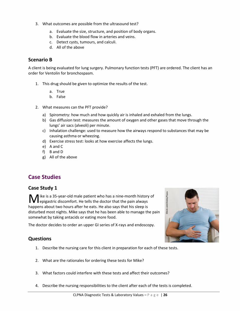
Case Study 1
ike is a 35-year-old male patient who has a nine-month history of epigastric discomfort. He tells the doctor that the pain always
happens about two hours after he eats. He also says that his sleep is disturbed most nights. Mike says that he has been able to manage the pain somewhat by taking antacids or eating more food.
The doctor decides to order an upper GI series of X-rays and endoscopy.
Questions
1. Describe the nursing care for this client in preparation for each of these tests.
2. What are the rationales for ordering these tests for Mike?
3. What factors could interfere with these tests and affect their outcomes?
4. Describe the nursing responsibilities to the client after each of the tests is completed.
M
CLPNA Diagnostic Tests & Laboratory Values – P a g e | 27
Case Study 2
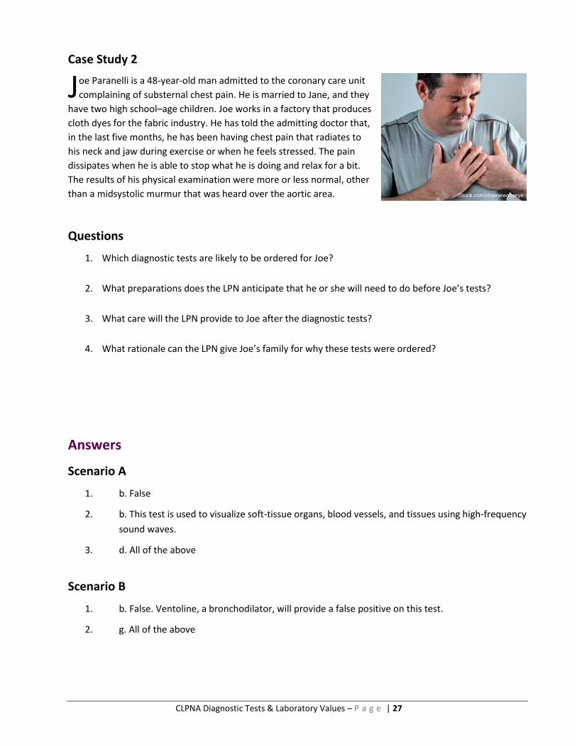
oe Paranelli is a 48-year-old man admitted to the coronary care unit
complaining of substernal chest pain. He is married to Jane, and they
have two high school–age children. Joe works in a factory that produces
cloth dyes for the fabric industry. He has told the admitting doctor that,
in the last five months, he has been having chest pain that radiates to
his neck and jaw during exercise or when he feels stressed. The pain
dissipates when he is able to stop what he is doing and relax for a bit.
The results of his physical examination were more or less normal, other
than a midsystolic murmur that was heard over the aortic area.
Questions
1. Which diagnostic tests are likely to be ordered for Joe?
2. What preparations does the LPN anticipate that he or she will need to do before Joe’s tests?
3. What care will the LPN provide to Joe after the diagnostic tests?
4. What rationale can the LPN give Joe’s family for why these tests were ordered?
Answers
Scenario A
1. b. False
2. b. This test is used to visualize soft-tissue organs, blood vessels, and tissues using high-frequency
sound waves.
3. d. All of the above
Scenario B
1. b. False. Ventoline, a bronchodilator, will provide a false positive on this test.
2. g. All of the above
J
CLPNA Diagnostic Tests & Laboratory Values – P a g e | 28
Answers
Case Study 1
Question 1: Describe the nursing care for this client in preparation for each of these tests.
X-rays
Clothes are removed to uncover the abdomen.
Instruct the client to lie in a supine position on the X-ray table.
Inform the client that the procedure takes approximately 15–20 minutes to complete.
Endoscopy
Provide client and family teaching.
Have the client sign the consent form.
Remove dentures, eyeglasses, jewellery, and clothing from the neck to the waist.
Encourage the client to void before the test.
Record pre-test vital signs.
Question 2: What are the rationales for ordering these tests for Mike?
X-rays
Study consists of a series of X-ray films of the lower esophagus, stomach, and duodenum using
barium contrast medium. The purpose of this exam is to detect ulcers, tumours, inflammations.
Endoscopy
Enables direct visualization of the upper GI tract using a long fiber-optic scope. The esophagus,
stomach, and duodenum are examined for tumours, varices, mucosal inflammations, hiatal
hernias, polyps, ulcers, and obstruction.
Question 3: What factors could interfere with these tests and affect their outcomes?
X-rays
Incorrect positioning of the client could produce distorted images.
Obesity and ascites may distort images.
Endoscopy
Barium from previous GI testing can interfere with visualization of the mucosa.

CLPNA Diagnostic Tests & Laboratory Values – P a g e | 29
Question 4: Describe the nursing responsibilities to the client after each of the tests is completed.
X-rays
Client aftercare consists of monitoring symptoms and providing reassurance.
Endoscopy
Record post-test vital signs.
Check gag reflex post-test before offering food and fluids.
Case Study 2
Question 1: Which diagnostic tests are likely to be ordered for Joe?
EKG, chest X-ray, exercise stress test, echocardiography, cardiac catheterization.
Question 2: What preparations does the LPN anticipate that he or she will need to do before Joe’s
tests?
EKG
Explain the procedure to the client, and provide support.
Assist in removing all jewellery from the neck, arms, and wrists.
Inform the client that tests usually take 5–10 minutes to complete.
Chest X-ray
Explain the purpose and procedure, and reassure the client.
Remove all jewellery and metal objects from the chest area.
Inform the client that the procedure takes approximately 10–15 minutes to complete.
Exercise stress test
Have the client sign the consent form.
Explain the procedure to the client, and provide support.
Advise the client not to smoke for at least four hours before the test.
Assess for signs and symptoms of respiratory distress.
Record vital signs pre-and post-test.
Echocardiography
Explain the procedure to the client, and provide support.
Inform the client that tests usually take 15–45 minutes to complete.

CLPNA Diagnostic Tests & Laboratory Values – P a g e | 30
Cardiac catheterization
Explain the procedure to the client, and provide support.
Check blood pressure and pulse.
Encourage the client to void before the test.
Assist client in removing all jewellery.
Inform the client that cardiac catheterization usually takes 30 minutes to 1 hour to
complete.
Question 3: What care will the LPN provide to Joe after the diagnostic tests?
EKG
If EKG is conducted under little or no sleep, plan to have the client get a ride after the
test.
Exercise stress test
Record vital signs post-test.
Cardiac catheterization
Monitor patient’s vital signs after procedure.
Question 4: What rationale can the LPN give Joe’s family for why these tests were ordered?
To assess cardiovascular functioning and injury, determine what is going on, and design a
treatment plan.

CLPNA Diagnostic Tests & Laboratory Values – P a g e | 31
Module 3: Overview of Laboratory Tests (Chemistry, Hematology, and Common Urine Tests)
Introduction
aboratory (lab) tests aid in evaluating clients’ health needs and provide health care providers with vital
information. A health practitioner may request lab tests to screen for disease, evaluate drug therapy,
confirm a diagnosis, or rule out a clinical problem. This module is intended to give the LPN a foundational
knowledge of commonly ordered laboratory tests.
The outcomes of lab tests affect clinical decision-making as well as client assessment and revisions to client
care plans. Interpretation of lab data is vital to safe, efficient, and competent care by the LPN. The LPN’s role
in infection control measures, collection of specimens, preparing clients for various tests, and teaching can
encourage collaboration with procedures and facilitate obtaining the most accurate results.
Module Outcomes
Upon completion of this module, the participant will be able to
define and describe the purpose of common laboratory tests;
identify the normal range values for common laboratory tests;
understand the interfering factors that may affect test results;
describe critical values and report findings appropriately; and
state the implications of laboratory test results for client health and nursing interventions.
Did You Know?
There is specific terminology related to lab tests that you should familiarize yourself with:
Normal Range vs. Reference Range
Established by testing a large population of healthy individuals. There is usually a range of “normal.”
Medical data must be interpreted in context (e.g., average heart rate is 60, but in a runner, 55 may be OK).
Reference range may vary with age and gender (e.g., creatinine tends to be higher in males).
Critical Values
These values alert the LPN to take prompt clinical intervention.
Critical results are dangerously abnormal. Must be reported to the responsible person, and the lab usually makes note of that.40
Post-Test Considerations
Certain laboratory tests will require the LPN to do follow-up health assessments and/or to restrict food and fluids until the gag reflex returns.
L
CLPNA Diagnostic Tests & Laboratory Values – P a g e | 32
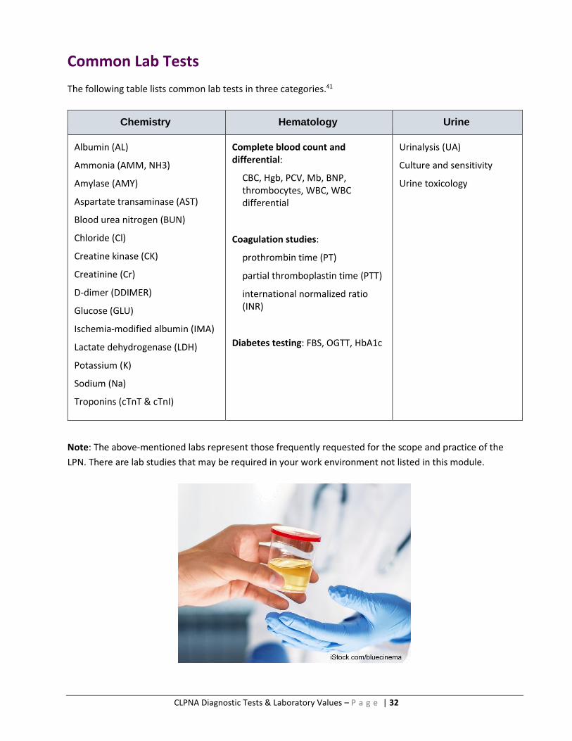
Common Lab Tests
The following table lists common lab tests in three categories.41
Chemistry Hematology Urine
Albumin (AL)
Ammonia (AMM, NH3)
Amylase (AMY)
Aspartate transaminase (AST)
Blood urea nitrogen (BUN)
Chloride (Cl)
Creatine kinase (CK)
Creatinine (Cr)
D-dimer (DDIMER)
Glucose (GLU)
Ischemia-modified albumin (IMA)
Lactate dehydrogenase (LDH)
Potassium (K)
Sodium (Na)
Troponins (cTnT & cTnI)
Complete blood count and differential:
CBC, Hgb, PCV, Mb, BNP, thrombocytes, WBC, WBC differential
Coagulation studies:
prothrombin time (PT)
partial thromboplastin time (PTT)
international normalized ratio (INR)
Diabetes testing: FBS, OGTT, HbA1c
Urinalysis (UA)
Culture and sensitivity
Urine toxicology
Note: The above-mentioned labs represent those frequently requested for the scope and practice of the
LPN. There are lab studies that may be required in your work environment not listed in this module.

CLPNA Diagnostic Tests & Laboratory Values – P a g e | 33
CHEMISTRY TESTS
ody chemistry is complex. There are many chemical elements in a human body that are finely balanced
to produce particular functions and processes, and maintain homeostasis. In the following tables, key
information of relevance to nurses is provided for each chemical element.
Albumin (AL)
Normal Range 35–50 g/L *
Indications Used to diagnose and monitor diseases of the liver, impaired nutrition,
chronic edema, and cancer.
Test Explanation Component of total serum protein (pre-albumin, albumin, and globulins).
Albumin is a protein that is made in the liver. Albumin maintains colloidal
osmotic pressure.
Purpose Used to check liver and kidney function.
Find out if diet contains enough protein.
Helps determine cause of edema to extremities, abdomen.
Interfering Factors Drugs that interrupt normal serum electrophoretic patterns (e.g., Aspirin,
bicarbonates, corticosteroids, salicylates).
Results and
Significance
Lower
Indicative of malnutrition
Liver disease
Ascites
Inflammatory disease
Autoimmune disorder
GI malabsorption syndromes
Higher
Severe dehydration
* All lab values are shown in SI format. If you need a refresher on SI units, you should take the SI Units of Measurement quiz in the Supplementary Practice Quizzes section of this online course. To ensure accuracy and to maintain optimal level of care for individuals receiving any lab tests or specimen collection, please refer to your workplace policies and procedures manual or contact the laboratory.
B

CLPNA Diagnostic Tests & Laboratory Values – P a g e | 34
Ammonia (AMM, NH3)
Normal Range 6–47 mcmol/L
Indications Used to diagnose severe liver diseases.
Test Explanation Measures the amount of ammonia in the blood. Ammonia in the body is
created by the breakdown of protein. The liver converts ammonia to urea,
which is excreted as urine in the kidneys.
Purpose Check liver function.
Check success of treatment for severe liver disease (e.g., cirrhosis).
Check levels in a person receiving total parenteral nutrition.
Interfering Factors Smoking.
Eating high-protein or low-protein diet.
Medications that increase blood ammonia (e.g., Loop diuretics and thiazides).
Strenuous exercise before test.
Results and
Significance
Lower
Essential or malignant hypertension
Higher
Liver disease (cirrhosis or hepatitis)
Reye syndrome
Heart failure
Kidney failure
Severe bleeding from stomach or
intestines
CLPNA Diagnostic Tests & Laboratory Values – P a g e | 35
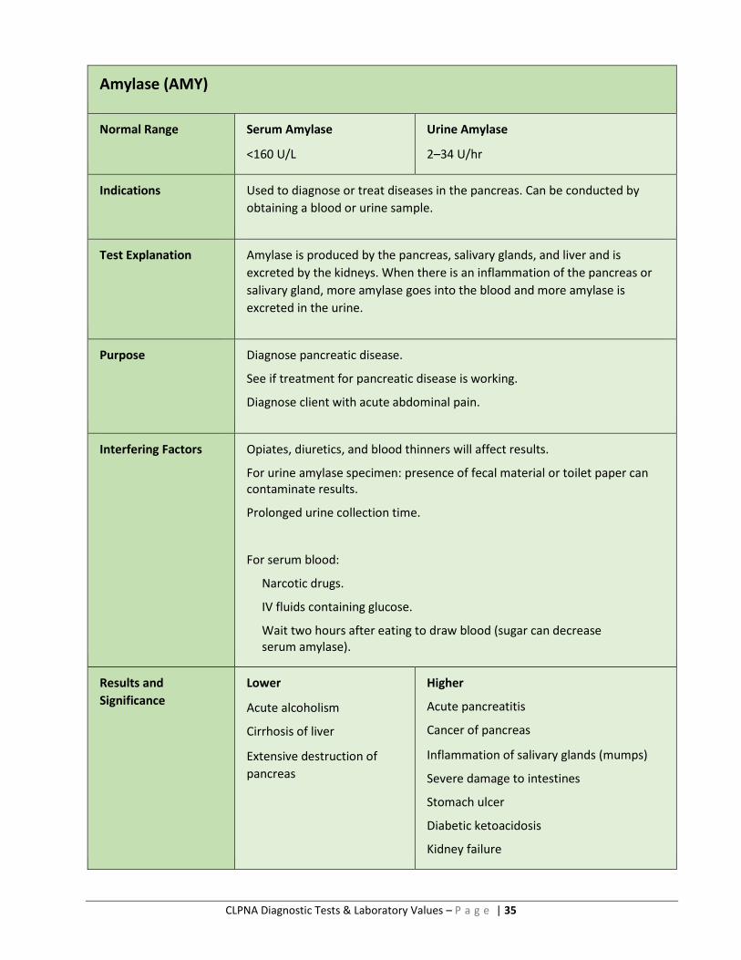
Amylase (AMY)
Normal Range Serum Amylase
<160 U/L
Urine Amylase
2–34 U/hr
Indications Used to diagnose or treat diseases in the pancreas. Can be conducted by
obtaining a blood or urine sample.
Test Explanation Amylase is produced by the pancreas, salivary glands, and liver and is
excreted by the kidneys. When there is an inflammation of the pancreas or
salivary gland, more amylase goes into the blood and more amylase is
excreted in the urine.
Purpose Diagnose pancreatic disease.
See if treatment for pancreatic disease is working.
Diagnose client with acute abdominal pain.
Interfering Factors Opiates, diuretics, and blood thinners will affect results.
For urine amylase specimen: presence of fecal material or toilet paper can contaminate results.
Prolonged urine collection time.
For serum blood:
Narcotic drugs.
IV fluids containing glucose.
Wait two hours after eating to draw blood (sugar can decrease serum amylase).
Results and
Significance
Lower
Acute alcoholism
Cirrhosis of liver
Extensive destruction of
pancreas
Higher
Acute pancreatitis
Cancer of pancreas
Inflammation of salivary glands (mumps)
Severe damage to intestines
Stomach ulcer
Diabetic ketoacidosis
Kidney failure

CLPNA Diagnostic Tests & Laboratory Values – P a g e | 36
Aspartate Transaminase (AST)
Normal Range Adult: 0–35 U/L
Indications This test is used in the evaluation of clients with suspected hepatocellular
diseases.
Test Explanation This enzyme is found in very high concentrations with highly metabolic
tissue, such as the heart muscle, liver cells, skeletal muscle cells, and, to a
lesser degree, in the kidneys, pancreas, and red blood cells. When disease or
injury affects the cells of these tissues, the cells lyse. AST is released and
picked up by the blood, and the serum level rises. The amount of AST
elevation is related directly to the number of cells affected by the disease or
injury. The degree of elevation depends on the length of time between the
injury and when the blood is collected.
Purpose Used to detect diseases such as acute hepatitis, gallstones, cirrhosis, liver
congestion, metastatic tumour of the liver, infectious mononucleosis, acute
pancreatitis, acute renal disease, musculoskeletal diseases or trauma.
Interfering Factors Pregnancy can cause decreased AST levels.
Exercise may increase AST levels.
Levels decreased by liver disease, uremia, or diabetic ketoacidosis.
Some drugs may cause increases in AST (e.g., antihypertensives, cholinergic agents, anticoagulants, contraceptives, opiates, and statins).
Results and
Significance
Lower
Acute renal disease
Beriberi
Diabetic ketoacidosis
Pregnancy
Chronic renal dialysis
Higher
Liver diseases such as hepatitis, hepatic
cirrhosis, drug-induced liver injury,
hepatic metastasis, mononucleosis
Skeletal muscle diseases such as muscle
trauma, surgery, burns, muscular
dystrophy, heat stroke
Other diseases such as acute hemolytic anemia and acute pancreatitis
Adapted from Pagana, K. D., and T. J. Pagana. Mosby’s Canadian Manual of Diagnostic and Laboratory Tests. 1st ed. Toronto, ON: Elsevier Canada, 2013.
CLPNA Diagnostic Tests & Laboratory Values – P a g e | 37
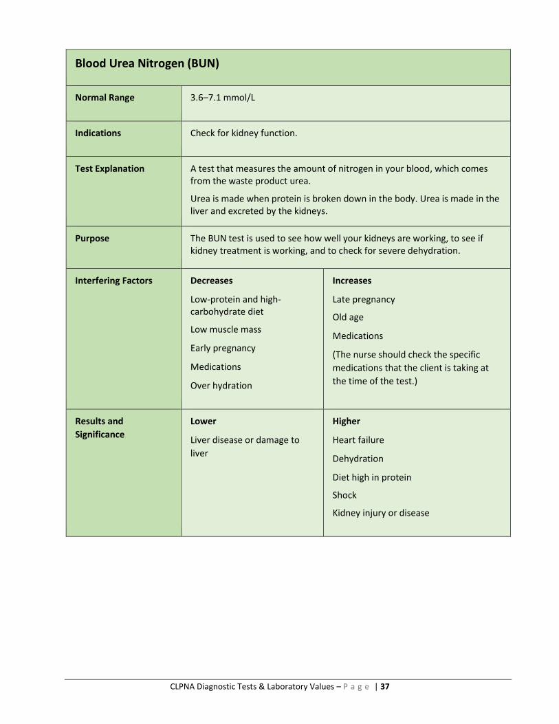
Blood Urea Nitrogen (BUN)
Normal Range 3.6–7.1 mmol/L
Indications Check for kidney function.
Test Explanation A test that measures the amount of nitrogen in your blood, which comes from the waste product urea.
Urea is made when protein is broken down in the body. Urea is made in the liver and excreted by the kidneys.
Purpose The BUN test is used to see how well your kidneys are working, to see if kidney treatment is working, and to check for severe dehydration.
Interfering Factors Decreases
Low-protein and high-carbohydrate diet
Low muscle mass
Early pregnancy
Medications
Over hydration
Increases
Late pregnancy
Old age
Medications
(The nurse should check the specific
medications that the client is taking at
the time of the test.)
Results and
Significance
Lower
Liver disease or damage to
liver
Higher
Heart failure
Dehydration
Diet high in protein
Shock
Kidney injury or disease

CLPNA Diagnostic Tests & Laboratory Values – P a g e | 38
Chloride (Cl)
Normal Range 96–106 mmol/L
Indications This test is usually included as one element in the test for electrolytes.
In conjunction with the other elements, chloride can provide an indication
of acid-base balance and hydration status.
Test Explanation Chloride’s purpose is to maintain water balance in the body and acid-base balance.
Purpose To check the chloride level in relation to potassium, sodium, and
bicarbonate balance. This helps form a differential with regard to acid-base
balance.
Interfering Factors Infusions of saline solutions can increase chloride levels.
Drugs that may cause increased serum chloride levels (e.g., cortisone,
estrogens, hydrochlorothiazide, and NSAIDS).
Results and
Significance
Lower
Heart failure
Ongoing vomiting
Over hydration
Higher
Dehydration (diarrhea or vomiting)
Increased sodium intake
Renal disease

CLPNA Diagnostic Tests & Laboratory Values – P a g e | 39
Creatine Kinase (CK)
Normal Range Male: 55–170 U/L Female: 30–135 U/L
Indications Cardiac enzyme study:
This test is used to support diagnosis of myocardial infarction.
Performed when client exhibits chest pain.
Test Explanation CK is found in the cardiac muscle, skeletal muscle, and brain. Serum CK levels are elevated when these muscle or nerve cells are injured.
Purpose Used to diagnose myocardial infarction or skeletal muscle disease.
Interfering Factors Drugs can cause increased levels of CK (e.g., ampicillin, anesthetics [some],
anticoagulants, aspirin, Decadron, Lasix, and morphine).
Results and
Significance
Higher
Acute myocardial infarction
Skeletal muscle disease
Cerebrovascular accident (CVA)
Severe angina
CLPNA Diagnostic Tests & Laboratory Values – P a g e | 40
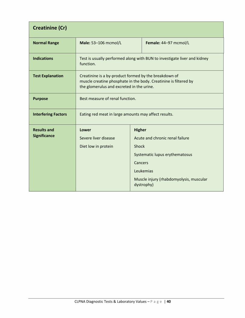
Creatinine (Cr)
Normal Range Male: 53–106 mcmol/L Female: 44–97 mcmol/L
Indications Test is usually performed along with BUN to investigate liver and kidney function.
Test Explanation Creatinine is a by-product formed by the breakdown of muscle creatine phosphate in the body. Creatinine is filtered by the glomerulus and excreted in the urine.
Purpose Best measure of renal function.
Interfering Factors Eating red meat in large amounts may affect results.
Results and
Significance
Lower
Severe liver disease
Diet low in protein
Higher
Acute and chronic renal failure
Shock
Systematic lupus erythematosus
Cancers
Leukemias
Muscle injury (rhabdomyolysis, muscular dystrophy)
CLPNA Diagnostic Tests & Laboratory Values – P a g e | 41
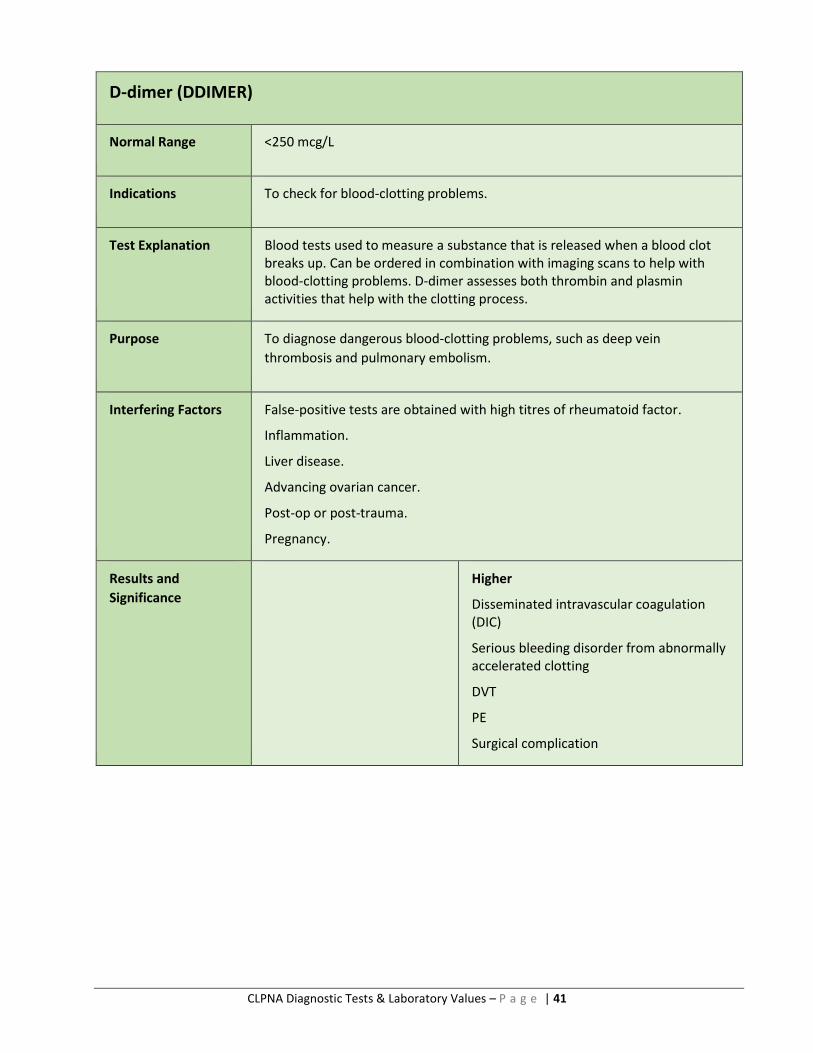
D-dimer (DDIMER)
Normal Range <250 mcg/L
Indications To check for blood-clotting problems.
Test Explanation Blood tests used to measure a substance that is released when a blood clot breaks up. Can be ordered in combination with imaging scans to help with blood-clotting problems. D-dimer assesses both thrombin and plasmin activities that help with the clotting process.
Purpose To diagnose dangerous blood-clotting problems, such as deep vein
thrombosis and pulmonary embolism.
Interfering Factors False-positive tests are obtained with high titres of rheumatoid factor.
Inflammation.
Liver disease.
Advancing ovarian cancer.
Post-op or post-trauma.
Pregnancy.
Results and
Significance
Higher
Disseminated intravascular coagulation (DIC)
Serious bleeding disorder from abnormally accelerated clotting
DVT
PE
Surgical complication

CLPNA Diagnostic Tests & Laboratory Values – P a g e | 42
Glucose (GLU)
(Fasting Blood Sugar, Random Blood Sugar)
Normal Range Fasting Blood
4.0–7.0 mmol/L
Random Glucose
<7.0 mmol/L
Indications To control blood glucose levels.
Used for persons with diabetes taking oral hypoglycemic medication or insulin.
Test Explanation Test that measures the amount of glucose in the blood. Glucose comes from carbohydrates, which are the main source of energy used by the body.
Purpose Used to check for diabetes, monitor treatment of diabetes.
Interfering Factors Vigorous exercise, stress, trauma, infection.
Use of cortisone drugs.
Intravenous fluids that contain dextrose.
Results and
Significance
Lower
Excessive doses of insulin
Inadequate food intake
Hypoglycemia
Liver disease
Malnutrition
Eating disorder
Hypothyroidism
Addison disease
Starvation
Higher
Diabetes
Prolonged corticosteroid therapy
Severe stress
CLPNA Diagnostic Tests & Laboratory Values – P a g e | 43
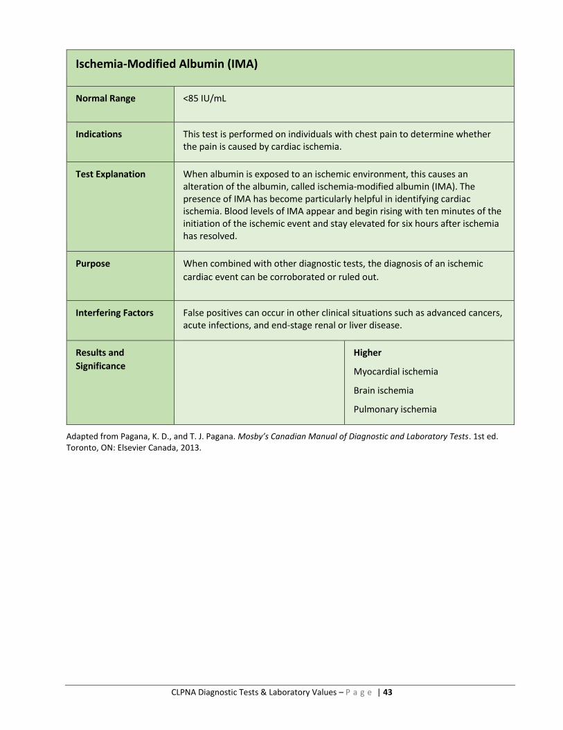
Ischemia-Modified Albumin (IMA)
Normal Range <85 IU/mL
Indications This test is performed on individuals with chest pain to determine whether the pain is caused by cardiac ischemia.
Test Explanation When albumin is exposed to an ischemic environment, this causes an alteration of the albumin, called ischemia-modified albumin (IMA). The presence of IMA has become particularly helpful in identifying cardiac ischemia. Blood levels of IMA appear and begin rising with ten minutes of the initiation of the ischemic event and stay elevated for six hours after ischemia has resolved.
Purpose When combined with other diagnostic tests, the diagnosis of an ischemic
cardiac event can be corroborated or ruled out.
Interfering Factors False positives can occur in other clinical situations such as advanced cancers, acute infections, and end-stage renal or liver disease.
Results and
Significance
Higher
Myocardial ischemia
Brain ischemia
Pulmonary ischemia
Adapted from Pagana, K. D., and T. J. Pagana. Mosby’s Canadian Manual of Diagnostic and Laboratory Tests. 1st ed. Toronto, ON: Elsevier Canada, 2013.

CLPNA Diagnostic Tests & Laboratory Values – P a g e | 44
Lactate Dehydrogenase (LDH)
Normal Range Total lactate dehydrogenase levels:
Newborn: 160–450 U/L Infant: 100–250 U/L
Child: 60–170 U/L Adult: 100–190 U/L
Isoenzymes (electrophoresis) for adults:
LDH-1: 0.17–0.27 LDH-2: 0.27–0.37
LDH-3: 0.18–0.25 LDH-4: 0.03–0.08
LDH-5: 0–0.05
Indications LDH is an intracellular enzyme used to diagnose injury or disease of the heart, liver, red blood cells, kidneys, skeletal muscle, brain, and lungs.
Test Explanation Because LDH is widely distributed through the body, the total level is not a specific indicator of any one disease or injury. When disease or injury affects the cells containing LDH, the cells lyse, and LDH is spilled into the bloodstream, in which it is identified in higher-than-normal levels. Five separate fractions (isoenzymes) make up the total LDH. Each tissue contains a predominance of one or more LDH enzymes.
Purpose A higher level of LDH-1 indicates myocardial injury.
Isolated elevation of LDH-5 indicates hepatocellular injury or disease.
Elevation of LDH- 2 and LDH-3 indicates pulmonary injury or disease.
Elevation of all LDH isoenzyme levels indicates multi-organ injury.
Interfering Factors Hemolysis of blood elevates LDH level.
Strenuous exercise may elevate LDH.
Some drugs (alcohol, anaesthetics, aspirin, clofibrate, fluorides) may increase
LDH levels.
Ascorbic acid may decrease LDH levels.
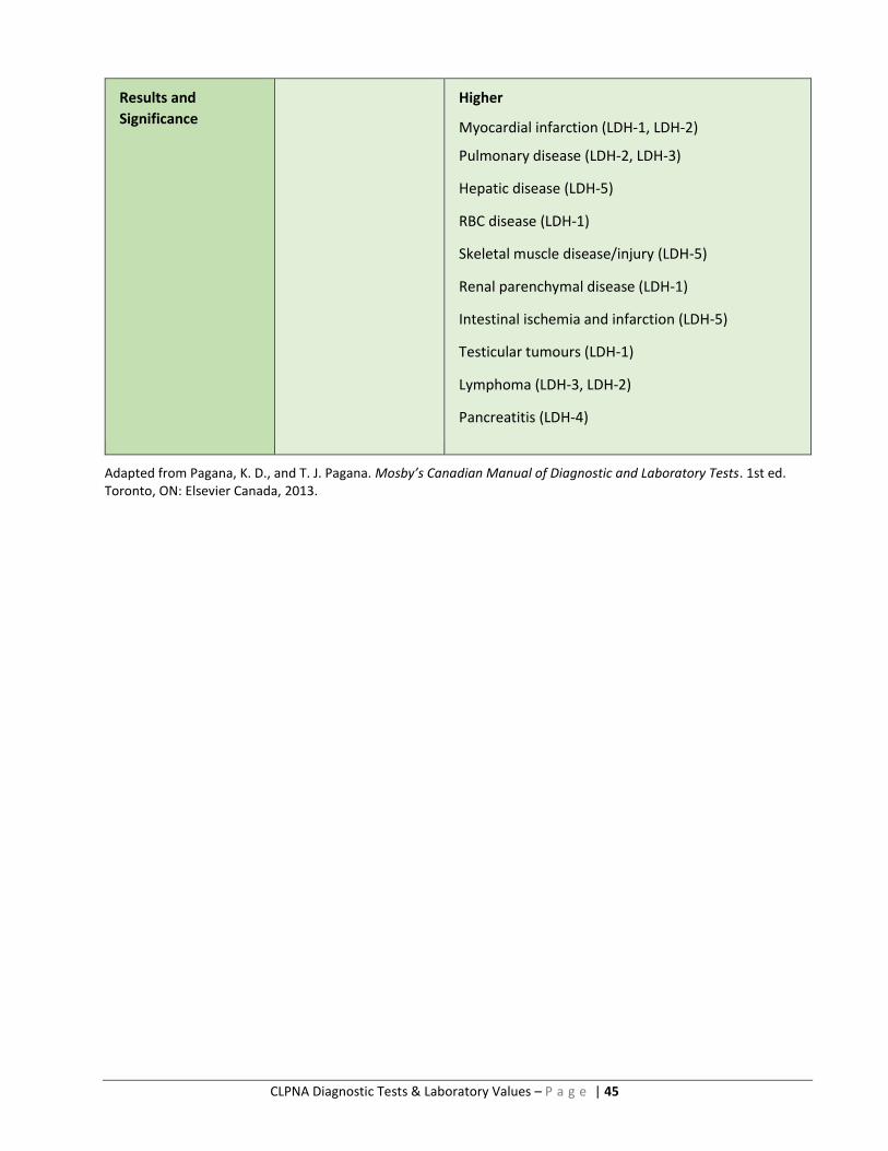
CLPNA Diagnostic Tests & Laboratory Values – P a g e | 45
Results and
Significance
Higher
Myocardial infarction (LDH-1, LDH-2)
Pulmonary disease (LDH-2, LDH-3)
Hepatic disease (LDH-5)
RBC disease (LDH-1)
Skeletal muscle disease/injury (LDH-5)
Renal parenchymal disease (LDH-1)
Intestinal ischemia and infarction (LDH-5)
Testicular tumours (LDH-1)
Lymphoma (LDH-3, LDH-2)
Pancreatitis (LDH-4)
Adapted from Pagana, K. D., and T. J. Pagana. Mosby’s Canadian Manual of Diagnostic and Laboratory Tests. 1st ed. Toronto, ON: Elsevier Canada, 2013.

CLPNA Diagnostic Tests & Laboratory Values – P a g e | 46
Potassium (K)
Normal Range 3.50–5.0 mmol/L
Indications Routine blood test that checks for cardiac and muscle function.
Can be used to diagnose disease in conjunction with sodium.
Test Explanation Potassium is essential in keeping the water and electrolyte balance of the body. Potassium is vital to how nerves and muscles work.
Potassium levels can change with sodium levels.
Purpose Used to check potassium level, detect the presence of hypo- or hyperkalemia.
Monitor potassium levels during renal insufficiency, with cancers, and with
certain drugs (diuretics).
Interfering Factors Taking potassium supplements.
Certain medications (e.g., antibiotics that contain potassium, NSAIDS, heparin, inulin, corticosteroids, non-potassium-sparing diuretics).
Overuse of laxatives.
Severe vomiting.
Results and
Significance
Lower
Overuse of diuretics
Hyperaldosteronism
Severe burns
Cystic fibrosis
Alcoholism
Dehydration
Higher
Damage or injury to the kidneys
Severe burns
Crushing injuries
Heart attack
Overconsuming potassium supplements
Medications (e.g., ACE inhibitors)
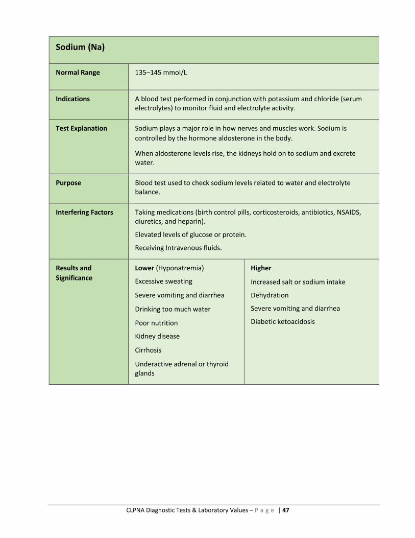
CLPNA Diagnostic Tests & Laboratory Values – P a g e | 47
Sodium (Na)
Normal Range 135–145 mmol/L
Indications A blood test performed in conjunction with potassium and chloride (serum electrolytes) to monitor fluid and electrolyte activity.
Test Explanation Sodium plays a major role in how nerves and muscles work. Sodium is
controlled by the hormone aldosterone in the body.
When aldosterone levels rise, the kidneys hold on to sodium and excrete water.
Purpose Blood test used to check sodium levels related to water and electrolyte balance.
Interfering Factors Taking medications (birth control pills, corticosteroids, antibiotics, NSAIDS, diuretics, and heparin).
Elevated levels of glucose or protein.
Receiving Intravenous fluids.
Results and
Significance
Lower (Hyponatremia)
Excessive sweating
Severe vomiting and diarrhea
Drinking too much water
Poor nutrition
Kidney disease
Cirrhosis
Underactive adrenal or thyroid glands
Higher
Increased salt or sodium intake
Dehydration
Severe vomiting and diarrhea
Diabetic ketoacidosis
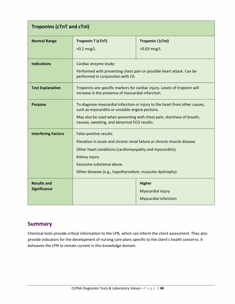
CLPNA Diagnostic Tests & Laboratory Values – P a g e | 48
Troponins (cTnT and cTnI)
Normal Range Troponin T (cTnT)
<0.2 mcg/L
Troponin I (cTnI)
<0.03 mcg/L
Indications Cardiac enzyme study:
Performed with presenting chest pain or possible heart attack. Can be performed in conjunction with CK.
Test Explanation Troponins are specific markers for cardiac injury. Levels of troponin will increase in the presence of myocardial infarction.
Purpose To diagnose myocardial infarction or injury to the heart from other causes, such as myocarditis or unstable angina pectoris.
May also be used when presenting with chest pain, shortness of breath, nausea, sweating, and abnormal ECG results.
Interfering Factors False-positive results.
Elevation in acute and chronic renal failure or chronic muscle disease.
Other heart conditions (cardiomyopathy and myocarditis).
Kidney injury.
Excessive substance abuse.
Other diseases (e.g., hypothyroidism, muscular dystrophy).
Results and
Significance
Higher
Myocardial injury
Myocardial infarction
Summary
Chemical tests provide critical information to the LPN, which can inform the client assessment. They also
provide indicators for the development of nursing care plans specific to the client’s health concerns. It
behooves the LPN to remain current in this knowledge domain.
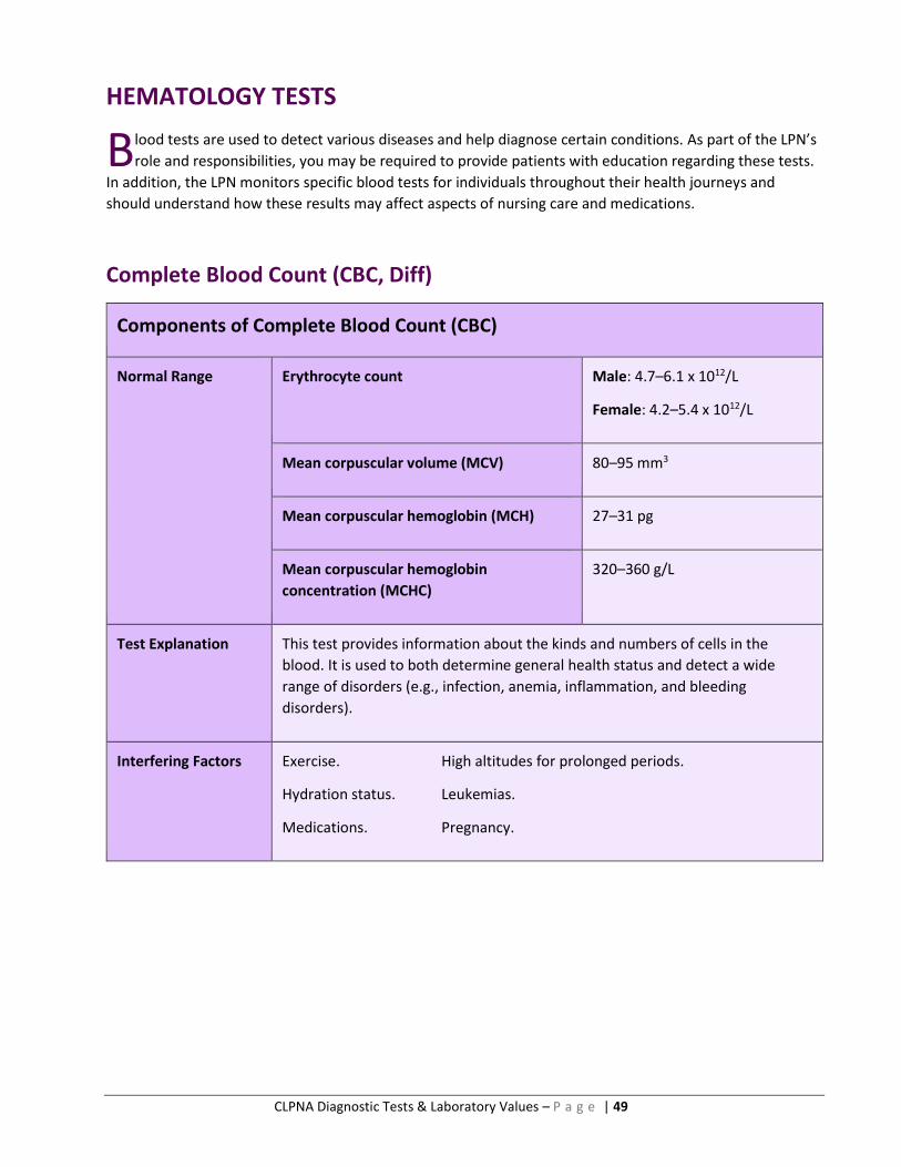
CLPNA Diagnostic Tests & Laboratory Values – P a g e | 49
HEMATOLOGY TESTS
lood tests are used to detect various diseases and help diagnose certain conditions. As part of the LPN’s
role and responsibilities, you may be required to provide patients with education regarding these tests.
In addition, the LPN monitors specific blood tests for individuals throughout their health journeys and
should understand how these results may affect aspects of nursing care and medications.
Complete Blood Count (CBC, Diff)
Components of Complete Blood Count (CBC)
Normal Range Erythrocyte count Male: 4.7–6.1 x 10¹²/L
Female: 4.2–5.4 x 10¹²/L
Mean corpuscular volume (MCV) 80–95 mm3
Mean corpuscular hemoglobin (MCH) 27–31 pg
Mean corpuscular hemoglobin
concentration (MCHC)
320–360 g/L
Test Explanation This test provides information about the kinds and numbers of cells in the
blood. It is used to both determine general health status and detect a wide
range of disorders (e.g., infection, anemia, inflammation, and bleeding
disorders).
Interfering Factors Exercise. High altitudes for prolonged periods.
Hydration status. Leukemias.
Medications. Pregnancy.
B

CLPNA Diagnostic Tests & Laboratory Values – P a g e | 50
Results and
Significance
LOWER HIGHER
Erythrocyte count
Anemia Leukemia
Post hemorrhage
Dehydration High altitudes
Polycythemia vera Severe diarrhea
Mean corpuscular volume (MCV)
Microcytic anemia Folic acid and vitamin B12 deficiency
Liver disease
Macrocytic anemia
Mean corpuscular hemoglobin (MCH)
Microcytic anemia Macrocytic anemia
Mean corpuscular hemoglobin concentration (MCHC)
Hypochromic anemia Intravascular hemolysis
Spherocytosis
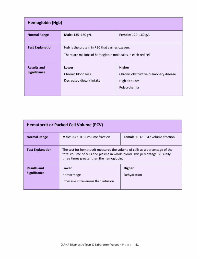
CLPNA Diagnostic Tests & Laboratory Values – P a g e | 51
Hemoglobin (Hgb)
Normal Range Male: 135–180 g/L Female: 120–160 g/L
Test Explanation Hgb is the protein in RBC that carries oxygen.
There are millions of hemoglobin molecules in each red cell.
Results and
Significance
Lower
Chronic blood loss
Decreased dietary intake
Higher
Chronic obstructive pulmonary disease
High altitudes
Polycythemia
Hematocrit or Packed Cell Volume (PCV)
Normal Range Male: 0.42–0.52 volume fraction Female: 0.37–0.47 volume fraction
Test Explanation The test for hematocrit measures the volume of cells as a percentage of the total volume of cells and plasma in whole blood. This percentage is usually three times greater than the hemoglobin.
Results and
Significance
Lower
Hemorrhage
Excessive intravenous fluid infusion
Higher
Dehydration

CLPNA Diagnostic Tests & Laboratory Values – P a g e | 52
Myoglobin (Mb)
Normal Range 1.0–5.3 nmol/L
Test Explanation This test is used in the early evaluation of a client with suspected acute myocardial infarction. It is also used to assist in the diagnosis of disease or injury in skeletal muscles.
Interfering Factors Recent administration of radioactive substances.
Myoglobin levels can increase after intramuscular injections.
Results and
Significance
Lower
Polymyositis
Higher
Myocardial infarction
Skeletal muscle inflammation (myositis)
Malignant hyperthermia
Muscular dystrophy
Skeletal muscle ischemia/trauma
Rhabdomyolysis
Adapted from Pagana, K. D., and T. J. Pagana. Mosby’s Canadian Manual of Diagnostic and Laboratory Tests. 1st ed. Toronto, ON: Elsevier Canada, 2013.
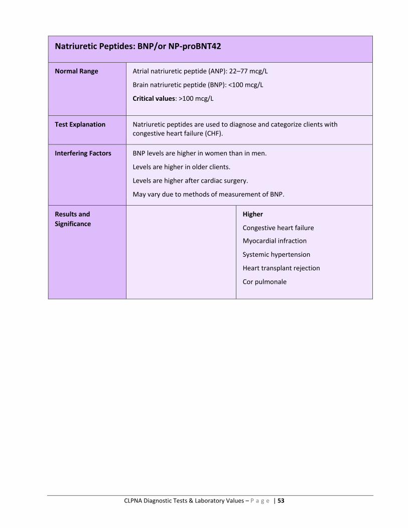
CLPNA Diagnostic Tests & Laboratory Values – P a g e | 53
Natriuretic Peptides: BNP/or NP-proBNT42
Normal Range Atrial natriuretic peptide (ANP): 22–77 mcg/L
Brain natriuretic peptide (BNP): <100 mcg/L
Critical values: >100 mcg/L
Test Explanation Natriuretic peptides are used to diagnose and categorize clients with congestive heart failure (CHF).
Interfering Factors BNP levels are higher in women than in men.
Levels are higher in older clients.
Levels are higher after cardiac surgery.
May vary due to methods of measurement of BNP.
Results and
Significance
Higher
Congestive heart failure
Myocardial infraction
Systemic hypertension
Heart transplant rejection
Cor pulmonale
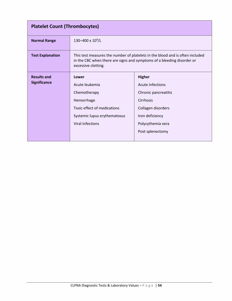
CLPNA Diagnostic Tests & Laboratory Values – P a g e | 54
Platelet Count (Thrombocytes)
Normal Range 130–400 x 109/L
Test Explanation This test measures the number of platelets in the blood and is often included in the CBC when there are signs and symptoms of a bleeding disorder or excessive clotting.
Results and
Significance
Lower
Acute leukemia
Chemotherapy
Hemorrhage
Toxic effect of medications
Systemic lupus erythematosus
Viral infections
Higher
Acute infections
Chronic pancreatitis
Cirrhosis
Collagen disorders
Iron deficiency
Polycythemia vera
Post splenectomy
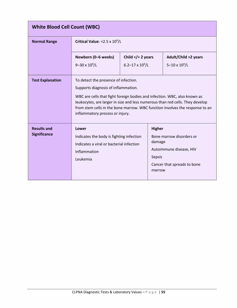
CLPNA Diagnostic Tests & Laboratory Values – P a g e | 55
White Blood Cell Count (WBC)
Normal Range Critical Value: <2.5 x 10⁹/L
Newborn (0–6 weeks)
9–30 x 109/L
Child </= 2 years
6.2–17 x 109/L
Adult/Child >2 years
5–10 x 109/L
Test Explanation To detect the presence of infection.
Supports diagnosis of inflammation.
WBC are cells that fight foreign bodies and infection. WBC, also known as
leukocytes, are larger in size and less numerous than red cells. They develop
from stem cells in the bone marrow. WBC function involves the response to an
inflammatory process or injury.
Results and
Significance
Lower
Indicates the body is fighting infection
Indicates a viral or bacterial infection
Inflammation
Leukemia
Higher
Bone marrow disorders or damage
Autoimmune disease, HIV
Sepsis
Cancer that spreads to bone
marrow
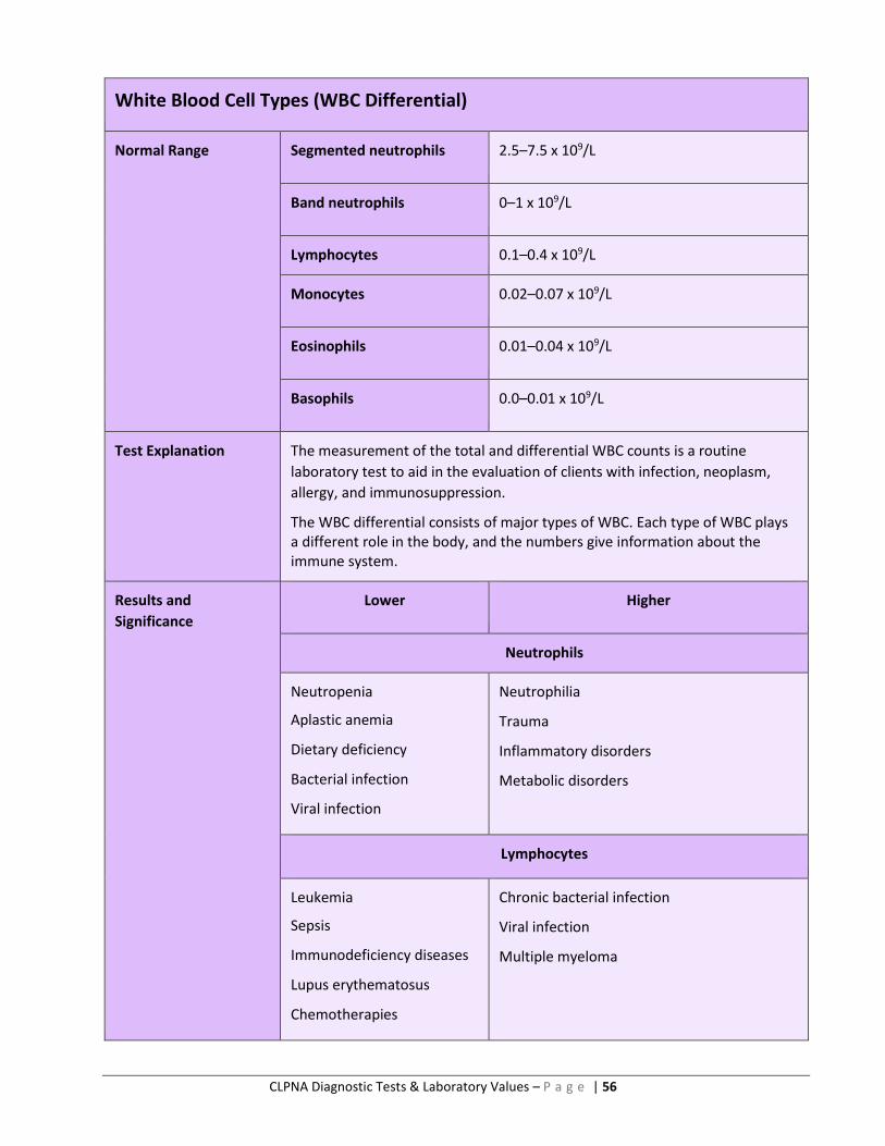
CLPNA Diagnostic Tests & Laboratory Values – P a g e | 56
White Blood Cell Types (WBC Differential)
Normal Range Segmented neutrophils 2.5–7.5 x 10⁹/L
Band neutrophils 0–1 x 10⁹/L
Lymphocytes 0.1–0.4 x 10⁹/L
Monocytes 0.02–0.07 x 10⁹/L
Eosinophils 0.01–0.04 x 10⁹/L
Basophils 0.0–0.01 x 10⁹/L
Test Explanation The measurement of the total and differential WBC counts is a routine
laboratory test to aid in the evaluation of clients with infection, neoplasm,
allergy, and immunosuppression.
The WBC differential consists of major types of WBC. Each type of WBC plays a different role in the body, and the numbers give information about the immune system.
Results and
Significance
Lower Higher
Neutrophils
Neutropenia
Aplastic anemia
Dietary deficiency
Bacterial infection
Viral infection
Neutrophilia
Trauma
Inflammatory disorders
Metabolic disorders
Lymphocytes
Leukemia
Sepsis
Immunodeficiency diseases
Lupus erythematosus
Chemotherapies
Chronic bacterial infection
Viral infection
Multiple myeloma
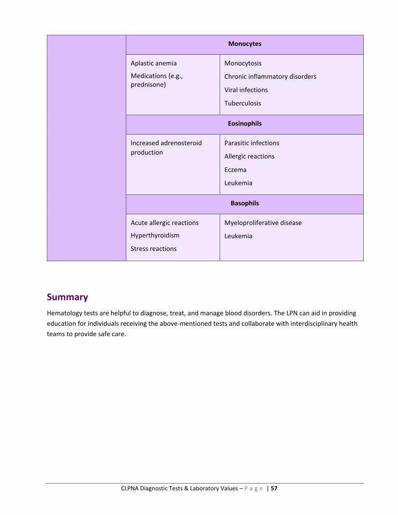
CLPNA Diagnostic Tests & Laboratory Values – P a g e | 57
Monocytes
Aplastic anemia
Medications (e.g., prednisone)
Monocytosis
Chronic inflammatory disorders
Viral infections
Tuberculosis
Eosinophils
Increased adrenosteroid
production
Parasitic infections
Allergic reactions
Eczema
Leukemia
Basophils
Acute allergic reactions
Hyperthyroidism
Stress reactions
Myeloproliferative disease
Leukemia
Summary
Hematology tests are helpful to diagnose, treat, and manage blood disorders. The LPN can aid in providing
education for individuals receiving the above-mentioned tests and collaborate with interdisciplinary health
teams to provide safe care.
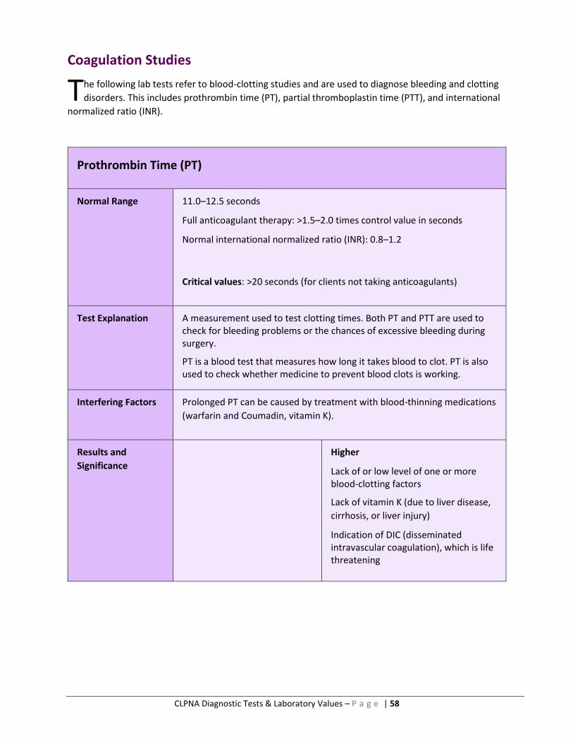
CLPNA Diagnostic Tests & Laboratory Values – P a g e | 58
Coagulation Studies
he following lab tests refer to blood-clotting studies and are used to diagnose bleeding and clotting
disorders. This includes prothrombin time (PT), partial thromboplastin time (PTT), and international
normalized ratio (INR).
Prothrombin Time (PT)
Normal Range 11.0–12.5 seconds
Full anticoagulant therapy: >1.5–2.0 times control value in seconds
Normal international normalized ratio (INR): 0.8–1.2
Critical values: >20 seconds (for clients not taking anticoagulants)
Test Explanation A measurement used to test clotting times. Both PT and PTT are used to check for bleeding problems or the chances of excessive bleeding during surgery.
PT is a blood test that measures how long it takes blood to clot. PT is also used to check whether medicine to prevent blood clots is working.
Interfering Factors Prolonged PT can be caused by treatment with blood-thinning medications
(warfarin and Coumadin, vitamin K).
Results and
Significance
Higher
Lack of or low level of one or more blood-clotting factors
Lack of vitamin K (due to liver disease,
cirrhosis, or liver injury)
Indication of DIC (disseminated intravascular coagulation), which is life threatening
T
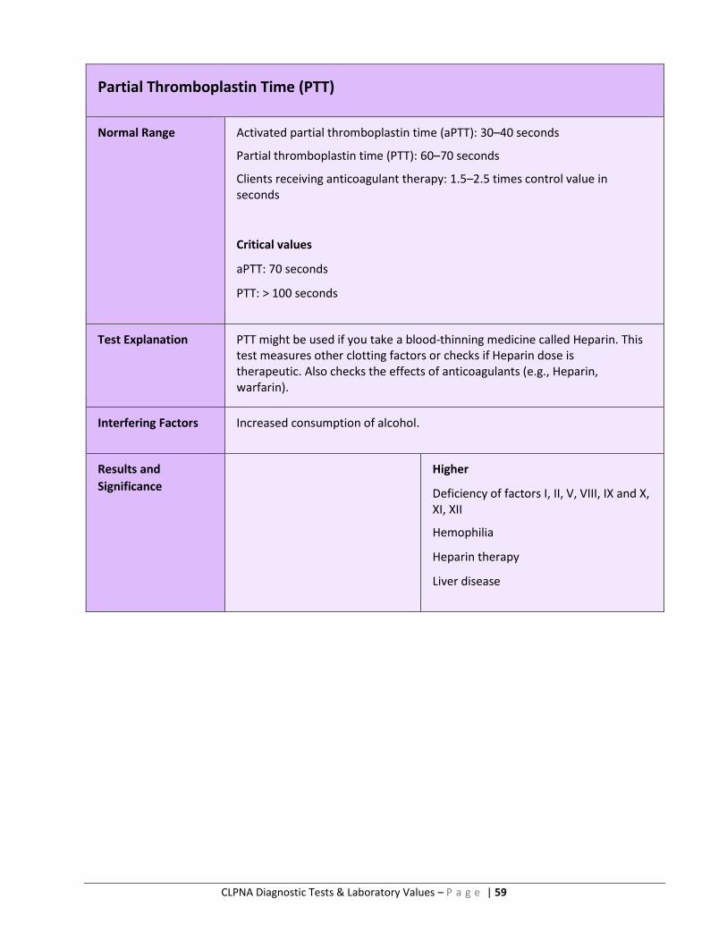
CLPNA Diagnostic Tests & Laboratory Values – P a g e | 59
Partial Thromboplastin Time (PTT)
Normal Range Activated partial thromboplastin time (aPTT): 30–40 seconds
Partial thromboplastin time (PTT): 60–70 seconds
Clients receiving anticoagulant therapy: 1.5–2.5 times control value in seconds
Critical values
aPTT: 70 seconds
PTT: > 100 seconds
Test Explanation PTT might be used if you take a blood-thinning medicine called Heparin. This test measures other clotting factors or checks if Heparin dose is therapeutic. Also checks the effects of anticoagulants (e.g., Heparin, warfarin).
Interfering Factors Increased consumption of alcohol.
Results and
Significance
Higher
Deficiency of factors I, II, V, VIII, IX and X, XI, XII
Hemophilia
Heparin therapy
Liver disease

CLPNA Diagnostic Tests & Laboratory Values – P a g e | 60
International Normalized Ratio (INR)
Normal Range 0.8–1.2
Test Explanation INR is a way of standardizing the results of prothrombin time tests, no matter the testing method. It lets the physician understand results regardless of different test methods.
Results and
Significance
Higher
Lack of or low level of one or more
blood-clotting factors
Lack of vitamin K (due to liver disease,
cirrhosis, or liver injury)
Indication of DIC (disseminated intravascular coagulation), which is life threatening
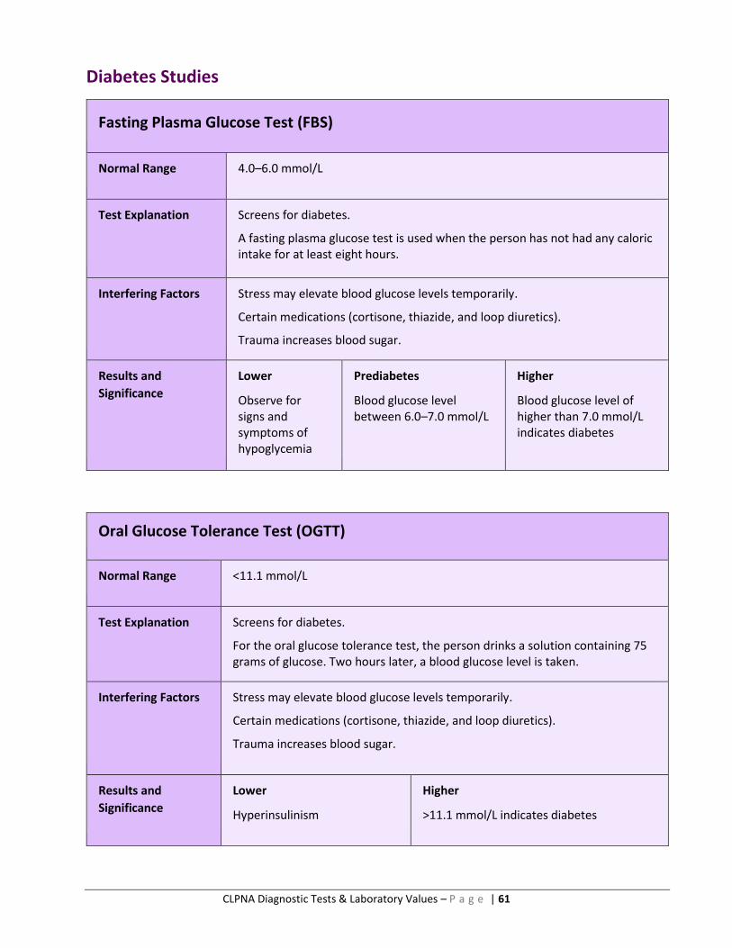
CLPNA Diagnostic Tests & Laboratory Values – P a g e | 61
Diabetes Studies
Fasting Plasma Glucose Test (FBS)
Normal Range 4.0–6.0 mmol/L
Test Explanation Screens for diabetes.
A fasting plasma glucose test is used when the person has not had any caloric intake for at least eight hours.
Interfering Factors Stress may elevate blood glucose levels temporarily.
Certain medications (cortisone, thiazide, and loop diuretics).
Trauma increases blood sugar.
Results and
Significance
Lower
Observe for signs and symptoms of hypoglycemia
Prediabetes
Blood glucose level between 6.0–7.0 mmol/L
Higher
Blood glucose level of higher than 7.0 mmol/L indicates diabetes
Oral Glucose Tolerance Test (OGTT)
Normal Range <11.1 mmol/L
Test Explanation Screens for diabetes.
For the oral glucose tolerance test, the person drinks a solution containing 75 grams of glucose. Two hours later, a blood glucose level is taken.
Interfering Factors Stress may elevate blood glucose levels temporarily.
Certain medications (cortisone, thiazide, and loop diuretics).
Trauma increases blood sugar.
Results and
Significance
Lower
Hyperinsulinism
Higher
>11.1 mmol/L indicates diabetes
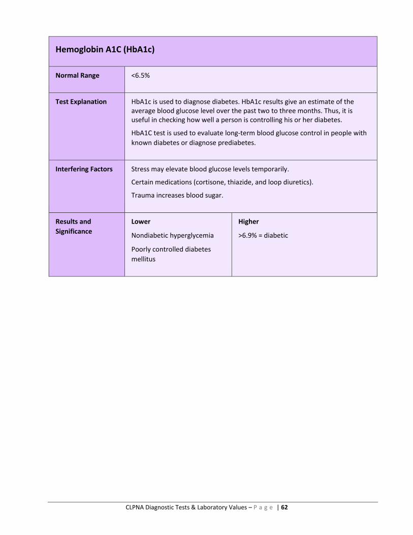
CLPNA Diagnostic Tests & Laboratory Values – P a g e | 62
Hemoglobin A1C (HbA1c)
Normal Range <6.5%
Test Explanation HbA1c is used to diagnose diabetes. HbA1c results give an estimate of the average blood glucose level over the past two to three months. Thus, it is useful in checking how well a person is controlling his or her diabetes.
HbA1C test is used to evaluate long-term blood glucose control in people with
known diabetes or diagnose prediabetes.
Interfering Factors Stress may elevate blood glucose levels temporarily.
Certain medications (cortisone, thiazide, and loop diuretics).
Trauma increases blood sugar.
Results and
Significance
Lower
Nondiabetic hyperglycemia
Poorly controlled diabetes
mellitus
Higher
>6.9% = diabetic
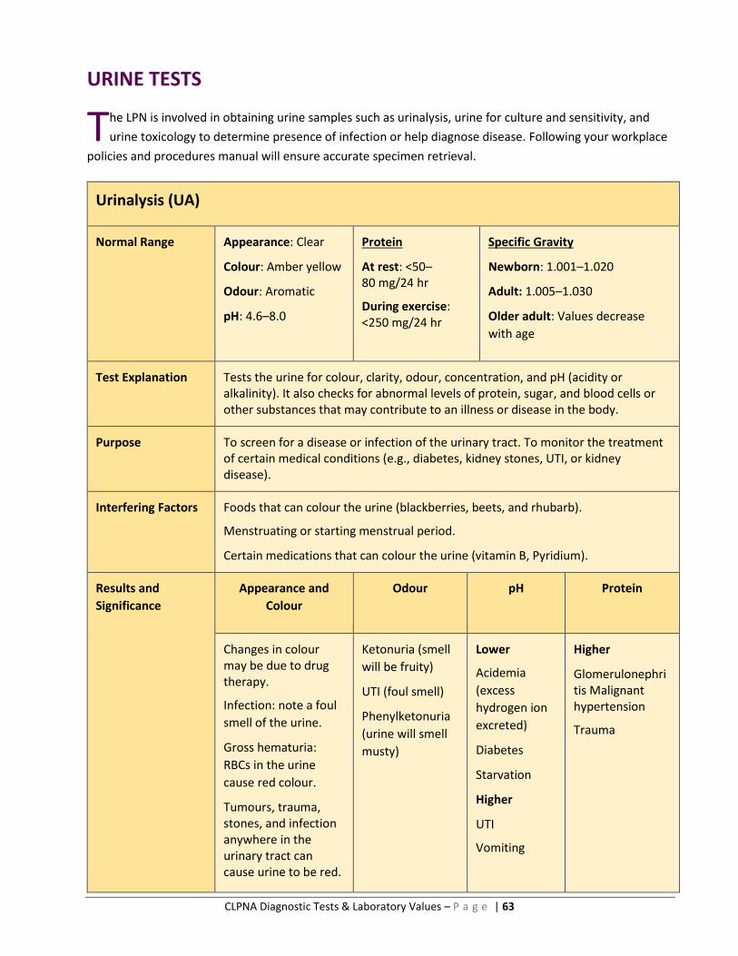
CLPNA Diagnostic Tests & Laboratory Values – P a g e | 63
URINE TESTS
he LPN is involved in obtaining urine samples such as urinalysis, urine for culture and sensitivity, and
urine toxicology to determine presence of infection or help diagnose disease. Following your workplace
policies and procedures manual will ensure accurate specimen retrieval.
Urinalysis (UA)
Normal Range Appearance: Clear
Colour: Amber yellow
Odour: Aromatic
pH: 4.6–8.0
Protein
At rest: <50–80 mg/24 hr
During exercise: <250 mg/24 hr
Specific Gravity
Newborn: 1.001–1.020
Adult: 1.005–1.030
Older adult: Values decrease
with age
Test Explanation Tests the urine for colour, clarity, odour, concentration, and pH (acidity or alkalinity). It also checks for abnormal levels of protein, sugar, and blood cells or other substances that may contribute to an illness or disease in the body.
Purpose To screen for a disease or infection of the urinary tract. To monitor the treatment of certain medical conditions (e.g., diabetes, kidney stones, UTI, or kidney disease).
Interfering Factors Foods that can colour the urine (blackberries, beets, and rhubarb).
Menstruating or starting menstrual period.
Certain medications that can colour the urine (vitamin B, Pyridium).
Results and
Significance
Appearance and
Colour
Odour pH Protein
Changes in colour may be due to drug therapy.
Infection: note a foul
smell of the urine.
Gross hematuria:
RBCs in the urine
cause red colour.
Tumours, trauma, stones, and infection anywhere in the urinary tract can cause urine to be red.
Ketonuria (smell
will be fruity)
UTI (foul smell)
Phenylketonuria
(urine will smell
musty)
Lower
Acidemia
(excess
hydrogen ion
excreted)
Diabetes
Starvation
Higher
UTI
Vomiting
Higher
Glomerulonephritis Malignant hypertension
Trauma
T

CLPNA Diagnostic Tests & Laboratory Values – P a g e | 64
Urine C & S (Culture and Sensitivity)
Normal Range No bacteria or other organisms (such as fungi) grow in the culture. The culture result is positive.
Test Explanation The urine C & S identifies the specific bacteria and tests its susceptibility to different antibiotic agents. This ensures that the proper antibiotic can be prescribed to clear up the infection.
Purpose To detect UTI.
Interfering Factors A urine specimen that has been sitting for an hour or longer at room temperature (should be kept in refrigerator until assessed by lab).
Feces or toilet paper in the urine specimen.
Drugs and food.
Use of antibiotics.
Results and
Significance
Lower
< infection is unlikely
Higher
>100,000 or more bacteria per milliliter (mL) of urine may indicate an infection
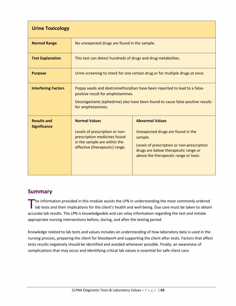
CLPNA Diagnostic Tests & Laboratory Values – P a g e | 65
Urine Toxicology
Normal Range No unexpected drugs are found in the sample.
Test Explanation This test can detect hundreds of drugs and drug metabolites.
Purpose Urine screening to check for one certain drug or for multiple drugs at once.
Interfering Factors Poppy seeds and dextromethorphan have been reported to lead to a false-
positive result for amphetamines.
Decongestants (ephedrine) also have been found to cause false-positive results for amphetamines.
Results and
Significance
Normal Values
Levels of prescription or non-prescription medicines found in the sample are within the effective (therapeutic) range.
Abnormal Values
Unexpected drugs are found in the
sample.
Levels of prescription or non-prescription drugs are below therapeutic range or above the therapeutic range or toxic.
Summary
he information provided in this module assists the LPN in understanding the most commonly ordered
lab tests and their implications for the client’s health and well-being. Due care must be taken to obtain
accurate lab results. The LPN is knowledgeable and can relay information regarding the test and initiate
appropriate nursing interventions before, during, and after the testing period.
Knowledge related to lab tests and values includes an understanding of how laboratory data is used in the
nursing process, preparing the client for bloodwork and supporting the client after tests. Factors that affect
tests results negatively should be identified and avoided whenever possible. Finally, an awareness of
complications that may occur and identifying critical lab values is essential for safe client care.
T

CLPNA Diagnostic Tests & Laboratory Values – P a g e | 66
Module 4: Nursing Implications and Applications of Lab Values
Introduction
aboratory values continue to be an area of difficulty for nurses to comprehend and apply in their nursing
practice. The nursing implications must be considered when delivering care for clients—assessment
before, during, and after any laboratory test as well as monitoring for signs and symptoms of complications.
As always, client teaching is a top priority and can encourage collaboration and prevent harm. With
continued education and practice with lab values, the LPN will strengthen nursing practice and support
clients through their health care experiences. This part of the course provides the opportunity, through the
learning activities, to challenge and expand your knowledge base in this regard.
Module Outcomes
Upon completion of this module, the participant will be able to
select the correct lab tests appropriate to particular client health challenges;
differentiate among chemistry, hematology, and urine test lab values;
recognize the significance of lab results; and
recognize critical values.
Questions
Before proceeding to the online quizzes, attempt to answer the following questions accurately.
1. The LPN is caring for a client who has vomiting and diarrhea that has lasted for 48 hours. He is
complaining of dizziness and leg cramps. Which lab test(s) will the LPN anticipate being ordered for
this client?
2. The LPN is managing care for a client with hematuria and complaints of dysuria. Which lab test(s)
will likely be ordered for this client?
3. The LPN is working in a community clinic for patients with diabetes. Marlon is a new client who has
been experiencing symptoms of hypoglycemia for some months now. The doctor has decided to
send Marlon to the clinic for a full assessment of his blood sugar levels and diabetic status. What lab
tests can the LPN expect to be ordered for Marlon, and what are the normal values for these tests?
L

CLPNA Diagnostic Tests & Laboratory Values – P a g e | 67
Answers
1. Query food poisoning, gastroenteritis: perform serum electrolytes (serum potassium), urea,
and creatinine, CBC count, stool studies for occult blood, stool for C-difficile.
2. CBC count, blood urea creatinine (BUN), urinalysis, urine culture and sensitivity, urine calcium.
3. FBS (4.0–7.0 mmol/L), RBS (< 7.0 mmol/L), HbA1C (<6.5%), urinalysis (0–0.8 mmol/L).
Case Study
A seven-year-old boy was brought to his pediatrician because he had developed hematuria, which
required hospitalization. About six weeks before his admission, he had had a severe sore throat but
received no treatment for it. Subsequently, he did well except for complaints of mild lethargy and
decrease in appetite. For the ten days before admission, he had an elevated temperature. He
complained of minimal bilateral back pain. Physical examination revealed a well-developed young boy
with moderate bilateral tenderness.
Question 1: List all the laboratory testing involved with this child’s disease process. Consider
rationale for each.
Answer:
Urinalysis (odour, glucose, blood, protein, RBCs, colour, specific gravity), BUN, and
creatinine test (test kidney function).
Question 2: What client teaching and nursing implications are involved with the above-mentioned
tests?
Answer:
Explain purpose and specific method of urine collection, determine if client can obtain urine
sample independently. If specimen cannot be tested immediately, cover and refrigerate it.

CLPNA Diagnostic Tests & Laboratory Values – P a g e | 68
Self-Assessment
ow that you have reviewed all the information on common laboratory tests and test values and
completed the case studies, you are ready to engage in self-assessment by completing the online
quizzes provided for this course. If you have difficulty with a question or you want to know more about a
test, choose from the resources in the endnotes at the end of this course. The resources identified in
this list provide comprehensive information on all the diagnostic and laboratory tests mentioned in this
course and in the online quizzes. A final examination is also available should you want a Certificate of
Completion.
When you have completed these tests, you can access the other supplementary quizzes on diagnostic
and laboratory tests at the following link: http://studywithclpna.com/diagnostictesting/
Summary
his module has provided the learner with an opportunity to evaluate his or her knowledge
concerning laboratory values. There are a multitude of laboratory tests that the LPN can learn about
and work with in practice. Interpretation of laboratory values is an important skill and can be developed
through continued practice and study. Since this is a critical component of the client assessment, LPNs
will continue to need this data to understand the care and treatment of the clients they work with.
N
T
CLPNA Diagnostic Tests & Laboratory Values – P a g e | 69
Endnotes
1 Government of Alberta, Competency Profile for Licensed Practical Nurses, 3rd ed., 2015, http://www.clpna.com/wp-content/uploads/2013/02/doc_Competency_Profile_for_LPNs_3rd_Ed_2015_COMPLETE.pdf.
2 K. D. Pagana and T. J. Pagana, Mosby’s Canadian Manual of Diagnostic and Laboratory Tests, 1st Cdn. ed., (Toronto, ON: Elsevier Canada, 2013).
3 Healthwise Staff, “Angiogram,” WebMD, Last modified August 21, 2015, http://www.webmd.com/heart-disease/angiogram#1.
4 Pagana and Pagana, Mosby’s Canadian.
5 Wolters Kluwer Health, Diagnostic Tests Made Incredibly Easy, 2nd ed. (Philadelphia: Lippincott Williams & Wilkins, 2009).
6 Pagana and Pagana, Mosby’s Canadian.
7 Wolters Kluwer Health, Diagnostic Tests.
8 Pagana and Pagana, Mosby’s Canadian.
9 Pagana and Pagana, Mosby’s Canadian.
10 Alberta Health Services, “Computed Topography,” http://www.albertahealthservices.ca/info/service.aspx?id=5953.
11 Pagana and Pagana, Mosby’s Canadian.
12 Alberta Health Services, “Cystoscopy,” http://www.albertahealthservices.ca/info/service.aspx?id=4074.
13 Pagana and Pagana, Mosby’s Canadian.
14 Wolters Kluwer Health, Diagnostic Tests.
15 Pagana and Pagana, Mosby’s Canadian.
16 Alberta Health Services, “Echocardiography,” http://www.albertahealthservices.ca/info/service.aspx?id=1066401.
17 Pagana and Pagana, Mosby’s Canadian.
18 Alberta Health Services, “Cardiology—Electrocardiogram Services,” http://www.albertahealthservices.ca/info/service.aspx?id=1000804.
19 Pagana and Pagana, Mosby’s Canadian.
20 Pagana and Pagana, Mosby’s Canadian.
21 Wolters Kluwer Health, Diagnostic Tests.
22 Kee J. LeFever, Laboratory and Diagnostic Tests with Nursing Implications, 10th ed. (Upper Saddle River, NJ: Pearson, 2017).
23 Pagana and Pagana, Mosby’s Canadian.
24 LeFever, Laboratory and Diagnostic Tests.
25 Pagana and Pagana, Mosby’s Canadian.
26 Wolters Kluwer Health, Diagnostic Tests.
CLPNA Diagnostic Tests & Laboratory Values – P a g e | 70
27 LeFever, Laboratory and Diagnostic Tests.
28 Alberta Health Services, “General Radiography (X-Ray),” http://www.albertahealthservices.ca/info/service.aspx?id=5952.
29 Pagana and Pagana, Mosby’s Canadian.
30 LeFever, Laboratory and Diagnostic Tests.
31 Pagana and Pagana, Mosby’s Canadian.
32 Wolters Kluwer Health, Diagnostic Tests.
33 Pagana and Pagana, Mosby’s Canadian.
34 Wolters Kluwer Health, Diagnostic Tests.
35 Pagana and Pagana, Mosby’s Canadian.
36 Wolters Kluwer Health, Diagnostic Tests.
37 Pagana and Pagana, Mosby’s Canadian.
38 A. M. van Leeuwen and M. L. Bladh, Textbook of Laboratory and Diagnostic Testing: Practical Application of Nursing Process at the Bedside (Philadelphia, PA: F. A. Davis Company, 2016).
39 Office of the Information and Privacy Commissioner of Alberta, “Legislation,” https://www.oipc.ab.ca/legislation.aspx
40 PKD Foundation, “Understanding Your Lab Results,” YouTube, April 12, 2013, https://www.youtube.com/watch?v=K9sAdDYmINM.
41 The information for these common lab tests was adapted from M. B. Dunning III and F. Fischbach, Nurses’ Quick Reference to Common Laboratory and Diagnostic Tests, 5th ed. (Philadelphia, PA: Lippincott Williams & Wilkins, 2011); L. M. Malarkey and M. E. McMorrow, Saunders Nursing Guide to Laboratory and Diagnostic Tests, 2nd ed. (St. Louis, MO: Elsevier, 2011); Medical Council of Canada, “Clinical laboratory tests: Normal values,” 2017, https://apps.mcc.ca/objectives_online/objectives.pl?lang=english&loc=values; K. D. Pagana and T. J. Pagana, Mosby’s Canadian Manual of Diagnostic and Laboratory Tests, 1st Cdn. ed., (Toronto, ON:
Elsevier Canada, 2013); P. A. Potter, A. G. Perry, P. A. Stockert, and A. M. Hall, Canadian Fundamentals of Nursing, 5th ed. (Toronto: Elsevier Canada, 2014); and A. M. van Leeuwen and M. L. Bladh, Textbook of Laboratory and Diagnostic Testing: Practical Application of Nursing Process at the Bedside (Philadelphia, PA: F. A. Davis Company, 2016).
42 Pagana and Pagana, Mosby’s Canadian.