diagnostic value of esophagoscopy and gastroscopy : an ......philadelphia broncho-esophagologic...
TRANSCRIPT

University of Nebraska Medical CenterDigitalCommons@UNMC
MD Theses College of Medicine
5-1-1964
Diagnostic value of esophagoscopy andgastroscopy : an analysis of 272 proceduresGerald C. MillerUniversity of Nebraska Medical Center
Follow this and additional works at: http://digitalcommons.unmc.edu/mdtheses
This Thesis is brought to you for free and open access by the College of Medicine at DigitalCommons@UNMC. It has been accepted for inclusion inMD Theses by an authorized administrator of DigitalCommons@UNMC. For more information, please contact [email protected].
Recommended CitationMiller, Gerald C., "Diagnostic value of esophagoscopy and gastroscopy : an analysis of 272 procedures" (1964). MD Theses. Paper 36.

~ ...
THE DIAGN!)STIC VALUE OF ESOPHAGOSCOPY AND GASTROSCOPY-AN ANALYSIS OF 272 PROCEDURES
Gerald Charles Miller
Submitted in Partial Fulfillment for the Degree of Doctor of Medicine
College of Medicine, University of l'lebraska
February 21, 1964
Omaha, Nebraska

TABLE OF CONTE.'NTS
PAGE
I. Introduction • • • • · • • • • • 1
II. jlf.ethod • • • · • • • • • 3
III. Results • • · • • • 3
(a) Esophagoscopy • • • · • • • · • 4
(b) Gastroscopy · • · • · • • • • 9
IV. Discussion • • • • • · . • • 17
,- V. Conclusions • • • • • • . • · • · • · 25
VI. Summary • . • . • .. • 27
VII. Bibliography • • • • · • . . 28
Diagnostic Value of Esophagoscopy and Gastroscopy-An Analysis of 272 Procedures
DfTRODUCTION
It is not well known that endoscopy was being performed on
the upper gastro-intestinal tract before the advent of radio-
1 logic procedures. The beginning efforts in esophagoscopy com-
menced in the nineteenth century. Initially it was associated
with a high morbidity and mortality. Credit is given to the
Philadelphia Broncho-Esophagologic Clinli~ for developing the
techniques necessary for safe examination during the early p~rt
of this century. Gastroscopy did not become clinically impor-
tant until 1932 when a semi-flexible instrument, the VV-olf-
SchindlEr gastroscope was introduced. In 1958, Hirschowitz at
al.2
described a new gastroscope, the IIfiberscope". It enables
visualization of the antrum and duodenal cavity without image
distortion, as its principal advantage. The instrument is com-
pletely flexible making it easier and safer to swallow. It
provides excellent light transmission making photography feas-
ible without excessive illumination; and it enables one to
view from contact to infinity so that the esophagus, stomach
and duodenum can all be visualized in one instrumentation.
In a recent re-evaluation of gastroscopy, Strub 3 cites
Schindler I S descr:lp:tion of the four blind areas in visualization
of the stomach using the semi-flexible. instrlLment. These
regions include; the fornix of the stomach, posterior wall,
-1-

lesser curvature of the antrQ~ and the lower pole of the ant-
rum. The fornix can sometimes be seen better when the patient
is lying on his back. The antrum is difficult to visualize
most of the time because the instrument looks out at right
angles. 4
Palm.er emphasizes the importance of roentgenographic stud-
ies before undertaking endoscopy, except in emergency situations.
He feels the two diagnostic procedures are supplementary. X-ray
examination gives shadow information about configurations, altered
motility features and precise localization of discrete lesions.
Endoscopy allows careful visualization of the inner wall of the
gut from different angles, so that configurations and colors of
a lesion can be directly inspected.
An analysis is presented of the esophagoscopy and gastro-
scopy procedures performed by the University of Nebraska Gastro-
intestinal Service at the University and Douglas County hospit-
als from July 1959 to July 1963. These studies were performed
by Dr. Frederick F. Paustian, B.S., M.D., Associate Professor
of Internal Medicine and residents in the department of Internal
1lIedicine •
. ~.
-2-

-
METHOD
The Eder-Hufford semi-flexible esophagoscope and the Gast
roflex gastroscope were used in all examinations. Esophageal
biopies were taken through the Eder-Hufford esophagoscope and
the Benedict operating scope was intrmduced separately for all
gastric biopies. Topical oral anesthesia, Pontocaine 2%, was
administered before all examinations. Preoperative medication
.vith Demerol 50 mg., Phenergan 25 mg., and Atropine 1/150 gr.
was used and the left decubitus position was used in most cases.
The medical records of the patients included in this study
were reviewed for information pertaining to: chief complaint on
admission, esophagoscopy and ~astroscopy findings, X-raY find
ings, procedure" complications, subsequent clinical course and
final diagnosis. Particular attention was directed to biopsy,
surgical and autopsy findings.
RESULTS
Fifty-one esophagoscopies, 101 gastroscopies and 60
esophago-gastroscopies were performed in 178 patients for a
total of 272 endoscopic studies. Three patients were ex
cluded from the study because their charts were unavailable.
Multiple examinations were performed on 22 patients.
-3-

Esophagoscopy
The general results of the 111 total esophagoscopies are
presented in table 1. There wene no procedure complications.
Table I-Results of 111 Esophagoscopies
Diagnosis No. ! Normal 39 35% Unsatisfactory 3 3% Incomplete 0 0% Pathologic lesion 69 62%
111 100%
Unsatis1'actory examinations represent inability to enter
the esophagus and incomplete studies the failure to see po~"
t'ions of this structure usually accessible to visualizatj_on.
Examinations were considered unsatisfactory or incomplete only
when the endoscopist reported an inadequate procedure in the
impression of the operative report. The unsatisfactory esophag-
oscopies resulted from resistance to oassage of the scope;
esophageal tortuosity was present in one patient and resistance
was met at the cardio-esophageal junction and mid-esophagus in
the other ~ffO cases.
Table 2 gives the number of times each diagnosis was made
by esophagoscopy and their relative frequency. In patients
WhO had multiple procedures performed on them, a diagnosis was
counted only once.
-4-
""~~-----------

Table 2-Relative Frequency of Abnormal Esophagoscopic Findings
Diagnosis No. %
Cardio-esophageal incompetence 38 42%
Varices 10 11% Esophagitis 8 9% Hiatus hernia 8 9% Carcinoma 18 20% Ulcer 5 5% Miscellaneous 4 4%
91 100%
The miscellaneous abnormal diagnoses include: Foreign body
2, leukoplakia 1, and ci~ha.g±a.lusoria 1.
Twenty patients had esophageal varices at esophagoscopy and/
or radiologic examination. Comparison of the findings in these
~ro procedures is made in table 3. This diagnosis was made by
esophagoscopy in one patient who had no X-ray studies.
Table 3-Comparison of Esophagoscopy and X-ray Diagnosis of Varices
Esophagoscopy • • • • · · • • · • · Esophagoscopy alone • • • • •• • · • • X-ray alone . . • · • • • .. • • • ·
5 12
2 19
-5-
(26%) (63%) (11%) 100%

Esophageal carcinoma was detected by esophagoscopy on
nine occasions. All were corrobora.ted by a biopsy specimen
showing malignant cell changes. A gastric carcinoma in the
region of the cardia was also described at esophagoscopy.
In two of the nine cases of esophageal carcinoma there were
no X-ray studies obtained. Roentgenography demonstrated a
carcinoma in the remaining seven cases.
At esophago-gastroscopy, 13 patients had findings of
distal esophageal inflammatory changes. Table 4 lists
these changes and also indicates the concomitant presence
or absence of hiatus hernia.
Table 4-Distal Esophagus Ir~lammatory Findings at Esophago-gastroscopy
Vlith WIthout
Findings Hiatus Hernia Hiatus Hernia
Peptic 3sophagitis
Hemorrhagic Errosive Esophagitis
Ulcerative Esophagi tis
3
3
5 II
2
o
o '2
Eight patients had diagnoses of non-ulcerative distal esoph-
agitis at endoscopy. In two of these cases the X-r~
-6-

findings were in agreement. One patient did not have radio-
logic studies. On the other hand, there were four radiolog-
ical diagnoses of this entity not confirmed at endoscopy.
Two of the procedures were unsatisfactory, one was reported
as a normal examination and inflammatory changes were not
observed in a hiatus hernia described roentgenographically in
the fourth patient.
Six patients had a diagnosis of distal esophageal ulcer
reported by either esophago-gastroscopy or X-rqy. In four
cases endoscopy alone made the diagnosis. One patient bad a
large distal ulcer reported by both~procedures. In another
patient, the presence of an ulcer was suggested on radio-
graphy but esophagoscopy showed inflammatory changes only.
Biopsy of one ulcer revealed a malignancy.
Hiatus hernia and car.di~aoph9;g.e)liL incompetency were
reported in 56 patients by either esophago-gastroscopy or
radiography. Table 5 compares the frequency of this diag-
nosis for these two methods. X-rays were not obtained in
one patient.
Table 5-Esophago-gastroscopic and Radiologic Diagnosis of Hiatus Hernia and Incompetency
Endoscopy, X-ray agree ••• Endoscopy alone ••••• X-ray alone • • • • • •
-7-
• • • • • • • • . . . . . 27 16 12
(49%) (29%) (22%)

Five patients had a die~nosis of esophageal diverti
culum at roentgenography. Four of these were pulsion
diverticula located in the upper esophagus B,nd one was a
traction type located in the mid-esophagus. Esophagoscopy
failed to demonstrate this entity in all four cases.
Two foreign bodies were encountered at endoscopy. An
almond was removed from one patient and extraction of a bone
failed in another case.
A patient presented with a radiologic impression of aber
rant right subclavian artery passing anterior to the esophagus,
just above the level of the aortic arch. At esophagoscopy a
persistent pulsating compression was observed at the 30 centi
meter level.
The pathologic impressions in 23 esophageal biopsies
were: chronic infla~~ation 10, adenocarcinoma 7, suspicious
for carcinoma 2, probably carcinoma 1, squ~~ous metaplasia 1,
superficial ulcer 1, and leukoplakia 1. Of the biopsies
interpreted as infl~~ation, two were from a patient suspected
of having a proximal stomach neoplasm. It was shown by a
third esophagoscopy that esophageal stenosis prevented gastric
tissue biopsy. There were no other false negative biopsies.
One of the patients I'd th a biopsy impression of "suspicious
for malignanoy" was lost to follow-up study. A chronic peptic
ulcer was found at autopsy in the other suspicious biopsy.
The pathologic diagnosis of probable carcir~ma was confirmed
-8-

at snrgery. Scleroderma was subsequently diagnosed in the
patient with leukoplakia changes.
Gastroscopy
Table 6 presents the general results in 161 gastros-
copies. There were no complications from instrumentation.
Table 6-Results of 161 Gastroscopies
Diagnosis No. <-<f iO
Normal 26 16% Unsatisfactory 11 8% Incomplete 6 3~6 Pathologic lesion 118 737(,
161 100%
T..f1e causes of uI'l.satisfactory instrumentation are listed
in table 7.
Table 7-Causes of Unsatisfactory Instrlli~entation
Inability to extend neck, kyphosis Active gag reflex •
Uncooperative patient Improper premedication • • Insufficient scope length. • Gastric stenosis • • [''{retching ••• •
•
•
•• 2 • • • • • e _ 1
• • • • • 1 • • • e 1
• • • • • . • 1 .• 1
• . • . • • . 1 -g
Table 8 presents the relative frequency of abnormal
gastroscopic findings.
-9-

'!'able 8-Relative Frequency of Abnormal Gastroscopic Findings
Dia~nosis No. %
Gastritis · • · • · • • · 56 35% Hypertrophic · • · • • • · • 21 Atrophic • · • · • · • · • • 20 Superficial · • • • • · • • 15
Ulcer • · · • · · · • · · 32 20% Benign • · · • • · · 28 Malignant · • · · • • · • • 4
Hiatus hernia • • · • · · · • 24 15% Carcinoma • • · • · · • • • 12 8% Polyp • • • . . • .. • • • • 3 2% Miscellaneous . . • · · • · 33 20%
IbO 100%
Forty-three patients had a diagnosis of gastric ulcer by
either gastroscopy or X-ray. Table 9 presents the detection
results by these two diagnostic methods.
Table 9-Gastroscopic and Radiologic Diagnoses of Gastric Ulcer
Gastroscopy, X-ray agree • • ••• 20 (46%) Gastroscopy alone •••••••••• 12 .(28%) X ... r ay (ilone ......... • • • •• 8. (19% ) Np.i ther procedure •• • • • • •• 3 C1%)
43 106%'
Twenty-two of these forty-three cases came to surgery or
autopsy. Four malignant and 18 benign ulcers were proven
to be present.
The four malignant ulcers included three adenocarcin-
omas and one reticulum cell sarcoma. The gastroscopic
impression was correct on three occasions; an error 'VaS made
in interpreting an adenocarcinoma as benign gastric ulcer.
-10-

-
The X-r~ interpretation was malignancy in all four patients,
but an adenocarcinoma was termed probable lymphoma. The
gastroscopic impression of this lesion was malignant ulcer.
In the case of the reticulum cell sarcoma, gastroscopic
observation was malignant ulcer and the radiologist termed
it an adenocarcinoma.
In the series of 18 proven benign gastric ulcers, X-ray
and gastroscopy agreed on the benignancy of these lesions in
nine (50%) cases. Each pr9cedure was alone correct in three
(16%) instances. ijeither was correct in three cases. Table
10 sma~arizes the findings in this group.
Table 10-Gastroscopic and X-ray Findings in 18 Proven Cases of Benign Ulcer
Findings
Benign gastric ulcer Malignant Ulcer Equivocal ulceration No diagnosis Unsatisfactory exam
Gastroscopy
12 1 1 3 1
W
X-ray
11 2 2 2 1
W
In the three cases gastroscopy alone diagnosed correctly, a
benign ulcer was visualized in one instance where radiologic
studies failed to demonstrate an ulcer niche. The X-ray was
labeled as "consistent with ulceration tl• An X-ray impression
of malignancy was disputed correctly in the two remaining
cases. Considering the three cases X-ray alone diagnosed as
benign ulcer, gastroscopy demonstrated stomach bleeding
-11-
f-
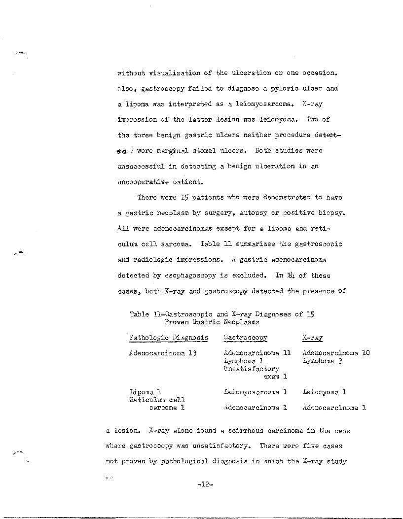
wi thout visualization of the ulcera-tion on one occasion.
Also, gastroscopy failed to diagnose a pyloric ulcer and
a lipoma was interpreted as a leiomyosarcoma. X-raY
impression 01' the latter lesion was leiomyoma. Two of
the three benign gastric ulcers neither procedure deteet-
~d:;,~ were marglimal stomal ulcers. Both studies were
unsuccessful in detecting a benign ulceration in an
uncooperative patient.
There were 15 patients who were demonstrated to have
a gastric neoplasm by surgerj, autopsy or positive biopsy.
All were adenocarcinomas except for a lipoma and reti-
culum cell sarcoma. Table 11 s~~~arizes the gastroscopic
and radiologic impressions. A gastric adenocarcinoma
detected by esophagoscopy is excluded. In 14 of these
cases, both X-raY and gastroscopy detected the presence of
Table Il-Gastroscopic and X-ray Diagnoses of IS Proven Gastric Neoplasms
Pathologic Diagnosis
Adenocarcinoma 13
Lipoma 1 Reticulum cell
sarcoma 1
Gastroscopy
Adenocarcinoma Lymphoma 1 Unsatisfactory
exam
Leiomyos8.rcoma
Adenocarcinoma
X-ray
11 Adenocarcinoma Lymphoma 3
1
1 Leiomyoma 1
1 Adenocarcinoma
a lesion. X-ray alone found a. scirrhous carcinoma in the case
where gastroscopy was unsatisfactory. There were five cases
not proven by pathological diagnosis in which the X-ray study
-12-
10
1
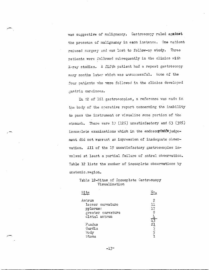
was suggestive of malignancy. Gastroscopy ruled against
the presence of malignancy in each instance. One oat,ient
refused surgery and was lost to follow-up study. Three
patients were followed subsequently in the clinics with
X-ray studies. A fifth patient had a repeat gastroscopy
many months later which was unsuccessful. None of the
four patients who we~e followed in the clinics developed
gastric carcinoma.
In ~2 of 161 gastroscopies, a reference was made in
the body of the operative report concerning the inability
to pass the instrument or visualize some portion of the
stomach. There were 19 (12%) unsatisfactory and 63 (39%)
incomplete examinations which in the endosccpi)s,:t~s judge-
ment did not warrant an impression of inadequate obser-
vation. All of the 19 unsatisfactory gastroscopies in-
volved at least a partial failure of antral observation.
Table 12 lists the number of incomplete observations by
al1atomic * region.
Table l2-Sites of Incomplete Gastroscopy Visualization
Site
Antru.m lesser curvature pyloruss greater curva.ture distal antrum
Fundus Cardia Body Stoma
2 11 17
9 4
4T 21 5 5 1

Twenty-three patients had diagnoses of an antral lesion
by either gastroscopy or X-r~. There were no proven
cases of m~lignancy. X-ray studies diagnosed four cases
as suggestive of carcinoma. In one of these patients,
gastroscopy was inadequate because the scope could not
be adv~~ced into antrum. The patient refused surgery
and the case was lost to follow-up study. On tli'1O oec as
ions gastroscopy ruled out the presence of a malignancy;
one examination was called normal and a diagnosis of
gastro-pancrea.tic cystostomy with secondary hypertrophic
gastritis was made in the other. The normal interpret
ation was made in a case with deformity and persistent
narrowing.:of the antrum by X-re,y study. The fourth
patient had a gastroscopic diagnosis of benign antral
ulcer which was proven to be correct by subsequent surgery.
There were no cases in vrhich gastroscopy alone diagnosed
malignancy ~
There were two proven benign antral ulcers. The
X-ray impression in one of them was malignancy, but
gastroscopy correctly diagnosed benignancy. ~~e other
ulcer was located on the lesser curvature and diagnosed
by gastroscopy alone.
The radiologic interpretation was IIconsistent with
a gastric ulcer, but without demonstration of an ulcer
niche" •
In two~;o'tb. er'. cases, there was a roentgenographic
-14-

impression of possible ~~tral ulcer. Both gastroscopic
studies were reported as inadequate because of failure
to visualize the antrum completely. Surgery failed to
demonstrate a lesion in one case. In the other, bleed
ing was reported as originating in the body of the
stomach at endoscopy, but an ulceration could not be
visualized.
Three additional diagnoses of benign antral ulcer
were made at gastroscopy. None of them were proven by
pathologic study. Only one was confirmed by X-ray.
There were 56 cases with gastric mucosal changes of
various types diagnosed at gastroscopy and/or X-ray.
Only five of these patients had similar diagnoses invol
v,in;gl.ihe observations of hypertrophic gastritis on three
occasions and atrophic gastritis in two instances.
Seven patients had an X-r8Y or gastroscopic diag
u'Osts of post-gastrectomy stoma changes other than ulcer
ation. By endoscopy, stenosis was diagnosed four times,
peristomal hypertrophic gastritis on two occasions and
a. stom.9.l polyp was seen t·w"ice. X-ray detected a sim
ilar lesion in all instances.
Bleeding noted at endoscopy as well as complaints
of hematemesis and melena were recorded in 71 patients.
Cases presenting a problem of anemia were also included
-15-

in this group. Esophago-gastroscopy gave no positive
findings in nine cases and seven examinations were un-
satisfactory or incomplete. The endoscopic diagnoses
and their frequency are shown in table 13.
Table 13-Abnormal Findings in Cases of Suspected and Established Upper Gastro-intestinal Bleeding.
Diagnosis
Gastritis Ulcer Hiatus hernia Tumor Varices Mallory-Weiss syndrome Errosion
No.
26 18 11
9 8 1 1
Three gastric biopsies were performed using the
operating gastroscope. The biopsy results were: infla-
mmation 2 and adenocarcinoma 1. One of the biopsies
reported as infl~~ation was taken from a patient who
had an adenocarcinoma demonstrated at subsequent surg-
ery. The gastroscopic impression of thi.s lesion was
multi-centric tumor, probably lymphoma.
There were no complications from esophagoscopy or
gastroscopy in 272 procedures, On withdr~«al of the
instrument in four examinations, slight bleeding was
noted bu.j,:,it:~was not vigorous in any of these patients,
nor was there any apparent subsequent hemorrhage.
-16-

DISCUSSION
.5 Sullivan and Myers present~ data perta.ining to the
general results of 860 esophagoscopies a.nd 892 gastros-
copies. Their yield of pathologic lesions in these two
procedures respectively was 59.3% and 48.7%. Normal exam-
ination constitutes close to 35% for both esophagoscopy
and gastroscopy in their analysis. Percentages for unsat-
isfactory and incomplete examinations in esophagoscopy and
gastroscopy respectively were: 6.2%, 0.8%; 10.3%, 5.5%.
'J.'he lack of success vn th gastroscopy in their series of
procedures as in this study, is about ~Nice that of esoph-
agoscopy. They attribute this primarily to the anatomy of
the esophagus which allows adequate, complete visualization
follo,ring successful instrumentation. Gastroscopy, on the
other hand, to be complete, requires adequate gastric air
retention and a lack of excess accumulated secretions. The
greater variations in ga.stric contour also act as a limi t-
lng factor.
The most frequent esophagoscopic diagnosis in this
analysis was cardio-esophagea.l incompetence. This finding
involves regurgttation of gastrio secretions into the
distal esophagus. Esophageal varices were just one-half as
frequent. At gastroscopy, gastritis was 1.7 times more
frequent than ulcer. 5 Sullivan and Myers found esophageal
-17-

varices the most rewarding diagnosis by esophagoscopy
with both esophagi tis and hiatus hernia present in respec-
table numbers. Gastritis accounted for over two-thirds of
their diagnoses by gastroscopy with gastric ulcer next
most frequent. Review of the relative incidence of gastritis 6,7, 8
and gastric ulcer in several gastroscopic analyses
shows varying statistics. In these studies gastritis was
diagnosed 1.6 to 6.2 times more often than ulceration.
Gastric ulcer accounted for 10-29% of the diagnoses. In
this analysis, gastritis accounted for only 35% of the gast-
roscopy diagnoses, gastric ulcer constituted 20% of the
diagnoses.
Esophagoscopy made a diagnosis of varices in 17 or 85%
of the known cases in this study. This compares with 7 or
35% by X-raY examination. Brick and Palmer9, in 172 biopsy
proven cirrhotics, demonstrated varices in 62.7% of the
toteS.l series by esophagoscopy as compared to only 14% by
roentgenography. They also showed that the percentage of
diagnoses increases for both X-ray and endoscopy in the
presence of a history of bleeding. Significantly, the X-
ray diagnosis was four times more accurate with a history
hemorrhage. This was attributed to the fact that larger
varices are most apt to be present in the cases of bleeding.
In the detection of esophageal carcinoma, both X-ray
and esophagoscopy were equally important. However, tissue
-18-
biopsy with the esophagoscop:e gave this procedure added
importance. The number of cases of distal esophagitis
and esophageal ulcers were too small to warrant compar-
ison between endoscopyaadd radiography but the data does
suggest the greater value of the former diagnostic method.
Stempien et al.10
, in a clinical and radiological correl-
ation with esophagoscopy in 172 patients, found the latter
procedure superior in the diagnosis of esophag~tjs, hemorr-
hagic errosions and gastritis "vi thin hernial pouches, telan-
giectasia and in certain instances of esophageal varices
and hiatus herp~a. Diverticula and certain instances of
varices and small hiatus hernia were concluded to be more
easily demonstrated by X-ray. In 65 cases of esophagitis,
Spiroll
reported that 53 were Jeefi by esophagoscopy alone
and X-ray alone reported no cases. More diagnoses of
esophagitis would be made if biopsies were performed more 12
1'requently in patients with hiatus hernia. Bernstein
ci tes a pathological study of ,00 unselected hospitaL
autopsies and 100 cases of sudden death. Gross and micro-
scopic study of the esophageal mucosa revealed an incidence
of esophagitis of 36% in the 500 autopsies. Esophagitis
was found in 8% of the 100 cases of sudden death.
Gastroscopy was instrumental in the detection of 32
or 74% of the ulcerative lesions described at either
endoscopy or roentgenography. X-ray examinations detected
-19-

28 61' 65% of the lesions. In a 15 year gastroscopy eva1-6
uation, Zaharias at al. report detection rates of 61.1%
and 94.7% for endoscopy and rqdiography respectively. 13
Comparable figures from r~ssien and Stanton are 71.4%
and (JO.9%.
In the dia_gnosis of benign gastric ulcer, neither
lie.thQd proved to be a superior procedure. !';ach was of aid
to the other in the differentiation of malignancy from
benignancy. Gastroscop¥" was especially effective in ~r:i-
f,y in g ~;J the presence of an benign ulcer as \vell as corr-
ecting erroneous radiologic impressions of malignancy.
Zaharias at al. 6 say that gastroscopy is generally accepted
as complementary to X-ray, not a substityt.e. They further
conclude that X-ray, is especially superior in demonstrating
ulcers in unusual locations and in finding shallow ulcers
and in determ.ining the benign or malignant appe8.l'anCe of
the ulcer ;::dges and tho membrane surrou,,'1ding it. Meadows
and Lefeber? stress that both. X-ray and gastroscopy freq-
uently commit errors of omission and COTIUnission. They
feel X-ray is superior as a screening procedure ,md that
gastroscopy is most effective in identifying the true nature
of a lesion.
Gastric neoplasm was detected equally well by gastros-
copy and radiography. As in the diagnosis of ulcer, each
supplied complementary information. Meadows and Lefeber?
report that the statistics in t'r.W:lr series are in favor of
-20-
-______ P_. ______ --------------------------____________________ __

X-ray. This ?rocedure revealed an abnormality in 89% of
patients with a gastroscopic diagnosis of carcinoma" On
the other hand, only 45% of patients with an X-ray diag-
n08is of carcinoma had':-s:tmil:iir l'esioiur $."j;)~ga:stroSQ'OPY. 8
Schultz et a1. report figures in which roentgenography
and endoscopy are about equal in di8.gn08ing gastric car-
cimoma. Both procedures were within two percentage points
of 70% correct for two 5 year periods analyzed.
The data compiled from the endoscopic report,s emphat-
ically confirms the relative lnace.seibil:±'tyof the antrU1Jl.
Despite tnis limitation though, gastroscopy proved itself
of value in the detection and differentiation of lesions
in certain cases. Degradi et a1. 14, on the basis of 100
cases with antral diagnoses proven either by surgery, autopsy
or clinically, came to the same conclusion. They believe
gastroscopy is especially valuable in the antrum observed
to be spagt.icj rigid or deformed on roentgenography.
Also stressed by this group is its value in correcting
erroneous X-ray interpretations of benign antral ulcers.
Some of the more important technical factors which make
antral visualization difficult are failure of perist"",
alsis in the region, antral adhesions and over-inflation
of the stomach with air. Occasionally inadequate antral
observation is caused by misinterpretation of pylorus
closure. vVhen the closure apparently occurs proximal
-21-

to the pylorus, distally located lesions may be hidden
from view. They conclude that gastroscopy is a valua;...·
1j1e adjunct to radiology in the differential diagnosis of
antral lesions and should be done in all such patients.
Chronic gilstl"it:iswas reported 56 times by gastroscopy.
By subtype, their relative frequencies were: hypertrophic
38%, atrophic 36% and superficial 27%. No cases of mixed
superficial and atrophic gastritis were reported as in 6,7,8
other analyses Review of relative frequency of the
various gastritis subtypes in these reports shows a marked
lack of clinical-histological correlation in chronic gast-15
ri tis. Schindler concludes that gastr!:8aop:ic·· and;
suction biopsies are insufficient in the. differentiation
of the subtypes of chronic gastritis with the exception
of V'ddespread atrophy. The biopsy specimen by these t-V'iO
techniques are too superficial to evaluate all patholog-
ical chauge:s; in the full thickness of the mucosa. Many
of the characteristic features of hypertrophic gastritis
are present below the plane of the biopsy_ Atrophic
changes often occur SpoI'adi~al1Y-;:SQ rthlitmu.c'osal :vi'SUa.li~
zation:c.onoomitantwitn;biopsy is necessary for valid results.
In an analysis of III patients with upper gastro-
intestinal bleeding of un.lmovm etiology, :Meadows and
Lefeber7 had no findings in 54% of the examinations.
They state they are reluctant to attribute bleeding to
-22-

gastritis.
Three cases of gastric polyp were described in 161
gastroscopies for an incidence of 1.9%. This compares
with an incidence of 2% found by Meadows and Lefeber. 6
Za,charias et 13.1. found that X-ray and gastroscopy
were both inefficient means of detecting marginal ulcer
which they state is characteristically jejunal in 10c-
ation and shallow. Table 14 presents their results in
71 patients examined 95 times. Both methods combined
made this diagnosis in only 26 or 37% of the patients
later Droven to have a marginal ulcer.
Table 14-Frequency of I,iarginal Ulcer Diagnosis from Zacharias at 13.1.
Both gastroscopy, Gastroscopy alone X-ray alone • • • Neither detected
X-ray detected • • • • Lt • • • 12
• • • • • • 10 . . . . . . . . .. ... 45 . . . . .
71
'Q d" t 16 t th t t i b' . ~ene 1C repor s . a gas rosoop c 10PSY 1S most
valuable in the diagnosis of lymphoma, carcinoma, and 17
gastritis. Both he and Shallenberger et 13.1. point
out that negative bi.opsy is absolu te only in ruling out
diffuse disease such as lymphoma. The latter author
gives a resume of 60 cases including the following biopsy
findings: gastritiS 20, normal 17, gastric ulcer 14,
malignancy 5, gastric polj-P 2, marginal ulcer:22, and
-23-

polycythemia vera 1. In a series of 310 biopsies in 18
198 patients ,:i"irts collected 30 cases of malignancy
proven subsequently by surgery. Biopsies were positive
in 11 or 37% of the cases. There were no false posit-
ives, 13 (43%) false negatives and 6 (20%) unsatisfact-
ory specimen$ • 19
Palmer a.nd {Virts surveyed for accidents by com-
piling 8. questionaire which they sent to practj.oing
endoscopiats. In a series of 267,175 gastroscopies and
40,540 esophagoscopies, an accident and fatality rate
were fo;~nd to be much less than 1%. The results are
shown in table 15.
Table 15-Accident and Fatality Rates From Pa.L'Tler and Vlirts
Accidents Fatalities
Esophago scopy Gastroscopy
.25%
.079% .059% .014%
The major complications noted in this survey were per-
foration, a~esthetic reaction and post examination
hemorrhage and/or myocardial inf~1ction.
-24-

CONCWSIONS
Esophagoscopy and gastroscopy proved to be useful and
safe diagnostic procedures in the cases presented in this
analysis. There was a yield of abnormal findings in appro
ximately two-thirds of t.he procedures performed. At gast
roscopy, unsatisfactory instrnmentation was most responsible
for inadequate observations but incomplete visualization was
much more £requent. Less difficulty in instrumentation and
visualization was met at esophagoscopy than gastroscopy.
The most frequent diagnoses in this study correlated
well with the results in another similar study. Ca!6dio
esophageal incompe tencs, varices, esophagi tis, carcinoma and
Ulceration were the most frequent esophagoscopic diagnoses.
Gastritis, benign ulcer, hiatus herr~a and carcinoma were the
most frequent diagnoses by gastroscopy.
The ~eriiJr:i:l;.}'" of esophagoscopy over roentgenography in
the detection of esophageal varices was established. This
procedure1]))roved especially important in the diagnosis of
esophageal carcinoma because of concomitant biopsy. Distal
esophageal ulceration when shallow was detected best by
esophagoscopy. In the detection of hiatus hernia, esoph
agoscopy generally gave an impression of cardio-esophageal
incompetency when the gastroscopy described a herniated
segment of stomach. The esophagoscope tends to reduce
the herniation on introduction of the instrument so all
-25-

that is seen is regurgitation of gastric secretions into
the distal esophagus. The air insufflation 13.t gastro
scopy has just the opposite effect on the gastric hernia.
Gastroscopy was shmm to be an aid in the detection
of gastric ulcers as well as in the differentiation of
benigna.'1cy from malignancy. This procedure "vas especially
adept in ~orrecting erroneous X-ray impressions of mal
ignancy. However, tho possibility of terming a malignant
lesion benign was dernomstrated in one caSG.
In this series the antrtLl1l proved a difficult locat
ion to completely Visualize. The lesser curvature and
pyloric areas were especially inaccessible. Despite this
limitation, gastroscopy was useful in the study of this
region of the stomach. Gastroscopy should prove to be of
even greater value in this area with the use of the flex
ible fiberscope.
There was marked lack of correlation between the end
oscopic and X-r~ diagnoses of musocal pathology. Endos
copy revealed these findings much more frequently than
X-ray because it is especially adept in demonstrating
superficial lesions.
In the cases presenting with evidence of upper gast
ro.;;ii.:n:testinal bleeding, inflammation and ulceration were
the most frequent findings with tumor and varices foll
owing in that order. Though a good yield of abnormal
-26-

findin..gs waS achieved by endoscopy, it is difficult to
ascribe bleeding to some of them.
Endoscopy was of least aid in this study in det
ecting esophageal diverticula and marginal ulceration.
1. An analysis of 111 esophagoscopy and 161 gastroscopy
procedures in 178 patients is' pres~ed.
2. Endoscopic findings are compared with X-ray studies
and the clinical course of the patient including,
surgery, autopsy, and biopsy.
3. Endoscopy was found to be an aid to X-ray studies in
obtaining information and in many cases it alone made
a diagno sis.
l~. The in§thrUtl1~;)11tation waS performed without 8Jly comp-
lications.
-27-

1. Bockus, ;t o L" , Company, 191~3.
4. ~. D., 41\ft.>klJ¢j,J.. I)lvieiot'l,
S. J. , Jr., Qnd Dbcl..l.s~ioi1 1"
Diairriet of Colwnbia
6. , L .. I;vGll1at1on,
7.
o , ..
10.
11 •
•
-28-

Association 168:27 1958.
13. Rosslen, A~ X. and Stanton, A., Critical Evaluation of Roentgenology and Gastroscopy in the Diagnosis of Gastric Disorders, American Journal of Gastroenterology 29:4 April 1958.
14. Dagradi, A. E. and others, The Value and Limitations of Gastroscopy in the Diagnosis of Antral Lesions, American Journal of Digestive Diseases 7=993-1000 1962.
15. Schindler, R., Uri tical Evaluation of Biopsy Techniques for the Diagnosis of the Gastritides, American Journal of Digestive Diseases 7:167-76 February 1962.
16. Benedict, E. G., The VifferentialDiagnosis of Benign and Malignant Lesions of the Stomach by Me.ans of the. Flexible Operating Gastroscope, Gastroenterology 14:275 1950.
17. Shallenberger, p. C. and others, Biopsy Through the Flexible Operating Gastroscope, Gastroenterology 16:327, 1950.
18. Wirts, C. N. and others, Experience With the Operating Gastroscope, Gastroenterology 19:777 1951.
19. Palmer, E. D. and Wirts, C .. V'f., Survey on Gastroscopic and Esophagoscopic Accidents: Report of Committee on Accidents of American Gastroscopic Society, Journal of the American Medical Association 164: 2012-5 1957.

. ,.,-..
ACKl\IOWli:DGMENT
I am grateful to Dr. Frederick F. Paustian, B.S., M.D., Associate Professor of Internal Medicine, University of Nebraska College of Medicine, for allowing me to review his endoscopy operative reports and for his advice in the preparation of this thesis. I appreciate the kindness of Merdyth EVans, medical records file clerk, in gratiously obtaining patient charts for me •