diagnostics in ebola virus disease in resource-rich and resource-limited...
TRANSCRIPT
REVIEW
Diagnostics in Ebola Virus Disease in
Resource-Rich and Resource-Limited Settings
Robert J Shorten1,2*, Colin S Brown3,4, Michael Jacobs5, Simon Rattenbury5, Andrew
J. Simpson2,6, Stephen Mepham5
1 Public Health Laboratory Manchester, Manchester Royal Infirmary, Manchester, United Kingdom,
2 University College London, Centre for Clinical Microbiology, Department of Infection, London United
Kingdom, 3 Hospital for Tropical Diseases, University College London Hospital, London, United Kingdom,
4 King’s Sierra Leone Partnership, King’s Centre for Global Health, King’s College London, and King’s
Health Partners, London, United Kingdom, 5 Department of Infection, Royal Free London NHS Foundation
Trust, London, United Kingdom, 6 Rare and Imported Pathogens Laboratory, Public Health England,
Salisbury, United Kingdom
Abstract
The Ebola virus disease (EVD) outbreak in West Africa was unprecedented in scale and
location. Limited access to both diagnostic and supportive pathology assays in both
resource-rich and resource-limited settings had a detrimental effect on the identification
and isolation of cases as well as individual patient management. Limited access to such
assays in resource-rich settings resulted in delays in differentiating EVD from other ill-
nesses in returning travellers, in turn utilising valuable resources until a diagnosis could be
made. This had a much greater impact in West Africa, where it contributed to the initial fail-
ure to contain the outbreak. This review explores diagnostic assays of use in EVD in both
resource-rich and resource-limited settings, including their respective limitations, and some
novel assays and approaches that may be of use in future outbreaks.
Introduction
The 2013–2016 Ebola virus (EBOV) outbreak centred in West Africa is the largest everrecorded and has resulted in a substantial global response, involving 77 centres across Guinea,Sierra Leone, and Liberia. The outbreak, which exceeds 28,500 cases [1], has seen sporadiccases imported to resource-rich settings, such as the United Kingdom, the United States, Spain,Switzerland, and Italy, either in the form of repatriation of confirmed cases or by case identifi-cation after travellers fell ill after returning from West Africa. Secondary transmission has beenrecorded in the US [2] and Spain [3]. Although the World Health Organization (WHO) hasdeclared the outbreak over in West Africa, enhanced surveillance has identified sporadic cases,all of which have been rapidly contained. These ongoing intermittent cases occur mostly fromsexual transmission and are likely to continue for some time.
Diagnostic and other assays play a vital role in confirming or excluding suspect cases, moni-toring disease progression and complications, and discharge planning. In both resource-rich,and resource-limited settings, this allows the appropriate segregation of confirmed cases andquarantine of contacts as well as optimal care and the appropriate allocation of resources.
PLOS Neglected Tropical Diseases | DOI:10.1371/journal.pntd.0004948 October 27, 2016 1 / 16
a11111
OPENACCESS
Citation: Shorten RJ, Brown CS, Jacobs M,
Rattenbury S, Simpson AJ, Mepham S (2016)
Diagnostics in Ebola Virus Disease in Resource-
Rich and Resource-Limited Settings. PLoS Negl
Trop Dis 10(10): e0004948. doi:10.1371/journal.
pntd.0004948
Editor: Manuel Schibler, University of Geneva
Hospitals, SWITZERLAND
Published: October 27, 2016
Copyright: © 2016 Shorten et al. This is an open
access article distributed under the terms of the
Creative Commons Attribution License, which
permits unrestricted use, distribution, and
reproduction in any medium, provided the original
author and source are credited.
Funding: The authors received no specific funding
for this work.
Competing Interests: The authors have declared
that no competing interests exist.
Sporadic outbreaks of human EBOV infection have appeared with increasing frequencysince the virus was first identified in 1976 [4,5]. Person-to-person transmission occurs viadirect contact with blood and body fluids [6], and nosocomial transmission is a prominent fea-ture of outbreaks [7]. Indeed, in the West African outbreak, at least 881 health care workersbecame infected, of which 513 died [8].
Following an incubation period of 2–21 days, patients initially present with nonspecificsymptoms, including fever, headache, malaise, and myalgia. By days 3–5 of the illness, a gastro-intestinal stage develops with epigastric pain, hiccups, nausea, vomiting, and diarrhoea [9].Large volume, watery diarrhoea of five litres or more daily has been reported [10]. By days7–10, neurological manifestations, including delirium, confusion, slowed cognition, or agita-tion and seizures, may present [10]. Purpuric rash, conjunctival injection, and oozing fromvenous catheter sites may occur. Massive haemorrhage from the gastrointestinal tract is rareand normally only occurs in fatal cases [11]. For this reason, the term Ebola virus disease(EVD) is now more widely used than the previous term Ebola haemorrhagic fever. Case fatalityrates (CFR) of previous outbreaks have been as high as 80%–90%, though most have beenbetween 35%–75% [12]. The CFR in this outbreak is estimated to be below 50% by WHO [1].
Several systemic manifestations are seen in EVD. These may be due to a combination of dehy-dration, endothelial damage, disseminated intravascular coagulation (DIC), septic shock, andorgan damage caused by direct viral infection and associated immune responses [13]. The abilityto monitor relevant laboratory parameters is essential for optimal care and can indicate individ-ual patient prognosis. Diarrhoea and volume depletion, likely combined with direct viral renaldamage, lead to electrolyte derangement, which may be severe and life-threatening [10,13,14].Hepatocellular damage is a common feature, and deranged liver enzymes are well documented inanimal models and human cases [15,16]. Raised serum levels of creatine kinase (CK), which maybe measured within a liver function test panel, are also noted to be a marker of severe disease inviral haemorrhagic fevers (VHF), such as Crimean Congo Haemorrhagic Fever (CCHF) [17].Disruption to normal coagulation processes is common [16,18]. Biomarkers of deranged coagu-lation, such as prolonged prothrombin and activated partial thromboplastin times (PT andAPTT, respectively), and DIC, such as the presence of fibrinogen degradation products, arenoted [15,16]. Aside from the enumeration of the number of platelets, a full blood count (FBC)may show both leukopenia [16] and neutrophilia, the latter in the presence of a subsequent bacte-rial sepsis or advanced disease. Anaemia and abnormal erythrocyte indices will be observedwithblood loss. An inability to monitor and correct the complications of EVD, in particular electrolyteimbalance, may have contributed to the high mortality rates early in the West Africa outbreak.
Access to rapid, accurate diagnostic assays is essential to enable appropriate patient manage-ment and may be used to allow effective discharge planning. In addition, effective outbreak con-trol requires the rapid diagnosis, isolation, and treatment of infected individuals, and the follow-up of their contacts. Early containment of the West Africa outbreak was undoubtedly hamperedby a lack of rapid diagnostics that could differentiate EVD from other diseases, given its nonspe-cific clinical features. In both resource-rich and resource-limited settings, the predominant issueat the outbreak onset was limited or absent access to diagnostics allowing EVD to be differenti-ated from other febrile illnesses. Whilst patients in resource-rich settings remained in isolationfacilities for significant periods of time, often blocking side rooms in busy emergency depart-ments, the impact in resource-limited settings was significantly more severe where case identifi-cation and subsequent outbreak response became even more challenging. Patient managementin Ebola Treatment Centres (ETC) in resource-limited settings is challenging for a number ofreasons: large numbers of patients, few staff, and oppressively hot and cumbersome personal pro-tective equipment that limits the time that clinical and support staff can spend in the clinicalareas. Additionally, note-keeping is difficult due to infection control limitations, and the use of
PLOS Neglected Tropical Diseases | DOI:10.1371/journal.pntd.0004948 October 27, 2016 2 / 16
needles and other sharps pose a significant risk to health care workers. Here, we review the assayscurrently available in resource-rich and resource-limited settings as well as some in developmentthat could play a role in the diagnosis and clinical management of EVD.
Diagnostic Methods in a Resource-rich setting
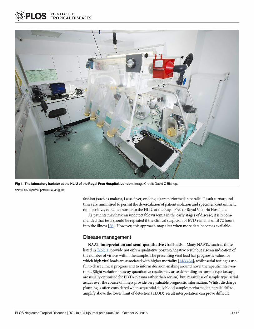
Laboratory Requirements. EBOV is categorised as a high-hazard pathogen that is han-dled at Biosafety Level 4 in the US [19] and is designated as a Hazard Group 4 Pathogen in theUK [20]. Complex facilities are required to examine samples that contain this and similarviruses. These laboratory suites consist of large, highly secure facilities with restricted access,carefully controlled air-handling systems with filtered air, and the meticulous control and inac-tivation of hazardous waste. Staff are highly skilled and often train for years to be able to workat this level of containment. The manipulation of these samples within these laboratories addi-tionally requires either the isolation of the scientist via suited systems or of the specimensthemselves with the use of microbiological safety cabinets. Building, commissioning, maintain-ing, and staffing these units is expensive and labour intensive, so work on such high-hazardpathogens is usually restricted to a few national or regional centres in resource-rich settings. Inthe UK, bespoke Containment Level 3+ pathogen laboratories are also located within the HighLevel Isolation Units (HLIU), Royal Free Hospital (London) (Fig 1), and the Royal VictoriaInfirmary (Newcastle), the national centres for managing confirmed cases of VHF. Whilstthese facilities do not provide Containment Level 4 viral diagnostics per se, they enable staff toprovide pathology assays to exclude alternative diagnoses, such as rapid malaria diagnosticsand blood cultures, and to support patient clinical management decisions. These facilities arestaffed by trained scientific staff and are available at all times.
Ebola-specific diagnostics and exclusion of alternative diagnoses
A febrile patient returning from an area endemic for EVD within 21 days of exposure requiresrapid, accurate, and safe diagnostic tests not only for VHF but also for more common diagno-ses, such as malaria, typhoid, and arboviruses, such as dengue. Samples are taken by medicalstaff wearing appropriate personal protective equipment and sent to a central facility in appro-priate packaging (UN2814 [category A] or UN 3373 [category B] depending on the risk assess-ment) and using a preapproved courier service [21].
Historically, a combination of antigen and antibody detection via enzyme-linked immuno-assay (EIA) and reverse transcriptase polymerase chain reaction (RT-PCR) was used to identifyan outbreak in May 1995 in Zaire [22,23]. Electron microscopy (EM) together with antigenand antibody analysis was used to identify a new variant of Ebola in the Ivory Coast in 1995[24]. The use of RT-PCR in the detection of EBOV has been further described in these out-breaks, as well as for the detection of Ebola Reston [25], and is now recommended [26]. Molec-ular assays have advantages over the previous technologies of EIA, EM, andimmunofluorescence (IF), including increased sensitivity, specificity, and faster turnaround,allowing clinical teams to act upon the results much more rapidly.
The preferred diagnostic method is via direct detection of viral RNA using a nucleic acidamplification test (NAAT) (Table 1). Viral nucleic acid is extracted inside the containment labo-ratory prior to amplification and detection. An additional advantage of this setup is that addi-tional differential diagnoses can be investigated at the same time. Travellers returning to the UKfrom VHF-endemic areas will be tested for a panel of likely pathogens (Table 2). The PublicHealth England Rare & Imported Pathogens Laboratory (RIPL) performs a range of these molec-ular assays according to patient epidemiology, including pan-Ebola–[27] and Ebola Zaire–spe-cific [28] NAATs. Assays for other geographically relevant diseases that may present in a similar
PLOS Neglected Tropical Diseases | DOI:10.1371/journal.pntd.0004948 October 27, 2016 3 / 16
fashion (such as malaria, Lassa fever, or dengue) are performed in parallel. Result turnaroundtimes are minimised to permit the de-escalation of patient isolation and specimen containmentor, if positive, expedite transfer to the HLIU at the Royal Free or Royal Victoria Hospitals.
As patients may have an undetectable viraemia in the early stages of disease, it is recom-mended that tests should be repeated if the clinical suspicion of EVD remains until 72 hoursinto the illness [26]. However, this approach may alter when more data becomes available.
Disease management
NAAT interpretation and semi-quantitative viral loads. Many NAATs, such as thoselisted in Table 1, provide not only a qualitative positive/negative result but also an indication ofthe number of virions within the sample. The presenting viral load has prognostic value, forwhich high viral loads are associated with higher mortality [14,33,34], whilst serial testing is use-ful to chart clinical progress and to inform decision-making around novel therapeutic interven-tions. Slight variation in assay quantitative results may arise depending on sample type (assaysare usually optimised for EDTA plasma rather than serum), but, regardless of sample type, serialassays over the course of illness provide very valuable prognostic information. Whilst dischargeplanning is often considered when sequential daily blood samples performed in parallel fail toamplify above the lower limit of detection (LLOD), result interpretation can prove difficult
Fig 1. The laboratory isolator at the HLIU of the Royal Free Hospital, London. Image Credit: David C Bishop.
doi:10.1371/journal.pntd.0004948.g001
PLOS Neglected Tropical Diseases | DOI:10.1371/journal.pntd.0004948 October 27, 2016 4 / 16
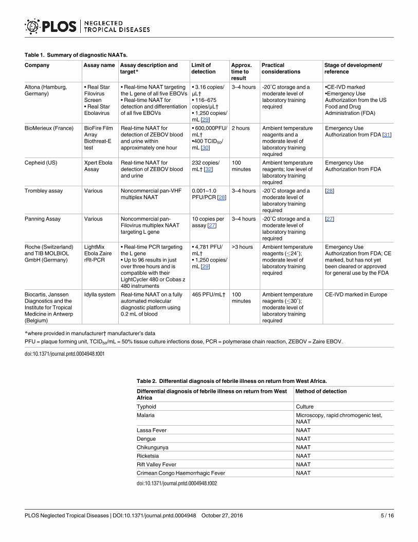
Table 1. Summary of diagnostic NAATs.
Company Assay name Assay description and
target*Limit of
detection
Approx.
time to
result
Practical
considerations
Stage of development/
reference
Altona (Hamburg,
Germany)
• Real Star
Filovirus
Screen
• Real Star
Ebolavirus
• Real-time NAAT targeting
the L gene of all five EBOVs
• Real-time NAAT for
detection and differentiation
of all five EBOVs
• 3.16 copies/
μL†
• 116–675
copies/μL†
• 1,250 copies/
mL [29]
3–4 hours -20˚C storage and a
moderate level of
laboratory training
required
•CE-IVD marked
•Emergency Use
Authorization from the US
Food and Drug
Administration (FDA)
BioMerieux (France) BioFire Film
Array
Biothreat-E
test
Real-time NAAT for
detection of ZEBOV blood
and urine within
approximately one hour
• 600,000PFU/
mL†
•400 TCID50/
mL [30]
2 hours Ambient temperature
reagents and a
moderate level of
laboratory training
required
Emergency Use
Authorization from FDA [31]
Cepheid (US) Xpert Ebola
Assay
Real-time NAAT for
detection of ZEBOV blood
and urine
232 copies/
mL† [32]
100
minutes
Ambient temperature
reagents; low level of
laboratory training
required
Emergency Use
Authorization from FDA
Trombley assay Various Noncommercial pan-VHF
multiplex NAAT
0.001–1.0
PFU/PCR [28]
3–4 hours -20˚C storage and a
moderate level of
laboratory training
required
[28]
Panning Assay Various Noncommercial pan-
Filovirus multiplex NAAT
targeting L gene
10 copies per
assay [27]
3–4 hours -20˚C storage and a
moderate level of
laboratory training
required
[27]
Roche (Switzerland)
and TIB MOLBIOL
GmbH (Germany)
LightMix
Ebola Zaire
rRt-PCR
• Real-time PCR targeting
the L gene
• Up to 96 results in just
over three hours and is
compatible with their
LightCycler 480 or Cobas z
480 instruments
• 4,781 PFU/
mL†
• 1,250 copies/
mL [29]
>3 hours Ambient temperature
reagents (�24˚);
moderate level of
laboratory training
required
Emergency Use
Authorization from FDA; CE
marked, but has not yet
been cleared or approved
for general use by the FDA
Biocartis, Janssen
Diagnostics and the
Institute for Tropical
Medicine in Antwerp
(Belgium)
Idylla system Real-time NAAT on a fully
automated molecular
diagnostic platform using
0.2 mL of blood
465 PFU/mL† 100
minutes
Ambient temperature
reagents (�30˚);
moderate level of
laboratory training
required
CE-IVD marked in Europe
*where provided in manufacturer† manufacturer’s data
PFU = plaque forming unit, TCID50/mL = 50% tissue culture infections dose, PCR = polymerase chain reaction, ZEBOV = Zaire EBOV.
doi:10.1371/journal.pntd.0004948.t001
Table 2. Differential diagnosis of febrile illness on return from West Africa.
Differential diagnosis of febrile illness on return from West
Africa
Method of detection
Typhoid Culture
Malaria Microscopy, rapid chromogenic test,
NAAT
Lassa Fever NAAT
Dengue NAAT
Chikungunya NAAT
Ricketsia NAAT
Rift Valley Fever NAAT
Crimean Congo Haemorrhagic Fever NAAT
doi:10.1371/journal.pntd.0004948.t002
PLOS Neglected Tropical Diseases | DOI:10.1371/journal.pntd.0004948 October 27, 2016 5 / 16
because a low-level viraemia can persist for many days (and the LLOD may vary between assays).As the patient improves clinically, NAAT testing of other anatomical sites may inform the infec-tion risk assessment prior to patient de-isolation, although more data are needed to understandthe relationship between positive tests for EBOV RNA in these sites and risk of transmission.These samples include sputum, throat swabs, sweat, urine, and breast milk, whilst the more inti-mate samples of semen and cervical samples can be deferred to the outpatient clinic setting.
Supportive pathology assays
Measuring biomarkers of the complications of EVD is imperative to monitoring disease progres-sion and assists clinical teams to correct such abnormalities. A nonexhaustive panel of suggestedparameters is listed in Table 3. The ability to analyse these parameters in resource-rich settings iswell established, either in centres of excellence that manage confirmed cases of VHF infection orin routine diagnostic laboratories. Whilst diagnostic assays are awaited, the authors feel that sup-portive pathology assays may be analysed safely using routine processes and autoanalysers fol-lowing appropriate risk assessment [21]. This would enable optimal patient care whilst EVDdiagnostic assays are performed. When reviewing previous imported cases of VHF to resource-rich settings, supportive assays have been performed using routine analysers in standard pathol-ogy laboratories, often prior to the diagnosis being made with no recorded transmissions to labo-ratory workers [35–39]. In addition, over 9,000 cases of CCHF were notified in the whole ofTurkey (2002–2014), with an estimated minimum of 180,000 blood samples processed in routinelaboratories with no additional precautions. A review was performed of 51 health care exposuresthat occurred in nine centres where 4,869 of these patients were managed. Only two cases in lab-oratory staff were identified.One may have been associated with phlebotomy and the other withhandling samples whilst not wearing protective gloves [40].
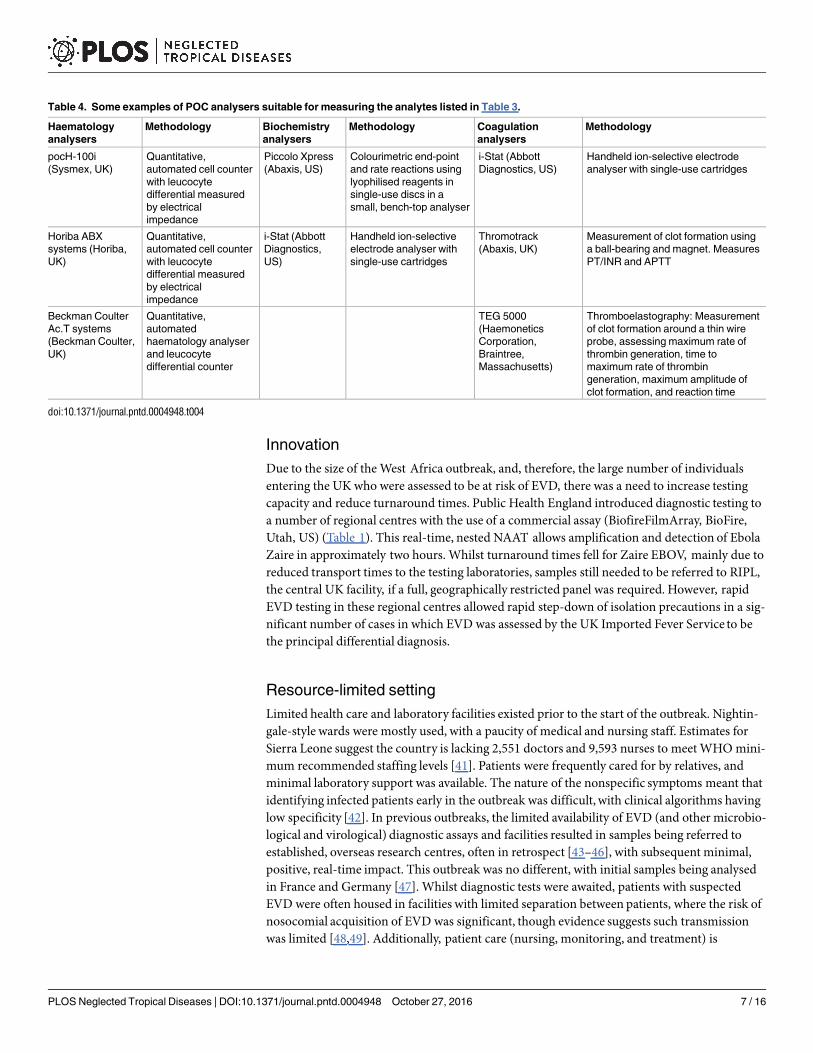
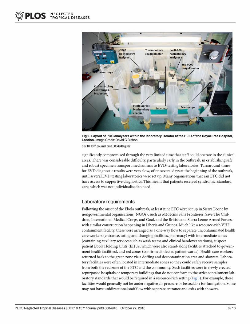
Stand-alone, discrete analysers may be used in high-level containment units either in com-bination with suited systems, safety cabinets, or isolators to protect staff. The HLIU at theRoyal Free uses small, Point of Care (POC) analysers in a designated laboratory to provide theassays listed in Table 3, and some suitable analysers that meet these needs are shown in Table 4and Fig 2. It is noteworthy that the performance of POC analysers may vary significantly fromroutine autoanalysers. The benefits of decreased size, portability, and ease of use should be con-sidered alongside the potentially poorer accuracy and precision of these assays. It is also impor-tant to remember that assay methodologies, and, therefore, normal ranges, vary betweeninstruments. Therefore, results obtained on an autoanalyser in a routine laboratory may not bedirectly comparable to the results obtained on POC instruments in a specialised centre.
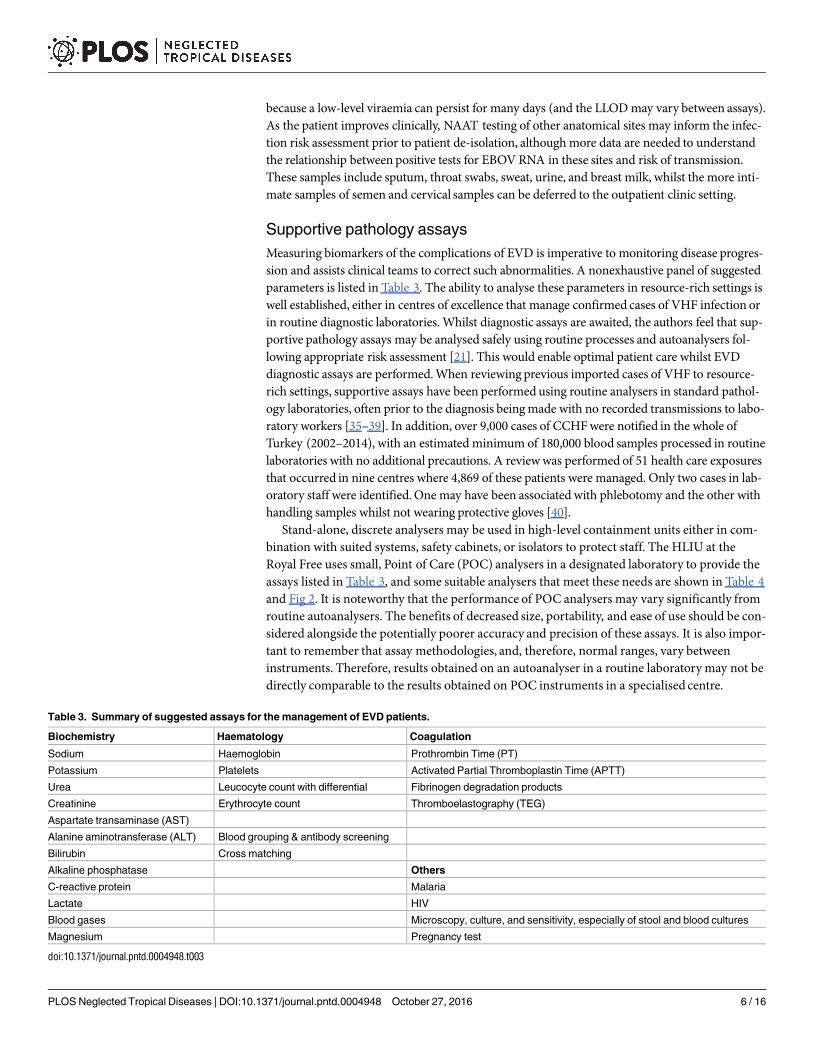
Table 3. Summary of suggested assays for the management of EVD patients.
Biochemistry Haematology Coagulation
Sodium Haemoglobin Prothrombin Time (PT)
Potassium Platelets Activated Partial Thromboplastin Time (APTT)
Urea Leucocyte count with differential Fibrinogen degradation products
Creatinine Erythrocyte count Thromboelastography (TEG)
Aspartate transaminase (AST)
Alanine aminotransferase (ALT) Blood grouping & antibody screening
Bilirubin Cross matching
Alkaline phosphatase Others
C-reactive protein Malaria
Lactate HIV
Blood gases Microscopy, culture, and sensitivity, especially of stool and blood cultures
Magnesium Pregnancy test
doi:10.1371/journal.pntd.0004948.t003
PLOS Neglected Tropical Diseases | DOI:10.1371/journal.pntd.0004948 October 27, 2016 6 / 16
Innovation
Due to the size of the West Africa outbreak, and, therefore, the large number of individualsentering the UK who were assessed to be at risk of EVD, there was a need to increase testingcapacity and reduce turnaround times. Public Health England introduced diagnostic testing toa number of regional centres with the use of a commercial assay (BiofireFilmArray, BioFire,Utah, US) (Table 1). This real-time, nested NAAT allows amplification and detection of EbolaZaire in approximately two hours. Whilst turnaround times fell for Zaire EBOV, mainly due toreduced transport times to the testing laboratories, samples still needed to be referred to RIPL,the central UK facility, if a full, geographically restricted panel was required. However, rapidEVD testing in these regional centres allowed rapid step-down of isolation precautions in a sig-nificant number of cases in which EVD was assessed by the UK Imported Fever Service to bethe principal differential diagnosis.
Resource-limited setting
Limited health care and laboratory facilities existed prior to the start of the outbreak. Nightin-gale-style wards were mostly used, with a paucity of medical and nursing staff. Estimates forSierra Leone suggest the country is lacking 2,551 doctors and 9,593 nurses to meet WHO mini-mum recommended staffing levels [41]. Patients were frequently cared for by relatives, andminimal laboratory support was available. The nature of the nonspecific symptoms meant thatidentifying infected patients early in the outbreak was difficult, with clinical algorithms havinglow specificity [42]. In previous outbreaks, the limited availability of EVD (and other microbio-logical and virological) diagnostic assays and facilities resulted in samples being referred toestablished, overseas research centres, often in retrospect [43–46], with subsequent minimal,positive, real-time impact. This outbreak was no different, with initial samples being analysedin France and Germany [47]. Whilst diagnostic tests were awaited, patients with suspectedEVD were often housed in facilities with limited separation between patients, where the risk ofnosocomial acquisition of EVD was significant, though evidence suggests such transmissionwas limited [48,49]. Additionally, patient care (nursing, monitoring, and treatment) is
Table 4. Some examples of POC analysers suitable for measuring the analytes listed in Table 3.
Haematology
analysers
Methodology Biochemistry
analysers
Methodology Coagulation
analysers
Methodology
pocH-100i
(Sysmex, UK)
Quantitative,
automated cell counter
with leucocyte
differential measured
by electrical
impedance
Piccolo Xpress
(Abaxis, US)
Colourimetric end-point
and rate reactions using
lyophilised reagents in
single-use discs in a
small, bench-top analyser
i-Stat (Abbott
Diagnostics, US)
Handheld ion-selective electrode
analyser with single-use cartridges
Horiba ABX
systems (Horiba,
UK)
Quantitative,
automated cell counter
with leucocyte
differential measured
by electrical
impedance
i-Stat (Abbott
Diagnostics,
US)
Handheld ion-selective
electrode analyser with
single-use cartridges
Thromotrack
(Abaxis, UK)
Measurement of clot formation using
a ball-bearing and magnet. Measures
PT/INR and APTT
Beckman Coulter
Ac.T systems
(Beckman Coulter,
UK)
Quantitative,
automated
haematology analyser
and leucocyte
differential counter
TEG 5000
(Haemonetics
Corporation,
Braintree,
Massachusetts)
Thromboelastography: Measurement
of clot formation around a thin wire
probe, assessing maximum rate of
thrombin generation, time to
maximum rate of thrombin
generation, maximum amplitude of
clot formation, and reaction time
doi:10.1371/journal.pntd.0004948.t004
PLOS Neglected Tropical Diseases | DOI:10.1371/journal.pntd.0004948 October 27, 2016 7 / 16
significantly compromised through the very limited time that staff could operate in the clinicalareas. There was considerable difficulty, particularly early in the outbreak, in establishing safeand robust specimen transport mechanisms to EVD-testing laboratories. Turnaround timesfor EVD diagnostic results were very slow, often several days at the beginning of the outbreak,until several EVD testing laboratories were set up. Many organisations that ran ETC did nothave access to supportive diagnostics. This meant that patients received syndromic, standardcare, which was not individualised to need.
Laboratory requirements
Following the onset of the Ebola outbreak, at least nine ETC were set up in Sierra Leone bynongovernmental organisations (NGOs), such as Médecins Sans Frontières, Save The Chil-dren, International Medical Corps, and Goal, and the British and Sierra Leone Armed Forces,with similar construction happening in Liberia and Guinea. Much like a resource-rich VHFcontainment facility, these were arranged as a one-way flow to separate uncontaminated healthcare workers (entrance, eating and changing facilities, pharmacy) with intermediate zones(containing auxiliary services such as wash teams and clinical handover stations), suspectpatient Ebola Holding Units (EHUs, which were also stand-alone facilities attached to govern-ment health facilities), and red zones (confirmed infected patient wards). Health care workersreturned back to the green zone via a doffing and decontamination area and showers. Labora-tory facilities were often located in intermediate zones so they could safely receive samplesfrom both the red zone of the ETC and the community. Such facilities were in newly erected,repurposedhospitals or temporary buildings that do not conform to the strict containment lab-oratory standards that would be required in a resource-rich setting (Fig 3). For example, thesefacilities would generally not be under negative air pressure or be sealable for fumigation. Somemay not have unidirectional staff flow with separate entrance and exits with showers.
Fig 2. Layout of POC analysers within the laboratory isolator at the HLIU of the Royal Free Hospital,
London. Image Credit: David C Bishop.
doi:10.1371/journal.pntd.0004948.g002
PLOS Neglected Tropical Diseases | DOI:10.1371/journal.pntd.0004948 October 27, 2016 8 / 16
The challenges of providing diagnostics in such settings are numerous, including powercuts that interrupt both diagnostic assays and cold storage of molecular reagents and high envi-ronmental temperatures that may interfere with the optimal performance of some NAAT plat-forms and other POC analysers. The provision of power generators and air conditioning wasnecessary to ensure continuity of laboratory services. Like the clinical facilities, laboratoriesproduce considerable volumes of hazardous waste, which must be stored safely until appropri-ate inactivation and disposal can be arranged.
Ebola-specific diagnostics and exclusion of alternative diagnoses
As a result of the international response, molecular methods for the detection of Ebola are nowwidely used and have been utilised in field hospitals set up in response to previous outbreaks(Table 1). Assays in use in West Africa include the Trombley assay [28] and the RealStar Filovi-rus Screen RT-PCR (Altona, Hamburg, Germany). Such technologies are not always easy touse in relatively unskilled hands, requiring infrastructure, training, and often the assistance, atleast initially, of international staff and resources.
Following the international response, NAATs were deployed in laboratories as they wereestablished by governmental agencies and nongovernmental organisations. Rapid turnaroundtimes were desirable to enable the isolation of infected patients and the discharge of negativepatients from high-risk exposure settings of the Ebola holding centres. Comparison of sequentialcycle threshold (Ct) values was difficult, as different platforms were often used between the refer-ring unit and receiving ETC. Unlike resource-rich settings, sequential Ct values were performedless frequently. Repeat NAATs were usually performed in ETC to assess patient recovery and to
Fig 3. Public Health England Laboratory at the Kerry Town ETC, Sierra Leone. The POC analysers are
situated in the isolator in the foreground. The isolator in the background is used for virus inactivation and
RNA extraction. Image supplied by A. J. Simpson.
doi:10.1371/journal.pntd.0004948.g003
PLOS Neglected Tropical Diseases | DOI:10.1371/journal.pntd.0004948 October 27, 2016 9 / 16
assess whether a patient could be discharged. As in resource-rich settings, an undetectable bloodviral load together with clinical resolution of disease was used as an indication for patient dis-charge in Monrovia, Liberia [10], and in Sierra Leone. Facilities to investigate alternative diagno-ses were largely absent. Other diagnostics were usually limited to malaria rapid diagnostic tests(RDT) [50], meaning that other potentially serious diagnoses such as typhoid, dengue, and Lassafever could well go unrecognised.EVD assays can also be used to assess the efficacyof environ-mental decontamination procedures within EHUs and ETC [51].
Supportive pathology assays
The provision of supportive assays in a resource-limited setting is challenging and is unavail-able in most settings due to expensive equipment and reagents, availability of trained staff toperform the assays and interpret the results, provision of continuous power supply, and refrig-eration and appropriate environmental temperatures. The Piccolo (Abaxis, US) (Table 4) orFuji Dri-Chem NX-500 were used to measure biochemistry parameters in this outbreak inKenema, Sierra Leone [14] as well as in Kerry Town, where it was used alongside the HoribaABX systems (Horiba, UK) for the measurement of FBC. The i-Stat (Abbott Diagnostics, US)was used in Conakry, Guinea to measure basic biochemistry, coagulation, and blood gases [52].However, this required the establishment of dedicated laboratories and containment facilities,including bespoke sealed, negative pressure, high efficiencyparticulate air (HEPA) filtered iso-lators, and trained staff from overseas (Fig 3).
Innovation
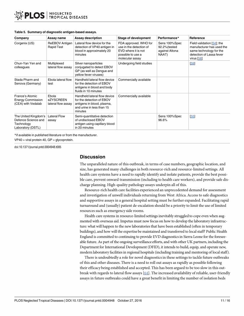
The unprecedented scale and duration of the West Africa outbreak has resulted in the develop-ment of new technologies being offered for trial and approval [53] (Tables 1 and 5). Such VHFdiagnostic POC tests are simple to use and ideal for resource-limited settings where outbreaksand infections are both sporadic and transient [54]. Handheld lateral flow assays that detectviral antigens in blood and body fluids require no electricity, can often be stored at ambienttemperatures, and can provide a result within approximately 10 to 20 minutes. Several arebeing trialled and marketed currently, examples of which are detailed in (Table 5). Given theirreported 100% sensitivity, one significant application is their use as a screening “rule-out” test.This attribute is of considerable importance given the likely need for repeated testing for sus-pect patients in the region [55]. Additionally, there is a need to perform these assays on samplesother than venous blood. When confirming EVD as the cause of death in cadavers, it is saferfor health care and public health staff to take oral swabs rather than to obtain blood samplesand risk a sharps-related injury. This could also apply to living patients for increased ease andsafety, and it has been successful [56,57]. Achieving suitable regulatory approvals for new POCdevices has, however, proved to be an obstacle to the rapid deployment of such assays.
Rapid molecular detection of EBOV has been demonstrated using Reverse Transcription-Loop-Mediated Isothermal Amplification (RT-LAMP) assay [56]. This assay allows amplifica-tion and detection in approximately 35 minutes with excellent sensitivity and specificity,although viral nucleic acid extraction must initially be performed. Other novel technologies,such as portable and low-cost molecular systems [60], handheld NAAT devices [61], and theuse of nanoparticles to detect isothermally amplified nucleic acids [62], have been described.Another innovative molecular assay showing promise is the feasibility of providing real-timegenome sequencing in an outbreak setting [63]. As equipment becomes more portable anddata analysis becomes faster, it is likely that real-time genome sequencing of future outbreakswill bring real benefit in terms of epidemiology and monitoring viral variation.
PLOS Neglected Tropical Diseases | DOI:10.1371/journal.pntd.0004948 October 27, 2016 10 / 16
Discussion
The unparalleled nature of this outbreak, in terms of case numbers, geographic location, andsize, has generated many challenges in both resource-rich and resource-limited settings. Allhealth care systems have a need to rapidly identify and isolate patients, provide the best possi-ble care, prevent onward transmission (including to health care workers), and provide safe dis-charge planning. High-quality pathology assays underpin all of this.
Resource-rich health care facilities experienced an unprecedented demand for assessmentand investigation of unwell individuals returning from West Africa. Access to safe diagnosticsand supportive assays in a general hospital setting must be further expanded. Facilitating rapidturnaround and (usually) patient de-escalation should be a priority to limit the use of limitedresources such as emergency side rooms.
Health care systems in resource-limited settings inevitably struggled to cope even when aug-mented with overseas aid. Impetus must now focus on how to develop the laboratory infrastruc-ture: what will happen to the new laboratories that have been established (often in temporarybuildings), and how will the expertisebe maintained and transferred to local staff? Public HealthEngland is committed to continuing to provide EVD diagnostics in Sierra Leone for the foresee-able future. As part of the ongoing surveillance efforts, and with other UK partners, including theDepartment for International Development (DFID), it intends to build, equip, and operate new,modern laboratory facilities in regional hospitals (including training and mentoring of local staff).
There is undoubtedly a role for novel diagnostics in these settings to tackle future outbreaksof this and other diseases. There is a need to roll out assays as rapidly as possible followingtheir efficacy being established and accepted. This has been argued to be too slow in this out-break with regards to lateral flow assays [64]. The increased availability of reliable, user-friendlyassays in future outbreaks could have a great benefit in limiting the number of isolation beds
Table 5. Summary of diagnostic antigen-based assays.
Company Assay name Assay description Stage of development Performance* Reference
Corgenix (US) ReEBOV Antigen
Rapid Test
Lateral flow device for the
detection of VP40 antigen in
blood in approximately 20
minutes
FDA approved. WHO for
use in the detection of
EVD where it is not
possible to use a
molecular assay
Sens 100%Spec
92.2%(tested
against Altona
NAAT)
Field validation [54]; the
manufacturer has used the
same technology for the
detection of Lassa fever
virus [58]
Chun-Yan Yen and
colleagues
Multiplexed
lateral flow assay
Silver nanoparticles
conjugated to detect EBOV
GP (as well as Dengue and
yellow fever viruses)
Undergoing field studies [59]
Stada Pharm and
Senova (Germany)
Ebola lateral flow
test
Handheld lateral flow device
for the detection of EBOV
antigens in blood and body
fluids in 10 minutes
Commercially available
France’s Atomic
Energy Commission
(CEA) with Vedalab
Ebola
eZYSCREEN
lateral flow assay
Handheld lateral flow device
for the detection of EBOV
antigens in blood, plasma,
and urine in less than 15
minutes
Commercially available
The United Kingdom’s
Defence Science and
Technology
Laboratory (DSTL)
Lateral Flow
assay
Semi-quantitative detection
of undisclosed EBOV
antigen using capillary blood
in 20 minutes
Sens 100%Spec
96.6%
[55]
*if available in published literature or from the manufacturer.
VP40 = viral protein 40, GP = glycoprotein.
doi:10.1371/journal.pntd.0004948.t005
PLOS Neglected Tropical Diseases | DOI:10.1371/journal.pntd.0004948 October 27, 2016 11 / 16
required for EVD testing, controlling transmission, and improving patient outcomes. More-over, traditional microbiological and virological assays need to be deployed across the region toallow for diagnosis of a variety of parasitic, bacterial, and viral pathogens. This will allow foralternative diagnoses of unexplained febrile illness to be rapidly made as well as provide a base-line for disease prevalence in the affected countries.
There is a continued need for investment in this area—new assays need to be developed, eval-uated, and embedded into local health care systems to allow the prompt control of future out-breaks. For instance, rapid, low-cost molecular detection of Zika virus has been achieved usingprogrammable, biosynthetic components, which may be applicable in an outbreak setting [65].Additional vigilance and surveillance is required to ensure that such assays meet the diagnosticneeds as new viruses such as Bundibugyo EBOV are discovered [66] and inevitable sequence var-iation occurs [67–70]. Finally, up-skilling and retention of laboratory staff, along with sustainedresourcing of basic pathology services,needs to be at the fore of long-term resilience plans. Therecent Ebola outbreak resulted in numerous lessons learned and significant innovation. As aresult, it is hoped that future outbreaks will be identified faster and ultimately terminated moreefficiently in part through greater access to portable, easy-to-use diagnostic assays.
Key Learning Points
• The initial identification and containment of this outbreak was hampered by pooraccess to diagnostic assays.
• Patients with Ebola in West Africa were often managed in the absence of supportivepathology assays, which may have led to suboptimal care.
• There is a need to roll out diagnostic and supportive assays much more quickly in thenext such outbreak, including the rapid establishment of clinical trials for newtechnologies.
• Unwell travellers who returned to resource-rich settings from West Africa were oftenisolated for some time (and investigations/treatment delayed) whilst Ebola assays wereperformed in reference facilities.
• Unwell travellers who are assessed to be at high risk for Ebola and other VHF can bemanaged safely in resource-rich settings whilst diagnostic results are awaited. Thisincludes the processing of routine pathology assays.
Top Five Papers
1. Baize S, Pannetier D, Oestereich L, et al. Emergence of Zaire Ebola virus disease inGuinea. N Engl J Med. 2014 Oct 9;371(15):1418–25. Epub 2014 Apr 16.
2. Kortepeter MG, Bausch DG, Bray M. Basic clinical and laboratory features of filoviralhemorrhagic fever. J Infect Dis. 2011;204 Suppl(Suppl 3):S810–6.
3. Fletcher TE, Fowler R a., Beeching NJ. Understanding organ dysfunction in Ebolavirus disease. Intensive Care Med. 2014;40(12):1936–9.
4. Schieffelin JS, Shaffer JG, Goba A, Gbakie M, Gire SK, Colubri A, et al. Clinical Illnessand Outcomes in Patients with Ebola in Sierra Leone. N Engl J Med. 2014;371:2092–100.
5. Racsa LD, Kraft CS, Olinger GG, Hensley LE. Viral Hemorrhagic Fever Diagnostics.Clin Infect Dis. 2016 Jan 15;62(2):214–9.
PLOS Neglected Tropical Diseases | DOI:10.1371/journal.pntd.0004948 October 27, 2016 12 / 16
Acknowledgments
We would like to thank David C. Bishop, Photography ServicesManager, University CollegeLondon Illustration Services,Royal Free Hospital, London, United Kingdom for creating Figs 1and 2.
References1. WHO. Ebola Situation Report. 2016;(February 3rd):1–16.
2. Chevalier MS, Chung W, Smith J, Weil LM, Hughes SM, Joyner SN, et al. Ebola Virus Disease Cluster
in the United States—Dallas County, Texas, 2014. Morb Mortal Wkly Rep. 2014; 63(46):1087–8.
3. Parra JM, Salmeron OJ, Velasco M. The First Case of Ebola Virus Disease Acquired outside Africa. N
Engl J Med [Internet]. 2014; 371(25):2439–40. doi: 10.1056/NEJMc1412662 PMID: 25409262
4. Johnson K, Webb P, Lange J, Murphy F. Isolation and partial characterisation of a new virus causing
acute haemorrhagic fever in Zaire. Lancet. 1977;(5: ):569–71.
5. Bowen ETW, Lloyd G, Harris WJ, Platt GS, Baskerville A, Vella EE. Viral haemorrhagic fever in South-
ern Sudan and Northern Zaire. Lancet. 1977;571–3.
6. Dowell SF, Mukunu R, Ksiazek TG, Khan a S, Rollin PE, Peters CJ. Transmission of Ebola hemor-
rhagic fever: a study of risk factors in family members, Kikwit, Democratic Republic of the Congo,
1995. Commission de Lutte contre les Epidemies à Kikwit. J Infect Dis. 1999; 179 Suppl: S87–91. doi:
10.1086/514284 PMID: 9988169
7. Fisher-Hoch SP. Lessons from nosocomial viral haemorrhagic fever outbreaks. Br Med Bull. 2005; 73–
74:123–37. doi: 10.1093/bmb/ldh054 PMID: 16373655
8. Public Health England. Ebola Epidemiological Update No. 62: 8 January 2016 Ebola virus disease out-
break in West Africa (December 2013 to present). Public Heal England; Ebola Epidemiol Updat. 2016;
(62):1–5.
9. Kortepeter MG, Bausch DG, Bray M. Basic clinical and laboratory features of filoviral hemorrhagic
fever. J Infect Dis [Internet]. 2011; 204 Suppl(Suppl 3):S810–6. doi: 10.1093/infdis/jir299 PMID:
21987756
10. Chertow DS, Kleine C, Edwards JK, Scaini R, Giuliani R, Sprecher A. Ebola Virus Disease in West
Africa—Clinical Manifestations. N Engl J Med. 2014;2054–7.
11. Beeching NJ, Fenech M, Houlihan CF. Ebola virus disease. BMJ [Internet]. 2014;7348(December):1.
12. Centers for Disease Control. Outbreaks Chronology: Ebola Virus Disease [Internet]. 2016.
13. Fletcher TE, Fowler R a., Beeching NJ. Understanding organ dysfunction in Ebola virus disease. Inten-
sive Care Med [Internet]. 2014; 40(12):1936–9. doi: 10.1007/s00134-014-3515-1 PMID: 25366120
14. Schieffelin JS, Shaffer JG, Goba A, Gbakie M, Gire SK, Colubri A, et al. Clinical Illness and Outcomes
in Patients with Ebola in Sierra Leone. N Engl J Med. 2014; 371:2092–100. doi: 10.1056/
NEJMoa1411680 PMID: 25353969
15. Rollin PE, Bausch DG, Sanchez A. Blood chemistry measurements and D-Dimer levels associated
with fatal and nonfatal outcomes in humans infected with Sudan Ebola virus. J Infect Dis. 2007; 196
Suppl (Suppl 2):S364–71. doi: 10.1086/520613 PMID: 17940972
16. Feldmann H, Geisbert TW. Ebola haemorrhagic fever. Lancet [Internet]. Elsevier Ltd; 2011; 377
(9768):849–62. doi: 10.1016/S0140-6736(10)60667-8 PMID: 21084112
17. Ergonul O, Celikbaş A, Dokuzoguz B, Eren S, Baykam N, Esener H. Characteristics of patients with
Crimean-Congo hemorrhagic fever in a recent outbreak in Turkey and impact of oral ribavirin therapy.
Clin Infect Dis. 2004; 39(2):284–7. doi: 10.1086/422000 PMID: 15307042
18. Wilson AJ, Martin DS, Maddox V, Rattenbury S, Bland D, Bhagani S, et al. Thromboelastography in
the management of coagulopathy associated with Ebola virus disease. Clin Infect Dis [Internet].
2015;1–3.
19. US Department of Health and Human Services. Biosafety in Microbiological and Biomedical Laborato-
ries. Public Heal Serv [Internet]. 1999;5th Editio(April):1–250.
20. Advisory Committee on Dangerous Pathogens. The Approved List of Biological Agents. Heal Saf
Exec. 2013;1–32.
21. Advisory Committee on Dangerous Pathogens. Management of Hazard Group 4 viral haemorrhagic
fevers and similar human infectious diseases of high consequence. 2015;(November).
22. CDC. Outbreak of Ebola Viral Hemorrhagic Fever—Zaire, 1995. Morb Mortal Wkly Rep. 1995; 44
(19):381–2.
PLOS Neglected Tropical Diseases | DOI:10.1371/journal.pntd.0004948 October 27, 2016 13 / 16
23. Muyembe T, Kipasa M, Fevers O behalf of the IS and TC and WCC for H. Ebola haemorrhagic fever in
Kikwit, Zaire. Lancet. 1995; 345(8962):1448.
24. Le Guenno B, Formenty P, Wyers M, Gounon P, Walker F, Boesch C. Isolation and partial characteri-
sation of a new strain of Ebola virus. Lancet [Internet]. 1995; 345(8960):1271–4. PMID: 7746057
25. Sanchez a, Ksiazek TG, Rollin PE, Miranda ME, Trappier SG, Khan a S, et al. Detection and molecular
characterization of Ebola viruses causing disease in human and nonhuman primates. J Infect Dis.
1999; 179 Suppl (Suppl 1):S164–9. doi: 10.1086/514282 PMID: 9988180
26. WHO. Laboratory diagnosis of Ebola virus disease. 2014;(September):1–4.
27. Panning M, Laue T, Olschlager S, Eickmann M, Becker S, Raith S, et al. Diagnostic reverse-transcrip-
tion polymerase chain reaction kit for filoviruses based on the strain collections of all European bio-
safety level 4 laboratories. J Infect Dis. 2007; 196 Suppl: S199–204. doi: 10.1086/520600 PMID:
17940950
28. Trombley AR, Wachter L, Garrison J, Buckley-Beason V a., Jahrling J, Hensley LE, et al. Short report:
Comprehensive panel of real-time TaqMan polymerase chain reaction assays for detection and abso-
lute quantification of filoviruses, arenaviruses, and new world hantaviruses. Am J Trop Med Hyg. 2010;
82(5):954–60. doi: 10.4269/ajtmh.2010.09-0636 PMID: 20439981
29. Cherpillod P, Schibler M, Vieille G, Cordey S, Mamin A, Vetter P, et al. Ebola virus disease diagnosis
by real-time RT-PCR: A comparative study of 11 different procedures. J Clin Virol [Internet]. Elsevier
B.V.; 2016; 77:9–14. doi: 10.1016/j.jcv.2016.01.017 PMID: 26874083
30. Southern TR, Racsa LD, Albari??o CG, Fey PD, Hinrichs SH, Murphy CN, et al. Comparison of FilmAr-
ray and quantitative real-time reverse transcriptase PCR for detection of zaire ebolavirus from con-
trived and clinical specimens. J Clin Microbiol. 2015; 53(9):2956–60. doi: 10.1128/JCM.01317-15
PMID: 26157148
31. Leski T a, Ansumana R, Taitt CR, Lamin JM, Bangura U, Lahai J, et al. Use of the FilmArray System
for Detection of Zaire ebolavirus in a Small Hospital in Bo, Sierra Leone. J Clin Microbiol [Internet].
2015; 53(7):2368–70. doi: 10.1128/JCM.00527-15 PMID: 25972415
32. Pinsky B a., Sahoo MK, Sandlund J, Kleman M, Kulkarni M, Grufman P, et al. Analytical performance
characteristics of the cepheid GeneXpert Ebola Assay for the detection of Ebola virus. PLoS ONE.
2015; 10(11):1–16.
33. Sanchez A, Lukwiya M, Bausch D, Mahanty S, Sanchez AJ, Wagoner KD, et al. Analysis of Human
Peripheral Blood Samples from Fatal and Nonfatal Cases of Ebola (Sudan) Hemorrhagic Fever: Cellu-
lar Responses, Virus Load, and Nitric Oxide Levels Analysis of Human Peripheral Blood Samples from
Fatal and Nonfatal Cases of Ebola (S. J Morphol. 2004; 78(19):10370–7.
34. Bah EI, Lamah M-C, Fletcher T, Jacob ST, Brett-Major DM, Sall AA, et al. Clinical Presentation of
Patients with Ebola Virus Disease in Conakry, Guinea. N Engl J Med [Internet]. 2015; 372(1):40–7.
doi: 10.1056/NEJMoa1411249 PMID: 25372658
35. Crowcroft NS, Meltzer M, Evans M, Shetty N, Maguire H, Bahl M, et al. The public health response to a
case of Lassa fever in London in 2000. J Infect. 2004; 48(3):221–8. doi: 10.1016/j.jinf.2003.11.009
PMID: 15001300
36. Atkin S, Anaraki S, Gothard P, Walsh A, Brown D, Gopal R, et al. The first case of Lassa fever imported
from Mali to the United Kingdom, Feruary 2009. Eurosurveillance. 2009; 14(10):2–4.
37. Kitching A, Addiman S, Cathcart S, Bishop L, Krahe D, Nicholas M, et al. A fatal case of Lassa fever in
London, Janyary 2009. Eurosurveillance. 2009; 14(6):2–4.
38. Haas WH, Breuer T, Pfaff G, Schmitz H, Kohler P, Asper M, et al. Imported Lassa fever in Germany:
surveillance and management of contact persons. Clin Infect Dis. 2003; 36(10):1254–8. doi: 10.1086/
374853 PMID: 12746770
39. Barr D a., Aitken C, Bell DJ, Brown CS, Cropley I, Dawood N, et al. First confirmed case of Crimean-
Congo haemorrhagic fever in the UK. Lancet. 2013; 382(9902):1458. doi: 10.1016/S0140-6736(13)
61718-3 PMID: 24243135
40. Leblebicioglu H, Sunbul M, Guner R, Bodur H, Bulut C, Duygu F, et al. Healthcare-associated Cri-
mean-Congo haemorrhagic fever in Turkey, 2002–2014: A multicentre retrospective cross-sectional
study. Clin Microbiol Infect [Internet]. Elsevier Ltd; 2016; 22(4):387.e1–387.e4.
41. Oxfam. Never Again? Oxfam Brief Note [Internet]. 2015;203(April).
42. Vogt F, Fitzpatrick G, Patten G, van den Bergh R, Stinson K, Pandolfi L, et al. Assessment of the MSF
triage system, separating patients into different wards pending ebola virus laboratory confirmation, Kai-
lahun, Sierra Leone, July to September 2014. Eurosurveillance. 2015; 20(50):1–9.
43. Jezek Z, Szczeniowski MY, Muyembe-Tamfum JJ, McCormick JB, Heymann DL. Ebola between out-
breaks: intensified Ebola hemorrhagic fever surveillance in the Democratic Republic of the Congo,
1981–1985. J Infect Dis. 1999; 179 Suppl (Suppl 1):S60–4. doi: 10.1086/514295 PMID: 9988166
PLOS Neglected Tropical Diseases | DOI:10.1371/journal.pntd.0004948 October 27, 2016 14 / 16
44. Leroy EM, Baize S, Lu CY, McCormick JB, Georges a. J, Georges-Courbot MC, et al. Diagnosis of
Ebola haemorrhagic fever by RT-PCR in an epidemic setting. J Med Virol. 2000; 60(4):463–7. PMID:
10686031
45. Leroy EM, Souquière S, Rouquet P, Drevet D. Re-emergence of ebola haemorrhagic fever in Gabon.
Lancet. 2002; 359(9307):712.
46. Okware SI, Omaswa FG, Zaramba S, Opio a., Lutwama JJ, Kamugisha J, et al. An outbreak of Ebola
in Uganda. Trop Med Int Heal. 2002; 7(12):1068–75.
47. Baize S, Pannetier D, Oestereich L, Rieger T, Koivogui L, Magassouba N, et al. Emergence of Zaire
Ebola Virus Disease in Guinea—Preliminary Report. N Engl J Med [Internet]. 2014;1–8.
48. Arkell P, Youkee D, Brown CS et al. Quantifying the risk of nosocomial infection within Ebola Holding
Units: a cohort study of negative patients discharged from five Ebola Holding Units in Western Area,
Sierra Leone. Submitted. 2016;
49. Fitzpatrick G, Vogt F, Moi Gbabai OB, Black B, Santantonio M, Folkesson E, et al. Describing readmis-
sions to an ebola case management centre (CMC), Sierra Leone, 2014. Eurosurveillance. 2014; 19
(40):1–6.
50. WHO. WHO Global Malaria Programme Information note on recommended selection criteria for pro-
curement of malaria rapid diagnostic tests (RDTs). 2012;2014(December):1–13.
51. Youkee D, Brown CS, Lilburn P, Shetty N, Brooks T, Simpson A, et al. Assessment of Environmental
Contamination and Environmental Decontamination Practices within an Ebola Holding Unit, Freetown,
Sierra Leone. PLoS ONE [Internet]. 2015; 10(12):e0145167. doi: 10.1371/journal.pone.0145167
PMID: 26692018
52. Cotte J, Janvier F, Cordier P-Y, Bordes J, Kaiser E. Letter to the Editor Letter to the Editor. Anaesth
Crit care pain Med Crit care pain Med [Internet]. 2015; 34:363–4.
53. Butler D. Ebola experts seek to expand testing. Nature. 2014; 516(7530):154–5. doi: 10.1038/516154a
PMID: 25503213
54. Broadhurst MJ, Kelly JD, Miller A, Semper A, Bailey D, Groppelli E, et al. ReEBOV Antigen Rapid Test
kit for point-of-care and laboratory-based testing for Ebola virus disease: a field validation study. Lan-
cet [Internet]. Elsevier Ltd; 2015; 6736(15):1–8.
55. Walker NF, Brown CS, Youkee D, Baker P, Williams N, Kalawa a., et al. Evaluation of a point-of-care
blood test for identification of Ebola virus disease at Ebola holding units, Western Area, Sierra Leone,
January to February 2015. Euro Surveill Bull Eur sur les Mal Transm = Eur Commun Dis Bull. 2015; 20
(12):1–6.
56. Kurosaki Y, Magassouba N, Oloniniyi OK, Cherif MS, Sakabe S, Takada A, et al. Development and
Evaluation of Reverse Transcription-Loop-Mediated Isothermal Amplification (RT-LAMP) Assay Cou-
pled with a Portable Device for Rapid Diagnosis of Ebola Virus Disease in Guinea. PLoS Negl Trop
Dis. 2016; 10(2):1–12.
57. Formenty P, Leroy EM, Epelboin A, Libama F, Lenzi M, Sudeck H, et al. Detection of Ebola virus in oral
fluid specimens during outbreaks of Ebola virus hemorrhagic fever in the Republic of Congo. Clin Infect
Dis. 2006; 42(11):1521–6. doi: 10.1086/503836 PMID: 16652308
58. Boisen ML, Schieffelin JS, Goba A, Oottamasathien D, Jones AB, Shaffer JG, et al. Multiple Circulating
Infections Can Mimic the Early Stages of Viral Hemorrhagic Fevers and Possible Human Exposure to
Filoviruses in Sierra Leone Prior to the 2014 Outbreak. Viral Immunol [Internet]. 2015; 28(1):19–31.
doi: 10.1089/vim.2014.0108 PMID: 25531344
59. Yen C-W, de Puig H, Tam JO, Gomez-Marquez J, Bosch I, Hamad-Schifferli K, et al. Multicolored sil-
ver nanoparticles for multiplexed disease diagnostics: distinguishing dengue, yellow fever, and Ebola
viruses. Lab Chip [Internet]. Royal Society of Chemistry; 2015; 15:1638–41. doi: 10.1039/c5lc00055f
PMID: 25672590
60. Chan K, Wong PY, Yu P, Hardick J, Wong KY, Wilson S a., et al. A rapid and low-cost PCR thermal
cycler for infectious disease diagnostics. PLoS ONE. 2016; 11(2):1–17.
61. Ahrberg CD, Manz A, Neuzil P. Palm-sized device for point-of-care Ebola detection. Anal Chem [Inter-
net]. 2016;acs.analchem.6b00278.
62. Wang Y, Zhu G, Qi W, Li Y, Song Y. A versatile quantitation platform based on platinum nanoparticles
incorporated volumetric bar-chart chip for highly sensitive assays. Biosens Bioelectron [Internet]. Else-
vier; 2016; 85:777–84. doi: 10.1016/j.bios.2016.05.090 PMID: 27285358
63. Quick J, Loman NJ, Duraffour S, Simpson JT, Severi E, Cowley L, et al. Real-time, portable genome
sequencing for Ebola surveillance. Nature [Internet]. Nature Publishing Group; 2016; 530(7589):228–
32. doi: 10.1038/nature16996 PMID: 26840485
64. Butler D. Researchers frustrated by failure to roll out “game-changing” Ebola test. Nature. 2015;
PLOS Neglected Tropical Diseases | DOI:10.1371/journal.pntd.0004948 October 27, 2016 15 / 16
65. Pardee K, Green A a., Takahashi MK, Braff D, Lambert G, Lee JW, et al. Rapid, low-cost detection of
Zika virus using programmable biomolecular components. Cell [Internet]. Elsevier Inc.; 2016; 165
(5):1–12.
66. Towner JS, Sealy TK, Khristova ML, Albariño CG, Conlan S, Reeder S a., et al. Newly discovered
Ebola virus associated with hemorrhagic fever outbreak in Uganda. PLoS Pathog. 2008; 4(11):3–8.
67. Carroll S a, Towner JS, Sealy TK, McMullan LK, Khristova ML, Burt FJ, et al. Molecular evolution of
viruses of the family Filoviridae based on 97 whole-genome sequences. J Virol [Internet]. 2013; 87
(5):2608–16. doi: 10.1128/JVI.03118-12 PMID: 23255795
68. Carroll MW, Matthews D a., Hiscox J a., Elmore MJ, Pollakis G, Rambaut A, et al. Temporal and spatial
analysis of the 2014–2015 Ebola virus outbreak in West Africa. Nature [Internet]. 2015;
69. Gire SK, Goba A, Andersen KG, Sealfon RSG, Park DJ, Kanneh L, et al. Genomic surveillance eluci-
dates Ebola virus origin and transmission during the 2014 outbreak. Science (80-). 2014; 345
(6202):1369–72.
70. Hoenen T, Safronetz D, Groseth A, Wollenberg KR, Koita OA, Diarra B, et al. Mutation rate and geno-
type variation of Ebola virus from Mali case sequences. Science (80-). 2015; 348(6230):117–9.
PLOS Neglected Tropical Diseases | DOI:10.1371/journal.pntd.0004948 October 27, 2016 16 / 16