dietary red palm oil-supplementation offers cardioprotection
TRANSCRIPT
DIETARY RED PALM OIL-SUPPLEMENTATION OFFERS CARDIOPROTECTION AGAINST
ISCHAEMIA/REPERFUSION INJURY: POSSIBLE CELLULAR MECHANISMS INVOLVED
by
Adriaan Johannes Esterhuyse
Dissertation presented for the Degree of
DOCTOR OF PHILOSOPHY (Physiology)
in the
Department of Physiological Sciences
at University of Stellenbosch
Stellenbosch
Promoter: Dr Jacques van Rooyen Department of Physiological Sciences University of Stellenbosch Co-promoter: Dr Eugene F du Toit Department of Medical Physiology and Biochemistry University of Stellenbosch December 2005
I
INDEX PAGE LIST OF FIGURES LIST OF TABLES LIST OF ABBREVIATIONS DECLARATION ACKNOWLEDGEMENTS ABSTRACT UITTREKSEL CHAPTER 1. INTRODUCTION 1.1 Aims of the study 8 CHAPTER 2. LITERATURE REVIEW 2.1 Dietary fats and oils in health 9 2.2 Fatty acids 10 2.2.1 Saturated, monounsaturated and polyunsaturated fatty acids in
cardiovascular health 10
2.2.2 Essential fatty acids 14 2.2.3 Physiological role of essential fatty acids 15 2.3 Cholesterol-enriched diets 19 . 2.3.1 Introduction to cholesterol-enriched diets 19 2.3.2 Mechanisms of myocardial effects of hyperlipidaemia 19 2.4 Palm oil 19 2.4.1 Introduction to palm oil 19 2.4.2 Composition of palm oil 20 2.4.3 Modulation of lipids and lipoproteins by dietary palm oil-
II
supplementation 20
2.4.4 Epidemiological Studies: lipids 23 2.4.5 Cardiovascular protection offered by palm oil components 25 2.4.5.1 Carotenoids 25 2.4.5.2 Vitamin E (tocopherols and tocotrienols) 26 2.4.5.3 Ubiquinones 29 2.4.6 Palm oil protection against breast cancer 29 2.4.6.1 Coenzyme Q10 protection 30 2.4.6.2 Tocotrienol protection 31 2.4.6.3 Vitamin E succinate protection 31 2.4.7 Effects of palm oil-supplementation on NO-cGMP signalling 31 2.5 Role of nitric oxide in myocardial ischaemia and reperfusion 32 2.5.1 Introduction to nitric oxide cardiovascular protection 32 2.5.2 Mechanisms for the cardiovascular protective effect of nitric oxide 34 2.5.2.1 NO-cGMP signalling 34 2.5.2.2 Cholesterol enriched diet and NO-cGMP signalling 36 2.5.2.3 Antioxidant properties of nitric oxide 38 2.5.2.4 Nitric oxide and production of cytoprotective prostanoids 38 . 2.5.2.5 Therapeutical potential of nitric oxide 38 2.6 Mitogen-activated protein kinases (MAPKs) 38 2.6.1 Apoptosis 38 2.6.2 Signalling pathways and apoptosis 39 2.6.3 Eicosapentaenoic acid (EPA) and docosapentaenoic acid (DHA)
modulate mitogen-activated protein kinase (MAPK) activity 40
III
2.6.4 The major multiple mitogen-activated protein kinase (MAPK) and
PKB/Akt signalling pathways 40
2.6.4.1 p38 MAPK 40 2.6.4.1.1 Inhibition of p38 MAPK 42 2.6.4.1.2 Activation of p38 MAPK 43 2.6.4.2 c-Jun N-terminal kinase (JNK) 44 2.6.4.2.1 The role of c-Jun N-kinase (JNK) in apoptosis 44 2.6.4.3 Extracellular signal-regulated kinases (ERK) 45 2.6.4.3.1 The role of extracellular signal-regulated kinases (ERK) in
apoptosis 46
. 2.6.4.4 Protein kinase B (PKB/Akt) pathway 46 2.6.5 Caspases 47 2.6.5.1 Mechanisms of caspase activation 48 2.6.5.2 Poly-(ADP-ribose) polymerse (PARP) 48 CHAPTER 3. MATERIALS AND METHODS 3.1 Animal Care 50 3.2 Experimental Model 1 50 3.2.1 Experimental groups 50 3.2.2 Working heart perfusion 51 3.2.3 Parameters measured and calculations used 53 3.2.3.1 Left ventricular developed pressure (mmHg) 53 3.2.3.2 Aortic output recovery (%) 54 3.2.3.3 Biochemical analyses 54 3.2.3.3.1 cGMP assay 54 3.2.3.3.2 cAMP assay 55
IV
3.2.3.4 Heart muscle total phospholipid fatty acids (%) 56 3.2.3.5 Serum lipids 56 3.2.3.6 Statistical methods 56 3.3 Experimental Model 2 58 3.3.1 Experimental groups 58 3.3.2 Working heart perfusion and study design 59 3.3.3 Parameters measured and calculations used 59 3.3.3.1 Rate pressure product recovery 59 3.3.3.2 Statistical methods 59 3.4 Experimental Model 3 60 3.4.1 Experimental groups 60 3.4.2 Working heart perfusion 60 3.4.3 Parameters measured and calculations used 61 3.4.3.1 Cardiac functional parameters and aortic output recovery 61 3.4.3.2 Measurement of cGMP 61 3.4.3.3 Measurement of cardiac nitric oxide concentrations 61 3.4.3.4 Measurement of cardiac nitric oxide synthase activity 62 3.4.3.5 Measurement of cardiac superoxide dismutase activity 63 3.4.3.6 Measurement of cardiac lipid hydroperoxide production 63 3.4.3.7 Statistical methods 64 3.5 Experimental Model 4 65 3.5.1 Experimental groups 65 3.5.2 Working heart perfusion 65 3.5.3 Parameters measured and calculations used 66 3.5.3.1 Aortic output recovery (%) 66
V
3.5.3.2 Western blot analysis 66 3.5.3.3 Statistical methods 67 CHAPTER 4 Dietary red palm oil supplementation protects against the
consequences of global ischaemia in the isolated perfused
rat heart 68
4.1 Abstract 69 4.2 Introduction 70 4.3 Materials and Methods 74 4.3.1 Experimental Model 74 4.3.2 Measurement of cardiac function 74 4.3.3 Biochemical analyses 74 4.3.4 Heart muscle total phospholipid fatty acid composition (%) 74 4.3.5 Serum lipids 75 4.3.6 Statistical methods 75 4.4 Results 75 4.4.1 Left ventricular developed pressure (LVDevP) 75 4.4.2 Aortic output recovery (%) 76 4.4.3 Effect of RPO-supplementation on ischaemic cAMP and cGMP
concentrations 77
4.4.4 Serum lipids 79 4.4.5 Heart muscle total phospholipid fatty acid composition (%) 80 4.5 Discussion 82 4.6 Conclusion 86
VI
CHAPTER 5 Dietary red palm oil improves reperfusion cardiac function
in the isolated perfused rat heart of animals fed a high-
cholesterol diet 87
5.1 Abstract 88 5.2 Introduction 89 5.3 Materials and Methods 93 5.3.1 Experimental Model 93 5.3.2 Measurement of cardiac function 93 5.3.3 Biochemical analyses 93 5.3.4 Heart muscle total phospholipid fatty acid composition (%) 93 5.3.5 Serum lipids 94 5.3.6 Statistical methods 94 5.4 Results 94 5.4.1 Percentage rate pressure product recovery 94 5.4.2 Aortic output recovery (%) 95 5.4.3 Effects of RPO-supplementation on ischaemic cAMP and
cGMP concentrations 96
5.4.4 Serum lipids 98 5.4.5 Heart muscle total phospholipid fatty acid composition before and
after ischaemia 101
5.5 Discussion 104 5.6 Conclusion 110
VII
CHAPTER 6 Proposed mechanisms for red palm oil induced cardio-
protection of the isolated perfused rat heart model 112
6.1 Abstract 113 6.2 Introduction 114 6.3 Materials and Methods 117 6.3.1 Experimental groups and model used 117 6.3.2 Functional parameters measured 117 6.3.3 Biochemical parameters measured 118 6.3.3.1 Measurement of cGMP concentrations 118 6.3.3.2 Measurement of cardiac nitric oxide concentrations 118 6.3.3.3 Measurement of cardiac nitric oxide synthase activity 118 6.3.3.4 Measurement of cardiac superoxide dismutase activity 118 6.3.3.5 Measurement of cardiac lipid hydroperoxide production 119 6.3.3.6 Statistical methods 119 6.4 Results 120 6.4.1 Cardiac functional parameters in isolated perfused rat hearts 120 6.4.1.1 Aortic output recovery (%) 120 6.4.2 Cardiac cGMP concentrations 121 6.4.3 Cardiac nitric oxide content 123 6.4.4 Cardiac nitric oxide synthase activity 125 6.4.5 Cardiac superoxide dismutase activity 126 6.4.6 Cardiac lipid hydroperoxide production 126 6.5 Discussion 127 6.5.1 Effects of cholesterol-enriched diet on baseline myocardial NO
concentrations 127
VIII
6.5.2 Nitric oxide production during ischaemia 128 6.5.3 Dietary vitamin E and generation of NO, O2 -, and ONOO- in
cholesterol-enriched diets 129
6.5.4 RPO-supplementation and NO-cGMP signalling 130 6.5.6 Effects of diets rich in PUFAs and SFAs in cardiovascular disease 131
6.6 Conclusion 132 CHAPTER 7 p38-MAPK and PKB/Akt, possible players in red palm oil
induced protection of the isolated perfused rat heart 133
7.1 Abstract 134
7.2 Introduction 135
7.3 Materials and Methods 137
7.3.1 Antibodies and chemicals 137 7.3.2 Experimental groups and model used 137 7.3.3 Functional parameters measured 137 7.3.4 Western blot analyses 137 7.3.5 Statistical methods 138 7.4 Results 138 7.4.1 Aortic output recovery (%) 138 7.4.2 The effect of RPO-supplementation on the phosphorylation of p38,
JNK and ERK in hearts subjected to and reperfusion 138
7.4.3 The effect of RPO-supplementation on the phosphorylation of
PKB/Akt in hearts subjected to ischaemia and reperfusion 143
7.4.4 The effect of RPO-supplementation on caspase-3 activation and

IX
PARP cleavage in hearts subjected to ischaemia and reperfusion 144
7.5 Discussion 145 CHAPTER 8. CONCLUSION 150 ADDENDUM 154 REFERENCES 156
X
LIST OF FIGURES Chapter 2 Figure 2.1 Biosynthesis pathway of long chain PUFAs in animals p 12
Figure 2.2 Biosynthesis of eicosanoids from dietary fatty acids p 16
Figure 2.3 Mechanism of the cholesterol suppressive action of
tocotrienol p 24
Figure 2.4 Cellular mechanisms of nitric oxide, superoxide and peroxy-
nitrite actions p 34
Chapter 3
Figure 3.1 Study design for Experiment Model 1 p 53
Figure 3.2 Study design for Experiment Model 3 p 60
Figure 3.3 Study design for Experiment Model 4 p 65
Chapter 4
Figure 4.1 The effect of RPO-supplementation on left ventricular
developed pressure during pre-ischaemic perfusion
and reperfusion p 76
Figure 4.2 Percentage aortic output recovery in RPO-supplemented
diet versus a control diet p 77
Figure 4.3 Myocardial cAMP concentrations for hearts of rats on RPO-
supplemented diet versus hearts of rats on control diet p 78
Figure 4.4 Myocardial cGMP concentrations for hearts of rats on RPO-
supplemented diet versus hearts of rats on control diet p 78
XI
Figure 4.5 % Change in serum lipid concentration in control and
RPO-supplemented groups after 6 weeks p 80
Figure 4.6 Heart muscle total phospholipid fatty acids 20 minutes
in perfusion and 10 minutes in reperfusion respectively,
for RPO-supplemented group versus control group p 81
Chapter 5
Figure 5.1 % Rate pressure product recovery in the 4 experimental
groups 25 minutes into reperfusion p 95
Figure 5.2 % Aortic output recovery in the 4 experimental groups 25
minutes into reperfusion p 96
Figure 5.3 % Change in myocardial cAMP concentrations in the 4
experimental groups before ischaemia, during ischaemia
and in reperfusion p 97
Figure 5.4 % Change in myocardial cGMP concentrations in the 4
experimental groups before ischaemia, during ischaemia
and in reperfusion p 98
Figure 5.5 % Change in serum total cholesterol in the 4 experimental
groups after a 6-week supplementation period p 99
Figure 5.6 % Change in serum high density lipoprotein cholesterol in
the 4 experimental groups after a 6-week supplement-
ation period p 100
Figure 5.7 % Change in serum triacylglycerol in the 4 experimental
groups after a 6-week supplementation period p 101
XII
Figure 5.8 % Change in phospholipid fatty acid composition in the
4 experimental groups after an ischaemic period of
25 minutes p 104
Chapter 6
Figure 6.1 % Aortic output recovery in the 4 experimental groups
25 minutes into reperfusion p 121
Figure 6.2 % Change in myocardial cGMP concentrations in the 4
experimental groups before ischaemia, during ischaemia
and in reperfusion p 122
Figure 6.3 Total myocardial NO concentrations in the 4 experi-
mental groups before ischaemia, during ischaemia and
in reperfusion p 123
Figure 6.4 Intracellular NO as detected by DAF fluorescence in
isolated cardiomyocytes A: for control group versus
RPO-supplemented group B: for cholesterol-versus
cholesterolRPO-supplemented group p 124
Figure 6.5 % Myocardial NOS activity in the 4 experimental groups
before ischaemia, during ischaemia and in reperfusion p 125
Chapter 7
Figure 7.1 The percentage aortic output recovery of RPO-
supplemented hearts versus control hearts p 138
XIII
Figure 7.2A The effect of dietary RPO-supplementation on p38
phosphorylation in hearts subjected to ischaemia
and reperfusion p 139
Figure 7.2B The effect of dietary RPO-supplementation on JNK
phosphorylation in hearts subjected to ischaemia
and reperfusion p 140
Figure 7.2C The effect of dietary RPO-supplementation on ERK
phosphorylation in hearts subjected to ischaemia
and reperfusion p 142
Figure 7.3 The effect of dietary RPO-supplementation on PKB
phosphorylation in hearts subjected to ischaemia
and reperfusion p 143
Figure 7.4A The effect of dietary RPO-supplementation on PARP
cleavage during ischaemia and reperfusion p 144
Figure 7.4B The effect of dietary RPO-supplementation on caspase-3
activation during ischaemia and reperfusion p 145
Chapter 8
Figure 8.1 Proposed mechanisms for dietary RPO-protection p 153
XIV
LIST OF TABLES
Chapter 2 Table 2.1 Saturated, monounsaturated and polyunsaturated fatty acids
in palm products, oils and fats p 10
Table 2.2 Fatty acid composition of palm oil and its effects on blood
cholesterol p 23
Table 2.3 Comparison between Carotino palm oil and other vegetable
oils p 25
Chapter 3 Table 3.1 Approximate energy and macronutrient content of rat diets p 51
Chapter 5
Table 5.1 Major fatty acids (%) of myocardial total phospholipid before
and after ischaemia p 102
Chapter 6 Table 6.1 Cardiac functional parameters in the 4 experimental groups
before ischaemia and after ischaemia p 120
Table 6.2 Baseline (20-minute perfusion), ischaemic and post-
ischaemic super oxide dismutase activity and lipid
hydroperoxide levels in the 4 experimental groups p 126
XV
LIST OF ABBREVIATIONS
α Alpha
AA Arachidonic acid
ALA α-Linolenic acid
AO Aortic output
BAD Pro-apoptotic protein BAD
β Beta
Ca2+ Calcium ion
cAMP Cyclic adenosine monophosphate
CF Coronary flow
cGMP Cyclic guanosine monophosphate
CHD Coronary heart disease
Chol Cholesterol
Chol/RPO Cholesterol/Red Palm Oil
CoQ10 Coenzyme Q10
COX-2 Cyclooxygenase-2
CVD Cardiovascular disease
DAF-2/DA Diaminofluorescein-2/diacetate
DHA Docosahexaenoic acid
Δ Delta
eNOS Endothelial nitric oxide synthase
EPA Eicosapentaenoic acid
ERK Extracellular signal-regulated protein kinase
γ Gamma
XVI
H2O2 Hydrogen peroxide
HMG-CoA 3-Hydroxy-3-methylglutaryl-coenzyme A
HR Heart rate
JNK c-JunN-terminal kinase
LA Linoleic acid
LDL Low density lipoprotein
LPO Lipid hydroperoxide
LTs Leukotrienes
LVDevP Left ventricular developed pressure
LVDP Left ventricular diastolic pressure
LVSP Left ventricular systolic pressure
MAPKs Mitogen-activated protein kinases
MKK6 MAPK6
MUFAs Monounsaturated fatty acids
NO Nitric oxide
NOS Nitric oxide synthase
O2- Superoxide
OH Hydroxyl radical
ONOO- Peroxynitrite
P/S Polyunsaturated/saturated ratio
p38 p38 Mitogen-activated protein kinase
PARP Poly(ADP-ribose)polymerase
PDK Phosphoinisitide-dependent protein kinase
PGIs Prostacyclines
PGs Prostaglandins
XVII
PKB/Akt Serine/threonine protein kinase, Protein kinase B or AKT
PKC Protein kinase C
PPM Parts per million
PUFAs Polyunsaturated fatty acids
RBDPO Bleached and deodorized palm oil
RC Rat chow
RNS Reactive nitogen species
ROS Reactive oxygen species
RPO Red Palm Oil
RPP Rate pressure product
SF Saturated animal fat
SFAs Saturated fatty acids
SOD Superoxide dismutase
SRC Standard rat chow
SSO Sunflower seed oil
TAG Triglycerides
TC Total cholesterol
Thr-X-Tyr Threonine-X-Tyrosine
TLC Thin layer chromatography
TNFα Tumour necrosis factor alpha
TX Thromboxanes
TXA2 Thromboxane A2
U/S Unsaturated/saturated ratio
WHO World Health Organization

XVIII
DECLARATION
I, the undersigned, hereby declare that the work contained in this dissertation
is my own original work and that I have not previously in its entirety or in part
submitted it at any university for a degree.
Signature:……………………………. Date: ………………………..
XIX
ACKNOWLEDGEMENTS
I would like to express my gratitude to my colleagues for encouraging me to
pursue the opportunity of furthering my studies and to the Rector and Council
of the Cape Peninsula University of Technology for financial assistance and
leave granted to complete this dissertation.
I am greatly indebted to my supervisor, Dr J van Rooyen and co-supervisor, Dr
EF du Toit, for their guidance and support throughout my study. Their interest
in my work and in particular their determination in never allowing me to give up
until the final draft was completed, is sincerely appreciated.
I sincerely wish to express my gratitude to:
♦ The University of Stellenbosch for providing the research facilities and
financial support
♦ The National Research Foundation (NRF) for financial support
♦ Dirk Bester, not only for his technical assistance, but also for his insight and
clarity of thought throughout this study. Without his help this study would
not have been possible
♦ Maléna van Zyl, Carina Pretorius, Petro Engelbrecht and Sonja Genade for
technical support
♦ Prof A Lochner from the Department of Medical Biochemistry and
Physiology, University of Stellenbosch, for temporary use of laboratory
facilities to finish this study
♦ Dr M Smuts for phospholipid analysis

XX
♦ Dr H Strijdom and Dr A-M Engelbrecht for assistance with intracellular nitric
oxide and MAPK analyses, respectively
♦ Mary Mattheyse for proof-reading of this thesis
Finally, I could not complete this thesis without the help of Jesus Christ and the
support of my wife Marietjie and my children Marianda, Charelle and Lizanne.
XXI
ABSTRACT
Activation of the NO-cGMP pathway is associated with myocardial protection
against ischaemia/reperfusion injury. However, high-cholesterol diets alter
function of this pathway and these alterations have been implicated in both
ischaemic/reperfusion injury and the development of ischaemic heart disease.
Little is known about the effects of supplements such as Red Palm Oil (RPO)
on the myocardial NO-cGMP-signalling pathway. RPO consists of saturated,
mono-unsaturated and poly-unsaturated fatty acids and is rich in antioxidants
such as β-carotene and Vitamin E (tocopherols and tocotrienols). The aims of
this study were: 1) to determine whether dietary RPO-supplemention protects
against ischaemia/reperfusion injury in rats fed a standard rat chow (control)
and cholesterol-enriched diets and 2) if so, to investigate possible mechanisms
for this protection.
Male Long-Evans rats were fed a standard rat chow or a standard rat chow
plus cholesterol and/or RPO-supplementation for 6 weeks. Myocardial
functional recovery was measured and hearts were freeze-clamped for
determination of myocardial phospholipid, cAMP/cGMP concentrations, total
myocardial nitric oxide concentrations, lipid hydroperoxide production and
superoxide dismutase- and nitric oxide synthase activity in isolated rat hearts
subjected to 25 minutes of normothermic total global ischaemia. In addition,
the degree of phosphorylation of extracellular signal-regulated kinase (ERK),
p38, c-Jun N-terminal protein kinase (JNK) and protein kinase B (PKB/Akt)
was investigated. Furthermore, the effect of RPO-supplementation on
caspase-3 activation and poly (ADP-ribose) polymerase (PARP)-cleavage in
hearts subjected to ischaemia and reperfusion was also investigated.
XXII
Our data show that dietary RPO-supplementation protects the hearts of rats on
a standard rat chow (control) and hypercholesterolaemic diet against
ischaemia/reperfusion injury as reflected by improved aortic output recovery.
Increased intracellular cardiomyocyte NO concentrations as observed in
control hearts supplemented with RPO after 120 minutes hypoxia may
contribute to the elevated cGMP concentration and may confer some of the
cardioprotection to the ischaemic/reperfused heart. Although improved
functional recovery with RPO-supplementation of a high-cholesterol diet was
also associated with an increase in intracellular cardiomyocyte NO production
after hypoxia compared to the non-hypoxic conditions, it could not be linked to
increased NO-cGMP signalling. These data are in agreement with other
studies, which showed that high-cholesterol diet impairs NO-cGMP signalling
and confirms our hypothesis that elevated cGMP concentrations may not be
the only mechanism of protection. We have also shown that RPO-
supplementation caused increased phosphorylation of p38 and PKB, reduced
phosphorylation of JNK and attenuation of PARP cleavage, which may
contribute to the protection of the cell against apoptosis.
Based on our results we propose that the myocardial protection offered by
RPO-supplementation of rats on a normal and hypercholesterolaemic diet may
be associated with either its antioxidant characteristics and/or changes in the
fatty acid composition of the myocardium during ischaemia/reperfusion.
Furthermore, we demonstrated for the first time that RPO-supplementation
protects the isolated perfused working rat heart during reperfusion from
ischaemia/reperfusion-induced injury through a MAPK-dependent pathway.
XXIII
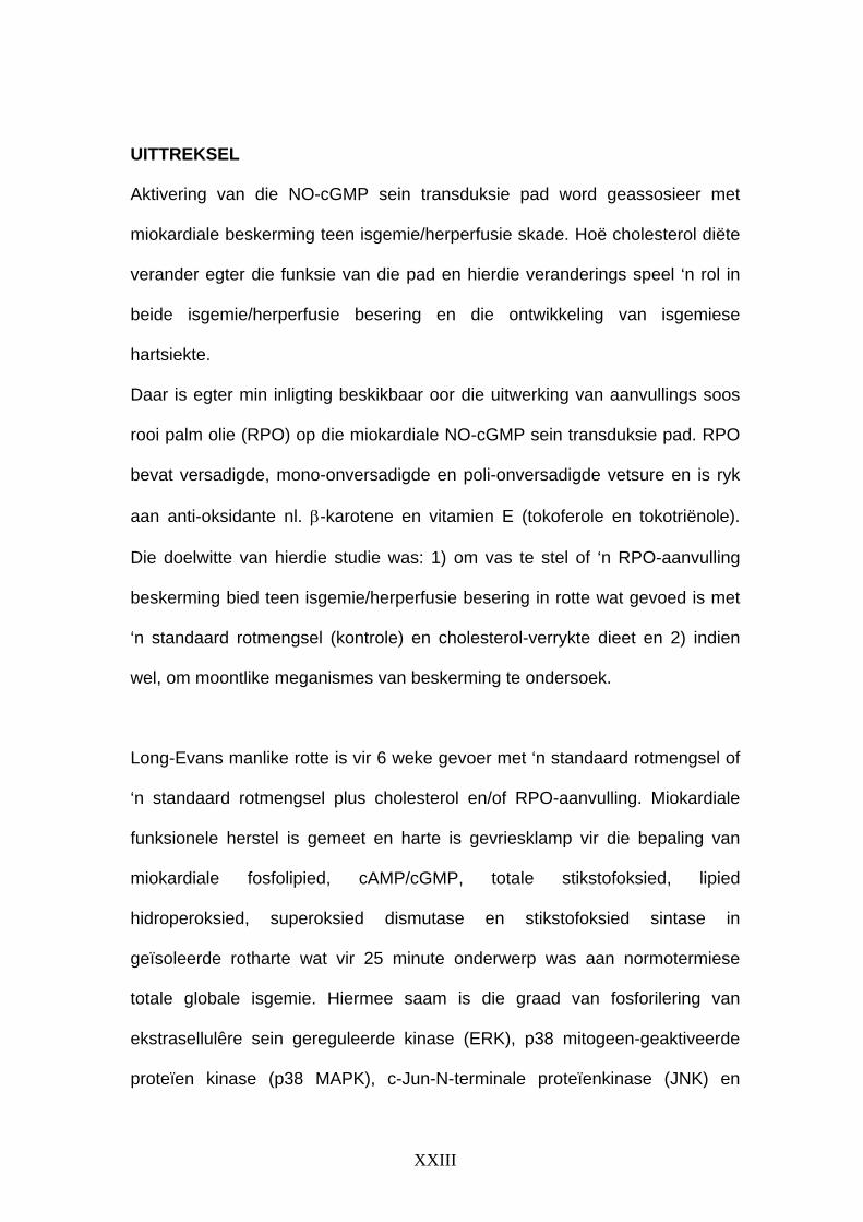
UITTREKSEL
Aktivering van die NO-cGMP sein transduksie pad word geassosieer met
miokardiale beskerming teen isgemie/herperfusie skade. Hoë cholesterol diëte
verander egter die funksie van die pad en hierdie veranderings speel ‘n rol in
beide isgemie/herperfusie besering en die ontwikkeling van isgemiese
hartsiekte.
Daar is egter min inligting beskikbaar oor die uitwerking van aanvullings soos
rooi palm olie (RPO) op die miokardiale NO-cGMP sein transduksie pad. RPO
bevat versadigde, mono-onversadigde en poli-onversadigde vetsure en is ryk
aan anti-oksidante nl. β-karotene en vitamien E (tokoferole en tokotriënole).
Die doelwitte van hierdie studie was: 1) om vas te stel of ‘n RPO-aanvulling
beskerming bied teen isgemie/herperfusie besering in rotte wat gevoed is met
‘n standaard rotmengsel (kontrole) en cholesterol-verrykte dieet en 2) indien
wel, om moontlike meganismes van beskerming te ondersoek.
Long-Evans manlike rotte is vir 6 weke gevoer met ‘n standaard rotmengsel of
‘n standaard rotmengsel plus cholesterol en/of RPO-aanvulling. Miokardiale
funksionele herstel is gemeet en harte is gevriesklamp vir die bepaling van
miokardiale fosfolipied, cAMP/cGMP, totale stikstofoksied, lipied
hidroperoksied, superoksied dismutase en stikstofoksied sintase in
geïsoleerde rotharte wat vir 25 minute onderwerp was aan normotermiese
totale globale isgemie. Hiermee saam is die graad van fosforilering van
ekstrasellulêre sein gereguleerde kinase (ERK), p38 mitogeen-geaktiveerde
proteïen kinase (p38 MAPK), c-Jun-N-terminale proteïenkinase (JNK) en
XXIV
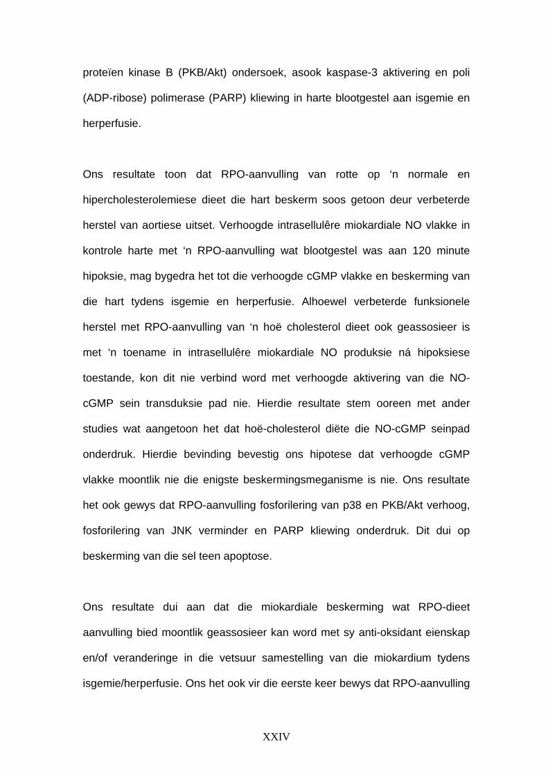
proteïen kinase B (PKB/Akt) ondersoek, asook kaspase-3 aktivering en poli
(ADP-ribose) polimerase (PARP) kliewing in harte blootgestel aan isgemie en
herperfusie.
Ons resultate toon dat RPO-aanvulling van rotte op ‘n normale en
hipercholesterolemiese dieet die hart beskerm soos getoon deur verbeterde
herstel van aortiese uitset. Verhoogde intrasellulêre miokardiale NO vlakke in
kontrole harte met ‘n RPO-aanvulling wat blootgestel was aan 120 minute
hipoksie, mag bygedra het tot die verhoogde cGMP vlakke en beskerming van
die hart tydens isgemie en herperfusie. Alhoewel verbeterde funksionele
herstel met RPO-aanvulling van ‘n hoë cholesterol dieet ook geassosieer is
met ‘n toename in intrasellulêre miokardiale NO produksie ná hipoksiese
toestande, kon dit nie verbind word met verhoogde aktivering van die NO-
cGMP sein transduksie pad nie. Hierdie resultate stem ooreen met ander
studies wat aangetoon het dat hoë-cholesterol diëte die NO-cGMP seinpad
onderdruk. Hierdie bevinding bevestig ons hipotese dat verhoogde cGMP
vlakke moontlik nie die enigste beskermingsmeganisme is nie. Ons resultate
het ook gewys dat RPO-aanvulling fosforilering van p38 en PKB/Akt verhoog,
fosforilering van JNK verminder en PARP kliewing onderdruk. Dit dui op
beskerming van die sel teen apoptose.
Ons resultate dui aan dat die miokardiale beskerming wat RPO-dieet
aanvulling bied moontlik geassosieer kan word met sy anti-oksidant eienskap
en/of veranderinge in die vetsuur samestelling van die miokardium tydens
isgemie/herperfusie. Ons het ook vir die eerste keer bewys dat RPO-aanvulling

XXV
die geïsoleerde geperfuseerde werkende rothart gedurende herperfusie
beskerm teen isgemie/herperfusie besering deur die aktivering en/of
deaktivering van die MAPK afhanklike pad.
1
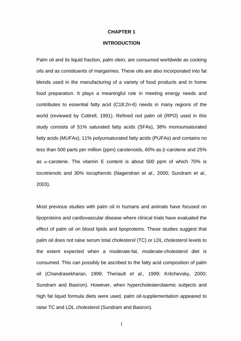
CHAPTER 1
INTRODUCTION
Palm oil and its liquid fraction, palm olein, are consumed worldwide as cooking
oils and as constituents of margarines. These oils are also incorporated into fat
blends used in the manufacturing of a variety of food products and in home
food preparation. It plays a meaningful role in meeting energy needs and
contributes to essential fatty acid (C18:2n-6) needs in many regions of the
world (reviewed by Cottrell, 1991). Refined red palm oil (RPO) used in this
study consists of 51% saturated fatty acids (SFAs), 38% monounsaturated
fatty acids (MUFAs), 11% polyunsaturated fatty acids (PUFAs) and contains no
less than 500 parts per million (ppm) carotenoids, 60% as β-carotene and 25%
as α-carotene. The vitamin E content is about 500 ppm of which 70% is
tocotrienols and 30% tocopherols (Nagendran et al., 2000; Sundram et al.,
2003).
Most previous studies with palm oil in humans and animals have focused on
lipoproteins and cardiovascular disease where clinical trials have evaluated the
effect of palm oil on blood lipids and lipoproteins. These studies suggest that
palm oil does not raise serum total cholesterol (TC) or LDL cholesterol levels to
the extent expected when a moderate-fat, moderate-cholesterol diet is
consumed. This can possibly be ascribed to the fatty acid composition of palm
oil (Chandrasekharan, 1999; Theriault et al., 1999; Kritchevsky, 2000;
Sundram and Basiron). However, when hypercholesterolaemic subjects and
high fat liquid formula diets were used, palm oil-supplementation appeared to
raise TC and LDL cholesterol (Sundram and Basiron).
2
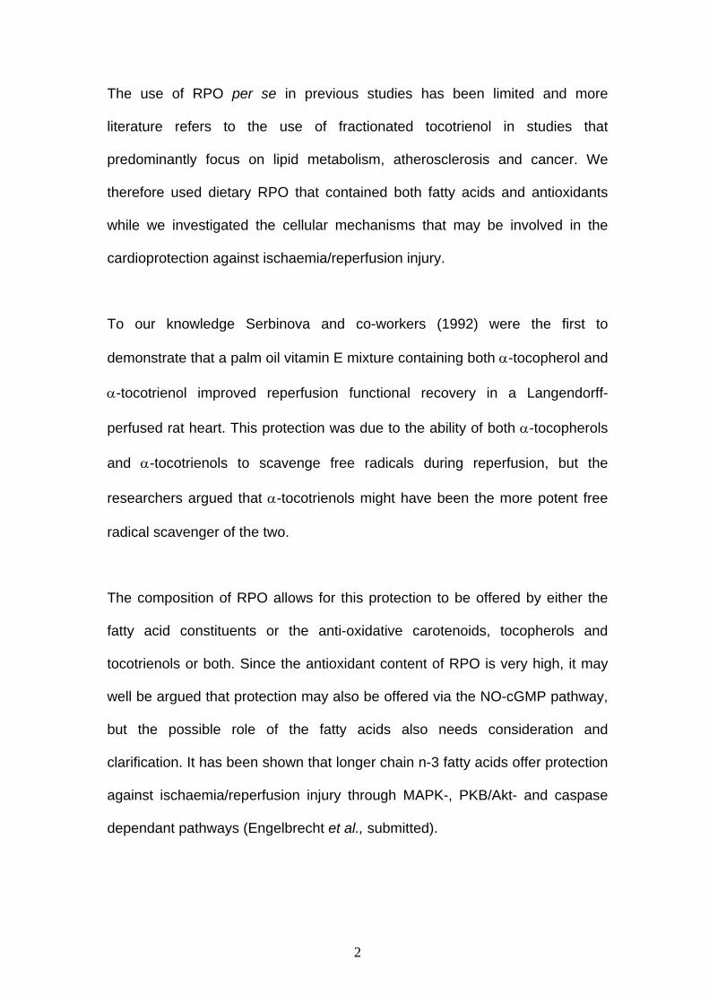
The use of RPO per se in previous studies has been limited and more
literature refers to the use of fractionated tocotrienol in studies that
predominantly focus on lipid metabolism, atherosclerosis and cancer. We
therefore used dietary RPO that contained both fatty acids and antioxidants
while we investigated the cellular mechanisms that may be involved in the
cardioprotection against ischaemia/reperfusion injury.
To our knowledge Serbinova and co-workers (1992) were the first to
demonstrate that a palm oil vitamin E mixture containing both α-tocopherol and
α-tocotrienol improved reperfusion functional recovery in a Langendorff-
perfused rat heart. This protection was due to the ability of both α-tocopherols
and α-tocotrienols to scavenge free radicals during reperfusion, but the
researchers argued that α-tocotrienols might have been the more potent free
radical scavenger of the two.
The composition of RPO allows for this protection to be offered by either the
fatty acid constituents or the anti-oxidative carotenoids, tocopherols and
tocotrienols or both. Since the antioxidant content of RPO is very high, it may
well be argued that protection may also be offered via the NO-cGMP pathway,
but the possible role of the fatty acids also needs consideration and
clarification. It has been shown that longer chain n-3 fatty acids offer protection
against ischaemia/reperfusion injury through MAPK-, PKB/Akt- and caspase
dependant pathways (Engelbrecht et al., submitted).
3
To our knowledge the cardioprotection offered by RPO and the role of NO-
cGMP signalling has not been investigated under normal and/or conditions
such as ischaemia or reperfusion. Recent studies suggested that the MAPK,
PKB/Akt and signal transduction caspases might also be involved in the
regulation of apoptosis in response to myocardial ischaemia/reperfusion.
Therefore, we used the ischaemic/reperfused working rat heart model to
elucidate whether the function and activity of these pathways (NO-cGMP,
MAPK, PKB/Akt and caspases) are altered and contribute to the red palm oil-
induced protection against ischaemia/reperfusion injury.
Nitric Oxide (NO) is, by virtue of its vasodilator, antioxidant, anti-platelet and
anti-neutrophil actions, an essential molecule for normal heart function
(reviewed by Ferdinandy and Schultz, 2003). It has been shown to be
cardioprotective in the ischaemic heart (reviewed by Bolli, 2001). However, NO
is detrimental when it is combined with superoxide (O2-) to form peroxynitrite
(ONOO-), which rapidly decomposes to highly reactive oxidant species, leading
to tissue injury (reviewed by Ferdinandy and Schultz, 2003). There is a critical
balance between cellular concentrations of NO, O2- and superoxide dismutase
(SOD) which physiologically favour NO production, but in pathological
conditions such as ischaemia and reperfusion injury, result in ONOO- formation
(reviewed by Ferdinandy and Schultz, 2003). A role for reactive oxygen
species, including the superoxide radical (O2-), hydrogen peroxide (H2O2) and
hydroxyl radical (.OH) have long been implicated in the pathogenesis of
ischaemia/reperfusion injury. These oxygen free radicals can react with nucleic
4
acids, proteins and lipids, resulting in damage to the cell membrane or
intracellular organelles (Gilham et al., 1997).
Myocardial NO formation is increased during ischaemia and reperfusion,
offering protection against ischaemia/reperfusion injury (Williams et al., 1995;
Araki et al., 2000; Bolli, 2001). Protective effects of NO are mediated through
the production of cGMP. NO is known to increase myocardial cGMP, and it has
been suggested that the protective effect of NO is related to a mechanism
secondary to the stimulation of guanylyl cyclase within the vascular wall or in
ventricular myocytes (Beresewics et al., 1995; Maulik et al., 1995; Depré et al.,
1996). NO donors given during ischaemia possibly protect the myocardium by
increasing tissue cGMP and decreasing cytosolic Ca2+ overload (Du Toit et al.,
2001). These investigators found that nitric oxide donor treatment reduces
ischaemia/reperfusion injury by increasing cGMP concentrations and
suggested that the cAMP-to-cGMP ratio might play an important role in NO
induced cardioprotection. Maulik and co-workers (1995) showed that NO plays
a significant role in transmembrane signalling in the ischaemic myocardium.
This group suggested that NO signalling is switched off due to inactivation of
NO by reactive oxygen species and was the first to suggest that reactive
oxygen species may alter NO-cGMP signalling.
Increases in cAMP concentrations associated with ischaemia would increase
Ca2+ concentrations and exacerbate ischaemic/reperfusion injury (Du Toit et
al., 2001). In this regard it is possible that cGMP may attenuate this type of
injury by inhibiting the cAMP induced increase in the slow inward calcium

5
current, thus leading to a decrease in cytosolic calcium levels (Summi and
Sperelakis, 1995). Therefore, cGMP appears to be an endogenous intracellular
cardioprotectant (Pabla et al., 1995).
Cardiac stress adaptation is possibly jeopardized in hyperlipidaemia due to
altered NO-cGMP pathway function in vascular and myocardial tissue.
Szilvassy and co-workers (2001) found that a cholesterol-enriched diet
decreased both vascular NO and cGMP concentrations and increased aortic
O2- production. Several other studies have also shown that a high cholesterol
diet impairs NO-cGMP signalling in both endothelium and non-endothelial cells
with a significant decrease in cardiac NO-concentrations (Deliconstantinos et
al., 1995; Ferdinandy et al., 1997; Scekeres et al., 1997). Giricz and co-
workers (2003) found an increase in O2- production with decreased cardiac
NO-concentrations in cholesterol-fed rats. However, nitric oxide synthase
(NOS) activity was unchanged. These data suggest that NO synthesis was not
impaired, but that increased O2- production was responsible for the decreased
NO-concentrations in the hyperlipidaemic myocardium. Furthermore,
hyperlipidaemia stimulates ONOO- generation in the heart, which leads to
myocardial dysfunction (Onody et al., 2003). Newaz and co-workers (2003)
showed an antioxidant protection by γ-tocotrienols in hypertensive rats when
compared with normotensive control animals. These authors suggested that
improved NOS activity in blood vessels and increased NO availability were
mediated through the antioxidant properties of γ-tocotrienol where it effectively
scavenges the free radicals. Venditti and co-workers (1999) also reported that
6
vitamin E treatment offers protection against ischaemia/reperfusion-induced
oxidative stress, but the precise mechanism of action is unclear.
Diniz and co-workers (2004) showed that changes in dietary fatty acid
composition affect cardiac oxidative stress. These authors showed that,
despite their beneficial effects on serum lipid concentrations, diets rich in
polyunsaturated fatty acids (PUFAs) are deleterious to the heart by increasing
cardiac susceptibility to lipid peroxidation. PUFA-fed rats also showed
diminished SOD activities as compared to saturated fatty acid (SFA)-fed rats in
this particular study. Although many animal feeding studies have shown that
fish oil diets rich in n-3 PUFAs prevent ischaemia-induced cardiac arrhythmias
(Nair et al., 1997; Kang and Leaf 2000; Jump, 2002), only a few reports have
been published on the protective effects of RPO-supplementation against
ischaemia/reperfusion injury (Abeywardena et al., 1991; Charnock et al., 1991;
Abeywardena and Charnock, 1995).
We speculate that compositional changes in myocardial phospholipid fatty
acids during ischaemia may be involved in the regulation of several signal
transduction pathways in the heart in direct response to ischaemia/reperfusion-
induced injury. One of the best-characterized signal transduction pathways in
the heart is the family of mitogen-activated protein kinases (MAPKs). The
MAPKs are a family of serine-threonine kinases that are activated in response
to a variety of extracellular stimuli (Robinson and Cobb, 1997; Ip and Davis,
1998). Three major MAPKs, including extracellular signal-regulated protein
kinase (ERK), p38, and c-Jun N-terminal protein kinase (JNK) have been
7
implicated in the response to ischaemia and reperfusion in the heart
(Bogoyevitch et al., 1996; Knight and Buxton, 1996). All three MAPKs have
been shown to play pivotal roles in transmission of signals from cell surface
receptors to the nucleus and are involved in cell growth, differentiation and
apoptosis (Mansour et al., 1994; Leppa et al., 1998; Nemoto et al., 1998).
Another potential target of RPO might be the serine/threonine kinase PKB/Akt.
PKB/Akt contains a pleckstrin homology (PH) domain that is part of a slightly
larger portion in the NH2 terminus, called the Akt homology domain. The
phosphoinositide 3-kinase (PI3-K) product phosphatidylinositiol-3,4-
bisphosphate bind in vitro directly to the PH domain and increases enzyme
activity (Downward, 1998). PKB/Akt has been shown to be activated by factors
that stimulate PI3-K including thrombin, platelet-derived growth factor, and
insulin (Downward, 1998). There is also increasing evidence that the PKB/Akt
pathway participates in ischaemia/reperfusion-induced injury (Brar et al., 2002;
Andreucci et al., 2003).
In order to assess the mechanisms of protection, the isolated perfused rat
heart model was used to determine whether dietary RPO-supplementation was
associated with changes in the regulation of the MAPKs and PKB/Akt in
ischaemia/reperfusion. To our knowledge no previous studies have
investigated the effect of dietary RPO-supplementation on these signalling
pathways during and after ischaemia.
8
1.1 Aims of the study The aims of this study were:
1) to determine whether dietary RPO-supplementation protects against
ischaemia/reperfusion injury in the isolated perfused rat heart from animals
on a standard rat chow diet
2) to determine whether dietary RPO-supplementation offers the same
protection when cholesterol is added to the diet
3) to elucidate the mechanisms of protection which include the NO-cGMP
signalling pathway, myocardial total phospholipid fatty acid compositional
changes during ischaemia and MAPK, PKB/Akt and caspase activities.
9
CHAPTER 2
LITERATURE REVIEW
2.1 Dietary fats and oils in health
A balanced diet, including oils and fats that supply energy and essential fatty
acids is needed for good health. The World Health Organization (WHO)
recommends that humans consume about 20-25 kg oils and fats per capita per
year (Ong and Goh, 2002).
There are many sources of oils and fats, but soybean, palm oil, sunflower seed
oil and rape seed oil constitute 60-70% of the world’s production (Ong and
Goh, 2002). Fats and oils are classified as saturated, monounsaturated and
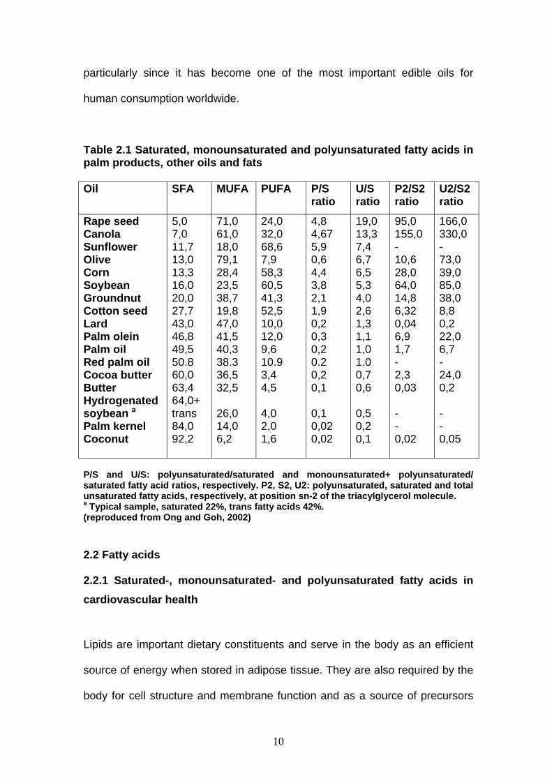
polyunsaturated depending on which fatty acids are dominant. The fatty acid
compositions of the most important oils and fats are summarized in Table 2.1.
Plant oils, like coconut oil and palm oil, can be considered highly structured,
usually having the sn-2 positional fatty acids unsaturated and the 1,3-fatty
acids saturated on the triacylglycerol molecule. The belief that palm oil should
be classified nutritionally with saturated fats is therefore debatable.
Furthermore, research has also confirmed that palm oil is a non-genetically
modified, cholesterol-free, trans-free oil that contains phytonutrients such as α-
carotene, β-carotene, vitamin E tocopherols and tocotrienols, lycopene and
other carotenoids (Goh et al., 1985; Sundram et al., 2003). These findings
merit a re-evaluation of the nutritional properties of palm oil and palm olein
10
particularly since it has become one of the most important edible oils for
human consumption worldwide.
Table 2.1 Saturated, monounsaturated and polyunsaturated fatty acids in palm products, other oils and fats Oil SFA MUFA PUFA P/S
ratio U/S ratio
P2/S2 ratio
U2/S2 ratio
Rape seed Canola Sunflower Olive Corn Soybean Groundnut Cotton seed Lard Palm olein Palm oil Red palm oil Cocoa butter Butter Hydrogenated soybean a
Palm kernel Coconut
5,0 7,0 11,7 13,0 13,3 16,0 20,0 27,7 43,0 46,8 49,5 50.8 60,0 63,4 64,0+ trans 84,0 92,2
71,0 61,0 18,0 79,1 28,4 23,5 38,7 19,8 47,0 41,5 40,3 38.3 36,5 32,5 26,0 14,0 6,2
24,0 32,0 68,6 7,9 58,3 60,5 41,3 52,5 10,0 12,0 9,6 10.9 3,4 4,5 4,0 2,0 1,6
4,8 4,67 5,9 0,6 4,4 3,8 2,1 1,9 0,2 0,3 0,2 0.2 0,2 0,1 0,1 0,02 0,02
19,0 13,3 7,4 6,7 6,5 5,3 4,0 2,6 1,3 1,1 1,0 1.0 0,7 0,6 0,5 0,2 0,1
95,0 155,0 - 10,6 28,0 64,0 14,8 6,32 0,04 6,9 1,7 - 2,3 0,03 - - 0,02
166,0 330,0 - 73,0 39,0 85,0 38,0 8,8 0,2 22,0 6,7 - 24,0 0,2 - - 0,05
P/S and U/S: polyunsaturated/saturated and monounsaturated+ polyunsaturated/ saturated fatty acid ratios, respectively. P2, S2, U2: polyunsaturated, saturated and total unsaturated fatty acids, respectively, at position sn-2 of the triacylglycerol molecule. a Typical sample, saturated 22%, trans fatty acids 42%. (reproduced from Ong and Goh, 2002)
2.2 Fatty acids
2.2.1 Saturated-, monounsaturated- and polyunsaturated fatty acids in cardiovascular health
Lipids are important dietary constituents and serve in the body as an efficient
source of energy when stored in adipose tissue. They are also required by the
body for cell structure and membrane function and as a source of precursors
11
for eicosanoid synthesis. Lipid components like cholesterol and phospholipids
regulate membrane-associated functions such as activities of membrane
bound enzymes, receptors and ion channels (Clandinin et al., 1991).
Lipids are composed of fatty acids of different chain lengths and degrees of
saturation. The differences in chain length and degrees of saturation are
known to influence cardiovascular health (Nair et al., 1997). Fatty acids are
classified into three types, namely saturated fatty acids (SFAs),
monounsaturated fatty acids (MUFAs) and polyunsaturated fatty acids
(PUFAs). The three major fatty acid families found in mammalian tissue are the
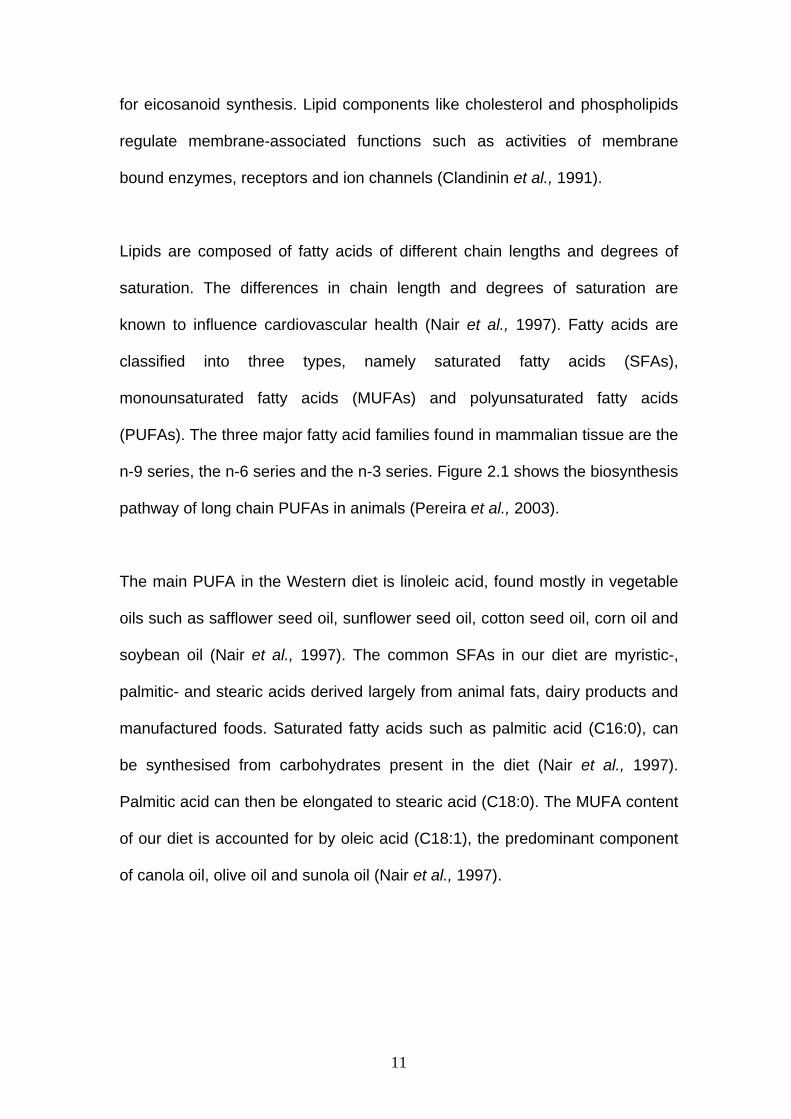
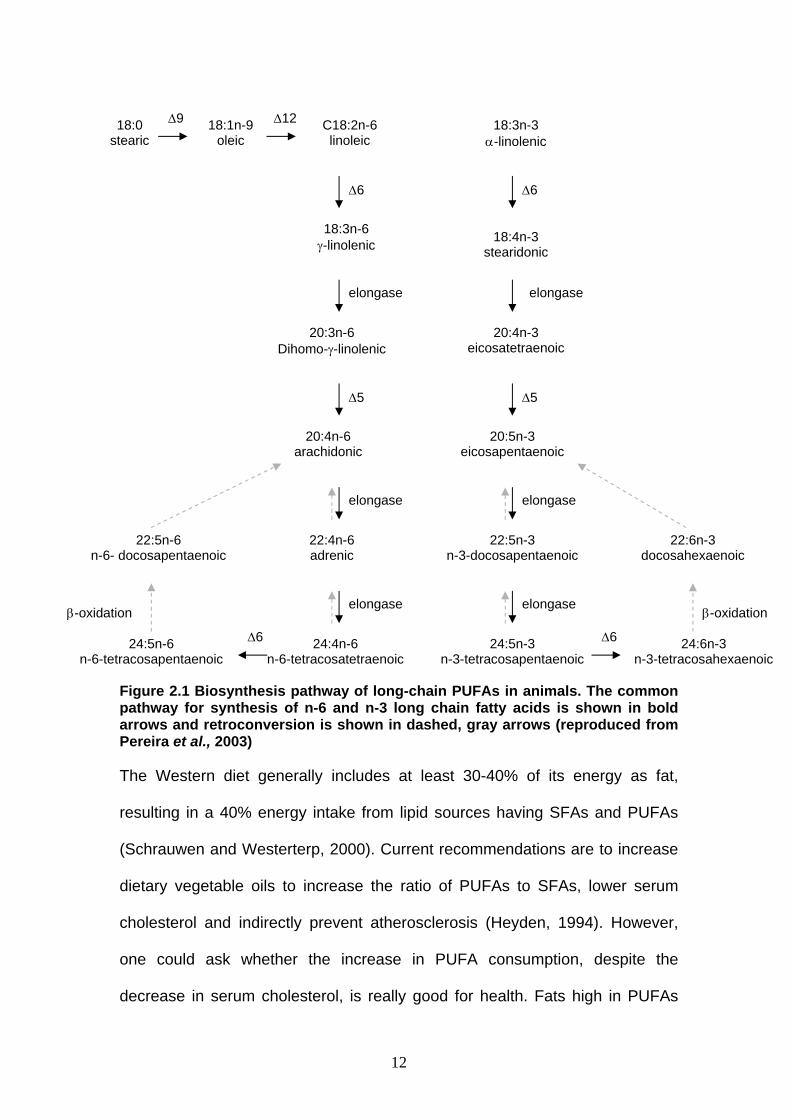
n-9 series, the n-6 series and the n-3 series. Figure 2.1 shows the biosynthesis
pathway of long chain PUFAs in animals (Pereira et al., 2003).
The main PUFA in the Western diet is linoleic acid, found mostly in vegetable
oils such as safflower seed oil, sunflower seed oil, cotton seed oil, corn oil and
soybean oil (Nair et al., 1997). The common SFAs in our diet are myristic-,
palmitic- and stearic acids derived largely from animal fats, dairy products and
manufactured foods. Saturated fatty acids such as palmitic acid (C16:0), can
be synthesised from carbohydrates present in the diet (Nair et al., 1997).
Palmitic acid can then be elongated to stearic acid (C18:0). The MUFA content
of our diet is accounted for by oleic acid (C18:1), the predominant component
of canola oil, olive oil and sunola oil (Nair et al., 1997).
12
Figure 2.1 Biosynthesis pathway of long-chain PUFAs in animals. The common pathway for synthesis of n-6 and n-3 long chain fatty acids is shown in bold arrows and retroconversion is shown in dashed, gray arrows (reproduced from Pereira et al., 2003) The Western diet generally includes at least 30-40% of its energy as fat,
resulting in a 40% energy intake from lipid sources having SFAs and PUFAs
(Schrauwen and Westerterp, 2000). Current recommendations are to increase
dietary vegetable oils to increase the ratio of PUFAs to SFAs, lower serum
cholesterol and indirectly prevent atherosclerosis (Heyden, 1994). However,
one could ask whether the increase in PUFA consumption, despite the
decrease in serum cholesterol, is really good for health. Fats high in PUFAs
24:5n-6 n-6-tetracosapentaenoic
β-oxidation
18:0 stearic
Δ9 18:1n-9 oleic
Δ12 C18:2n-6 linoleic
18:3n-3 α-linolenic
18:3n-6 γ-linolenic
Δ6 Δ6
18:4n-3 stearidonic
elongase elongase
20:3n-6 Dihomo-γ-linolenic
20:4n-3 eicosatetraenoic
Δ5 Δ5
20:4n-6 arachidonic
20:5n-3 eicosapentaenoic
elongase elongase
22:4n-6 adrenic
22:5n-3 n-3-docosapentaenoic
elongase elongase
24:4n-6 n-6-tetracosatetraenoic
24:5n-3 n-3-tetracosapentaenoic
Δ6 24:6n-3 n-3-tetracosahexaenoic
Δ6
β-oxidation
22:6n-3 docosahexaenoic
22:5n-6 n-6- docosapentaenoic
13
are more susceptible to oxidation than SFAs. Therefore, PUFAs in the
absence of adequate antioxidants increase oxidative stress in the heart and
contribute to cardiac dysfunction and myocardial damage by increasing cardiac
susceptibilty to lipid peroxidation. (Mehta et al., 1994; Esposito et al., 1999;
Hart et al., 1999; Droge, 2002; Faine et al., 2002; Novelli et al., 2002; Diniz et
al., 2004).
Little is known about the metabolic effects of dietary fatty acids on markers
used to evaluate oxidative stress in the heart. However, Diniz and co-workers
(2004) showed that rats on a high SFA diet had lower myocardial
hydroperoxide concentrations than did PUFA-supplemented animals. This
demonstrates the importance of the PUFA:SFA ratio on lipid peroxidation. The
readiness with which fatty acids peroxidize is proportional to the number of
double bonds, and a positive correlation between the amount of PUFA in the
diet and the rate of microsomal lipid peroxidation has been demonstrated in
rats (Mehta et al., 1994). The oxidative stability of the cardiac cells is
determined by the balance between factors such as PUFAs, which change the
PUFA:SFA ratio in the membrane and enhance the sensitivity to lipid
peroxidation and the levels of antioxidants (Diniz et al., 2004). These
observations support the concept that the sensitivity of cardiac tissue to
oxidative stress may depend on dietary factors.
There are many nutritional qualities and benefits of the dietary use of palm oil.
Palm oil, like other vegetable oils, is cholesterol free. Having a moderate level
of saturation, it does not require hydrogenation for use as a fat component in

14
foods and, as such, does not contain trans fatty acids (Cottrell, 1991). It is rich
in natural antioxidants, which makes it a safe, stable and versatile oil with
many positive health and nutritional attributes (Nagendran et al., 2000).
2.2.2 Essential fatty acids
Essential fatty acids (EFAs) are defined as those fatty acids which cannot be
biosynthesised or are synthesised in inadequate amounts by animals and
humans that require these nutrients for various physiological processes such
as growth and maintenance of health (Horrobin, 1990).
Linoleic acid (C18:2n-6; LA) and α-linolenic acid (C18:3n-3; ALA), the parents
of the n-6 and n-3 family of fatty acids, respectively, are essential fatty acids
that cannot be synthesised in the body and have to be supplied by diet (Figure
2.2; Nair et al., 1997). Linoleic acid is found mostly in vegetable oils such as
safflower seed oil, sunflower seed oil, cotton seed oil, corn oil and soybean oil.
Other long chain PUFAs, like arachidonic acid (C20:4n-6; AA), are synthesised
in human tissue via desaturation and chain elongation from LA which is by far
the dominant precursor fatty acid for eicosanoid formation provided by the
Western diet. Significant amounts of α-linolenic acid are found in green
vegetables and in vegetable oils like linseed oil, canola seed oil and soybean
oil. Eicosapentaenoic acid (C20:5n-3; EPA) and docosahexaenoic acid
(C22:6n-3; DHA) are synthesised via a series of alternating desaturation and
chain elongation steps from ALA and are also found in high concentrations in
fish oils. LA and ALA compete for desaturation and chain elongation, therefore
15
a proper balance is essential to optimize AA and DHA in membranes (Nair et
al., 1997).
2.2.3 Physiological role of essential fatty acids
Although many animal feeding studies have shown that fish oil diets rich in n-3
PUFAs prevent ischaemia-induced cardiac arrhythmias (Nair et al., 1997; Kang
and Leaf, 2000; Jump, 2002), only a few reports have been published on the
protective effects of palm oil-supplementation against ischaemia/reperfusion
injury (Abeywardena et al., 1991, Charnock et al., 1991; Abeywardena and
Charnock, 1995). Several mechanisms for protection by fish oil diets have
been proposed. However, all the mechanisms suggested appear to be
interrelated and it is not known whether their effects are independent or are
compounded and also in what sequence they take place (Nair et al., 1997).
The polyunsaturated fatty acids, arachidonic acid (C20:4n-6; AA) and
eicosapentaenoic acid (C20:5n-3; EPA) are metabolised through
cyclooxygenase and lipoxygenase pathways to form eicosanoids, including
prostaglandins (PGs), thromboxanes (TXs), prostacyclins (PGIs) and
leukotrienes (LTs) (Figure 2.2).

16
Figure 2.2 Biosynthesis of eicosanoids from dietary fatty acids. LT=leukotrienes; PG=prostaglandins; TX=thromboxane (reproduced from Nair et al., 1997)
Eicosanoids are known to have a variety of cardiovascular effects: 1) all
prostaglandins derived from AA are arrhythmogenic, of which PGF2 is the most
potent, 2) PGE1 from Dihomo-γ-linolenic acid (C20:3n-6; DGLA) appears to
have concentration-dependent effects with low concentrations being
antiarrhythmic and high concentrations arrhythmogenic, 3) the precursor fatty
Diet Diet
Linoleic acid
Dihomo γ-linolenic acid
Prostanoids PGE1 PGE2 PGF1 TXA1
Leukotrienes LTA3 LTC3 LTD3
α-Linolenic acid
Eicosatetraenoic acid
MEMBRANE PHOSPHOLIPIDS
Arachidonic acid Eicosapentaenoic acid
n-6 series n-3 series
Prostanoids PGD2 PGE2 PGF2 PGI2 TXA2
Leukotrienes LTA4 LTB4 LTC4 LTD4 LTE4
Prostanoids PGD3 PGE3 PGF3 PGI3 TXA3
Leukotrienes LTA3 LTB3 LTC3
17
acids (free n-6 series AA and EPA n-3 series) are able to prevent eicosanoid-
induced arrhythmias, 4) the eicosanoids derived from EPA are generally less
arrhythmogenic, 5) lipoxygenase metabolites of both AA and EPA are neither
arrhythmogenic nor antiarrhythmic (Li et al., 1997; Yunyuan et al., 1997). The
role of AA in arrhythmias is of particular interest. Most investigations on the link
between fish oils and cardiovascular disease have demonstrated competition
between AA and EPA to become substrates in the production of eicosanoids.
When fish oils are included in the diet, the n-3 PUFAs (EPA and DHA)
compete with AA in several ways: 1) they inhibit delta 6 (Δ6) activity to
decrease AA biosynthesis (Garg et al., 1988), 2) they compete with AA for the
sn-2 position in triacylglycerols and membrane phospholipids and thereby
reduce plasma and cellular levels of AA (Siess et al., 1988), 3) EPA competes
with AA as the substrate for the cyclooxygenase enzyme thus inhibiting the
production of thromboxane A2 (TXA2) by platelets (Fischer and Weber, 1984).
Research has shown that dietary supplementation of different edible oils may
influence cardiovascular function due to compositional changes in the PUFAs
of the myocardial membrane phospholipids after ischaemia and reperfusion
(Abeywardena et al., 1991; Abeywardena and Charnock, 1995). The presence
of fish oil in the diet results in increased incorporation of n-3 PUFAs (EPA and
DHA), mainly at the expense of n-6 unsaturated arachidonic acid. The
significant increase in DHA associated with fish oil supplementation is likely to
be due not only to a direct incorporation of DHA from the diet, but also to an
increased elongation and further desaturation of EPA (Abeywardena and
Charnock, 1995). Both fish oil- and RPO-supplementation caused a significant
18
inhibition of myocardial thromboxane A2 production. Abeywardena and co-
workers (1991) speculated that n-3 PUFAs (EPA and DHA) might act as
specific inhibitors of thromboxane synthases, whereas the effect of RPO is
unlikely to be mediated via fatty acids. Charnock and co-workers (1991)
investigated the effect of long-term feeding with various dietary fats and oils on
cardiac arrhythmias in an animal model. These authors showed that dietary
supplementation with saturated animal fat (SF) increased the susceptibility to
develop cardiac arrhythmias under ischaemic stress whereas the
polyunsaturated fatty acids of sunflower seed oil (SSO) reduced this
susceptibility. RPO-supplementation produced results that lay between those
for the SF and SSO groups. Furthermore, the number of animals displaying
severe ventricular fibrillation was reduced after RPO-supplementation when
compared with SF feeding (Charnock et al., 1991). From the limited data
available, it is unclear whether these results are related to the ratio of
polyunsaturated to saturated fatty acids of the diets, or to the fatty acid
composition of the myocardial membranes. These effects may even been
mediated by differential actions of the dietary fats on myocardial eicosanoid
production. In addition, it has been suggested by Gapor and co-workers (1989)
that palm oil antioxidants in a palm oil/fish oil-supplementation may also prove
to be useful for protection of the less stable polyunsaturated fatty acids in fish
oils.
19
2.3 Cholesterol-enriched diets
2.3.1 Introduction to cholesterol-enriched diets
A high-cholesterol diet is regarded as an important factor in the development of
cardiovascular disease, since it leads to development of hyperlipidaemia,
atherosclerosis and ischaemic heart disease (Puskas et al., 2004).
2.3.2 Mechanisms of myocardial effects of hyperlipidaemia
The exact biochemical mechanisms of the direct effects of high-cholesterol diet
hyperlipidaemia on the myocardium are still a question of debate. However,
the following mechanisms have been shown to play a role in the cardiac
effects of hyperlipidaemia: 1) inhibition of the mevalonate pathway (Ferdinandy
et al., 1998), 2) decrease in NO bioavailability and cGMP metabolism
(Ferdinandy et al., 1997; Szekeres et al., 1997), 3) increase in free radical and
peroxynitrite production (Onody et al., 2003), 4) inhibition of heat shock
response (Csont et al., 2002), 5) expression of oxidized low-density lipoprotein
receptors which induced apoptosis (Chen et al., 2002). Recent studies
identified gene activity changes in atherosclerotic plaques in human and
animal blood vessels and rat hearts (Puskas et al., 2004).
2.4 Palm oil
2.4.1 Introduction to palm oil
Crude palm oil is produced from the fruit of the Elaeis guineensis tree
(Nagendran et al., 2000; Sundram et al., 2003) and has a long history of food
use dating back over 5 000 years. Palm oil is one of the 16 edible oils

20
possessing an FAO/WHO Food standard under the Codex Alimentarius
Commission Programme, which comprises 122 member countries (Codex
Alimentarius, 1983). Palm oil is a traditional food source native to West Africa.
From its origin in Africa, oil palm has crossed the oceans of the world to
become an important plantation crop in countries like Malaysia. Here it
emerged as the most prolific oil bearing crop in the world. A single tree has an
economic lifespan of 20-30 years and annually bears 10-12 fruit bunches, each
weighing between 20-30 kg.
2.4.2 Composition of palm oil
Crude palm oil consists of glycerides and small quantities of non-glyceride
components including free fatty acids, trace metals, moisture and impurities,
and minor components. The minor components in crude palm oil are
carotenoids, tocopherols, tocotrienols, sterols, phospholipids, squalene and
hydrocarbons (Goh et al., 1985, Sundram et al., 2003). Of these the
carotenoids, tocopherols and tocotrienols are the most important minor
components and together they contribute to the stability and nutritional
properties of palm oil (Ooi et al., 1996). A novel process involving pre-
treatment of crude palm oil, followed by deacidification and deodorization using
molecular distillation, can be used to produce a carotene-rich refined edible
palm oil. The product is a refined red palm oil that meets standard refined
edible oil specifications and retains up to 80% of the carotene and vitamin E
originally present in the crude palm oil. The oil contains no less than 500 ppm
carotene, 90% of which is present as α- and β-carotene. The vitamin E content
is about 500 ppm of which 70% is in the form of tocotrienols (mainly as α-, β-
21
and γ tocotrienols). Other important minor components present in this oil are
ubiquinones and phytosterols (Nagendran et al., 2000; Sundram et al., 2003).
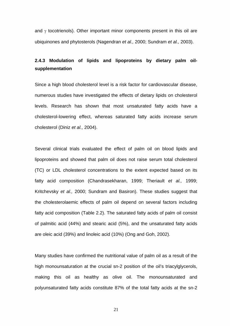
2.4.3 Modulation of lipids and lipoproteins by dietary palm oil-supplementation Since a high blood cholesterol level is a risk factor for cardiovascular disease,
numerous studies have investigated the effects of dietary lipids on cholesterol
levels. Research has shown that most unsaturated fatty acids have a
cholesterol-lowering effect, whereas saturated fatty acids increase serum
cholesterol (Diniz et al., 2004).
Several clinical trials evaluated the effect of palm oil on blood lipids and
lipoproteins and showed that palm oil does not raise serum total cholesterol
(TC) or LDL cholesterol concentrations to the extent expected based on its
fatty acid composition (Chandrasekharan, 1999; Theriault et al., 1999;
Kritchevsky et al., 2000; Sundram and Basiron). These studies suggest that
the cholesterolaemic effects of palm oil depend on several factors including
fatty acid composition (Table 2.2). The saturated fatty acids of palm oil consist
of palmitic acid (44%) and stearic acid (5%), and the unsaturated fatty acids
are oleic acid (39%) and linoleic acid (10%) (Ong and Goh, 2002).
Many studies have confirmed the nutritional value of palm oil as a result of the
high monounsaturation at the crucial sn-2 position of the oil’s triacylglycerols,
making this oil as healthy as olive oil. The monounsaturated and
polyunsaturated fatty acids constitute 87% of the total fatty acids at the sn-2

22
position (Ong and Goh, 2002). Comparison of palm oil with a variety of
monounsaturated edible oils including rape seed, canola and olive oils has
shown that serum cholesterol and LDL-cholesterol are not elevated by palm oil
(Sundram and Basiron). Furthermore, substitution of palmitic acid from palm oil
for the lauric acid and myristic acid combination of palm kernel oil and coconut
oil leads to a decrease in serum cholesterol and LDL-cholesterol (Ong and
Goh, 2002).
Research has shown that the contribution of dietary fats to blood lipids and
cholesterol modulation is a sequence of the digestion, absorption and
metabolism of the fats. Lipolytic hydrolysis of palm oil’s glyceride containing
predominantly oleic acid at the sn-2 position and palmitic and stearic acids at
the sn-1 and sn-3 positions allow for the ready absorption of the sn-2
monoglycerols, while the saturated free fatty acids remain poorly absorbed.
Therefore, dietary palm oil in balanced diets (when a moderate-fat, moderate-
cholesterol diet is consumed) generally reduces blood cholesterol and
triacylglycerol, while raising the HDL-cholesterol (Ong and Goh, 2002).
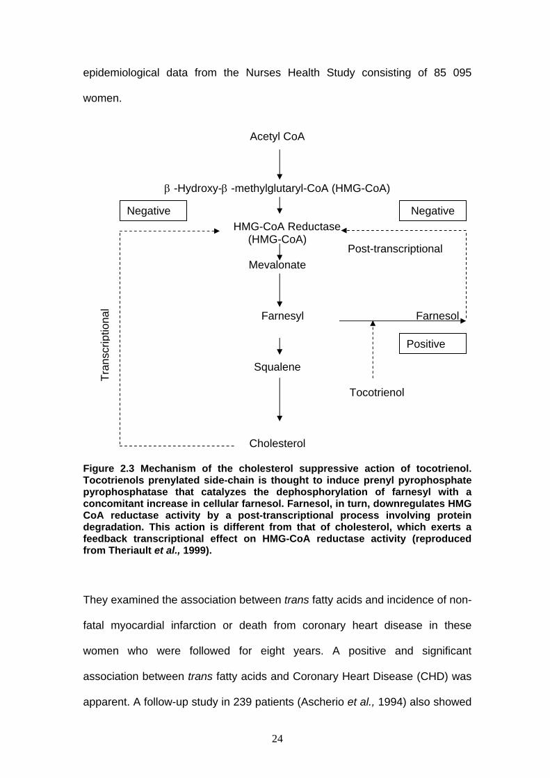
Apart from these fatty acids (Table 2.2), there is evidence that the tocotrienols
in palm oil products may have a hypocholesterolaemic effect. This is mediated
by the ability of the tocotrienols to suppress 3-hydroxy-3-methylglutaryl-
coenzyme A (HMG-CoA) reductase, a rate-limiting enzyme in cholesterol
biosynthesis (Khor et al., 1995; Theriault et al., 1999; Sundram and Basiron)
(Figure 2.3).
23
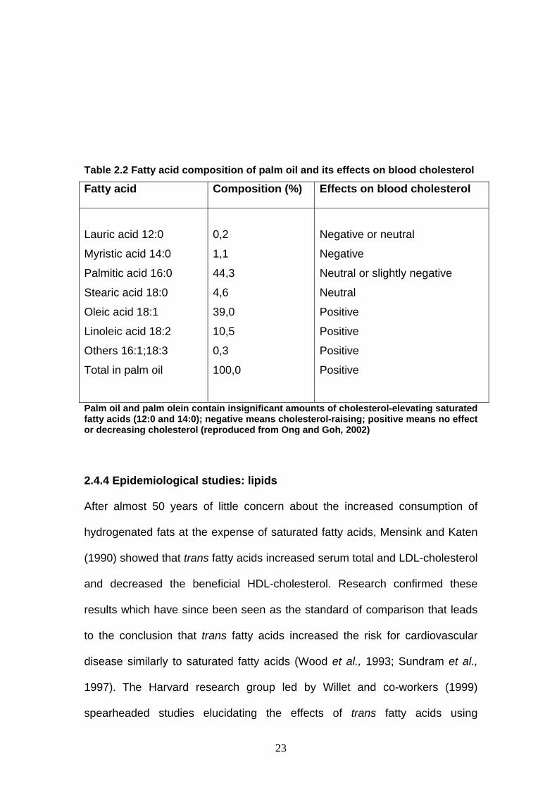
Table 2.2 Fatty acid composition of palm oil and its effects on blood cholesterol
Fatty acid Composition (%) Effects on blood cholesterol
Lauric acid 12:0
Myristic acid 14:0
Palmitic acid 16:0
Stearic acid 18:0
Oleic acid 18:1
Linoleic acid 18:2
Others 16:1;18:3
Total in palm oil
0,2
1,1
44,3
4,6
39,0
10,5
0,3
100,0
Negative or neutral
Negative
Neutral or slightly negative
Neutral
Positive
Positive
Positive
Positive
Palm oil and palm olein contain insignificant amounts of cholesterol-elevating saturated fatty acids (12:0 and 14:0); negative means cholesterol-raising; positive means no effect or decreasing cholesterol (reproduced from Ong and Goh, 2002)
2.4.4 Epidemiological studies: lipids
After almost 50 years of little concern about the increased consumption of
hydrogenated fats at the expense of saturated fatty acids, Mensink and Katen
(1990) showed that trans fatty acids increased serum total and LDL-cholesterol
and decreased the beneficial HDL-cholesterol. Research confirmed these
results which have since been seen as the standard of comparison that leads
to the conclusion that trans fatty acids increased the risk for cardiovascular
disease similarly to saturated fatty acids (Wood et al., 1993; Sundram et al.,
1997). The Harvard research group led by Willet and co-workers (1999)
spearheaded studies elucidating the effects of trans fatty acids using
24
epidemiological data from the Nurses Health Study consisting of 85 095
women.
Acetyl CoA
β -Hydroxy-β -methylglutaryl-CoA (HMG-CoA)
HMG-CoA Reductase (HMG-CoA)
Mevalonate
Farnesyl Farnesol
Squalene
Tocotrienol
Cholesterol Figure 2.3 Mechanism of the cholesterol suppressive action of tocotrienol. Tocotrienols prenylated side-chain is thought to induce prenyl pyrophosphate pyrophosphatase that catalyzes the dephosphorylation of farnesyl with a concomitant increase in cellular farnesol. Farnesol, in turn, downregulates HMG CoA reductase activity by a post-transcriptional process involving protein degradation. This action is different from that of cholesterol, which exerts a feedback transcriptional effect on HMG-CoA reductase activity (reproduced from Theriault et al., 1999).
They examined the association between trans fatty acids and incidence of non-
fatal myocardial infarction or death from coronary heart disease in these
women who were followed for eight years. A positive and significant
association between trans fatty acids and Coronary Heart Disease (CHD) was
apparent. A follow-up study in 239 patients (Ascherio et al., 1994) also showed
Tran
scrip
tiona
l
Post-transcriptional
Positive
Negative Negative
25
a positive association between trans fatty acids (in margarine) and myocardial
infarction. Trans fatty acid intake was associated with increased serum total
and LDL-cholesterol and negatively related to HDL-cholesterol in men suffering
a myocardial infarction.
The relative risk for Cardiovascular Disease (CVD) was increased by 27% as a
result of trans fatty acid consumption. These studies clearly established an
association between trans fatty acid consumption and increased incidence and
death from CVD and it was estimated that almost 80 000 deaths in the United
States alone are associated with continued consumption of foods rich in trans
fatty acids.
2.4.5 Cardiovascular protection offered by palm oil components
2.4.5.1 Carotenoids
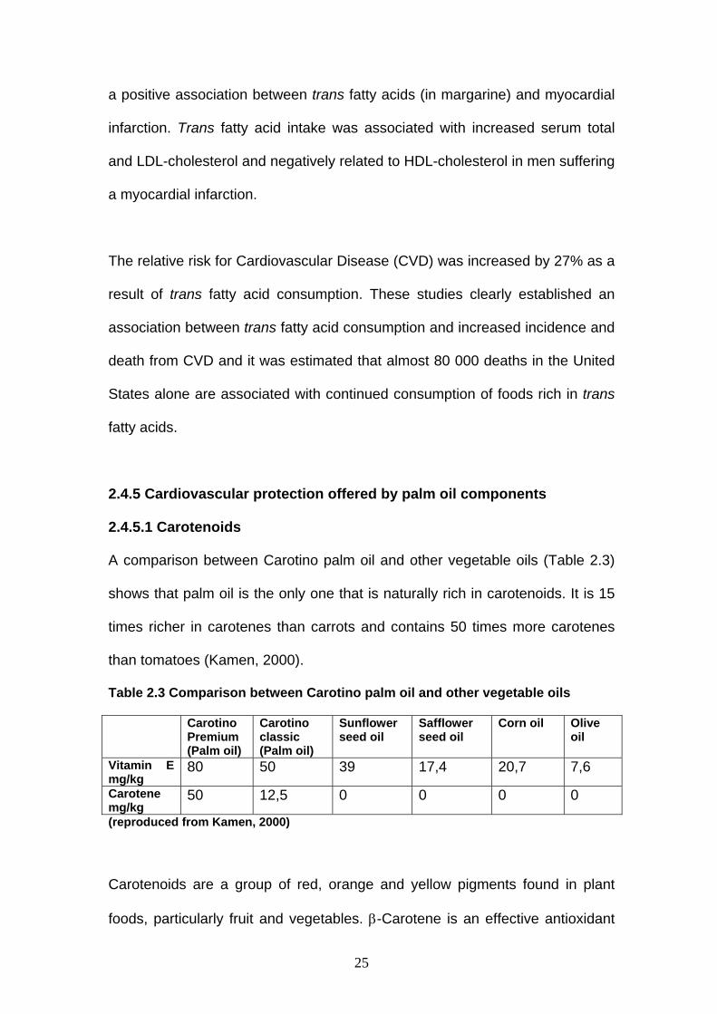
A comparison between Carotino palm oil and other vegetable oils (Table 2.3)
shows that palm oil is the only one that is naturally rich in carotenoids. It is 15
times richer in carotenes than carrots and contains 50 times more carotenes
than tomatoes (Kamen, 2000).
Table 2.3 Comparison between Carotino palm oil and other vegetable oils
Carotino Premium (Palm oil)
Carotino classic (Palm oil)
Sunflower seed oil
Safflower seed oil
Corn oil Olive oil
Vitamin E mg/kg
80 50 39 17,4 20,7 7,6
Carotene mg/kg
50 12,5 0 0 0 0
(reproduced from Kamen, 2000)
Carotenoids are a group of red, orange and yellow pigments found in plant
foods, particularly fruit and vegetables. β-Carotene is an effective antioxidant
26
as it is one of the most powerful singlet oxygen quenchers. It can dissipate the
energy of singlet oxygen, thus preventing this active molecule from generating
free radicals (Bagchi and Puri, 1998).
2.4.5.2 Vitamin E (tocopherols and tocotrienols)
Vitamin E is the collective name for eight compounds, namely four tocopherols
and four tocotrienols (Bagchi and Puri, 1998). The biological activity of vitamin
E has generally been associated with its well-defined antioxidant property,
especially against lipid peroxidation in biological membranes (Theriault et al.,
1999). Vitamin E is highly lipophilic and is believed to be the major lipid-soluble
chain-breaking antioxidant found in blood plasma and protects polyunsaturated
fatty acids in cell membranes from peroxidation (Bagchi and Puri, 1998).
Within biological membranes, vitamin E is believed to intercalate with
phospholipids and provide protection to PUFAs. Oxidation of PUFAs leads to
disturbances in membrane structure and function and is damaging to cell
structure. Vitamin E is highly efficient at preventing the auto-oxidation of lipids
and it appears as if this is its primary function in biological tissue (Burton and
Ingold, 1981). It is a singlet oxygen quencher and neutralises these highly
reactive and unstable molecules (Kamal-Eldin and Appelqvist, 1996). α-
Tocopherol is considered to be the most active form of vitamin E. However,
research has suggested tocotrienol to be a better antioxidant (Serbinova et al.,
1991; Suzuki et al., 1993).
More recently, alternative non-antioxidant functions of vitamin E have been
proposed and in particular that of a “gene regulator”. Effects of vitamin E have

27
been observed at the level of mRNA or protein and could be consequent to
regulation of gene transcription, mRNA stability, protein translation, protein
stability and post-translational events (Ricciarelli et al., 2001; Assi et al., 2002).
Moreover, tocotrienol has been shown to possess novel hypocholesterolaemic
effects together with an ability to reduce the atherogenic apolipoprotein B and
lipoprotein(a) serum concentrations (Hood, 1995). In addition, tocotrienol has
been suggested to have an anti-thrombotic and anti-tumour effect indicating
that tocotrienol may serve as an effective agent in the prevention and/or
treatment of cardiovascular disease and cancer (Guthrie et al., 1995; Qureshi
et al., 1997).
Vitamin E is known to afford protection against ischaemia and reperfusion
injury (Bagchi and Puri, 1998). Epidemiological evidence strongly associates
high vitamin E intake with reduced risk of coronary heart disease. Stephens
and co-workers (1996) showed that vitamin E treatment significantly reduced
the risk of cardiovascular deaths as well as non-fatal myocardial infarctions.
While all vegetable oils have tocopherols, palm oil has also tocotrienols in
abundance. In fact, among vegetables, palm oil is the only rich source of
tocotrienols (Kamen, 2000). In rat ischaemia/reperfusion studies, α-tocotrienol
protected more efficiently against oxidative stress than α-tocopherol as shown
by improved reperfusion function recovery in a Langendorff perfused rat heart
(Serbinova et al., 1992). This higher antioxidant activity of the tocotrienols has
been attributed to a number of mechanisms including efficient interaction with
free radical species, higher recycling efficiency of the chromanoxyl radical and

28
uniform distribution of tocotrienols in membrane bilayers (Serbinova et al.,
1991; Theriault et al., 1999).
Yoshida and co-workers (2003) carried out a comparative study on the action
of tocopherols and tocotrienols as antioxidants and found that: 1) the
tocopherols and tocotrienols exerted the same effects on free radical
scavenging and lipid-peroxidation in solution and liposomal membranes, 2)
tocopherols increased the rigidity of liposomal membranes more significantly
than tocotrienols, 3) tocopherols and tocotrienols showed similar mobilities
within the liposomal membranes, but tocotrienols were more readily transferred
between the membranes and incorporated into the membranes than
tocopherols. These findings are in agreement with data reported previously by
Sen and co-workers (2000) that tocotrienols were more readily incorporated
into cultured cells than tocopherols. Therefore, tocotrienols appear to be more
effective antioxidants than tocopherols due to higher uptake.
Although both tocopherols and tocotrienols are natural antioxidants, the
apparent ‘antioxidant activities’ of tocopherols and tocotrienols may vary
depending on the experimental conditions applied. The inconsistent results
reported previously for the antioxidant activities of tocopherols and tocotrienols
may be ascribed partly to the different experimental conditions and evaluation
methods used (Yoshida et al., 2003).
Toxicological and pharmacological studies in rats found that palm tocotrienols
are safe without adverse side effects when consumed at doses as high as
29
2500 mg per kg body weight. The recommended human dosage is 50-100 mg
per 60 kg body weight (Oo et al., 1992).
2.4.5.3 Ubiquinones
Crude palm oil contains small quantities of ubiquinones of which coenzyme Q10
(CoQ10) is the most common. Although it is present at a relatively low
concentration in crude palm oil, CoQ10 has been reported to boost the immune
system, relieve angina and afford protection against heart disease and
reduction of high blood pressure (Nagendran et al., 2000).
CoQ10 plays a major role in the mitochondrial electron-transport system and as
an antioxidant protects the ischaemic/reperfused myocardium in rats (Hano et
al., 1994). Yokoyama and co-workers (1996) proposed that CoQ10 is a free
radical scavenger and preserves coronary vessel mechanical function
following ischaemia/reperfusion injury via a direct antioxidant mechanism.
2.4.6 Palm oil protection against breast cancer
Components of palm oil such as coenzyme Q10, tocotrienols and vitamin E
succinate have possible protective effects against breast cancer (Guthrie et al.,
1995).
30
2.4.6.1 Coenzyme Q10 (CoQ10) protection
Since the 1960’s studies have shown that cancer patients often have
decreased blood levels of CoQ10 due to increased consumption of this
coenzyme by oxygen free radical scavenging. In particular, breast cancer
patients who underwent radical mastectomy were found to have significantly
decreased tumour concentrations of CoQ10 compared to levels in normal
surrounding tissues. Therefore, CoQ10 may have a protective effect on breast
tissue (Portakal et al., 2000).
CoQ10 has also demonstrated promise in treating breast cancer. In a clinical
study 32 patients were treated with CoQ10 (90 mg) in addition to other
antioxidants and fatty acids (Lockwood et al., 1994). Six of these patients
showed partial tumour regression. In one of these cases the dose of CoQ10
was increased to 390 mg and within one month the tumour was no longer
palpable. Within two months the mammography confirmed the absence of
tumour. In another case, a patient took 300 mg of CoQ10 for residual tumour
tissue (post non-radical surgery) and within three months there was non-
residual tumour tissue. This overt complete regression of breast tumours in
these two cases, coupled with further reports of disappearance of breast
cancer metastases (liver and elsewhere) in several other cases (Lockwood et
al., 1995), demonstrates the potential of CoQ10 therapy in breast cancer.
Furthermore, there are promising results for the use of CoQ10 in protecting
against heart damage related to chemotherapy. Animal studies found that

31
CoQ10 could reduce the adverse cardiac effects of these chemotherapy drugs
(Folkers, 1996).
2.4.6.2 Tocotrienol protection
Tocotrienols elicit powerful anticancer properties and studies have confirmed
tocotrienol activity to be much stronger than that of tocopherols (Schwenke et
al., 2002). Tocotrienols possess the ability to stimulate the selective killing of
cancer cells through programmed cell death (apoptosis) and to reduce cancer
cell proliferation while leaving normal cells unaffected (Kline et al., 2001).
2.4.6.3 Vitamin E succinate protection
Vitamin E succinate, a derivative of fat-soluble vitamin E, has been shown to
inhibit tumour cell growth in vitro and in vivo (Cameron et al., 2003). Since
vitamin E is considered the main chain breaking lipophilic antioxidant in serum
and tissue, its role as a potential chemopreventative agent and its use in the
adjuvant treatment of aggressive human breast cancer appears reasonable.
2.4.7 Effects of palm oil-supplementation on NO-cGMP signalling
Nitric Oxide (NO) is an important regulator of both cardiac and vascular
function and tissue reperfusion (reviewed by Ferdinandy and Schultz, 2003).
Little is known about the effects and possible protective mechanisms of palm
oil-supplementation against myocardial ischaemia/reperfusion injury,
particularly pertaining to N0-cGMP signalling.
32
2.5 Role of nitric oxide in myocardial ischaemia and reperfusion
2.5.1 Introduction to nitric oxide cardiovascular protection
Ischaemic heart disease, which is characterized by insufficient blood supply to
regions of the myocardium, develops as a consequence of many pathological
conditions including hypertension, atherosclerosis, hyperlipidaemia and
diabetes. The development of cardioprotective agents which improve
myocardial function, decrease the incidence of arrthythmias, lessen the
necrotic tissue mass and delay the onset of necrosis during
ischaemia/reperfusion, is of great clinical importance (reviewed by Ferdinandy
and Schultz, 2003).
Over the past decade many studies have focused on the role of NO in
myocardial ischaemia. The overwhelming majority of the studies published,
support a cytoprotective role for NO (either endogenous or exogenous) in
myocardial ischaemia/reperfusion injury, both in vitro and in vivo (reviewed by
Bolli, 2001).
Sources of basal NO production by Ca2+-dependent NO synthases (NOS)
include coronary endothelium, endocardial endothelium, cardiac nerves and
cardiomyocytes of the normal heart (Curtis and Pabla, 1997). NO serves a
number of important physiological roles in the regulation of cardiac function
including coronary vasodilation, inhibiting platelet and neutrophil actions,
antioxidant effects, modulation of cardiac contractile function and inhibition of
cardiac contractile energy consumption (Hare and Comerford, 1995; Xie and
Wolin, 1996).
33
NO offers cardioprotection against ischaemia/reperfusion injury (Maulik et al.,
1995; Williams et al., 1995; Araki et al., 2000; Bolli, 2001). Several
mechanisms of cardioprotection include stimulation of soluble guanylate
cyclase and thus bringing about reduction of Ca2+, partly through activation of
cGMP-dependent protein kinase and termination of chain propagating lipid
radical reactions caused by oxidative stress (Rubbo et al., 1994).
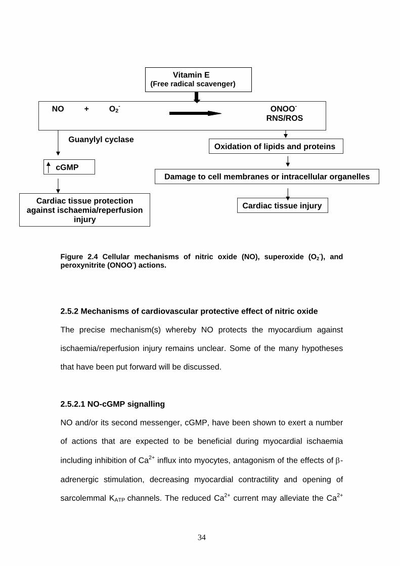
However, NO is detrimental when it is combined with superoxide (O2-) to form
peroxynitrite (ONOO-), which rapidly decomposes to highly reactive oxidant
species leading to tissue injury (Figure 2.4). There is a critical balance between
cellular concentrations of NO, O2- and superoxide dismutase (SOD) which
physiologically favours NO production, but in pathological conditions such as
ischaemia and reperfusion, results in ONOO- formation (reviewed by
Ferdinandy and Schultz, 2003). Illarion and co-workers (2002) reviewed the
role of reactive oxygen species, focusing mainly on superoxide radicals (O2-),
hydrogen peroxide (H2O2) and hydroxyl radical (.OH), which have long been
implicated in the pathogenesis of ischaemia/reperfusion injury. These oxygen
free radicals can react with nucleic acids, proteins and lipids, resulting in
damage to the cell membrane or intracellular organelles. Vitamin E acts as a
free radical scavenger that can react with oxygen, superoxide anion radicals
and hydroxyl radicals (Wall, 2000; Abuda et al., 2004) (Figure 2.4).
34
Figure 2.4 Cellular mechanisms of nitric oxide (NO), superoxide (O2-), and
peroxynitrite (ONOO-) actions.
2.5.2 Mechanisms of cardiovascular protective effect of nitric oxide
The precise mechanism(s) whereby NO protects the myocardium against
ischaemia/reperfusion injury remains unclear. Some of the many hypotheses
that have been put forward will be discussed.
2.5.2.1 NO-cGMP signalling
NO and/or its second messenger, cGMP, have been shown to exert a number
of actions that are expected to be beneficial during myocardial ischaemia
including inhibition of Ca2+ influx into myocytes, antagonism of the effects of β-
adrenergic stimulation, decreasing myocardial contractility and opening of
sarcolemmal KATP channels. The reduced Ca2+ current may alleviate the Ca2+
NO + O2- ONOO-
RNS/ROS
cGMP
Cardiac tissue protection against ischaemia/reperfusion
injury
Oxidation of lipids and proteins
Damage to cell membranes or intracellular organelles
Cardiac tissue injury
Guanylyl cyclase
Vitamin E (Free radical scavenger)
35
overload associated with acute myocardial ischaemia - one of the major
mechanisms of ischaemic injury (reviewed by Bolli, 2001).
NO is known to increase myocardial cGMP and it can be speculated that the
protective effect of NO is related to a mechanism secondary to the stimulation
of guanylyl cyclase within the vascular wall or in ventricular myocytes
(Beresewics et al., 1995; Maulik et al., 1995; Depré et al., 1996) (Figure 2.4).
NO donors administered during ischaemia possibly protect the myocardium by
increasing tissue cGMP and decreasing cytosolic Ca2+ overload (Du Toit et al.,
2001). Du Toit and co-workers (2001) found that nitric oxide donor treatment
reduces ischaemia/reperfusion injury by increasing cGMP concentrations and
suggested that the cAMP-to-cGMP ratio might play an important role in
cardioprotection. Maulik and co-workers (1995) showed that NO plays a
significant role in transmembrane signalling in the ischaemic myocardium. This
group suggested that NO signalling is switched off due to inactivation of NO by
reactive oxygen species and was the first to suggest that reactive oxygen
species may alter NO-cGMP signalling.
Increases in cAMP concentrations associated with ischaemia would increase
Ca2+ concentrations and exacerbate ischaemic/reperfusion injury (Du Toit et
al., 2001). In this regard it is possible that cGMP may attenuate this type of
injury by inhibiting the cAMP induced increase in slow inward calcium current,
thus leading to a decrease in cytosolic calcium levels (Sumii and Sperelakis,
1995). Therefore, cGMP appears to be an endogenous intracellular
cardioprotectant (Pabla et al., 1995; Pabla and Curtis, 1995).
36
2.5.2.2 Cholesterol-enriched diet and NO-cGMP signalling
It is well known that high cholesterol concentrations influence the NO-cGMP
signalling pathway. Therefore, cardiac stress adaptation is possibly
jeopardized in hyperlipidaemia due to altered NO-cGMP pathway function in
vascular and myocardial tissue. Szilvassy and co-workers (2001) found that a
cholesterol-enriched diet decreased both vascular NO and cGMP
concentrations and increased aortic O2- and ONOO- production. Several other
studies have also shown that a high-cholesterol diet impairs NO-cGMP
signalling in both endothelium (Deliconstantinos et al., 1995) and non-
endothelial cells with a significant decrease in cardiac NO concentrations
(Ferdinandy et al., 1997; Szekeres et al., 1997). Giricz and co-workers (2003)
found that although cardiac NO-concentration in cholesterol-fed rats was
decreased, nitric oxide synthase (NOS) activity was unchanged which may
suggest that NO synthesis was not impaired. The mechanism leading to
decreased cardiac NO level in hyperlipidaemia remains unknown. However, it
is well known that hyperlipidaemia leads to increased production of reactive
oxygen species (ROS) in the vasculature (Kojda and Harrison, 1999). For
example, hyperlipidaemia stimulates ONOO- generation in the heart, which
leads to myocardial dysfunction (Onody et al., 2003). It could be speculated
that increased O2- production (Giricz and co-workers, 2003) is responsible for
the decreased NO concentrations in the hyperlipidaemic myocardium since
NADPH oxidase activity due to hyperlipidaemia is a major source of increased
O2- production (Warnholtz et al., 1999).
37
Natural antioxidants can act as scavengers of damaging oxygen free radicals
(Cotrell, 1991; Chandrasekharan, 1999; Theriault et al., 1999; Kritchevsky,
2000; Wall, 2000). Vitamin E acts as a free radical scavenger that can react
with oxygen, superoxide anion radicals and hydroxyl radicals (Wall, 2000;
Abuda et al., 2004). Due to its lipid solubility, it is predominantly a chain
breaking antioxidant within the lipoprotein (Wall, 2000; Abuda et al., 2004).
Chow and co-workers (2002) reported that dietary vitamin E is capable of
reducing the production and/or availibilty of not only O2-, but also NO and
ONOO-. By reducing available O2- and NO, vitamin E may alleviate nitric
toxicity via reduced formation of reactive ONOO-. However, it is not clear if the
action of vitamin E to reduce the generation of O2- and other ROS species is
independent of its antioxidant function (Chow and Hong, 2002). Newaz and co-
workers (2003) showed an antioxidant protection by γ-tocotrienols in
hypertensive rats when compared with control animals. These authors
suggested that improved NOS activity in blood vessels and increased NO
availability were mediated through the antioxidant properties of γ-tocotrienol
where it effectively scavenges the free radicals. Venditti and co-workers (1999)
also reported that vitamin E treatment offers protection against
ischaemia/reperfusion-induced oxidative stress. However, the precise
mechanism of action is unknown.
38
2.5.2.3 Antioxidant properties of nitric oxide
By virtue of its antioxidant properties, NO offers protection specifically through
its ability to attenuate the deleterious free radical actions of O2- and to
terminate ONOO- mediated lipid radical chain propagation (Rubbo et al., 1994).
2.5.2.4 Nitric oxide and production of cytoprotective prostanoids
A previously unrecognized mechanism by which NO protects the ischaemic
myocardium has recently emerged, namely, stimulation of cyclooxygenase-2
(COX-2) activity with consequent production of cytoprotective prostanoids such
as prostaglandin (PGE2 and PGI2) (Shinmuri et al., 2000).
2.5.2.5 Therapeutical potential of nitric oxide
Research has shown that NO plays a fundamental biological role in protecting
the heart against ischaemia/reperfusion injury. The recognition that production
of NO represents nature’s own protective mechanism against ischaemia offers
fertile practical implications. Many opportunities loom on the horizon for
enhancing NO availability in a manner that would be therapeutically desirable
(Bolli, 2001).
2.6 Mitogen-activated protein kinases (MAPKs)
2.6.1 Apoptosis
Programmed cell death (apoptosis) is essential to many biological processes in
multicellular organisms including embryonic development, immune responses,
tissue homeostasis and normal cell turnover (reviewed by Lin, 2003).
39
Research has shown that both necrosis and apoptosis play a significant role in
myocardial ischaemia/reperfusion injury (Kajstura et al., 1996). Stress
conditions such as ischaemia (especially when followed by reperfusion) and
oxygen free radicals can elicit cardiomyocyte apoptosis, as has been
demonstrated in both cell cultures and isolated perfusion (reviewed by
Feuerstein and Young, 1999).
2.6.2 Signalling pathways and apoptosis
Regulatory mechanisms controlling proliferation, differentiation or apoptosis of
cells involve intracellular protein kinases that can transduce signals detected
on the cell’s surface into changes in gene expression. There is growing
evidence that multiple mitogen-activated protein (MAP) kinases and protein
kinase B (PKB/Akt) are activated during ischaemia and/or reperfusion and may
contribute to the structural and functional changes after myocardial
ischaemia/reperfusion injury (Abe et al., 2000). However, reports on their
precise roles are still conflicting (reviewed by Steenbergen, 2002).
Three major MAP kinase cascades that have been extensively studied in the
heart include extracellular signal regulated kinases (ERKs), c-Jun N-terminal
kinases (JNK 1 and JNK 2), and p38 kinases, of which the p38α and p38β
isoforms are found to play a role in myocardial tissue (Takeishi et al., 1999).
MAP kinases are proline-directed serine/threonine kinases that are activated in
response to a wide variety of stimuli including growth factors, G-protein
coupled receptors and environmental stresses (Robinson and Cobb, 1997;
Sugden and Clerk, 1998). The MAP kinases themselves require dual
phosphorylation on a Threonine-X-Tyrosine (Thr-X-Tyr) motif to become active

40
and each of these enzymes is a target for discrete but closely related
phosphorylation cascades in which the sequential activation of three kinases
constitutes a common signalling mode (Gartner et al., 1992; Fanger et al.,
1997; Siow et al., 1997).
2.6.3 Eicosapentaenoic acid (EPA) and docosahexaenoic acid (DHA)
modulate mitogen-activated protein kinases (MAPK) activity
Mirnikjoo and co-workers (2001) reported that n-3 fatty acids modulate
activation of extracellular signalling pathways by inhibiting the in vitro activities
of cAMP-dependent protein kinase, protein kinase C (PKC), Ca2+/calmodulin
dependent protein kinase 2 and the mitogen activated protein kinases
(MAPKs). These authors showed that DHA and EPA act as broad-spectrum
kinase inhibitors, blocking not only the activity of PKC but also the activity of
MAPK. These results were confirmed by Denys and co-workers (2002) who
showed that EPA and DHA regulate human T-cell function by modulating
MAPK enzyme activity. Other studies have shown that EPA also inhibits
hypoxia-reoxygenation-induced injury by attenuating upregulation of MMP-1,
thereby reducing phosphorylation of p38 (Cheng et al., 2003).
2.6.4 The major multiple mitogen-activated protein kinase (MAPK) and
PKB/Akt signalling pathways
2.6.4.1 p38 MAPK
At least 6 isoforms of the p38 MAPK group have been cloned, purified and
characterized, namely p38 α1/α2 (Han et al., 1994; Lee et al., 1994), p38
41
β1/β2 (Jiang et al., 1996), p38 γ (Li et al., 1996; Mertens et al., 1996) and p38δ
(Jiang et al., 1997; Wang et al., 1997)
Ultraviolet light, mechanical stress, hyper-osmotic shock,
hypoxia/reoxygenation, reactive oxygen species, pro-inflammatory cytokines
such as interleukin-1 and tumour necrosis factor alpha (TNF-α), and to a lesser
extent growth factors (Sugden and Clerk, 1998; Bogoyevitch, 2000), are some
of the extracellular stresses that activate p38 MAPK during ischaemia and
remain active during reperfusion (Pombo et al., 1994; Bogoyevitch et al., 1996;
Kyriakis and Avruch, 1996). Activation of p38 has been implicated in
transcription, protein synthesis, cell surface receptor expression and
cytoskeleton integrity, ultimately affecting cell survival or leading to apoptosis
and may even mediate cardiac hypertrophy (Obata et al., 2000). Huot and co-
workers (1997) reported that the p38 MAPK pathway may participate in several
oxyradical-activated functions of the endothelium that are associated with
reorganization of the microfilament network.
There is considerable controversy regarding the role of p38 in ischaemic injury
and in preconditioning. Research has shown both protective and detrimental
effects for p38 activation in ischaemia/reperfusion injury, where the p38α
isoform may be pro-apoptotic and the p38β isoform may be anti-apoptotic
(reviewed by Steenbergen, 2002).
42
2.6.4.1.1 Inhibition of p38 MAPK
A strong correlation between p38 activation and induction of apoptosis has
been reported in cardiac myocytes exposed to ischaemia/reperfusion
(Bogoyevitch et al., 1996). Research has shown that inhibition of p38 MAPK
activity during extended periods of ischaemia is cardioprotective (Mackay and
Mochly-Rosen, 1999; Barancik et al., 2000; Saurin et al., 2000). In a cultured
neonatal rat cardiac myocyte model, inhibition of p38 MAPK protects against
ischaemic injury as evidenced by a reduction in LDH release (Mackay and
Mochly-Rosen, 1999; Saurin et al., 2000). Barancik and co-workers (2000)
reported that the SB203580 inhibitor of p38 protected pig myocardium against
ischaemic injury, suggesting that p38 activation may accelerate ischaemic cell
death. Several studies indicated that p38 MAPK activation also plays a critical
role in promoting myocardial apoptosis (Ma et al., 1999; Mackay and Mochly-
Rosen, 1999; Yue et al., 2000). Ma and co-workers (1999) showed that p38
MAPK activity increased during ischaemia in isolated perfused rabbit heart, but
administration of SB203580 before ischaemia and during reperfusion
completely inhibited p38 MAPK activation, suggesting that inhibiting p38 was
cardioprotective. Yue and co-workers (2000) also showed that SB203580
improved cardiac contractile function and offered cardioprotection in isolated
ischaemic rat hearts by inhibiting p38 MAPK.
These studies show a correlation between inhibition of p38 MAPK activity and
cardioprotection against ischaemia/reperfusion injury in myocytes as well as in
isolated hearts.
43
2.6.4.1.2 Activation of p38 MAPK
On the other hand, research has also shown a protective role for p38 MAPK
activation in the heart. Weinbrenner and co-workers (1997) demonstrated that
tyrosine 182 phosphorylation of p38 MAPK was increased during a sustained
period of ischaemia in preconditioned hearts compared to non-preconditioned
hearts, which correlates with protection of preconditioning in the rabbit heart.
Communal and co-workers (2000) reported that increased p38 MAPK activity
together with decreased JNK1/2 activity, protects adult rat ventricular myocytes
against beta-adrenergic receptor-stimulated apoptosis. Nakano and co-
workers (2000) associated the protective effect of ischaemic preconditioning
with the activation of MAPKAPK2 (a substrate of p38 MAPK) in the isolated
rabbit heart. Zechner and co-workers (1998) reported myocardial protection
from apoptosis with overexpression of MAPK kinase 6 (MKK6), an upstream
activator of p38 MAPK. It appears that a distinct isoform of p38 MAPK, p38β,
participates in mediating cell survival (Wang et al., 1998). Therefore,
differential activation of p38 MAPK isoforms may exert opposing effects, where
p38α is implicated in cell death while p38β may mediate myocardial survival.
Although differential activation of p38 isoforms was observed in a study of
simulated ischaemic injury in cultured neonatal cardiomyocytes (Saurin et al.,
2000), more research is needed on the effects of p38 isoforms in the intact
adult myocardium. Differences in experimental protocol could also have
contributed to these conflicting results of p38 isoforms.
44
2.6.4.2 c-Jun N-terminal kinases (JNK)
Although JNK phosphorylation appears to be pro-apoptotic in many cell types,
their exact role in regulating cell death is unclear (Obata et al., 2000; Park et
al., 2000). Wang and co-workers (1998) reported that activation of JNK by
transfection of cultured rat neonatal cardiomyocytes with mitogen activated
protein kinase (MKK7) induced hypertrophy rather than apoptosis, and co-
activation of both JNK and p38 led to apoptosis.
The activation of JNK MAPKs has now been observed in a variety of cell types
of the cardiovascular system. JNKs are important for cytokine biosynthesis and
are involved in cell transformation and stress responses (Kyriakis and Avruch,
1996; Dong et al., 1998; Kuan et al., 1999). In almost all instances, the same
stimuli that activate p38 also activate the JNKs (Kyriakis and Avruch, 1996).
One exception is myocardial ischaemia/reperfusion where p38 is activated
during ischaemia, and JNK during reperfusion (Pombo et al., 1994;
Bogoyevitch et al., 1996; Kyriakis and Avruch, 1996). Furthermore, it has been
reported by Laderoute and Webster (1997) that antioxidants inhibit the
activation of JNKs in cultured myocytes subjected to hypoxia/reoxygenation.
2.6.4.2.1 The role of c-Jun N-terminal kinases (JNK) in apoptosis
Reoxygenation and reperfusion are known to activate JNK in the heart
(Bogoyevitch et al., 1996; Laderoute and Webster, 1997; Yue et al., 1998;
Davis, 2000), which is then translocated to the nucleus during ischaemia
where it is phosphorylated during the reperfusion period (Pombo et al., 1994;
Yue et al., 1998). Aoki and workers (2002) reported that the JNK pathway is a
45
direct activator of mitochondrial death machinery without other cellular
components and provides a molecular linkage from oxidative stress to the
mitochondrial apoptosis machinery. Lin (2003) proposed that JNK might be a
modulator rather than an intrinsic component of the apoptotic machinery. Thus,
JNK facilitates but does not induce apoptosis. These authors hypothesized that
activated JNK inactivates supressors of the apoptotic machinery. According to
their model, JNK activation contributes to apoptosis only if the apoptotic
process has already been activated.
In conclusion, both JNK and p38 are activated by ischaemia/reperfusion in the
intact heart, but neither has been shown definitely to be pro- or anti-apoptotic.
2.6.4.3 Extracellular signal-regulated kinases (ERK)
Extracellular signal-regulated kinase-1 (ERK 1) and ERK 2, also known as p44
and p42 MAPKs, represent the prototypical MAPKs in mammalial cells. A wide
variety of growth-promoting or hypertrophic agents activate these kinases in
cardiac myocytes, fibroblasts, smooth muscle cells and endothelial cells
(Bogoyevitch, 2000). Recently it has been shown that ERK activation also
takes place in response to mitochondria-derived superoxide production
secondary to the mitochondrial KATP opening (Samavati et al., 2002).
46
2.6.4.3.1 The role of extracellular signal-regulated kinases (ERK) in
apoptosis
The ERK pathway is important for survival of cells by protecting them from
programmed cell death caused by stress-induced activation of JNK and p38
(Yue et al., 2000).
Research has shown that inhibition of ERK enhances ischaemia/reperfusion-
induced apoptosis and that sustained activation of this kinase during simulated
ischaemia mediates adaptive cytoprotection in cultured neonatal
cardiomyocytes (Punn et al., 2000; Yue et al., 2000). Omura and co-workers
(1999) reported that both p42/44-MAPK and JNK were activated in the isolated
Langendorff perfused rat heart exposed to global ischaemia/reperfusion,
whereas in another experimental model of ischaemia/reperfusion in the intact
heart, ERK1/2 activation was shown to attenuate the extent of apoptosis
subsequent to reperfusion injury (Yue et al., 2000).
Although research has shown that stress or agonist induced ERK1/2 activation
is associated with protection of cardiac myocytes from apoptosis, little is known
as how ERK signalling results in cellular protection. Cyclooxygenase-2 (COX-
2) has been identified as a possible downstream mediator of protection in
association with ERK 1/2 signalling in cardiomyocytes (Adderley et al., 1999).
2.6.4.4 Protein kinase B (PKB/Akt) pathway
The serine/threonine protein kinase, protein kinase B or AKT (PKB/Akt), has
emerged as a crucial regulator of cellular processes including apoptosis,
proliferation and differentiation. PKB/Akt is activated downstream of PI-3-

47
kinase by the phosphoinisitide-dependent protein kinases (PDK) PDK-1 and
PDK-2 (Anderson et al., 1998). PKB/Akt in turn phosphorylates a number of
downstream targets relevant to cell survival function, including the pro-
apoptotic Bcl-2 family member BAD (Del Peso et al., 1997). Phosphorylation of
BAD on Ser136 by PKB/Akt inhibits its pro-apoptotic function, thus promoting
cell survival (Datta et al., 1997).
PKB/Akt has been suggested to be involved in cell survival pathways. Fujio
and co-workers (2000) showed that PKB/Akt promotes survival of
cardiomyocytes in vitro and protects against ischaemia/reperfusion injury in the
mouse heart. Dominant negative alleles of PKB/Akt reduce ability of growth
factors and other stimuli to maintain cell survival whereas over-expression of
wild type or activated PKB/Akt can rescue cells from apoptosis induced by
various stress signals (Kauffmann-Zeh et al., 1997; Kennedy et al., 1997). The
mechanisms involved in cell survival have only recently begun to emerge. One
way in which PKB/Akt may promote cell survival, is through direct
phosphorylation of transcription factors that control the expression of pro- and
anti-apoptotic genes.
2.6.5 Caspases
Activation of caspases is associated with apoptotic cell death. Caspases have
been grouped into an upstream (caspases -1,-2,-4,-5,-8,-9,-10) and a
downstream (caspases -3,-6,-7) subgroup (Nicholson and Thornberry, 1997).
All caspases are composed of a prodomain and an enzymatic region.
Upstream caspases are characterized by long prodomains that appear to
48
contain essential regulatory proteins. However, downstream caspases
sensitive to DEVD (aspartate-glutamine-valine-aspartate) oligopeptides
(caspase-3 and caspase-7) normally lead to the lethal proteolytic breakdown of
cellular target proteins. For activation, the caspase proform has to be cleaved
into a large subunit and a small subunit within the enzymatic domain that finally
reassociates to form a complex comprising of two small and two large
subunits. Substrates for caspases comprise many different proteins including
nuclear proteins, proteins involved in signal transduction, and cytoskeletal
targets (Cardone, 1997; Kothakota et al., 1997). Most of these protein
substrates appear to be cleaved by caspase -3 and -7. Although many of the
target proteins defined to date have a nuclear localization, apoptotic cell death
does not depend on the presence of a cell nucleus, as the characteristic
cytoplasmic features of apoptosis can be observed in anucleate cytoplasts
(Jacobson et al., 1994).
2.6.5.1 Mechanisms of caspase activation
Recent data suggest that activation of caspases may take place either within
death receptor complexes of the cytoplasmic membrane or by a
mitochondrion-dependent mechanism within the cytosol (Zou et al., 1997).
2.6.5.2 Poly-(ADP-ribose) polymerase (PARP)
A variety of stimuli induce apoptosis associated with cleavage of PARP by
caspases (Kaufmann, 1989; Kaufmann et al., 1993). Cleavage and inactivation
of PARP prevents energy depletion and induction of necrosis. However, acute
and massive DNA damage induces hyper-activation of PARP leading to
49
necrosis. Therefore, PARP cleavage has a function in the prevention of
necrotic cell death that would otherwise lead to pathological inflammatory
responses (Earnshaw, 1995).
Reperfusion injury is associated with caspase-3 activation, which results in
cleavage of PARP to its proteolysed products (Engelbrecht et al., 2004).
50
CHAPTER 3
MATERIALS AND METHODS
3.1 Animal Care
All animals received humane care in accordance with the Principle of
Laboratory Animal Care of the National Society of Medical Research and the
Guide for the Care and use of Laboratory Animals of the National Academy of
Sciences. (National Institutes of Health Publications no. 80-23, revised 1978).
3.2 Experimental Model 1
3.2.1 Experimental groups Male Long-Evans rats were divided into two groups: a control group receiving
standard rat chow and an experimental group receiving standard rat chow plus
7g RPO/kg diet for 6 weeks. The rat chow was supplied by Atlas Animal
Foods, Cape Town, South Africa and regularly analyzed to monitor possible
variations between batches. Red palm oil used for experimental work was
supplied by the Malaysian Palm Oil Board. The approximate energy and
macronutrient content of the two diets are indicated in Table 3.1.
The rats consumed an average of 25g food/day standard rat chow, containing
0.625g fat, which provides 8,7% of the energy intake. Protein intake was 4.5g
(28% of energy intake). In the experimental group, 0.175g RPO baking fat was
supplemented every morning, for 6 weeks, before they received their daily
allowance of rat chow. Thus, there was a 21% increase in fat intake in the
RPO-supplemented experimental group.
The red palm oil used in this study provided 70,0 μg carotenoids and 87,5 μg
vitamin E (tocotrienols and tocopherols) additional to any other antioxidants
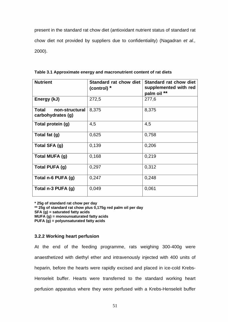
51
present in the standard rat chow diet (antioxidant nutrient status of standard rat
chow diet not provided by suppliers due to confidentiality) (Nagadran et al.,
2000).
Table 3.1 Approximate energy and macronutrient content of rat diets
Nutrient Standard rat chow diet (control) *
Standard rat chow diet supplemented with red palm oil **
Energy (kJ) 272,5 277,6
Total non-structural carbohydrates (g)
8,375 8,375
Total protein (g) 4,5 4,5
Total fat (g) 0,625 0,758
Total SFA (g) 0,139 0,206
Total MUFA (g) 0,168 0,219
Total PUFA (g) 0,297 0,312
Total n-6 PUFA (g) 0,247 0,248
Total n-3 PUFA (g) 0,049 0,061
* 25g of standard rat chow per day ** 25g of standard rat chow plus 0,175g red palm oil per day SFA (g) = saturated fatty acids MUFA (g) = monounsaturated fatty acids PUFA (g) = polyunsaturated fatty acids
3.2.2 Working heart perfusion
At the end of the feeding programme, rats weighing 300-400g were
anaesthetized with diethyl ether and intravenously injected with 400 units of
heparin, before the hearts were rapidly excised and placed in ice-cold Krebs-
Henseleit buffer. Hearts were transferred to the standard working heart
perfusion apparatus where they were perfused with a Krebs-Henseleit buffer
52
equilibrated with 95% O2 and 5% CO2 at 37 °C (118,5 mM NaCI; 4,75 mM KCI;
1,2 mM MgCI 6 H2O; 1,36 mM CaCI2;; 25,0 mM NaHCO3; 1,2 mM KH2PO4;;
11,0 mM glucose) at a perfusion pressure of 100 cm H2O.
The aorta was cannulated and retrograde perfusion with Krebs-Henseleit
buffer was initiated. During this initial perfusion in the Langendorff mode,
excess tissue was removed from the heart and the opening to the left atrium
was cannulated.
Following a 5-minute stabilisation period in the Langendorff mode, hearts were
switched to the working mode (Figure 3.1). The temperature of both the
perfusate and the air surrounding the heart was thermostatically controlled and
checked at regular intervals to ensure that the temperature was maintained at
37 °C irrespective of coronary flow. A cannula, connected to a pressure
transducer, was inserted through the apex of the heart into the left ventricle.
Left ventricular systolic and diastolic pressure, coronary flow (CF), heart rate
(HR) and aortic output (AO) were measured at 25 minutes into perfusion.
Hearts were then subjected to 25 minutes of global ischaemia. At the end of
ischaemia, hearts were reperfused in the Langendorff mode for 10 minutes. In
order to reduce the incidence of reperfusion arrhythmias, 2% lignocaine
solution was used for the initial 3 minutes of reperfusion of all hearts. This was
followed by a 15-minute working heart perfusion period during which cardiac
function was measured. To assess fatty acid composition and NO-cGMP
pathway activity, hearts were freeze-clamped with Wollenberger clamps pre-
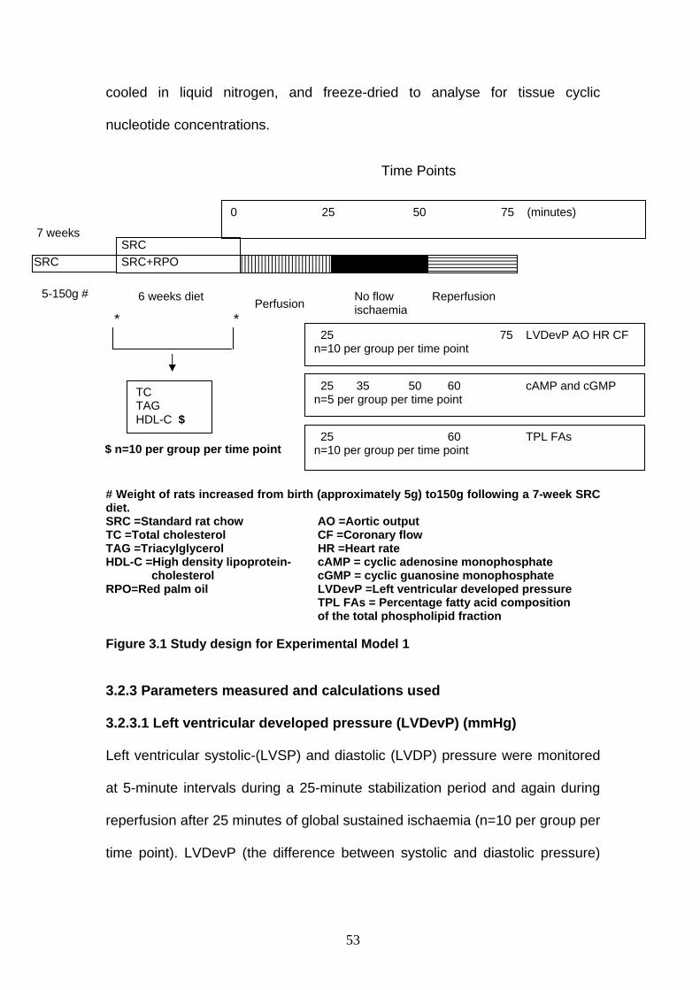
53
cooled in liquid nitrogen, and freeze-dried to analyse for tissue cyclic
nucleotide concentrations.
# Weight of rats increased from birth (approximately 5g) to150g following a 7-week SRC diet. SRC =Standard rat chow AO =Aortic output TC =Total cholesterol CF =Coronary flow TAG =Triacylglycerol HR =Heart rate HDL-C =High density lipoprotein- cAMP = cyclic adenosine monophosphate cholesterol cGMP = cyclic guanosine monophosphate RPO=Red palm oil LVDevP =Left ventricular developed pressure
TPL FAs = Percentage fatty acid composition of the total phospholipid fraction
Figure 3.1 Study design for Experimental Model 1 3.2.3 Parameters measured and calculations used 3.2.3.1 Left ventricular developed pressure (LVDevP) (mmHg)
Left ventricular systolic-(LVSP) and diastolic (LVDP) pressure were monitored
at 5-minute intervals during a 25-minute stabilization period and again during
reperfusion after 25 minutes of global sustained ischaemia (n=10 per group per
time point). LVDevP (the difference between systolic and diastolic pressure)
SRC SRC+RPO SRC
6 weeks diet Perfusion No flow ischaemia
Reperfusion
0 25 50 75 (minutes)
* * 25 75 LVDevP AO HR CF n=10 per group per time point
TC TAG HDL-C $
25 35 50 60 cAMP and cGMP n=5 per group per time point
25 60 TPL FAs n=10 per group per time point
5-150g #
7 weeks
Time Points
$ n=10 per group per time point
54
was used to measure mechanical function of the heart. It was calculated by
comparing the LVDevP before and after ischaemia.
3.2.3.2 Aortic output recovery (AO)(%)
In order to compare the functional recovery of the hearts in the different
groups, the heart rate (beats/min), coronary flow (ml/min) and aortic flow
(ml/min) were measured by collecting 1-minute samples of the respective
effluent (n=10 per group per time point). Aortic output (AO) recovery was
calculated by dividing the AO after ischaemia by AO before ischaemia and
expressing these values as a percentage recovery (see Figure 3.1 for time
points used).
3.2.3.3 Biochemical analyses
The cAMP and cGMP concentrations were determined at regular intervals (see
Figure 3.1) using radioimmunoassay kits obtained from Amersham Corp.
(Amersham, UK).
3.2.3.3.1 cGMP assay
The assay is based on the competition between unlabelled cGMP and a fixed
quantity of 125 I-labeled cGMP for a limited number of binding sites on a cGMP-
specific antibody. Measurement of the radioactivity in the pellet enables the
amount of labelled cGMP in the bound fraction to be calculated. The
concentration of unlabelled cGMP in the sample is then determined by
interpolation from a standard curve.
55
For cGMP assays freeze-clamped hearts (n=5 per group per time point, see
Figure 3.1) were freeze-dried and 20-25 mg of dry tissue was extracted in 5%
trichloroacetic acid. The extracted sample was ether-washed 3 times during 5-
minute wash cycles. These samples were diluted 1:10 (V/V) with assay buffer
and acetylated for the 125 I-labeled cGMP assay. The IC50 for the cCMP assay
was 25 pmol/tube (Du Toit et al., 1999).
3.2.3.3.2 cAMP assay
The assay is based on the competition between unlabelled cAMP and a fixed
quantity of the tritium labelled compound for binding to a protein which has a
high specificity and affinity for cAMP. The amount of labelled protein-cAMP
complex formed is inversely related to the amount of unlabelled cAMP present
in the assay sample. Measurement of the protein-bound radioactivity enables
the amount of unlabelled cAMP in the sample to be calculated. Separation of
the protein bound cAMP from the unbound nucleotide is achieved by
adsorption of the free nucleotide on to coated charcoal, followed by
centrifugation. An aliquot of the supernatant is then removed for liquid
scintillation counting. The concentration of unlabelled cAMP in the sample is
then determined from a linear standard curve.
For the cAMP assays, 20-25 mg freeze-dried tissue was extracted with
perchloric acid, neutralized and assayed (n=5 per group per time point, see
Figure 3.1). The IC50 for this assay was 1,92 mmol/tube (Du Toit et al., 1999).
56
3.2.3.4 Heart muscle total phospholipid fatty acid composition (%)
Hearts isolated from rats fed standard rat chow or standard rat chow and 7g
RPO per kg food for 6 weeks were perfused, freeze clamped and freeze-dried.
The freeze-dried tissue was used to determine myocardial total phospholipid
fatty acid composition (n=10 per group per time point). Tissue samples were
extracted with chloroform: methanol (2:1; v/v) according to a modified method
of Folch and co-workers (1957). Neutral lipids were separated from total
phospholipids by thin-layer chromatography and the total phospholipid fraction
analysed for fatty acid composition by gas chromatography. A fatty acid
mixture was prepared from individual fatty acids (Sigma. St. Louis, MO, USA)
and used as a standard. (Smuts et al., 1992; Van Jaarsveld et al., 2000).
3.2.3.5 Serum lipids
Rats were weighed weekly during the RPO-supplementation period and blood
was collected from the tail vein before and after the 6-week period for both the
standard rat chow (control) and the RPO-supplemented groups (n=10 per
group per time point). The blood was centrifuged for 10 minutes at 3 000x g to
obtain serum and analysed for serum lipid profiles i.e. total cholesterol (TC),
high-density lipoprotein (HDL)-cholesterol and triacylglycerol. The serum lipid
profile was determined using a Beckman Synchron Cx 9-instrument and
Beckman Synchron Cx reagents (Beckman Coulter) employing enzymatic and
colorimetric methods.
3.2.3.6 Statistical methods Values are presented as mean ± SEM. Some values are presented as
percentage change from the baseline values. To calculate the percentage

57
change, the values were devided by baseline values and multiplied by 100. To
adjust the X-axis to zero, 100 were subtracted from the values obtained. For
paired comparisons the Student’s t test was used. P<0,05 was considered
statistically significant.

58
3.3 Experimental Model 2
3.3.1 Experimental groups
Seven-week old Long-Evans rats were randomly allocated to 4 groups
according to the dietary supplementation they received.
Group 1: Standard rat chow.
Group 2: Standard rat chow plus RPO (7 g RPO per kg diet).
Group 3: Standard rat chow, containing 2% cholesterol.
Group 4: Standard rat chow containing 2% cholesterol plus RPO (7 g RPO per
kg diet).
The rats were fed a standard rat chow diet or 2% cholesterol-enriched diet
(based on previous studies by Giricz et al., 2003) for 6 weeks. Rats of the
control group consumed an average of 25g food/day standard rat chow,
containing 0.625g fat, which provides 8,7% of the energy intake. Protein intake
was 4,5g (28% of energy intake). In two additional groups RPO-baking fat (7g
RPO per kg diet) was supplemented every morning for 6 weeks to cholesterol-
enriched and standard rat chow diet, respectively, before they received their
daily allowance of rat chow. Thus, there was a 21% increase in fat intake in the
RPO-supplemented experimental groups. The red palm oil used in this study
provided 70,0 μg carotenoids and 87,5 μg vitamin E (tocotrienols and
tocopherols) additional to that present in the standard rat chow diet
(antioxidant nutrient status of standard rat chow diet not provided by supplier
due to confidentiality) (Nagadran et al., 2000).
59
3.3.2 Working heart perfusion and study design
The same protocol and study design (Figure 3.1 p.53) were used as discussed
in Experimental Model 1 (see working heart perfusion 3.2.2 p.51).
3.3.3 Parameters measured and calculations used The same measurements and calculations were used as discussed under
paragraph 3.2.3 p.53 in Experimental Model 1. In addition, rate pressure
product recovery was calculated for this study.
3.3.3.1 Rate pressure product recovery (RPP)(%)
Left ventricular developed pressure (LVDevP) was used to assess mechanical
function of the heart (n=10 per group per time point). LVDevP recovery (%) =
LVDevP 25 minutes into reperfusion (post-ischaemic)/ LVDevP 25 minutes into
perfusion (pre-ischaemic) x 100.
Functional recovery was expressed as the percentage rate pressure product
(RPP) recovery using the following formulae:
RPP =Heart Rate (HR) X LVDevP
% RPP recovery =(RPP reperfusion/RPP pre-ischaemic) x 100
3.3.3.2 Statistical Methods Statistical methods were discussed in 3.2.3.6 p.56 (Experimental Model 1).

60
3.4 Experimental Model 3 3.4.1 Experimental groups
Four groups with the same dietary regimens as described in 3.3.1 p.58
(Experimental Model 2) were used. Male Wistar rats were used as
experimental animals.
# Weight of rats increased from birth (approximately 5g) to150g following a 7-week SRC diet. NO= Nitric oxide HR= Heart rate NOS= Nitric oxide synthase CF= Coronary flow SOD= Superoxide dismutase AO= Aortic output LPO= Lipid hydroperoxide LVDevP=Left ventricular developed pressure SRC= Standard rat chow cGMP= cyclic guanosine monophosphate Chol= Cholesterol RPO=Red palm oil Figure 3.2 Study design for Experimental Model 3 3.4.2 Working heart perfusion
The protocol followed has been discussed in detail in 3.2.2 p.51 (Experimental
Model 1). However, for assessment of myocardial nitric oxide concentrations,
nitric oxide synthase- and superoxide dismutase activities and lipid
hydroperoxide production, hearts were freeze-clamped with Wollenberger
clamps pre-cooled in liquid nitrogen at the following times: at the end of the
SRC
6 weeks diet Perfusion No flow
Ischaemia Reperfusion
0 25 50 75 (minutes)
25 75 HR, CF, AO, LVDevP n=7 per group per time point
25 35 50 60 cGMP n=5 per group per time point
25 35 60 NO NOS SOD LPOn=5 per group per time point
SRC+ CholSRC+ Chol +RPO
SRC + RPOSRC
5-150g#
7 week
Time Points
61
pre-ischaemic working heart perfusion, after 10 minutes ischaemia and 10
minutes into reperfusion. Samples were stored at -80 °C for biochemical
analyses (Figure 3.2 p.60).
3.4.3 Parameters measured and calculations used 3.4.3.1 Cardiac functional parameters and aortic output recovery (AO)(%)
Measurement of cardiac functional parameters and calculation of aortic output
recovery have been discussed in section 3.2.3 p.53 under Materials and
Methods in Chapter 3 (Experimental Model 1).
3.4.3.2 Measurement of cGMP
Determination of cGMP concentrations has been discussed in 3.2.3.3 p.54
(Experimental Model 1).
3.4.3.3 Measurement of cardiac nitric oxide concentrations Approximately 200 mg of cardiac tissue was homogenized in 0,5 ml PBS (pH
7,4) and centrifuged at 10 000 x g for 20 minutes. The supernatant was ultra-
filtered using a 30 kDa molecular weight cut-off filter (Millipore) and 40 µl of the
filtrate was assayed.
Myocardial NO concentrations were determined using a Nitrate/Nitrite kit
(Cayman Chemicals, Cayman Islands) which provides an accurate and
convenient method for measurement of total nitrate/nitrite concentration in a
simple two-step process. The first step is the conversion of nitrate to nitrite
utilizing nitrate reductase. The second step is the addition of the Griess
Reagents, which convert nitrite into a deep purple azo compound. Photometric
62
measurement of the absorbance due to this chromophore accurately
determines nitrite concentration.
For direct measurement of intracellular NO, cardiomyocytes from RPO-
supplemented and control hearts were isolated by collagenase perfusion,
followed by incubation in a Krebs-Henseleit buffer containing 2% bovine serum
albumin in the presence of 10 μM diaminofluorescein-2/diacetate (DAF-2/ DA)
(-0,5 x 10⁶ cells/ml). Following incubation and washing, intracellular
fluorescence of DAF-triazol (DAF-2T, oxidized from DAF-DAF-2/DA) was
analyzed by flow cytometry (Strijdom et al., 2004).
3.4.3.4 Measurement of cardiac nitric oxide synthase activity
Approximately 100 mg of cardiac tissue was homogenized in 0,5 ml
homogenization buffer (1:10 dilution of stock solution, supplied by Cayman
Chemicals) and centrifuged at 10 000 x g for 15 minutes at 4 °C. For the
assay, 250 µl of supernatant was used.
Total NOS activity was measured using a NOS assay-kit which is based on the
biochemical conversion of L-arginine to L-citrulline by NOS (Cayman
Chemicals, Cayman Islands). This reaction, which represents a novel
enzymatic process, involves a 5-electron oxidation of guanidino nitrogen of L-
arginine to NO, together with the stoichiometric production of L-citrulline.
Advantages of this NOS kit include the use of radioactive substrate [14C]
arginine that enables sensitivity in the picomole range, as well as the specificity
of the assay for the NOS pathway due to the direct enzymatic conversion of
arginine to citrulline in eukaryotic cells. The NOS activity is quantified by
63
counting the radioactivity in the eluate (i.e. the flowthrough after reaction
samples were added to spin cups and centrifuged for 30 seconds) after
performing the assay.
3.4.3.5 Measurement of cardiac superoxide dismutase activity
Approximately 200 mg of cardiac tissue was homogenized in 1,0 ml ice-cold
HEPES buffer and centrifuged at 1 500 x g for 5 minutes at 4 °C. For the
assay, 10 µl of the supernatant was used.
Total activity of SOD was measured using a SOD kit (Cayman Chemicals,
Cayman Islands) that utilizes a tetrazolium salt for detection of superoxide
radicals generated by xanthine oxidase and hypoxanthase. One unit of SOD is
defined as the amount of enzyme required to exhibit 50% dismutation of the
superoxide radical. The SOD assay measures all three types of SOD.
3.4.3.6 Measurement of cardiac lipid hydroperoxide production
Approximately 200 mg of cardiac tissue was homogenized in 0,5 ml HPLC-
grade water. An equal volume of Extract R saturated methanol (Cayman
Chemicals, Cayman Islands) was added and mixed thoroughly by vortexing.
Cold chloroform (1,0 ml) was then added, mixed thoroughly and centrifuged at
1,500 x g for 5 minutes at 0 °C. The bottom chloroform layer was collected and
500 µl of this extract was used for the assay.
Lipid hydroperoxide (LPO) production in cardiac muscle was assessed using a
LPO assay kit (Cayman Chemicals, Cayman Islands) which measures the
hydroperoxides directly utilizing the redox reaction with ferrous ions.
64
Hydroperoxides are highly unstable and react readily with ferrous ions to
produce ferric ions. The resulting ferric ions are detected using thiocyanate ion
as the chromagen.
3.4.3.7 Statistical Methods Statistical methods were discussed in 3.2.3.6 p.56 (Experimental Model 1).

65
3.5 Experimental Model 4 3.5.1 Experimental groups
Two groups with the same dietary regimens as described in 3.2.1 p.50 were
used. Experimental design is given in Figure 3.3. Male Wistar rats were used
as experimental animals.
3.5.2 Working heart perfusion
Protocol followed has been discussed in detail in 3.2.2 p.51 (Experimental
Model 1). To assess the activity of myocardial MAPKs, hearts were freeze-
clamped with Wollenberger clamps pre-cooled in liquid nitrogen at the end of
the pre-ischaemic working heart perfusion after 10 minutes ischaemia and 10
minutes into reperfusion and samples were stored at -80 °C (Figure 3.3).
# Weight of rats increased from birth (approximately 5g) to150g following a 7-week SRC diet. SRC= Standard rat chow RPO= Red palm oil AO= Aortic output MAPKs= Mitogen-activated protein kinases Figure 3.3 Study design for Experimental Model 4
SRC SRC+RPO SRC
6 weeks diet Perfusion No flow
IschaemiaReperfusion
0 25 50 75 (minutes)
25 75 AO n=7 per group per time point
25 35 60 MAPKs n=4 for 4 independent experiments
5-150g #
7 weeks
Time Points
66
3.5.3 Parameters measured and calculations used 3.5.3.1 Aortic output recovery (AO)(%)
Measurement of AO has been discussed in 3.2.3.2 p.54.
3.5.3.2 Western blot analysis
Cardiac MAPKs and PKB/Akt, as well as caspase-3 and poly (ADP-ribose)
polymerase (PARP) protein, were extracted with a lysis buffer containing: Tris,
20 mM; p-nitrophenylphosphate, 20 mM; EGTA, 1mM; sodium fluoride (NaF),
50 mM; sodium orthovanadate, 0.1 mM; phenylmethyl sulphonyl fluoride
(PMSF), 1 mM; dithiothreitol (DTT), 1 mM; aprotinin, 10 µg/ml; leupeptin, 10
µg/ml. Mitogen-activated protein kinase phosphatase-1 (MKP-1) protein was
extracted with a lysis buffer containing: Hepes, 50 mM; EDTA, 10 mM; EGTA,
10 mM; PMSF, 1 mM; Aprotinin, 1µg/ml; Leupeptin, 1µg/ml; Triton, 0,5%. The
tissue lysates were diluted in Laemmli sample buffer and boiled for 5 minutes
after the lysate protein content was determined using the Bradford technique
(Bradford, 1976). For MAPKs and PKB/Akt 10µg protein, and 50µg protein for
caspase-3 and PARP, were separated by 10% PAGE-SDS-gel
electrophoresis. The separated proteins were transferred to a PVDF
membrane (ImmobilonTM P, Millipore). These membranes were routinely
stained with Ponceau Red for visualization of proteins. Non-specific binding
sites on the membranes were blocked with 5% fat-free milk in Tris-buffered
saline, 0.1% Tween 20 (TBST) and then incubated with the primary antibodies
that recognize phosphospecific ERK p42/p44 (Thr202/Tyr204), p38-MAPK
(Thr180/Tyr182), JNK p54/p46 (Thr183/Tyr185), PKB (Ser473and Thr308), caspase-3
(fragment pAb) and PARP (fragment pAb). Membranes were subsequently
67
washed with large volumes of TBST 5 x 5-minutes and the immobilized
antibody conjugated with a diluted horseradish peroxidase-labelled secondary
antibody (Amersham Life Sciences). After thorough washing with TBST,
membranes were covered with ECLTM detection reagents and quickly exposed
to an autoradiography film (Hyperfilm ECL, RPN 2103) to detect light emission
through a non-radioactive method (ECLTM Western blotting). Films were
densitometrically analysed (UN-SCAN-IT, Silkscience) and phosphorylated
protein values were corrected for minor differences in protein loading, if
required. Experiments were performed (data not shown) to ensure that all
signals were within the linear range of detection on the autoradiographs under
our assay and gel loading conditions.
Antibodies were purchased from Cell Signalling Technology and all other
chemicals were obtained from Sigma (St Louis, MO).
3.5.3.3 Statistical methods Data are presented as mean ± SEM. Statistical significance was determined in
multiple comparisons among independent groups of data in which ANOVA and
the Bonferroni’s post-hoc test indicated the presence of significant differences.
A P-value < 0.05 was considered statistically significant.
68
CHAPTER 4
Dietary red palm oil supplementation protects against the consequences
of global ischaemia in the isolated perfused rat heart.
Asia Pacific Journal of Clinical Nutrition (accepted 2004)

69
4.1 Abstract
Introduction: Activation of the NO-cGMP pathway is associated with
myocardial protection against ischaemia. During ischaemia the function of this
pathway is disturbed. Little is known about the effects of supplements such as
red palm oil (RPO) on the myocardial NO-cGMP-signalling pathway. RPO
consists of saturated (SFAs), monounsaturated (MUFAs) and polyunsaturated
(PUFAs) fatty acids and is rich in natural β-carotene and Vitamin E
(tocopherols and tocotrienols). Aims: To determine whether dietary RPO-
supplemention protects against the consequences of ischaemia/reperfusion
and to identify a possible mechanism for this protection. Methods: Long-Evans
rats were fed a standard rat chow (control) diet or standard rat chow diet plus
7g RPO per kg diet for 6 weeks. Hearts were excised and mounted on a
working heart perfusion apparatus. Cardiac function was measured before and
after hearts were subjected to 25 minutes of global ischaemia. Left ventricular
systolic (LVSP) and diastolic pressure (LVDP), coronary flow (CF), heart rate
(HR) and aortic output (AO) were measured. To assess NO-cGMP pathway
activity, hearts subjected to the same conditions, were freeze-clamped and
analysed for tissue cAMP and cGMP concentrations using a RIA method.
Furthermore, composition of myocardial total phospholipid fatty acids was
analysed by gas chromatography and blood samples were collected for serum
lipid determinations. Results: The percentage aortic output recovery of hearts
of the group supplemented with RPO was 72,9 ± 3,5% versus 55,4 ± 2,5% for
controls (P<0,05). Ten minutes into ischaemia the cGMP concentrations of the
RPO-supplementation group were significantly higher than the control group
70
(26,5 ± 2,8 pmol/g versus 10,1 ± 1,8 pmol/g). Myocardial total phospholipid
PUFA content in the group supplemented with RPO increased from 54,5 ±
1,1% before ischaemia to 59,0 ± 0,3% after ischaemia (P<0,05). Conclusions:
Our results demonstrate that dietary RPO-supplementation protected hearts
against ischaemia/reperfusion injury. These findings suggest that dietary RPO
protect via the NO-cGMP pathway and/or changes in phospholipid PUFA
composition during ischaemia/reperfusion.
4.2 Introduction
Palm oil and its liquid fraction, palm olein, are consumed worldwide as cooking
oils and as constituents of margarines. These oils are also incorporated into fat
blends used in the manufacturing of a variety of food products and in home
food preparation. It plays a useful role in meeting energy needs and
contributing to essential fatty acid (C18: 2n-6) needs in many regions of the
world (Cottrell, 1991). Red palm oil (RPO) used in this study, contains a
mixture of SFAs (51%), MUFAs (38%) and PUFAs (11%) (Nagendran et al.,
2000). Several clinical trails have evaluated palm oil’s effects on blood lipids
and lipoproteins. These studies suggest that palm oil and palm olein diets do
not raise serum total cholesterol (TC) and LDL cholesterol concentrations to
the extent expected from their fatty acid composition when a moderate-fat,
moderate-cholesterol diet is consumed (Therialt et al., 1999; Kritchevsky,
2000; Sundram and Basiron).
Although many animal feeding studies have shown that fish oil diets rich in n-3
PUFAs prevent ischaemia-induced cardiac arrhythmias (Nair et al., 1997; Kang
71
and Leaf, 2000; Jump, 2002), only a few reports have been published on the
protective effects of RPO-supplementation against ischaemia/reperfusion
injury (Abeywardena et al., 1991; Charnock et al., 1991; Abeywardena and
Charnock, 1995).
Palm oil and palm oil products are also natural occurring sources of the
antioxidant vitamin E constituents tocopherols and tocotrienols. These natural
antioxidants act as scavengers of damaging oxygen free radicals. Two studies
published in the New England Journal of Medicine show that both men and
women who supplement their diet with at least 100 IU of vitamin E (67μg
natural α-tocopherol) per day for at least two years have a 37-41% drop in the
risk of heart disease (Rimm et al., 1993; Stampfer et al., 1993). Vitamin E is
believed to be the major lipid-peroxidation chain-breaking antioxidant found in
blood plasma and membranes. The insight into the mechanism of heart injury
has suggested that administration of antioxidants may lessen oxidative
damage of the heart. It has been shown that a palm oil vitamin E mixture
containing both α-tocopherol and α-tocotrienol, was more efficient in the
protection of the isolated Langendorff heart against ischaemia/reperfusion
injury than tocopherol alone as measured by its mechanical recovery
(Serbinova et al., 1992). Palm oil vitamin E completely suppressed LDH
enzyme leakage from ischaemic hearts, prevented the decrease in ATP and
creatine phosphate levels, and inhibited the formation of endogenous lipid
peroxidation products (Serbinova et al., 1992).

72
Nitric Oxide (NO) is an important regulator of both cardiac and vascular
function and tissue reperfusion (reviewed by Ferdinandy and Schultz, 2003).
Myocardial NO formation is increased during ischaemia and reperfusion,
offering protection against ischaemia/reperfusion injury (Maulik et al., 1995;
Williams et al., 1995; Araka et al., 2000). However, when NO plummets during
reperfusion, due to rapid quenching by superoxide, the vascular protective
regulatory properties become dysfunctional. This may lead to exacerbation of
tissue injury. Reactive oxygen species such as the superoxide radical (O2-),
hydrogen peroxide (H2O2) and hydroxyl radical (.OH) have long been
implicated in the pathogenesis of ischaemia/reperfusion injury. These radicals
are predominant both during ischaemia and at the time of reperfusion, and can
react with nucleic acids, proteins and lipids, resulting in damage to the cell
membrane or intracellular organelles (Gilham et al., 1997). Peroxynitrite
(ONOO-) is generated by a diffusion-limited reaction between O2- and NO
(Rubbo et al., 1994; Naseem et al., 1995; Ferdinandy and Schultz, 2003).
Protective effects of NO are mediated through the production of cGMP. NO
donors given during ischaemia possibly protect the myocardium by increasing
tissue cGMP and decreasing cytosolic Ca2+ overload. Increases in cAMP
concentrations associated with ischaemia would increase Ca2+ concentrations
and exacerbate ischaemic and reperfusion injury (Du Toit et al., 2001). In this
regard it is possible that cGMP may attenuate this type of injury by inhibiting
the cAMP induced slow inward calcium current, thus leading to a decrease in
cytosolic calcium concentrations (Sumii and Sperelakis, 1995).
73
Because little is known about the protective effect of RPO and the mechanisms
involved, our aim was to determine whether dietary RPO-supplementation
protects against the consequences of ischaemia and reperfusion.
74
4.3 Materials and methods
4.3.1 Experimental Model
Long-Evans rats were fed a standard rat chow (control) diet or standard rat
chow diet plus 7g RPO per kg diet for 6 weeks. The working heart perfusion
method used in this experimental model is described in Chapter 3 under
Materials and Methods (Experimental Model 1 p.50) and the protocol that was
followed is summarized in Figure 3.1 p 53.
4.3.2 Measurement of cardiac function Left ventricular systolic (LVSP) and diastolic pressure (LVDP), coronary flow
(CF), heart rate (HR) and aortic output (AO) were measured as described in
Chapter 3 under Materials and Methods (Experimental Model 1 p.50).
4.3.3 Biochemical analyses
The cAMP and cGMP concentrations were determined using
radioimmunoassay kits obtained from Amersham Corp. (Amersham, UK). The
principle and methods were discussed in Chapter 3 under Materials and
Methods (Experimental Model 1 p.50).
4.3.4 Heart muscle total phospholipid fatty acid composition (%)
Composition of myocardial total phospholipid fatty acids was analysed by gas
chromatography as described in Chapter 3 under Materials and Methods
(Experimental Model 1 p.50).
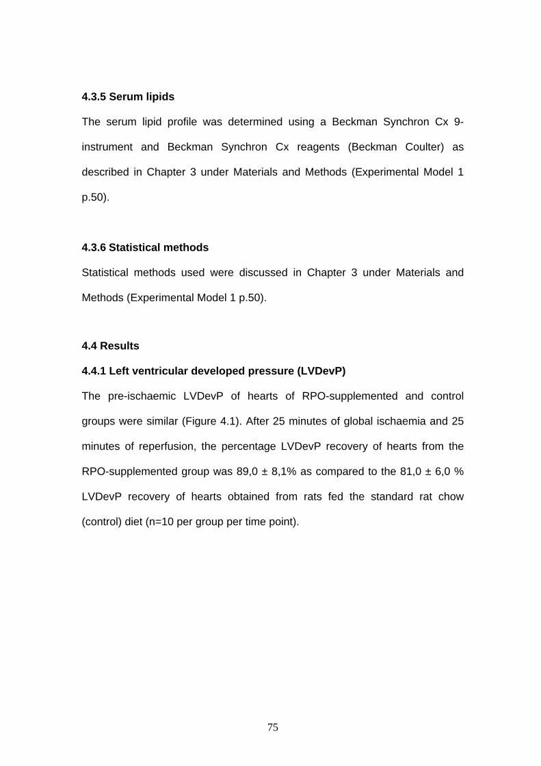
75
4.3.5 Serum lipids
The serum lipid profile was determined using a Beckman Synchron Cx 9-
instrument and Beckman Synchron Cx reagents (Beckman Coulter) as
described in Chapter 3 under Materials and Methods (Experimental Model 1
p.50).
4.3.6 Statistical methods
Statistical methods used were discussed in Chapter 3 under Materials and
Methods (Experimental Model 1 p.50).
4.4 Results
4.4.1 Left ventricular developed pressure (LVDevP)
The pre-ischaemic LVDevP of hearts of RPO-supplemented and control
groups were similar (Figure 4.1). After 25 minutes of global ischaemia and 25
minutes of reperfusion, the percentage LVDevP recovery of hearts from the
RPO-supplemented group was 89,0 ± 8,1% as compared to the 81,0 ± 6,0 %
LVDevP recovery of hearts obtained from rats fed the standard rat chow
(control) diet (n=10 per group per time point).
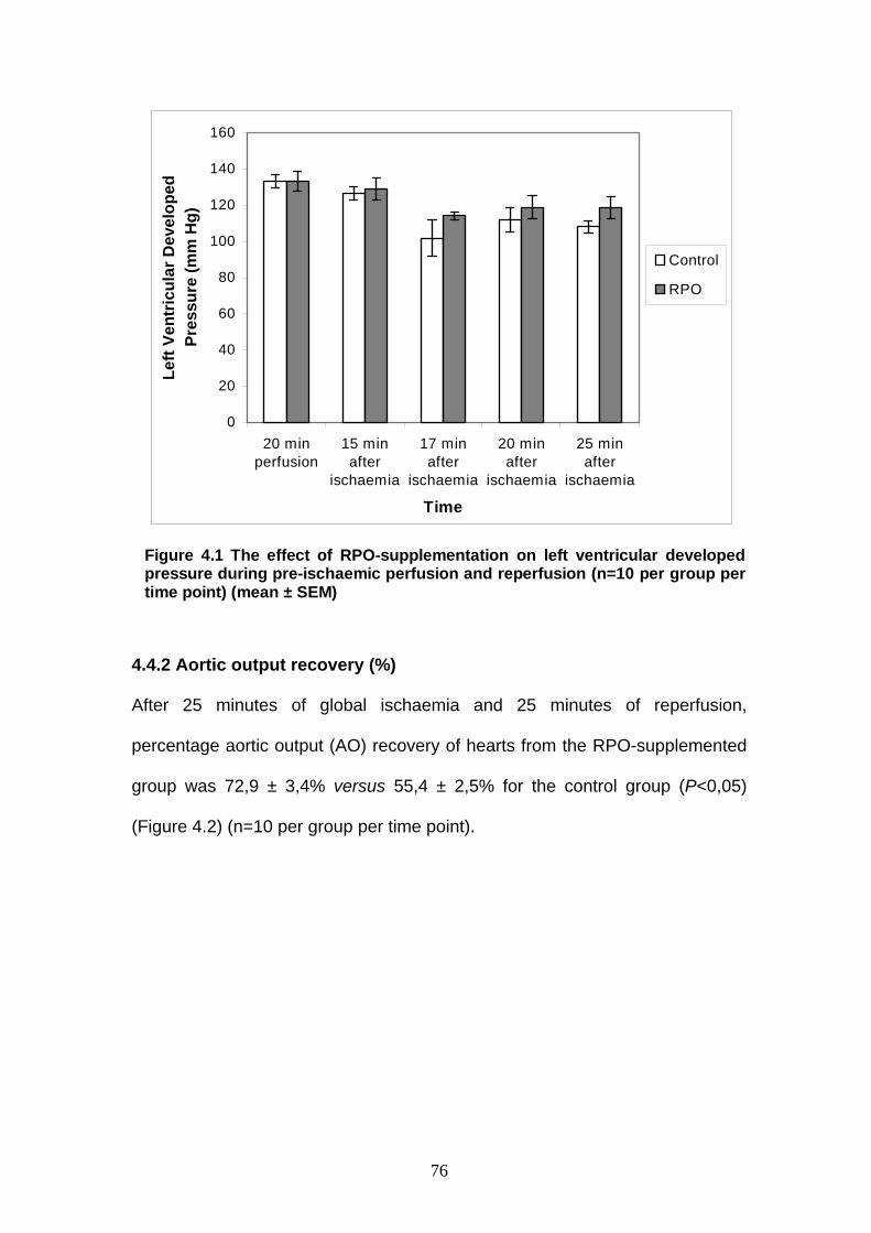
76
Figure 4.1 The effect of RPO-supplementation on left ventricular developedpressure during pre-ischaemic perfusion and reperfusion (n=10 per group pertime point) (mean ± SEM)
0
20
40
60
80
100
120
140
160
20 minperfusion
15 minafter
ischaemia
17 minafter
ischaemia
20 minafter
ischaemia
25 minafter
ischaemia
Time
Left
Vent
ricul
ar D
evel
oped
Pr
essu
re (m
m H
g)
Control
RPO
4.4.2 Aortic output recovery (%)
After 25 minutes of global ischaemia and 25 minutes of reperfusion,
percentage aortic output (AO) recovery of hearts from the RPO-supplemented
group was 72,9 ± 3,4% versus 55,4 ± 2,5% for the control group (P<0,05)
(Figure 4.2) (n=10 per group per time point).
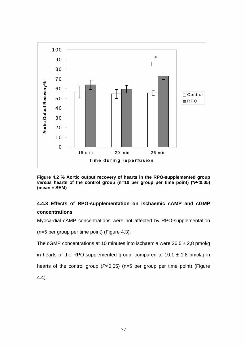
77
Figure 4.2 % Aortic output recovery of hearts in the RPO-supplemented group versus hearts of the control group (n=10 per group per time point) (*P<0.05) (mean ± SEM)
4.4.3 Effects of RPO-supplementation on ischaemic cAMP and cGMP concentrations Myocardial cAMP concentrations were not affected by RPO-supplementation
(n=5 per group per time point) (Figure 4.3).
The cGMP concentrations at 10 minutes into ischaemia were 26,5 ± 2,8 pmol/g
in hearts of the RPO-supplemented group, compared to 10,1 ± 1,8 pmol/g in
hearts of the control group (P<0,05) (n=5 per group per time point) (Figure
4.4).
0
1 0
2 0
3 0
4 0
5 0
6 0
7 0
8 0
9 0
100
15 m in 20 m in 25 m in
Tim e d u r in g r e p e r fu s io n
Aor
tic O
utpu
t Rec
over
y%
Contro lRP O
*
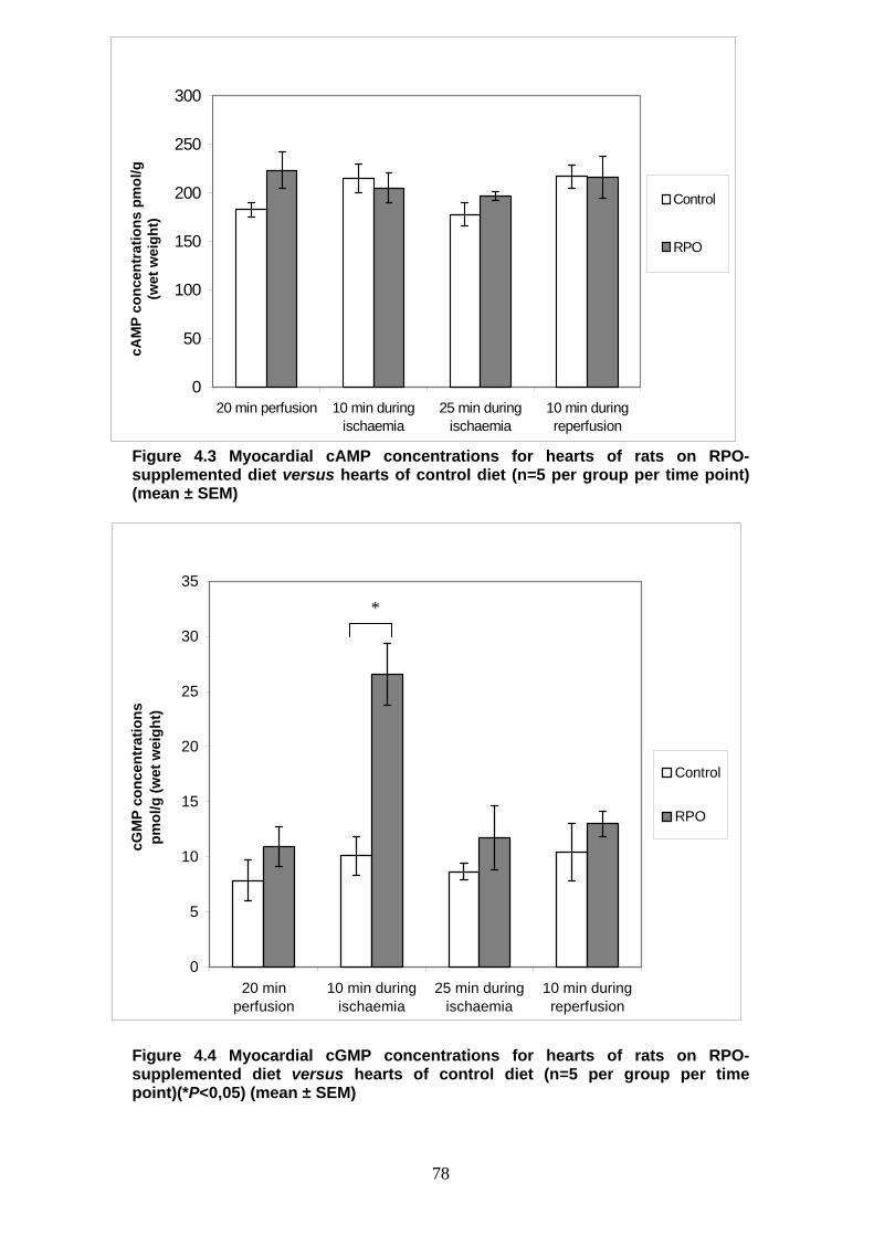
78
Figure 4.3 Myocardial cAMP concentrations for hearts of rats on RPO-supplemented diet versus hearts of control diet (n=5 per group per time point) (mean ± SEM)
Figure 4.4 Myocardial cGMP concentrations for hearts of rats on RPO-supplemented diet versus hearts of control diet (n=5 per group per time point)(*P<0,05) (mean ± SEM)
0
5
10
15
20
25
30
35
20 minperfusion
10 min duringischaemia
25 min duringischaemia
10 min duringreperfusion
cGM
P co
ncen
trat
ions
pm
ol/g
(wet
wei
ght)
Control
RPO
0
50
100
150
200
250
300
20 min perfusion 10 min duringischaemia
25 min duringischaemia
10 min duringreperfusion
cAM
P co
ncen
trat
ions
pm
ol/g
(w
et w
eigh
t)
Control
RPO
*

79
4.4.4 Serum lipids
Weight increase of rats remained stable between the control group and RPO-
supplemented group after the 6-week supplementation period.
All values in Figures 4.5 represent the percentage change in serum total
cholesterol-, high density lipoprotein cholesterol and triacylglycerol
concentrations between the baseline value and the value of the corresponding
experimental group after the 6-week supplementation period (n=10 per group).
Although there were baseline differences between the control and RPO-
supplemented groups (1,2 ± 0,04 mmol/l versus 1,4 ± 0,04 mmol/l, P<0,05),
the % change in serum total cholesterol showed no difference between the two
experimental groups after the 6-week diet period (n=10 per group).
HDL-cholesterol followed a similar trend with baseline differences between
control and RPO-supplemented groups (1,0 ± 0,03 mmol/l versus 1,2 ± 0,05
mmol/l, P<0,05), but the % change in HDL-cholesterol showed no difference
between the two groups after the 6-week diet period (n=10 per group) (Figure
4.5).
Serum concentrations of triacylglycerol were significantly increased in the
RPO-supplemented group after 6 weeks on the diet (before: 0,6 ± 0,1 mmol/l
versus after: 0,9 ± 0,1 mmol/l, P<0,05) (n=10 per group) (Figure 4.5). However,
percentage change in serum triacylglycerol between the control- and RPO-
supplemented groups showed no difference.
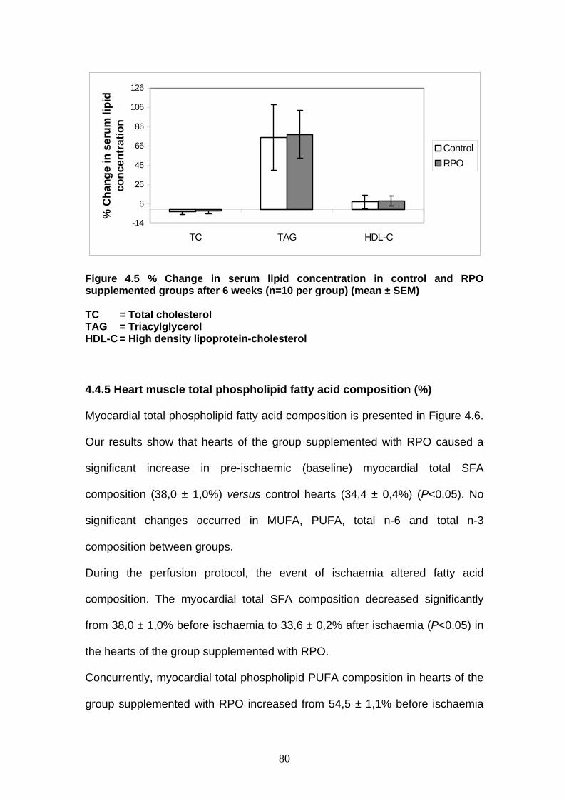
80
Figure 4.5 % Change in serum lipid concentration in control and RPO supplemented groups after 6 weeks (n=10 per group) (mean ± SEM) TC = Total cholesterol TAG = Triacylglycerol HDL-C = High density lipoprotein-cholesterol
4.4.5 Heart muscle total phospholipid fatty acid composition (%)
Myocardial total phospholipid fatty acid composition is presented in Figure 4.6.
Our results show that hearts of the group supplemented with RPO caused a
significant increase in pre-ischaemic (baseline) myocardial total SFA
composition (38,0 ± 1,0%) versus control hearts (34,4 ± 0,4%) (P<0,05). No
significant changes occurred in MUFA, PUFA, total n-6 and total n-3
composition between groups.
During the perfusion protocol, the event of ischaemia altered fatty acid
composition. The myocardial total SFA composition decreased significantly
from 38,0 ± 1,0% before ischaemia to 33,6 ± 0,2% after ischaemia (P<0,05) in
the hearts of the group supplemented with RPO.
Concurrently, myocardial total phospholipid PUFA composition in hearts of the
group supplemented with RPO increased from 54,5 ± 1,1% before ischaemia
-14
6
26
46
66
86
106
126
TC TAG HDL-C
% C
hang
e in
ser
um li
pid
conc
entr
atio
n
ControlRPO

81
to 59,0± 0,3% after ischaemia (P<0,05), with no changes in myocardial total
phospholipid MUFA composition.
Figure 4.6 Heart muscle total phospholipid fatty acids 20 minutes in perfusion and 10 minutes in reperfusion respectively, for RPO supplemented group versus control group (n=10 per group) (*P<0,05 for the group before and after ischaemia) (#P<0,05 for RPO supplemented group versus the corresponding control group) (mean ± SEM) SFA= Total saturated fatty acids MUFA= Total monounsaturated fatty acids PUFA= Total polyunsaturated fatty acids Tn-6= Total n-6 polyunsaturated fatty acids Tn-3= Total n-3 polyunsaturated fatty acids
0
10
20
30
40
50
60
70
80
SFA MUFA PUFA Tn-6 Tn-3
% P
hosp
holip
id fa
tty a
cids Control group before
ischaemia
Control group afterischaemia
RPO group beforeischaemia
RPO group afterischaemia
*
#
*
82
4.5 Discussion
Our results demonstrate that RPO-supplementation offered protection against
ischaemia/reperfusion injury in the isolated perfused working heart as reflected
by improved aortic output recovery. These data support the findings of
Serbinova and co-workers (1992) who showed that palm oil vitamin E was
more effective in the protection against ischaemia/reperfusion injury in the
isolated Langendorff perfused heart than tocopherol alone.
Based on our results we propose that the protective effect of RPO may be
associated with either its antioxidant characteristics and/or changes in the
myocardial total phospholipid fatty acid composition during
ischaemia/reperfusion. Abeywardena and co-workers (1991) also argued that
RPO protection lies in an alliance of fatty acids and endogenous antioxidants
during ischaemia/reperfusion.
Dietary RPO-supplementation for 6 weeks caused significant changes in heart
muscle total phospholipid fatty acid composition. The decrease in total
phospholipid SFA composition of the heart muscle was accompanied by an
increase in the total phospholipid PUFA composition, which could be
associated with improved reperfusion aortic output recovery. Normally, linoleic
acid (LA) undergoes a series of elongations and desaturations to yield
arachidonic acid (AA). To our knowledge no data exist on the effect of
ischaemia on elongase and desaturase activity. Many studies have focused on
the effect of FA on arrhythmias. Contradictory results exist on the effect of AA

83
on the development of arrhythmias. Li and co-workers (1997) found that free
AA is able to prevent arrhythmias, but the major cyclooxygenase metabolites
(PGD2, PGE2, PGF2 and TXA2) derived from AA are arrhythmogenic. However,
prostacyclins (PGI2), also synthesized from AA, are anti-thrombotic agents that
act as a vasodilator in blood vessels and have an antiarrhythmogenic effect.
No clear relationship exists between the availability of AA in myocardial
phospholipids and eicosanoid profile. Abeywardena and co-workers (1991)
showed that a chemically refined palm oil-supplementation for 12 months had
no effect on prostacyclin production from AA in rat myocardial tissue.
Concurrently, thromboxane A2 production was inhibited. Another palm oil
supplement (with a near-identical fatty acid profile) in the same study showed
no thromboxane A2 inhibition. This argues that thromboxane A2 production is
unlikely to be mediated via fatty acids. In another study (Abeywardena et al.,
1995) myocardial prostacyclin production was increased after ischaemia with
refined, bleached and deodorized palm oil (RBD-PO)-supplementation for 9
months. However, these authors argue that this increase in prostacyclin
production may not be mediated by fatty acids alone but that endogenous
antioxidants may also play a role.
The increase in PUFAs during ischaemia/reperfusion in the current study,
suggests that PUFAs may be involved in the protection against ischaemia/
reperfusion injury. The mechanism of phospholipid fatty acid protection
remains elusive and needs further investigation.
84
Generally, the mechanism of eicosanoid action is to bind to membrane
receptors, leading to generation of second messengers such as cAMP and
Ca2+ (Charnock et al., 1991; Mohan et al., 1995; Li et al., 1997). As myocardial
cAMP concentrations were not significantly affected throughout the protocol in
the hearts of the group supplemented with RPO, prevention of fatal cardiac
arrhythmias may depend on the net effect of these metabolites, which is
determined by the status of AA metabolism and the contents of n-6 and other
fatty acids (Abeywardena et al., 1991; Charnock et al., 1991; Mohan et al.,
1995; Li et al., 1997).
Interaction between cardiac endothelium-derived prostaglandins and NO
determines myocardial performance (Mohan et al., 1995). Our results suggest
that the NO-cGMP pathway is involved in the protective effect of RPO. The
elevated cGMP concentrations early in ischaemia may suggest that RPO-
supplementation protected the isolated rat heart against ischaemia/reperfusion
injury via the NO-cGMP pathway. NO is known to increase myocardial cGMP
and it can be speculated that the protective effect of NO is related to a
mechanism secondary to the stimulation of guanylyl cyclase within the
vascular wall or in ventricular myocytes (Beresewics et al., 1995). Besides its
effects on myocardial contractility, the NO-cGMP pathway stimulation during
ischaemia may protect the heart against ischaemia/reperfusion-induced
calcium overload. In this regard it is possible that cGMP may attenuate this
type of injury by inhibiting the cAMP-induced slow inward calcium current, thus
leading to a decrease in cytosolic calcium levels (Du Toit et al., 2001). Another
protective pathway might include the NO-cGMP dependent pathway in which
85
the sarcolemmal- K ATP channels are opened and the cystolic calcium
concentrations are lowered (Du Toit et al., 2001). Therefore, cGMP appears to
be an endogenous intracellular cardioprotectant (Pabla et al., 1995).
Studies have shown that reactive oxygen species (ROS) can oxidize lipids and
proteins and contribute to myocardial injury (reviewed by Illarion et al., 2002).
Peroxynitrite (ONOO-) is generated by a diffusion-limited reaction between O2-
and NO (Naseen et al., 1995; Ferdinandy et al., 2003). Palm oil and palm oil
products are naturally occurring sources of the antioxidant vitamin E
constituents tocopherol and tocotrienols, which act as scavengers of these
damaging oxygen free radicals which may lessen oxidative damage to the
heart (Serbinova et al., 1991). This may suggest that NO, synthesized from L-
arginine by NO-synthase (NOS), results in enhanced synthesis of cGMP. NO
synthase activity in the heart is rapidly stimulated by ischaemia and this
stimulation is maintained during the whole ischaemic episode (Moncado and
Higgs, 1993; Depré et al., 1996). Our data show that RPO-supplementation
increases cGMP concentrations that may confer some of the cardioprotection
to the ischaemic and reperfused rat heart.
Although there were baseline differences between the control and RPO-
supplemented groups, the serum total cholesterol concentration was not
effected by the 6-week diet. These data are in agreement with other studies
which showed that not all saturated dietary fats raise serum total cholesterol
concentration (Theriault et al., 1999; Kritchevsky, 2000; Sundram and
Basiron). Serum triacylglycerols concentrations were increased in both groups
86
after the 6-week period with a significant difference within the RPO-
supplemented group. The baseline difference in serum total cholesterol
between the control and RPO-supplemented groups cannot be explained and
requires further investigation, since rats were randomly allocated to groups.
Currently little documented data exist on the effects of dietary RPO-
supplementation and post ischaemic recovery linked to the NO-cGMP
pathway, heart muscle total phospholipid fatty acid composition and
antioxidant status. The findings of this study create opportunities for further
investigations to elucidate mechanisms involved in cardiac protection.
4.6 Conclusion
Dietary RPO-supplementation protects against the consequences of global
ischaemia in the isolated perfused rat heart from animals on a standard rat
chow (control) diet. The mechanism of protection may involve the NO-cGMP
pathway and/or myocardial phospholipid fatty acid compositional changes
during ischaemia/reperfusion.
Little knowledge exists on the protective effects of RPO-supplementation
against ischaemia/reperfusion injury, especially when supplemented with a
high-cholesterol diet. It was therefore our next aim to investigate whether RPO-
supplementation offers the same protection against ischaemia/reperfusion
injury when supplemented with a high-cholesterol diet, which simulates an
unhealthy dietary intake.
87
CHAPTER 5
Dietary red palm oil improves reperfusion cardiac function in the isolated
perfused rat heart of animals fed a high-cholesterol diet.
Prostaglandins, Leukotrienes and Essential Fatty Acids (published 2005)
88
5.1 Abstract
Introduction: It has been shown that dietary red palm oil (RPO)-
supplementation of rats on a standard rat chow (SRC) diet improved
reperfusion function. However, no exact cellular protective mechanisms have
been established. Aims: The aims of this study were to determine whether
dietary RPO-supplementation offers the same protection when cholesterol was
added to the diet and if so, to determine a possible cellular mechanism and a
role for fatty acids. Methods: Rats were fed a standard rat chow, standard rat
chow plus cholesterol and/or RPO-supplementation for 6 weeks. Functional
recovery, myocardial total phospholipid fatty acid composition and
cAMP/cGMP concentrations were determined in isolated rat hearts subjected
to 25 minutes of normothermic total global ischaemia. Results: Dietary RPO in
the presence of cholesterol improved aortic output recovery (63,2 ± 3,1%)
versus cholesterol only (36,5 ± 6,2%, P<0,05). The improved functional
recovery of hearts from rats supplemented with RPO versus control group was
preceded by an elevation in the cGMP concentrations early in ischaemia (RPO
132,9 ± 36,3% versus control 42,7 ± 24,4%, P<0,05). Concurrently, cAMP
concentrations decreased (RPO: -8,3 ± 6,9% versus control: 19,9 ± 7,7%,
P<0,05). Conclusions: Our data suggest that dietary RPO-supplementation
improved reperfusion aortic output through mechanisms that may include
activation of the NO-cGMP and inhibition of the cAMP pathway.
89
5.2 Introduction
Red palm oil consists of 51% saturated fatty acids (SFAs), 38%
monounsaturated fatty acids (MUFAs), 11% polyunsaturated fatty acids
(PUFAs) and a spectrum of antioxidants with carotenoids, tocopherols and
tocotrienols as the major constituents (Nagendran et al., 2000). It has been
shown by Serbinova and co-workers (1992) that a palm oil vitamin E mixture
containing both α-tocopherol and α-tocotrienol, improved reperfusion functional
recovery in a Langendorff perfused rat heart. They argued that the protection
was induced by the ability of both α-tocopherols and α-tocotrienols to
scavenge free radicals during reperfusion, but that α-tocotrienols offered more
efficient protection. Apart from this finding little knowledge exist on the exact
mechanism of dietary RPO-induced cardioprotection against
ischaemia/reperfusion injury. The composition of RPO allows for protection to
be offered by either the fatty acid constituents or the anti-oxidative carotenoids,
tocopherols and tocotrienols (Abeywardena et al., 1991; Charnock et al., 1991;
Cottrell, 1991; Abeywardena and Charnock, 1995; Chandrasekharan, 1999;
Theriault et al., 1999) or both. A possible mechanism that has not been
investigated may include the myocardial NO-cGMP pathway.
Reactive oxygen species, including superoxide radical (O2-), hydrogen
peroxide (H2O2) and hydroxyl radical (.OH), have long been implicated in the
pathogenesis of ischaemia/reperfusion injury. These radicals are generated
both during ischaemia and at the time of reperfusion and can react with nucleic
acids, proteins and lipids, resulting in damage to the cell membrane or
90
intracellular organelles (Gilham et al., 1997). Peroxynitrite (ONOO-) is
generated by a diffusion-limited reaction between O2- and nitric oxide (NO)
(Rubbo et al., 1994; Naseen et al., 1995; Ferdinandy et al., 1999; Ferdinandy
and Schultz, 2003).
NO is an important regulator of both cardiac and vascular function and tissue
reperfusion (reviewed by Ferdinandy and Schultz, 2003). Myocardial NO
formation is increased during ischaemia and reperfusion, offering protection
against ischaemia/reperfusion injury (Williams et al., 1995; Araki et al., 2000;
Bolli, 2001). Protective effects of NO are mediated through the production of
cGMP. NO is known to increase myocardial cGMP, and it can be speculated
that the protective effect of NO is related to a mechanism secondary to the
stimulation of guanylyl cyclase within the vascular wall, or in ventricular
myocytes (Maulik et al., 1995; Beresewics et al., 1995; Depré et al., 1997). NO
donors given during ischaemia possibly protect the myocardium by increasing
tissue cGMP and decreasing cytosolic Ca2+ overload. Increases in cAMP
concentrations associated with ischaemia would increase Ca2+ concentrations
and exacerbate ischaemia/reperfusion injury (Du Toit et al., 2001). In this
regard it is possible that cGMP may attenuate this type of injury by inhibiting
the cAMP-induced increase in slow inward calcium current, thus leading to a
decrease in cytosolic calcium concentrations (Sumii and Sperelakis, 1995).
Therefore, cGMP appears to be an endogenous intracellular cardioprotectant
(Pabla and Curtis, 1995; Pabla et al., 1995).

91
Cardiac stress adaptation is possibly compromised in hyperlipidaemia due to
altered NO-cGMP pathway function. Research showed that high-cholesterol
diets impair NO-cGMP signalling in both endothelium and non-endothelial cells
with a significant decrease in cardiac NO concentration. Giricz and co-workers
(2003) found an increase in O2- production in cholesterol-fed rats, which may
explain the decreased cardiac NO-concentration in hyperlipidaemia.
Despite the SFA content of RPO, several clinical trials have evaluated the
effect of RPO on blood lipids and lipoproteins. These studies suggest that RPO
in a moderate fat, moderate cholesterol diet, despite its high content of SFA,
does not raise serum total cholesterol (TC) or LDL cholesterol concentrations
(Chandrasekharan, 1999; Theriault et al., 1999; Kritchevsky, 2000; Sundram
and Basiron). However, when hypercholesterolaemic subjects and high fat
liquid formula diets were used, RPO-supplementation appeared to raise TC
and LDL cholesterol (Sundram and Basiron). Little knowledge exists on the
protective effects of RPO-supplementation against ischaemia/reperfusion
injury, especially with a high-cholesterol diet (Esterhuyse et al., 2003).
Serbinova and co-workers (1992) focused on the functional effects of palm oil
vitamin E in the Langendorff perfused rat heart subjected to global ischaemia,
while the current study investigated a possible role for the cellular signalling
pathways. We therefore designed a study to investigate the effect of RPO on
ischaemia/reperfusion injury in the working rat heart model. We also added
cholesterol to the diet to simulate an unhealthy dietary-cholesterol intake.
92
Our main aims were to determine whether: 1) dietary RPO-supplementation of
rats on a hypercholesterolaemic diet protected against ischaemia/reperfusion
injury, 2) the NO-cGMP pathway can be considered as a possible mechanism
of protection and 3) changes in the composition of myocardial total
phospholipid fatty acids during ischaemia contributed to this protection.
93
5.3 Materials and Methods
5.3.1 Experimental Model
Wistar rats were fed a standard rat chow (SRC), or a SRC plus cholesterol
diet. In two additional groups, these diets were supplemented with RPO for 6
weeks. The working heart perfusion method used in this Experimental Model is
described in Chapter 3 under Materials and Methods (Experimental Model 2
p.58) and the protocol that was followed is summarized in Figure 3.1 p.53.
5.3.2 Measurement of cardiac function Left ventricular systolic-(LVSP) and diastolic pressure (LVDP), coronary flow
(CF), heart rate (HR) and aortic output (AO) were measured as described in
Chapter 3 under Materials and Methods (Experimental Model 1 p.50).
Functional recovery was expressed as the percentage rate pressure product
(RPP) recovery as described in Chapter 3 under Materials and Methods
(Experimental Model 2 p.58).
5.3.3 Biochemical analyses
The cAMP and cGMP concentrations were determined using
radioimmunoassay kits obtained from Amersham Corp. (Amersham, UK). The
principle and methods were discussed in Chapter 3 under Materials and
Methods (Experimental Model 1 p.50).
5.3.4 Heart muscle total phospholipid fatty acid composition (%)
Freeze-dried myocardial tissue from all the groups was used to determine total
phospholipid fatty acid composition by gas chromatography as described in
Chapter 3 under Materials and Methods (Experimental Model 1 p.50).
94
5.3.5 Serum lipids
Blood was collected from the tail vein of the rats on the SRC diet and groups
supplemented with cholesterol and RPO before and after the 6-week
supplementation period (n=10 per group). The serum lipid profile was
determined using a Beckman Synchron Cx 9-instrument and Beckman
Synchron Cx reagents (Beckman Coulter) as described in Chapter 3 under
Materials and Methods (Experimental Model 1 p.50).
5.3.6 Statistical methods
Statistical methods used were described in Chapter 3 under Materials and
Methods (Experimental Model 1 p.50).
5.4 Results
5.4.1 % Rate pressure product recovery
Rate pressure product (RPP) recovery was increased in the RPO-
supplemented group when compared with the control group (89,4 ± 4,7%
versus 75,0 ± 4,7%, P<0,05) 25 minutes into reperfusion (n=5 per group)
(Figure 5.1). RPO also improved RPP recovery in the presence of cholesterol
(cholesterol/RPO: 99,9 ± 4,0% versus cholesterol only: 72,1 ± 2,7%, P<0,05).
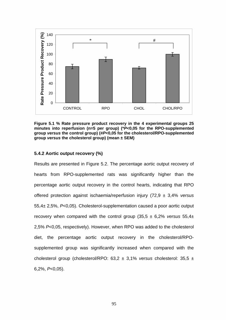
95
0
20
40
60
80
100
120
140
CONTROL RPO CHOL CHOL/RPO
Rat
e Pr
essu
re P
rodu
ct R
ecov
ery
(%)
Figure 5.1 % Rate pressure product recovery in the 4 experimental groups 25 minutes into reperfusion (n=5 per group) (*P<0,05 for the RPO-supplemented group versus the control group) (#P<0,05 for the cholesterol/RPO-supplemented group versus the cholesterol group) (mean ± SEM)
5.4.2 Aortic output recovery (%)
Results are presented in Figure 5.2. The percentage aortic output recovery of
hearts from RPO-supplemented rats was significantly higher than the
percentage aortic output recovery in the control hearts, indicating that RPO
offered protection against ischaemia/reperfusion injury (72,9 ± 3,4% versus
55,4± 2,5%, P<0,05). Cholesterol-supplementation caused a poor aortic output
recovery when compared with the control group (35,5 ± 6,2% versus 55,4±
2,5% P<0,05, respectively). However, when RPO was added to the cholesterol
diet, the percentage aortic output recovery in the cholesterol/RPO-
supplemented group was significantly increased when compared with the
cholesterol group (cholesterol/RPO: 63,2 ± 3,1% versus cholesterol: 35,5 ±
6,2%, P<0,05).
#*
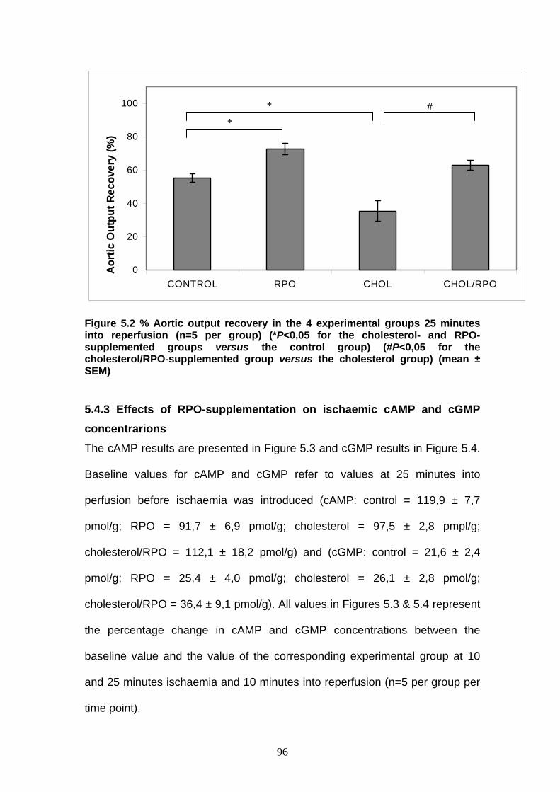
96
0
20
40
60
80
100
CONTROL RPO CHOL CHOL/RPO
Aor
tic O
utpu
t Rec
over
y (%
)
Figure 5.2 % Aortic output recovery in the 4 experimental groups 25 minutes into reperfusion (n=5 per group) (*P<0,05 for the cholesterol- and RPO-supplemented groups versus the control group) (#P<0,05 for the cholesterol/RPO-supplemented group versus the cholesterol group) (mean ± SEM)
5.4.3 Effects of RPO-supplementation on ischaemic cAMP and cGMP concentrarions The cAMP results are presented in Figure 5.3 and cGMP results in Figure 5.4.
Baseline values for cAMP and cGMP refer to values at 25 minutes into
perfusion before ischaemia was introduced (cAMP: control = 119,9 ± 7,7
pmol/g; RPO = 91,7 ± 6,9 pmol/g; cholesterol = 97,5 ± 2,8 pmpl/g;
cholesterol/RPO = 112,1 ± 18,2 pmol/g) and (cGMP: control = 21,6 ± 2,4
pmol/g; RPO = 25,4 ± 4,0 pmol/g; cholesterol = 26,1 ± 2,8 pmol/g;
cholesterol/RPO = 36,4 ± 9,1 pmol/g). All values in Figures 5.3 & 5.4 represent
the percentage change in cAMP and cGMP concentrations between the
baseline value and the value of the corresponding experimental group at 10
and 25 minutes ischaemia and 10 minutes into reperfusion (n=5 per group per
time point).
* # *
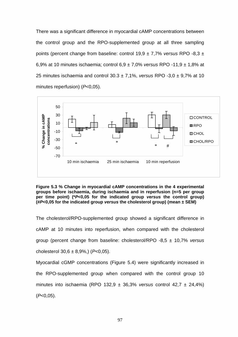
97
There was a significant difference in myocardial cAMP concentrations between
the control group and the RPO-supplemented group at all three sampling
points (percent change from baseline: control 19,9 ± 7,7% versus RPO -8,3 ±
6,9% at 10 minutes ischaemia; control 6,9 ± 7,0% versus RPO -11,9 ± 1,8% at
25 minutes ischaemia and control 30.3 ± 7,1%, versus RPO -3,0 ± 9,7% at 10
minutes reperfusion) (P<0,05).
-70
-50
-30
-10
10
30
50
10 min ischaemia 25 min ischaemia 10 min reperfusion
% C
hang
e in
cA
MP
conc
entr
atio
ns
CONTROL
RPO
CHOL
CHOL/RPO
Figure 5.3 % Change in myocardial cAMP concentrations in the 4 experimental groups before ischaemia, during ischaemia and in reperfusion (n=5 per group per time point) (*P<0,05 for the indicated group versus the control group) (#P<0,05 for the indicated group versus the cholesterol group) (mean ± SEM)
The cholesterol/RPO-supplemented group showed a significant difference in
cAMP at 10 minutes into reperfusion, when compared with the cholesterol
group (percent change from baseline: cholesterol/RPO -8,5 ± 10,7% versus
cholesterol 30,6 ± 8,9%,) (P<0,05).
Myocardial cGMP concentrations (Figure 5.4) were significantly increased in
the RPO-supplemented group when compared with the control group 10
minutes into ischaemia (RPO 132,9 ± 36,3% versus control 42,7 ± 24,4%)
(P<0,05).
* * # *
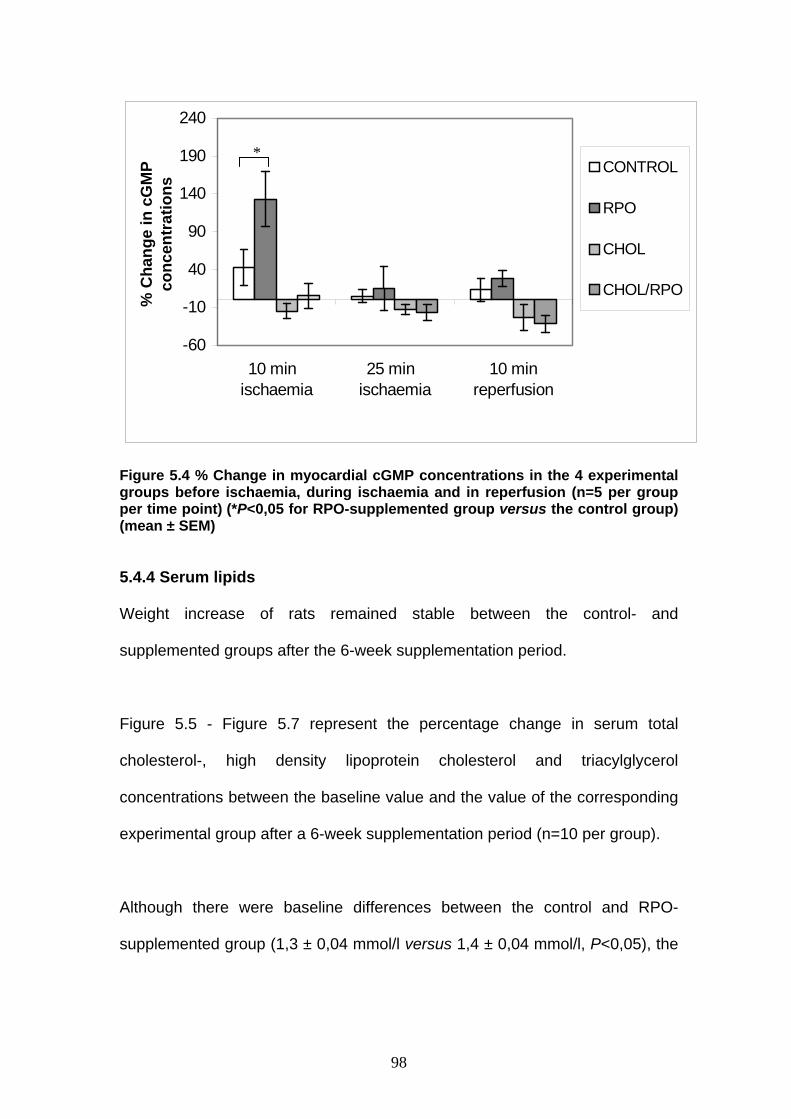
98
-60
-10
40
90
140
190
240
10 min ischaemia
25 min ischaemia
10 minreperfusion
% C
hang
e in
cG
MP
conc
entr
atio
nsCONTROL
RPO
CHOL
CHOL/RPO
Figure 5.4 % Change in myocardial cGMP concentrations in the 4 experimental groups before ischaemia, during ischaemia and in reperfusion (n=5 per group per time point) (*P<0,05 for RPO-supplemented group versus the control group) (mean ± SEM)
5.4.4 Serum lipids
Weight increase of rats remained stable between the control- and
supplemented groups after the 6-week supplementation period.
Figure 5.5 - Figure 5.7 represent the percentage change in serum total
cholesterol-, high density lipoprotein cholesterol and triacylglycerol
concentrations between the baseline value and the value of the corresponding
experimental group after a 6-week supplementation period (n=10 per group).
Although there were baseline differences between the control and RPO-
supplemented group (1,3 ± 0,04 mmol/l versus 1,4 ± 0,04 mmol/l, P<0,05), the
*
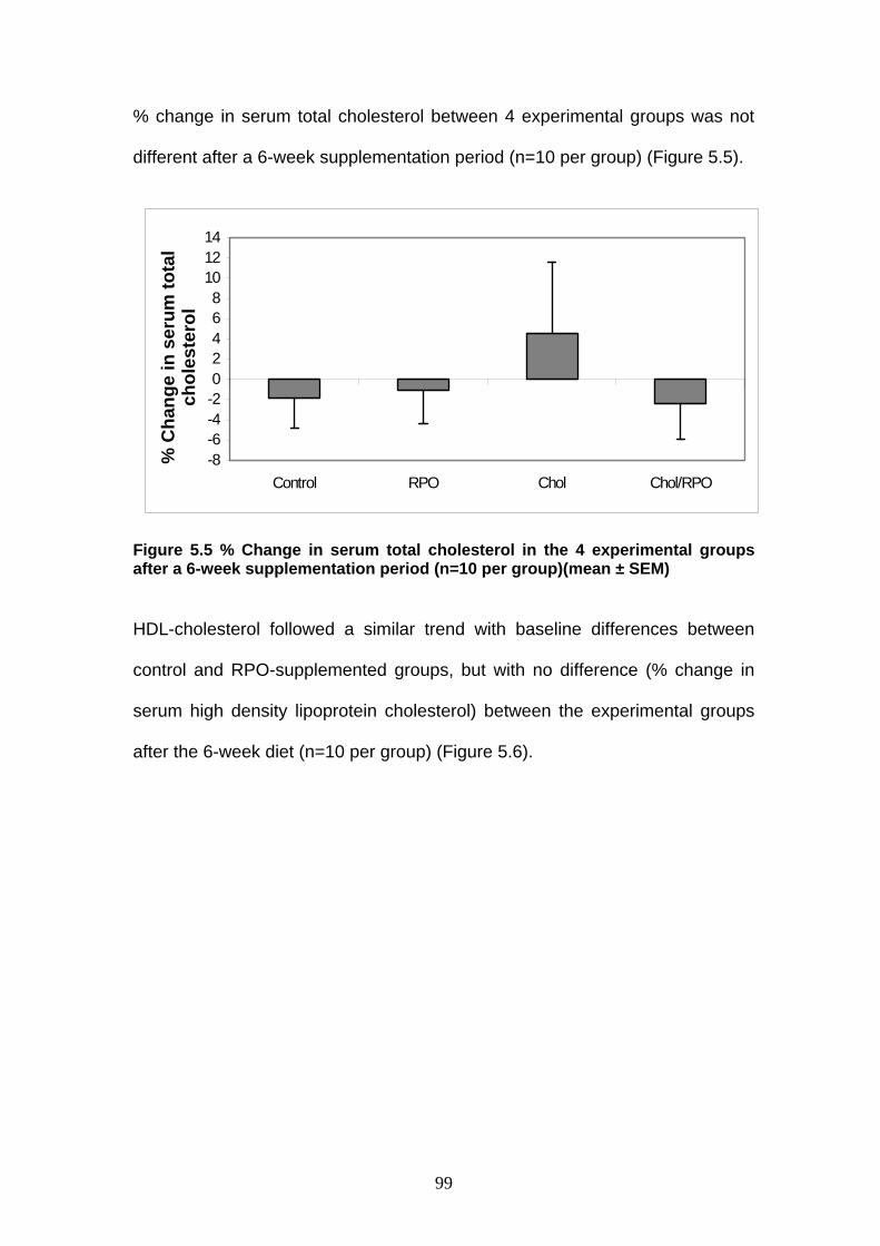
99
% change in serum total cholesterol between 4 experimental groups was not
different after a 6-week supplementation period (n=10 per group) (Figure 5.5).
Figure 5.5 % Change in serum total cholesterol in the 4 experimental groups after a 6-week supplementation period (n=10 per group)(mean ± SEM) HDL-cholesterol followed a similar trend with baseline differences between
control and RPO-supplemented groups, but with no difference (% change in
serum high density lipoprotein cholesterol) between the experimental groups
after the 6-week diet (n=10 per group) (Figure 5.6).
-8-6-4-202468
101214
Control RPO Chol Chol/RPO
% C
hang
e in
ser
um to
tal
chol
este
rol
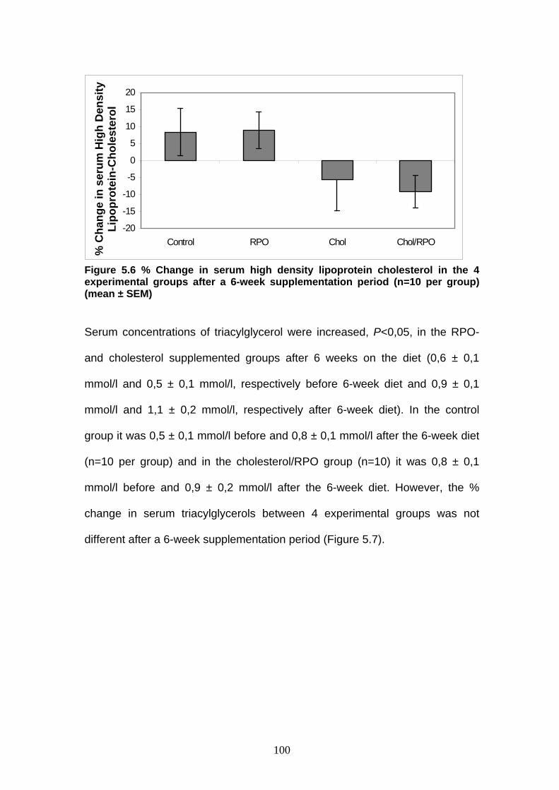
100
Figure 5.6 % Change in serum high density lipoprotein cholesterol in the 4 experimental groups after a 6-week supplementation period (n=10 per group) (mean ± SEM) Serum concentrations of triacylglycerol were increased, P<0,05, in the RPO-
and cholesterol supplemented groups after 6 weeks on the diet (0,6 ± 0,1
mmol/l and 0,5 ± 0,1 mmol/l, respectively before 6-week diet and 0,9 ± 0,1
mmol/l and 1,1 ± 0,2 mmol/l, respectively after 6-week diet). In the control
group it was 0,5 ± 0,1 mmol/l before and 0,8 ± 0,1 mmol/l after the 6-week diet
(n=10 per group) and in the cholesterol/RPO group (n=10) it was 0,8 ± 0,1
mmol/l before and 0,9 ± 0,2 mmol/l after the 6-week diet. However, the %
change in serum triacylglycerols between 4 experimental groups was not
different after a 6-week supplementation period (Figure 5.7).
-20
-15
-10
-5
0
5
10
15
20
Control RPO Chol Chol/RPO
% C
hang
e in
ser
um H
igh
Den
sity
Li
popr
otei
n-C
hole
ster
ol
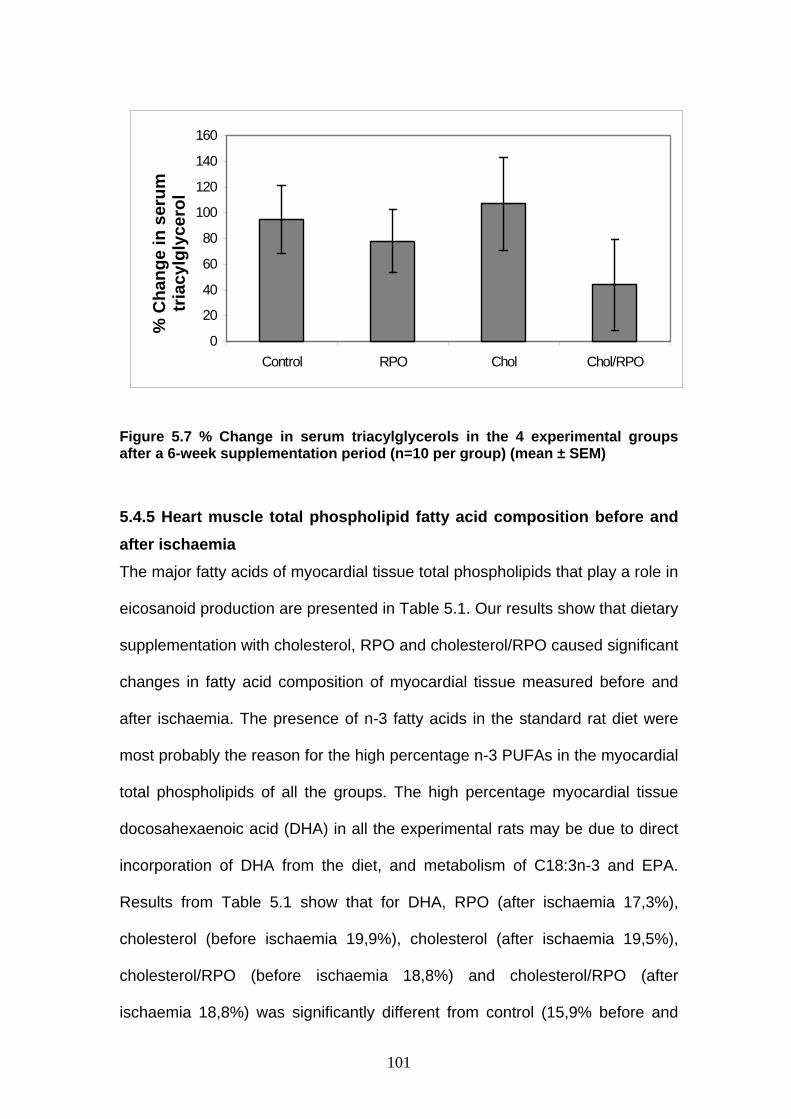
101
Figure 5.7 % Change in serum triacylglycerols in the 4 experimental groups after a 6-week supplementation period (n=10 per group) (mean ± SEM) 5.4.5 Heart muscle total phospholipid fatty acid composition before and after ischaemia The major fatty acids of myocardial tissue total phospholipids that play a role in
eicosanoid production are presented in Table 5.1. Our results show that dietary
supplementation with cholesterol, RPO and cholesterol/RPO caused significant
changes in fatty acid composition of myocardial tissue measured before and
after ischaemia. The presence of n-3 fatty acids in the standard rat diet were
most probably the reason for the high percentage n-3 PUFAs in the myocardial
total phospholipids of all the groups. The high percentage myocardial tissue
docosahexaenoic acid (DHA) in all the experimental rats may be due to direct
incorporation of DHA from the diet, and metabolism of C18:3n-3 and EPA.
Results from Table 5.1 show that for DHA, RPO (after ischaemia 17,3%),
cholesterol (before ischaemia 19,9%), cholesterol (after ischaemia 19,5%),
cholesterol/RPO (before ischaemia 18,8%) and cholesterol/RPO (after
ischaemia 18,8%) was significantly different from control (15,9% before and
0
20
40
60
80
100
120
140
160
Control RPO Chol Chol/RPO
% C
hang
e in
ser
um
tria
cylg
lyce
rol
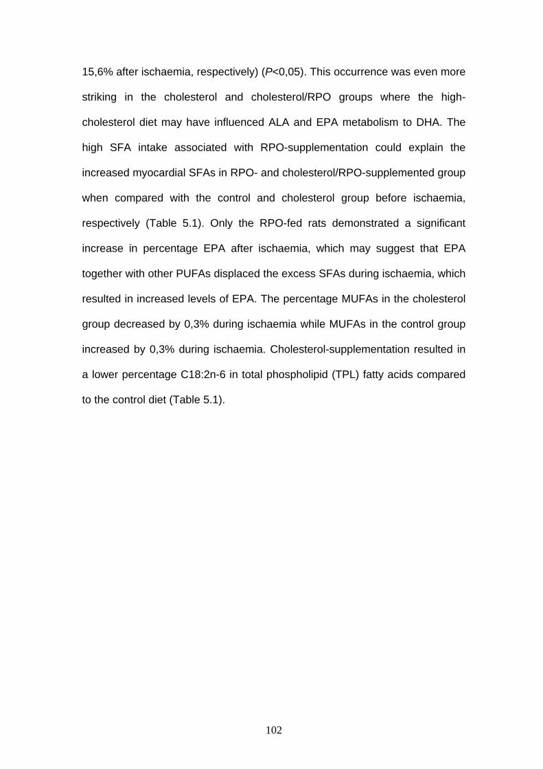
102
15,6% after ischaemia, respectively) (P<0,05). This occurrence was even more
striking in the cholesterol and cholesterol/RPO groups where the high-
cholesterol diet may have influenced ALA and EPA metabolism to DHA. The
high SFA intake associated with RPO-supplementation could explain the
increased myocardial SFAs in RPO- and cholesterol/RPO-supplemented group
when compared with the control and cholesterol group before ischaemia,
respectively (Table 5.1). Only the RPO-fed rats demonstrated a significant
increase in percentage EPA after ischaemia, which may suggest that EPA
together with other PUFAs displaced the excess SFAs during ischaemia, which
resulted in increased levels of EPA. The percentage MUFAs in the cholesterol
group decreased by 0,3% during ischaemia while MUFAs in the control group
increased by 0,3% during ischaemia. Cholesterol-supplementation resulted in
a lower percentage C18:2n-6 in total phospholipid (TPL) fatty acids compared
to the control diet (Table 5.1).
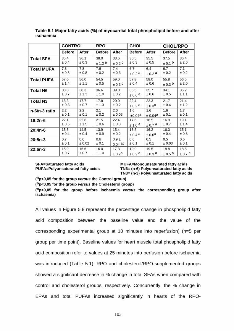
103
Table 5.1 Major fatty acids (%) of myocardial total phospholipid before and after ischaemia.
CONTROL RPO CHOL CHOL/RPO Before After Before After Before After Before After
Total SFA 35.4 ± 0.4
36.1 ± 0.3
38.0 ± 1.3 a
33.6 ± 0.2 c
35.5 ± 0.3
35.5 ± 0.5
37.5 ± 0.1 b
36.4 ± 2.0
Total MUFA 7.5 ± 0.3
7.8 ± 0.8
7.6 ± 0.2
7.4 ± 0.3
6.7 ± 0.2 a
6.4 ± 0.2 a
6.7 ± 0.2
7.1 ± 0.2
Total PUFA 57.0 ± 1.4
56.0 ± 1.1
54.5 ± 0.5
59.0 ± 0.3 c
57.8 ± 0.4
58.0 ± 0.6
55.8 ± 0.3 b
56.5 ± 2.0
Total N6 38.8 ± 0.7
38.3 ± 1.3
36.6 ± 1.0
39.0 ± 0.2
35.5 ± 0.6 a
35.7 ± 0.6
34.1 ± 0.5
35.2 ± 1.1
Total N3 18.3 ± 0.8
17.7 ± 0.7
17.8 ± 1.3
20.0 ± 0.2
22.4 ± 0.2 a
22.3 ± 0.3a
21.7 ± 0.4
21.4 ± 1.2
n-6/n-3 ratio 2.2 ± 0.1
2.2 ± 0.1
2.1 ± 0.2
2.0 ± 0.03
1.6 ±0.04a
1.6 ± 0.04a
1.6 ± 0.1
1.7 ± 0.1
18:2n-6 22.1 ± 0.6
22.6 ± 1.5
21.5 ± 0.6
22.4 ± 0.3
17.6 ± 1.0 a
18.5 ± 0.7 a
16.9 ± 0.7
19.1 ± 1.4
20:4n-6 15.5 ± 0.4
14.5 ± 0.4
13.9 ± 0.9
15.4 ± 0.2
16.8 ± 0.4 a
16.2 ± 0.6a
16.3 ± 0.4
15.1 ± 0.8
20:5n-3 0.7 ± 0.1
0.6 ± 0.02
0.6 ± 0.1
0.9 ± 0.04 ac
0.6 ± 0.1
0.5 ± 0.1
0.5 ± 0.03
0.6 ± 0.1
22:6n-3 15.9 ± 0.7
15.6 ± 0.7
16.0 ± 1.0
17.3 ± 0.2a
19.9 ± 0.2 a
19.5 ± 0.3 a
18.8 ± 0.5 a
18.8 ± 0.7 a
SFA=Saturated fatty acids MUFA=Monounsaturated fatty acids PUFA=Polyunsaturated fatty acids TN6= (n-6) Polyunsaturated fatty acids
TN3= (n-3) Polyunsaturated fatty acids (ap<0,05 for the group versus the Control group) (bp<0,05 for the group versus the Cholesterol group) (cp<0,05 for the group before ischaemia versus the corresponding group after ischaemia) All values in Figure 5.8 represent the percentage change in phospholipid fatty
acid composition between the baseline value and the value of the
corresponding experimental group at 10 minutes into reperfusion) (n=5 per
group per time point). Baseline values for heart muscle total phospholipid fatty
acid composition refer to values at 25 minutes into perfusion before ischaemia
was introduced (Table 5.1). RPO and cholesterol/RPO-supplemented groups
showed a significant decrease in % change in total SFAs when compared with
control and cholesterol groups, respectively. Concurrently, the % change in
EPAs and total PUFAs increased significantly in hearts of the RPO-

104
supplemented group when compared with hearts of the control group. Hearts
of the cholesterol-supplemented group showed a significant increase in %
change of total SFAs when compared with hearts of the RPO-supplemented
group, which was associated with a decrease in % change in EPA and total
PUFAs in hearts of cholesterol-supplemented group when compared with
hearts of RPO-supplemented group.
-10
-8
-6
-4
-2
0
2
4
6
8
10
SFA PUFA EPA
% C
hang
e in
pho
spho
lipid
fatty
aci
dco
mpo
sitio
n ControlRPOCHOLChol/RPO
Figure 5.8 % Change in phospholipid fatty acid composition in the 4 experimental groups after an ischaemic period of 25 minutes (n=5 for each group) (#P<0,05 for the indicated group versus the control group)(*P<0,05 for the group versus the RPO group)(^P<0,05 for the indicated group versus the cholesterol group) (mean ± SEM)
5.5 Discussion
Diets high in fat and cholesterol are associated with hypercholesterolaemia
and are considered a major risk factor for the development of ischaemic heart
disease (Ferdinandy et al., 1998; Giricz et al., 2003). To our knowledge little
data exist on the effects of dietary RPO-supplementation on post-ischaemic
cardiac recovery and the role of 1) the NO-cGMP pathway, 2) total heart
#
#
#
*
*
*
^ ^
105
muscle phospholipid fatty acid composition and 3) antioxidant status on this
recovery.
The protective properties of NO-cGMP pathway activation could be
responsible for improved aortic output recovery of hearts of the RPO-
supplemented group when compared with the control-fed animals. This
improved functional recovery was associated with elevated cGMP and
decreased cAMP concentrations early in ischaemia. However, although our
data indicate that RPO-supplementation of a high-cholesterol diet improved
functional recovery of the reperfused heart, this improvement could not be
linked with altered NO-cGMP signalling. Based on our results we propose that
the protective effect of RPO in high-cholesterol diets may be associated with
either RPO antioxidant characteristics and/or changes in the phospholipid fatty
acid composition of the myocardium during ischaemia/reperfusion.
Abeywardena and co-workers (1995) provided supportive evidence for the
existence of a complex interaction between fatty acids and antioxidants in
RPO-induced protection during ischaemia/reperfusion. These data support the
findings of Serbinova and co-workers (1992) which showed that palm oil
vitamin E mixture containing both α-tochopherol and α-tocotrienol may be
more effective in protecting the heart against ischaemia/reperfusion injury in
the isolated Langendorff perfused rat heart than α-tocopherol alone. This
higher antioxidant activity of the tocotrienols has been attributed to a number of
mechanisms including efficient interaction with free radical species, higher
recycling efficiency of chromanoxyl radical and uniform distribution in
membrane bilayers (Theriault et al., 1999).

106
We have previously reported that RPO-supplementation increased total
myocardial PUFAs over the 25-minute period of ischaemia (Esterhuyse et al.,
2003). In the present study we found that the % EPA in phospholipid (n-3
PUFA) increased from 0,6 ± 0,1% to 0,9 ± 0,04% over the same period in the
RPO-supplemented group. These findings suggest that EPA, together with
other PUFAs, displaced the excess SFAs during ischaemia and thus leads to
increased % EPA with subsequent production of DHA. The pathway leading to
the biosynthesis of DHA from docosapentaenoic acid (C22:5n-3, DPA) has
only recently been deciphered in mammals (Pereira et al., 2003). DPA is
elongated to tetracosapentaenoic acid (C24:5n-3), which is then desaturated
by a Δ6 desaturase to generate tetracosahexaenoic acid (C24:6n-3), which is
then thought to be transported to the peroxisomes where it undergoes β-
oxidation to generate DHA (Figure 2.1 p.12). Cholesterol and cholesterol/RPO-
supplemented groups showed increased % DHA phospholipid before
ischaemia when compared with control group. All the supplemented groups
showed a similar DHA increase after ischaemia when compared with the
corresponding control group. The relatively high % DHA in hearts in all the
groups, including the control group, could be explained by the composition of
the standard rat chow which contains a high percentage of n-3 PUFAs. It is
well known that when on high cholesterol diets, membrane fatty acid
composition shows increases in the longer chain PUFAs (such as DHA) in
order to compensate for, and maintain membrane fluidity to some extent as a
result of increased cholesterol molecules in the membrane. Van Rooyen and
co-workers (1998) reported that the metabolic rate of EPA in the cell
membranes of the erythrocyte decreased in animals consuming a typical
107
Western diet with high cholesterol content, which would lead to higher levels of
EPA. Although our results showed % EPA in phospholipid to be unchanged in
cholesterol and cholesterol/RPO-supplemented groups, decreased metabolic
rate could explain the increased % DHA in hearts of the cholesterol and
cholesterol/RPO-supplemented groups when compared with the other groups.
Abeywardena and co-workers (1995) reported that EPA is either poorly
incorporated in rat myocardium or futher elongated and desaturated to yield
DHA, which may explain the high % DHA compared to EPA. Research has
shown that DHA, EPA and free AA are antiarrhythmic and protect the heart
against ischaemia/reperfusion injury (Li et al., 1997; Nair et al., 1997; Kang
and Leaf, 2000; Pepe et al., 2002; Jump, 2003). Our results showed improved
aortic output recovery in hearts of rats supplemented with RPO versus control
hearts and hearts of rats fed cholesterol/RPO versus cholesterol-
supplementation, although DHA levels were increased in the cholesterol-
supplemented group as well. These findings suggest that protection may not
only be associated with a change in fatty acid composition and eicosanoid
production from dietary intake, but that accompanying non-fatty acid
constituents may also be important. Furthermore, research has shown that
thromboxane A2 and prostacyclin production are unlikely to be mediated via
fatty acids alone, but that endogenous antioxidants may also play an important
role (Abeywardena et al., 1991; Abeywardena and Charnock, 1995; Theriault
et al., 1999).
Generally, the mechanism of eicosanoid action is to bind to membrane-
receptors, leading to generation of second messengers such as cAMP and
108
Ca2+ (Mohan et al., 1995; Li et al., 1997). In the current study myocardial cAMP
concentrations were decreased in the RPO-supplemented group, compared to
the other groups. Therefore, prevention of fatal cardiac arrhythmias may
depend on the net effect of eicosanoids, which is determined by the status of
AA metabolism and the contents of n-6 and other fatty acids (Abeywardena et
al., 1991; Charnock et al., 1991; Nair et al., 1997; Kang and Leaf, 2000). Both
NO and prostaglandins have important independent effects on cardiovascular
function, but the interaction between them determines myocardial performance
(Mohan et al., 1995). The mechanism of fatty acid protection remains elusive
and needs further investigation.
Research has shown that cholesterol-enriched diets impair NO-cGMP
signalling in both endothelial and nonendothelial cells. Girics and co-workers
(2003) showed that cholesterol diet-induced hyperlipidaemia decreased
cardiac NO concentration, but it does not change nitric oxide synthases (NOS)
activity. Therefore, the decreased NO-concentrations were not due to impaired
NO synthesis, but caused by breakdown of cardiac NO due to increased ROS
production in hyperlipidaemic rats (Girics et al., 2003). These authors also
found an increased O2- production in hyperlipidaemic hearts, which support the
assumption that elevated ROS production is responsible for decreased NO
concentration in hyperlipidaemic myocardium.
We have previously shown that RPO-supplementation increased cGMP
concentrations early in ischaemia when compared with the control-fed rats,
which may confer some of the cardioprotection to the ischaemia/reperfused
heart (Esterhuyse et al., 2003). Although not indicated in Figure 5.4, the
109
present study has shown that cholesterol-supplementation for 6 weeks
influenced the NO-cGMP signalling pathway by decreasing % change in cGMP
concentrations in both the cholesterol and cholesterol/RPO-supplemented
groups when compared with the RPO-supplemented group; yet it was not
different from the control group. These data are in agreement with other
studies, which showed that high-cholesterol diet impairs NO-cGMP signalling
in both endothelial and non-endothelial cells (Deliconstantinos et al., 1995).
The decreased myocardial cAMP concentrations in the RPO-supplemented
group may be related with increased cGMP concentrations. In this regard it is
possible that cGMP may attenuate this type of injury by inhibiting the cAMP-
induced increase in the slow inward calcium current, thus leading to a
decrease in ischaemic cytosolic calcium concentrations (Sumii et al., 1995; Du
Toit et al., 2001).
High cholesterol-diets are also known to inhibit the formation of ubiquinone
(coenzyme Q10) production. This coenzyme is a key polyprenyl derivative
molecule in the mitochondrial electron transport system and as an endogenous
antioxidant it protects the ischaemia/reperfused myocardium (Ferdinandy et
al., 1998; Wall, 2000; Girics et al., 2003). CoQ10 is found naturally in small
amounts in RPO (Nagendran et al., 2000), which may explain improved aortic
output recovery in RPO and cholesterol/RPO-supplemented groups when
compared with the control and cholesterol fed groups, respectively.
110
Supplementation with RPO appeared to raise TC and LDL cholesterol when
hypercholesterolaemic subjects and high fat liquid formula diets were used
(Sundram and Basiron). However, our results have shown that RPO-
supplementation in the presence of potentially harmful cholesterol did not lead
to a significant difference in serum total cholesterol. We know that tocotrienols
in RPO not only suppress cholesterol production in the liver and lower its
serum concentration, but also lower the damaging LDL-cholesterol.
Tocotrienols prenylated side-chains are thought to induce prenyl
pyrophosphate pyrophosphatase that catalyzes the dephosphorylation of
fernysal with a concomitant increase of cellular farnesol. Farnesol
downregulates HMG CoA reductase activity by a post-transcriptional process
involving protein degradation. This mechanism is different from cholesterol,
which exerts a feedback transcriptional effect on HMG CoA reductase activity
(Theriault et al., 1999). These data are in agreement with other studies, which
showed that not all saturated dietary fats raise total serum cholesterol
(Theriault et al., 1999; Kritchevsky, 2000; Sundram and Basiron).
The findings of this study create opportunities for further investigations to
elucidate RPO mediated mechanisms involved in cardiac protection.
5.6 Conclusion
Dietary RPO-supplementation offered protection of hearts from rats on a
standard rat chow (control) and hypercholesterolaemic diet against
ischaemia/reperfusion injury as reflected by improved aortic output recovery
and rate pressure product. This was associated with an increase in cGMP
early in ischaemia and a decrease in cAMP during ischaemia in the RPO-
111
supplemented versus control group. However, with addition of cholesterol,
cGMP concentrations in ischaemia were decreased, but RPO still offered
protection. This suggests that cGMP may not be the only mechanism of
protection. The improved functional recovery in the cholesterol/RPO-
supplemented group was associated with a significant decrease of cAMP
concentrations during reperfusion, suggesting that RPO may also protect via
the inhibition of the cAMP pathway.
Furthermore, % total PUFAs in myocardial phospholipids were increased by
ischaemia in the RPO-supplemented group, but not in the cholesterol/RPO-
supplemented group. This suggests that PUFAs may have influenced the
tissue concentrations of cGMP and cAMP.
Evidence in this study suggests that both antioxidants and fatty acids play a
role in the cardioprotective mechanisms of dietary RPO and our next aim was
to search for the cellular mechanisms whereby antioxidants and fatty acids
offer this cardioprotection. We therefore aimed to investigate the effects of
RPO-supplementation of rats on a standard rat chow and cholesterol-enriched
diets on myocardial NO-synthesis and NO-cGMP signalling.
112
CHAPTER 6
Proposed mechanisms for red palm oil-induced cardioprotection in a
hyperlipidaemic perfused rat heart model.
(Submitted 2005)
113
6.1 Abstract
Introduction: High-cholesterol diets alter function of the myocardial and
vascular NO-cGMP signalling pathway. These alterations have been
implicated in both ischaemic/reperfusion injury and the development of
ischaemic heart disease. We have previously shown that the cardioprotection
offered by red palm oil (RPO)-supplementation could be related to NO-cGMP
signalling in the control, but not in the hyperlipidaemic group. Aims: We
investigated the effects of RPO-supplementation of rats on a standard rat chow
(control)- and cholesterol-enriched diet on myocardial nitric oxide synthesis,
superoxide dismutase- and nitric oxide synthase activity and lipid
hydroperoxide production in the rat heart. Materials and Methods: Wistar rats
were fed a standard rat chow (SRC), or a SRC plus cholesterol diet. In 2
additional groups, these diets were supplemented with RPO for 6 weeks. Post-
ischaemic mechanical function, total myocardial nitric oxide concentrations,
lipid hydroperoxide production and superoxide dismutase- and nitric oxide
synthase activity were determined in isolated working rat hearts subjected to
25 minutes of normothermic total global ischaemia. Results: Dietary RPO-
supplementation of the standard rat chow (control) diet and the cholesterol-
enriched diet improved aortic output recovery (72,1 ± 3,2% versus 54,0± 3,2%
and 64,0 ± 2,5% versus 39,3 ± 4,9% respectively, P<0,05). The improved
aortic output recovery of hearts from rats supplemented with RPO was
associated with an elevation in the cGMP concentration early in ischaemia.
Baseline myocardial nitric oxide content was significantly decreased in the
cholesterol-enriched diet group when compared to the control group (1,6 ± 0,4
mmol/l versus 2.8 ± 0,2 mmol/l, P<0,05). Direct intracellular nitric oxide
114
detection in isolated rat cardiomyocytes showed a significant increase in nitric
oxide production after 120-minute simulated ischaemia (hypoxia) in both the
RPO- and cholesterol/RPO-supplemented group when compared with non-
hypoxic control and cholesterol-supplemented groups, respectively (2,9 ± 0,1
arbitrary units and 2,5 ± 0,1 arbitrary units versus 2,4 ± 0,1 arbitrary units and
2,1 ± 0,1 arbitrary units, respectively, P<0,05). Conclusions: Our data show
that dietary RPO-supplementation protects the hearts of animals on a standard
rat chow or hypercholesterolaemic diet against ischaemia/reperfusion injury.
Mechanisms for this cardioprotection may include RPO-induced preservation
of myocardial ischaemic NO-concentrations, which causes elevated cGMP
concentrations in hearts of rats on a standard rat chow diet.
Key words: Hyperlipidaemia - Working heart perfusion - Ischaemia - RPO
protection - Nitric Oxide - Free radicals
6.2 Introduction
Nitric oxide (NO) is, by virtue of its vasodilator, antioxidant, anti-platelet and
anti-neutrophil actions, an essential molecule for normal heart function
(reviewed by Ferdinandy and Schultz, 2003). It has also been shown to be
cardioprotective in the ischaemic heart (reviewed by Bolli, 2002). However, NO
is detrimental when it is combined with superoxide (O2-) to form peroxynitrite
(ONOO-), which rapidly decomposes to highly reactive oxidant species leading
to tissue injury (Figure 2.4). There is a critical balance between cellular
concentrations of NO, O2- and superoxide dismutase (SOD) which
physiologically favour NO production, but in pathological conditions such as
ischaemia and reperfusion, result in ONOO- formation (reviewed by

115
Ferdinandy and Schultz, 2003). These oxygen free radicals can react with
nucleic acids, proteins and lipids, resulting in damage to the cell membrane or
intracellular organelles (Gilham et al., 1997).
Protective effects of NO are also mediated through the production of cGMP.
Du Toit and co-workers (2001) found that nitric oxide donor treatment reduces
ischaemia/reperfusion injury by increasing cGMP concentrations and
suggested that the cAMP-to-cGMP ratio might play an important role in this
cardioprotection. Maulik and co-workers (1995) showed that NO plays a
significant role in transmembrane signalling in the ischaemic myocardium. This
group suggested that NO signalling is switched off due to inactivation of NO by
reactive oxygen species and were the first to suggest that reactive oxygen
species may alter NO-cGMP signalling.
Cardiac stress adaptation is possibly jeopardized in hyperlipidaemia due to
altered NO-cGMP pathway function in vascular and myocardial tissue.
Szilvassy and co-workers (2001) found that a cholesterol-enriched diet
decreased both vascular NO and cGMP concentrations and increased aortic
O2- production. Several other studies have also shown that a high-cholesterol
diet impairs NO-cGMP signalling in both endothelium (Deliconstantinos et al.,
1995) and non-endothelial cells with a significant decrease in cardiac NO-
concentrations (Ferdinandy et al., 1997; Szekeres et al., 1997). Girics and co-
workers (2003) found an increase in O2- production with decreased cardiac
NO-concentration in cholesterol-fed rats. Nitric oxide synthase (NOS) activity
was unchanged which may suggest that NO synthesis was not impaired and
116
that increased O2- production was responsible for the decreased NO-
concentrations in the hyperlipidaemic myocardium. Furthermore,
hyperlipidaemia stimulates ONOO- generation in the heart, which leads to
myocardial dysfunction (Onody et al., 2003).
Diniz and co-workers (2004) showed that changes in dietary fatty acid
composition also affect cardiac oxidative stress. They showed that, despite
their beneficial effects on serum lipid levels, diets rich in polyunsaturated fatty
acids (PUFAs) were deleterious to the heart by increasing cardiac
susceptibility to lipid peroxidation. PUFA-fed rats also showed diminished SOD
activities when compared to saturated fatty acid (SFA)-fed rats.
We have previously shown that dietary RPO-supplementation protects against
the harmful consequences of global ischaemia in the isolated perfused rat
heart on a standard rat chow or cholesterol-enriched diet (Esterhuyse et al.,
2005). RPO contains a mixture of SFAs (51%), MUFAs (38%), PUFAs (11%)
and natural antioxidants, tocopherols and tocotrienols (Nagendran et al.,
2000). These natural antioxidants can act as scavengers of damaging oxygen
free radicals (Cottrell, 1991; Serbinova et al., 1991; Chandrasekharan, 1999;
Theriault et al., 1999; Kritchevsky, 2000). Newaz and co-workers (2003)
showed an antioxidant protection by γ-tocotrienols in hypertensive rats when
compared with control animals. This group suggested that improved NOS
activity in blood vessels and increased NO availability were mediated through
the antioxidant properties of γ-tocotrienol where it effectively scavenges the
free radicals. Venditti and co-workers (1999) also reported that vitamin E
117
treatment offers protection against ischaemia/reperfusion-induced oxidative
stress. However, the precise mechanism of action remains unknown.
Little is known about the effects and possible protective mechanisms of RPO-
supplementation against ischaemia/reperfusion injury, particularly when used
as a supplement with a high-cholesterol diet.
The aims of this study were to determine whether RPO-supplementation
influences 1) myocardial susceptibility to ischaemia/reperfusion injury in hearts
from rats on a standard rat chow or hypercholesterolaemic diet, 2) myocardial
NO-cGMP signalling pathway function, 3) production of cardiac NO and lipid
hydroperoxides (LPO) and 4) enzymatic NOS and SOD activities.
6.3 Materials and methods 6.3.1 Experimental groups and model used Wistar rats were fed a standard rat chow (SRC), or a SRC plus cholesterol
diet. In 2 additional groups, these diets were supplemented with RPO for 6
weeks. The protocol used has been discussed in Chapter 3 under Materials
and Methods (Experimental Model 3 p.60).
6.3.2 Functional parameters measured
Described in Chapter 3 under Materials and Methods (Experimental Model 1
p.50).
118
6.3.3 Biochemical parameters measured 6.3.3.1 Measurement of cGMP
Myocardial cGMP concentrations were determined using a radioimmunoassay
method as described in Chapter 3 under Materials and Methods (Experimental
Model 1 p.50).
6.3.3.2 Measurement of cardiac nitric oxide concentrations Myocardial NO concentrations were determined using a Nitrate/Nitrite kit
(Cayman Chemicals, Cayman Islands) as described in Chapter 3 under
Materials and Methods (Experimental Model 3 p.60). We also measured
intracellular myocyte NO-production with DAF-2/DA by flow cytometry
(Strijdom et al., 2004) as described in Chapter 3 under Materials and Methods
(Experimental Model 3 p.60).
6.3.3.3 Measurement of cardiac nitric oxide synthase activity
Total nitric oxide (NOS) activity was measured using a NOS assay-kit which is
based on biochemical conversion of L-arginine to L-citrulline by NOS (Cayman
Chemicals, Cayman Islands). For detail about principle and method used, see
full description in Chapter 3 under Materials and Methods (Experimental Model
3 p.60).
6.3.3.4 Measurement of cardiac superoxide dismutase activity
Total activity of superoxide dismutase (SOD) was measured as described in
Chapter 3 under Materials and Methods (Experimental Model 3 p.60) using a
SOD kit (Cayman Chemicals, Cayman Islands).
119
6.3.3.5 Measurement of cardiac lipid hydroperoxide production
Lipid hydroperoxide (LPO) production in cardiac muscle was assessed using a
LPO assay kit (Cayman Chemicals, Cayman Islands) as described in Chapter
3 under Methods and Materials (Experimental Model 3 p.60).
6.3.3.6 Statistical methods Statistical methods used in this experiment have been discussed in Chapter 3
under Materials and Methods (Experimental Model 1 p.50).
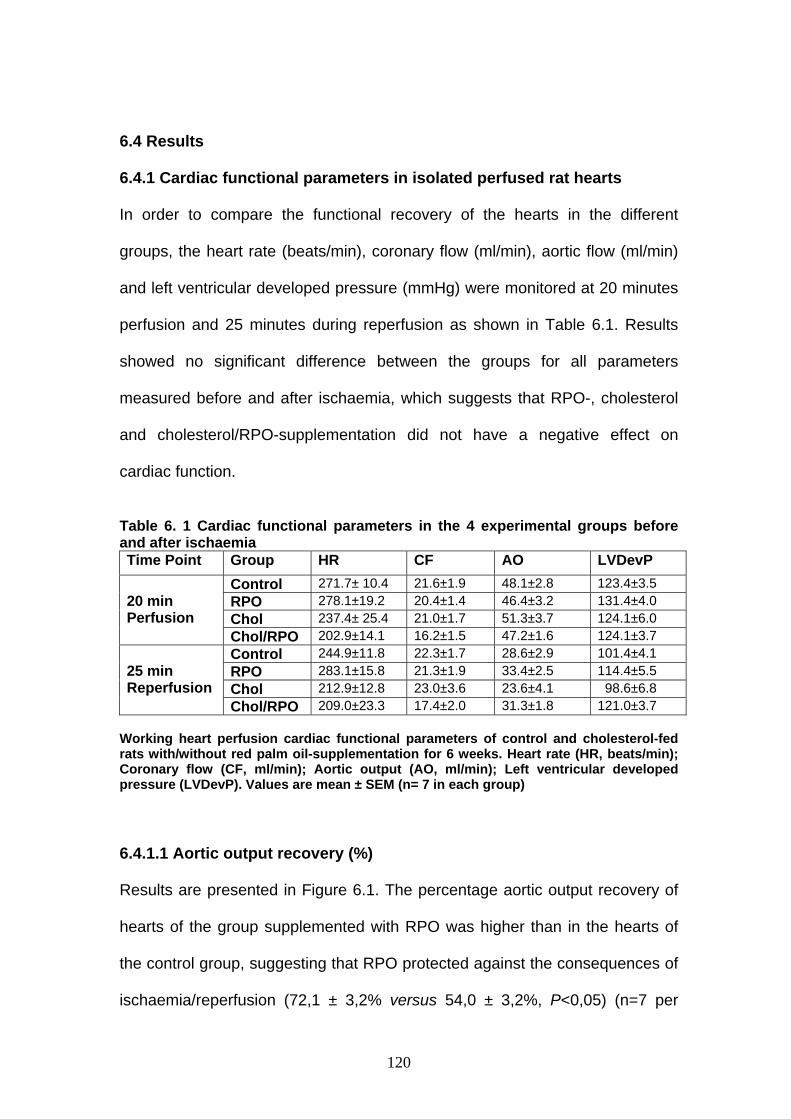
120
6.4 Results
6.4.1 Cardiac functional parameters in isolated perfused rat hearts In order to compare the functional recovery of the hearts in the different
groups, the heart rate (beats/min), coronary flow (ml/min), aortic flow (ml/min)
and left ventricular developed pressure (mmHg) were monitored at 20 minutes
perfusion and 25 minutes during reperfusion as shown in Table 6.1. Results
showed no significant difference between the groups for all parameters
measured before and after ischaemia, which suggests that RPO-, cholesterol
and cholesterol/RPO-supplementation did not have a negative effect on
cardiac function.
Table 6. 1 Cardiac functional parameters in the 4 experimental groups before and after ischaemia Time Point Group HR CF AO LVDevP
Control 271.7± 10.4 21.6±1.9 48.1±2.8 123.4±3.5 RPO 278.1±19.2 20.4±1.4 46.4±3.2 131.4±4.0 Chol 237.4± 25.4 21.0±1.7 51.3±3.7 124.1±6.0
20 min Perfusion
Chol/RPO 202.9±14.1 16.2±1.5 47.2±1.6 124.1±3.7 Control 244.9±11.8 22.3±1.7 28.6±2.9 101.4±4.1 RPO 283.1±15.8 21.3±1.9 33.4±2.5 114.4±5.5 Chol 212.9±12.8 23.0±3.6 23.6±4.1 98.6±6.8
25 min Reperfusion
Chol/RPO 209.0±23.3 17.4±2.0 31.3±1.8 121.0±3.7 Working heart perfusion cardiac functional parameters of control and cholesterol-fed rats with/without red palm oil-supplementation for 6 weeks. Heart rate (HR, beats/min); Coronary flow (CF, ml/min); Aortic output (AO, ml/min); Left ventricular developed pressure (LVDevP). Values are mean ± SEM (n= 7 in each group)
6.4.1.1 Aortic output recovery (%)
Results are presented in Figure 6.1. The percentage aortic output recovery of
hearts of the group supplemented with RPO was higher than in the hearts of
the control group, suggesting that RPO protected against the consequences of
ischaemia/reperfusion (72,1 ± 3,2% versus 54,0 ± 3,2%, P<0,05) (n=7 per
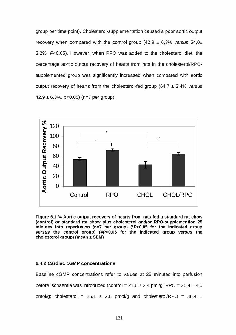
121
group per time point). Cholesterol-supplementation caused a poor aortic output
recovery when compared with the control group (42,9 ± 6,3% versus 54,0±
3,2%, P<0,05). However, when RPO was added to the cholesterol diet, the
percentage aortic output recovery of hearts from rats in the cholesterol/RPO-
supplemented group was significantly increased when compared with aortic
output recovery of hearts from the cholesterol-fed group (64,7 ± 2,4% versus
42,9 ± 6,3%, p<0,05) (n=7 per group).
Figure 6.1 % Aortic output recovery of hearts from rats fed a standard rat chow (control) or standard rat chow plus cholesterol and/or RPO-supplemention 25 minutes into reperfusion (n=7 per group) (*P<0,05 for the indicated group versus the control group) (#P<0,05 for the indicated group versus the cholesterol group) (mean ± SEM)
6.4.2 Cardiac cGMP concentrations
Baseline cGMP concentrations refer to values at 25 minutes into perfusion
before ischaemia was introduced (control = 21,6 ± 2,4 pml/g; RPO = 25,4 ± 4,0
pmol/g; cholesterol = 26,1 ± 2,8 pmol/g and cholesterol/RPO = 36,4 ±
0
20
40
60
80
100
120
Control RPO CHOL CHOL/RPO Aor
tic O
utpu
t Rec
over
y %
*
*#
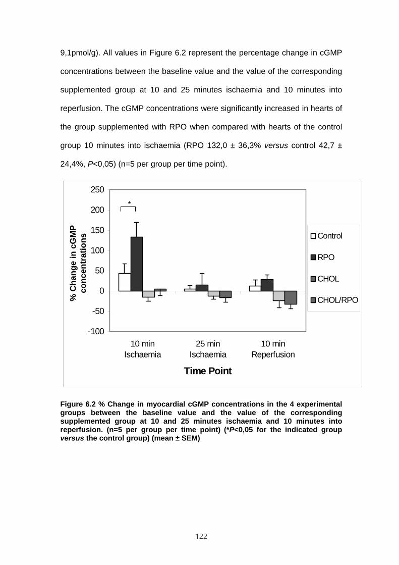
122
9,1pmol/g). All values in Figure 6.2 represent the percentage change in cGMP
concentrations between the baseline value and the value of the corresponding
supplemented group at 10 and 25 minutes ischaemia and 10 minutes into
reperfusion. The cGMP concentrations were significantly increased in hearts of
the group supplemented with RPO when compared with hearts of the control
group 10 minutes into ischaemia (RPO 132,0 ± 36,3% versus control 42,7 ±
24,4%, P<0,05) (n=5 per group per time point).
-100
-50
0
50
100
150
200
250
10 minIschaemia
25 minIschaemia
10 minReperfusion
Time Point
% C
hang
e in
cG
MP
conc
entr
atio
ns Control
RPO
CHOL
CHOL/RPO
Figure 6.2 % Change in myocardial cGMP concentrations in the 4 experimental groups between the baseline value and the value of the corresponding supplemented group at 10 and 25 minutes ischaemia and 10 minutes into reperfusion. (n=5 per group per time point) (*P<0,05 for the indicated group versus the control group) (mean ± SEM)
*
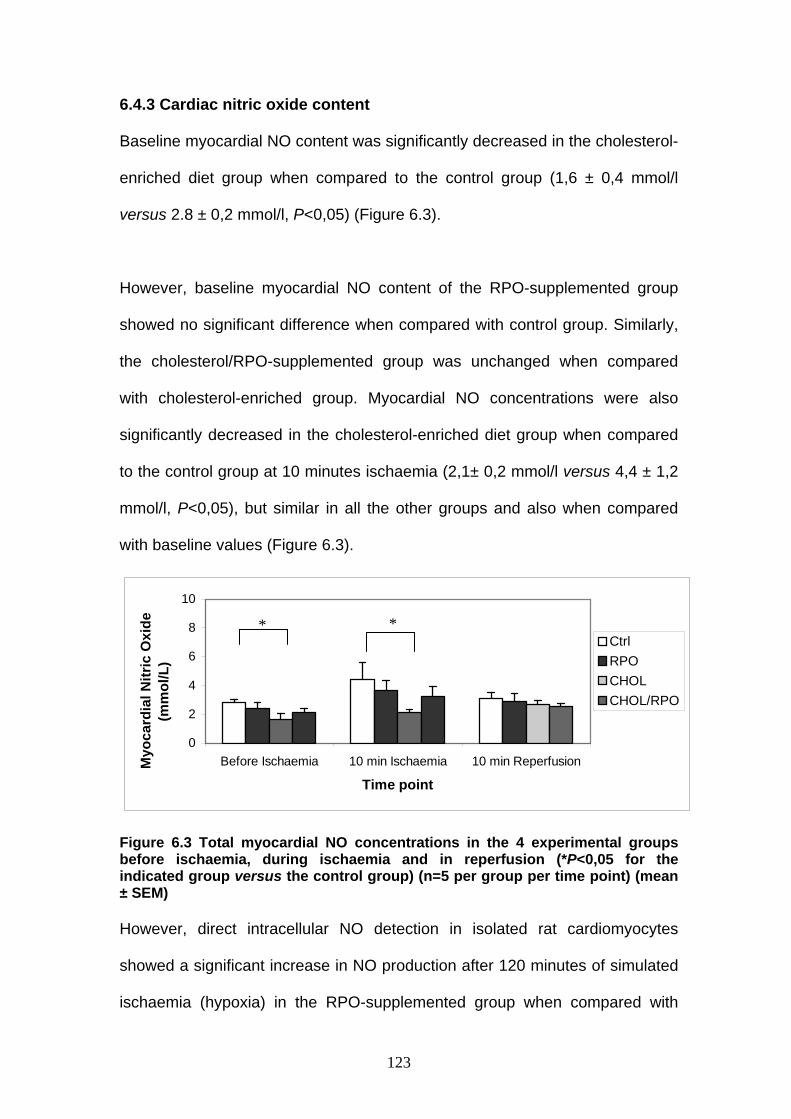
123
6.4.3 Cardiac nitric oxide content
Baseline myocardial NO content was significantly decreased in the cholesterol-
enriched diet group when compared to the control group (1,6 ± 0,4 mmol/l
versus 2.8 ± 0,2 mmol/l, P<0,05) (Figure 6.3).
However, baseline myocardial NO content of the RPO-supplemented group
showed no significant difference when compared with control group. Similarly,
the cholesterol/RPO-supplemented group was unchanged when compared
with cholesterol-enriched group. Myocardial NO concentrations were also
significantly decreased in the cholesterol-enriched diet group when compared
to the control group at 10 minutes ischaemia (2,1± 0,2 mmol/l versus 4,4 ± 1,2
mmol/l, P<0,05), but similar in all the other groups and also when compared
with baseline values (Figure 6.3).
0
2
4
6
8
10
Before Ischaemia 10 min Ischaemia 10 min Reperfusion
Time point
Myo
card
ial N
itric
Oxi
de
(mm
ol/L
)
CtrlRPOCHOLCHOL/RPO
Figure 6.3 Total myocardial NO concentrations in the 4 experimental groups before ischaemia, during ischaemia and in reperfusion (*P<0,05 for the indicated group versus the control group) (n=5 per group per time point) (mean ± SEM) However, direct intracellular NO detection in isolated rat cardiomyocytes
showed a significant increase in NO production after 120 minutes of simulated
ischaemia (hypoxia) in the RPO-supplemented group when compared with
* *
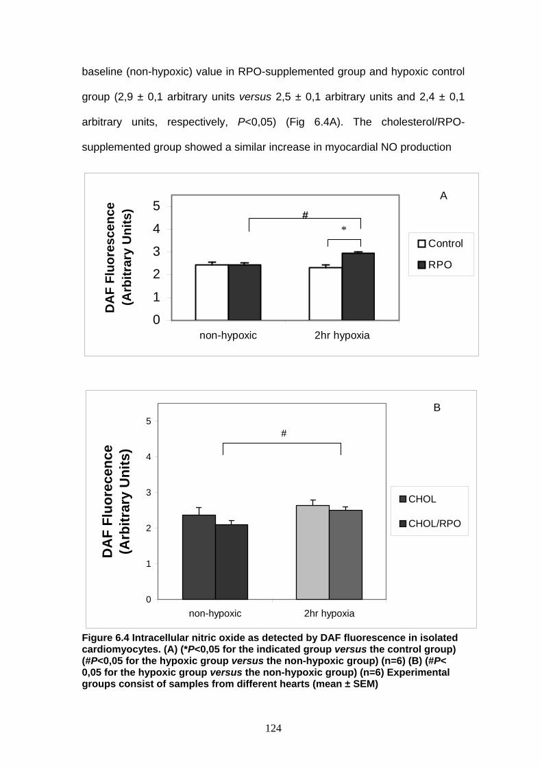
124
0
1
2
3
4
5
non-hypoxic 2hr hypoxia
DA
F Fl
uore
scen
ce
(Arb
itrar
y U
nits
)
Control
RPO
baseline (non-hypoxic) value in RPO-supplemented group and hypoxic control
group (2,9 ± 0,1 arbitrary units versus 2,5 ± 0,1 arbitrary units and 2,4 ± 0,1
arbitrary units, respectively, P<0,05) (Fig 6.4A). The cholesterol/RPO-
supplemented group showed a similar increase in myocardial NO production
0
1
2
3
4
5
non-hypoxic 2hr hypoxia
DA
F Fl
uore
cenc
e (A
rbitr
ary
Uni
ts)
CHOL
CHOL/RPO
Figure 6.4 Intracellular nitric oxide as detected by DAF fluorescence in isolated cardiomyocytes. (A) (*P<0,05 for the indicated group versus the control group) (#P<0,05 for the hypoxic group versus the non-hypoxic group) (n=6) (B) (#P< 0,05 for the hypoxic group versus the non-hypoxic group) (n=6) Experimental groups consist of samples from different hearts (mean ± SEM)
#*
#
A
B

125
after 120 minutes of simulated ischaemia (hypoxia) when compared with
baseline NO-concentration in cholesterol/RPO-supplemented group (2,5 ± 0,1
arbitrary units versus 2,1 ± 0,1 arbitrary units, P<0,05) (Fig 6.4B).
6.4.4 Cardiac nitric oxide synthase activity
Myocardial NOS activity was significantly decreased in the control group at 10
minutes ischaemia when compared with baseline values in control group (5,7 ±
0,3% versus 3,9 ± 0,5%, P<0,05) (n=5 per group per time point) (Figure 6.5).
0
1
2
3
4
5
6
7
8
9
Before Ischaemia 10 min Ischaemia 10 min Reperfusion
Time Point
% N
itric
Oxi
de S
ynth
ase
Act
ivity
ControlRPOCHOLCHOL/RPO
Fig 6.5 % Myocardial NOS activity in the 4 experimental groups before ischaemia, during ischaemia and in reperfusion (n=5 per group per time point)(*P<0,05 for the indicated group versus the same group before ischaemia) (#P<0,05 for the indicated group versus the cholesterol group) (mean ± SEM)
Baseline myocardial NOS activity was unchanged for cholesterol-,
cholesterol/RPO- and RPO-supplemented groups. However, myocardial NOS
activity was significantly increased in cholesterol/RPO-supplemented group
* #
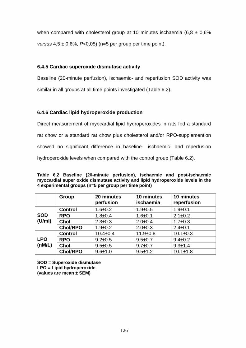
126
when compared with cholesterol group at 10 minutes ischaemia (6,8 ± 0,6%
versus 4,5 ± 0,6%, P<0,05) (n=5 per group per time point).
6.4.5 Cardiac superoxide dismutase activity
Baseline (20-minute perfusion), ischaemic- and reperfusion SOD activity was
similar in all groups at all time points investigated (Table 6.2).
6.4.6 Cardiac lipid hydroperoxide production
Direct measurement of myocardial lipid hydroperoxides in rats fed a standard
rat chow or a standard rat chow plus cholesterol and/or RPO-supplemention
showed no significant difference in baseline-, ischaemic- and reperfusion
hydroperoxide levels when compared with the control group (Table 6.2).
Table 6.2 Baseline (20-minute perfusion), ischaemic and post-ischaemic myocardial super oxide dismutase activity and lipid hydroperoxide levels in the 4 experimental groups (n=5 per group per time point) Group 20 minutes
perfusion 10 minutes ischaemia
10 minutes reperfusion
Control 1.6±0.2 1.9±0.5 1.9±0.1 RPO 1.8±0.4 1.6±0.1 2.1±0.2 Chol 2.3±0.3 2.0±0.4 1.7±0.3
SOD (U/ml)
Chol/RPO 1.9±0.2 2.0±0.3 2.4±0.1 Control 10.4±0.4 11.9±0.8 10.1±0.3 RPO 9.2±0.5 9.5±0.7 9.4±0.2 Chol 9.5±0.5 9.7±0.7 9.3±1.4
LPO (nM/L)
Chol/RPO 9.6±1.0 9.5±1.2 10.1±1.8 SOD = Superoxide dismutase LPO = Lipid hydroperoxide (values are mean ± SEM)
127
6.5 Discussion Our data show that RPO-supplementation of a standard rat chow (control) or
cholesterol-enriched diet improves aortic output recovery when compared with
control and cholesterol-fed groups, respectively. The improved functional
recovery in hearts from rats on a control diet supplemented with RPO was
associated with an elevation in ischaemic cGMP concentration. Cholesterol-
supplementation decreased baseline myocardial NO-concentration when
compared with the control group, but the decrease was not associated with
changes in cardiac NOS activity. Intracellular NO concentrations in isolated
cardiomyocytes from RPO-supplemented rats were increased after 120
minutes of simulated ischaemia (hypoxia) when compared with baseline (non-
hypoxic) value in RPO-supplemented group and hypoxic control group. The
cholesterol/RPO-supplemented group showed a significant increase in
myocardial NO production after hypoxia compared to the non-hypoxic
conditions, a trend that was not observed in hearts of cholesterol-fed rats
without RPO-supplementation. Ischaemia decreased myocardial NOS activity
in the control hearts. However, hearts of the cholesterol/RPO-supplemented
group showed a significant increase in ischaemic NOS activity compared to
hearts of the cholesterol-supplementated group.
6.5.1 Effects of cholesterol-enriched diet on baseline myocardial NO concentrations It is well known that hyperlipidemia leads to increased production of ROS in
the vasculature which in turn leads to formation of ONOO- (Deliconstantinos et
al., 1995). Onody and co-workers (2003) showed that a cholesterol-enriched
128
diet reduces cardiac NO concentrations, enhances cardiac formation of O2-
and stimulates ONOO- generation in the heart, which leads to myocardial
dysfunction. Another study by the same group confirmed these results, but
also showed that a cholesterol-enriched diet does not change NOS activity
(Girics et al., 2003). They speculated that the decreased myocardial NO
concentrations were not due to impaired NO synthesis, but was possibly
caused by increased breakdown of cardiac NO due to hyperlipidaemia
(Ferdinandy et al., 1998; Girics et al., 2003). The mechanism of decreased
myocardial NO level in hyperlipidaemia is still unknown. Girics and co-workers
(2003) further propose that increased superoxide production in hyperlipidaemic
hearts would promote the reaction of ROS with NO to form ONOO- and support
the assumption that elevated ROS production is responsible for decreased NO
concentrations in the hyperlipidaemic myocardium.
6.5.2 NO production during ischaemia
Our results showed that myocardial NOS activity was impaired under
ischaemic conditions in control hearts when compared with baseline (non-
ischaemic) values of the same group. Surprisingly, ischaemic myocardial NOS
activity of hearts from the cholesterol/RPO-supplemented group was increased
when compared with cholesterol-supplemented group. These findings are
supported by the data obtained with the isolated cardiomyocytes. The hearts of
rats supplemented with cholesterol/RPO showed an increased intracellular
cardiomyocyte NO production after 120 minutes of simulated ischaemia
(hypoxia) when compared with baseline NO-concentration in cholesterol/RPO-
supplemented group. Newaz and co-workers (2003) demonstrated antioxidant-
129
mediated protection by γ-tocotrienols in hypertensive rats. This group
suggested that improved NOS activity in blood vessels and increased NO
availability were mediated through the antioxidant properties of γ-tocotrienol,
which effectively scavenges the free radicals. Previous studies (Onody et al.,
2003) have demonstrated that a cholesterol-enriched diet increased basal
myocardial superoxide generation. Based on these data we speculate that free
radical generation would be further increased during ischaemia. Elevated
concentrations of free radicals may contribute to the suppressed NOS activity
in the ischaemic hearts of the cholesterol fed rats. The inclusion of RPO
containing tocopherols and tocotrienols, could potentially improve NOS activity
in the cholesterol/RPO-supplemented group by reducing free-radical induced
NOS enzyme damage.
Although there appears to be a discrepancy between ischaemic NOS activity
and NO concentrations in the hearts, these data may be explained by the
findings of Zweier and co-workers (1999). These authors have shown that NO
generation from nitrite via a NOS-independent pathway can be the major
source of NO during ischaemia.
6.5.3 Dietary vitamin E and generation of NO, O2- and ONOO- in
cholesterol-enriched diets Although our data indicate that RPO-supplementation of rats on a high-
cholesterol diet improved functional recovery of the ischaemic/reperfused
heart, this improvement could not be linked with increased NO-cGMP
signalling, despite the increased intracellular cardiomyocyte NO concentrations
as observed in the cholesterol/RPO-supplemented group after 120-minute
130
hypoxia when compared with the non-hypoxic cholesterol/RPO-supplemented
group. Research has shown that cholesterol-enriched diets impair NO-cGMP
signalling in both endothelial and non-endothelial cells (Deliconstantinos et al.,
1995; Ferdinandy et al., 1997; Szekeres et al., 1997; Szilvassy et al., 1997).
Therefore, based on our results we propose that the protective effect of RPO in
high-cholesterol diets may be due to the dietary RPO vitamin E antioxidant
characteristics. Vitamin E acts as a free radical scavenger that can react with
oxygen, superoxide anion radical and hydroxyl radical (Abudu et al., 2004).
Due to its lipid solubility, it is predominantly a chain breaking antioxidant within
the lipoprotein (Abudu et al., 2004).
Chow and co-workers (2002) reported that dietary vitamin E is capable of
reducing the production and/or availability of not only O2-, but also NO and
ONOO-. By reducing available O2- and NO, vitamin E may alleviate nitric oxide
toxicity via reduced formation of reactive ONOO-.
However, it is not clear if the action of vitamin E to reduce the generation of O2-
and other ROS is independent of its antioxidant function.
6.5.4 RPO-supplementation and NO-cGMP signalling
Improved functional recovery of hearts of rats supplemented with RPO when
compared with hearts of rats on a control diet was associated with elevated
cGMP concentrations early in ischaemia. Increased intracellular cardiomyocyte
NO concentrations as observed in the RPO-supplemented group after 120-
minute hypoxia may contribute to the elevated cGMP concentration and may
confer some of the cardioprotection to the ischaemic/reperfused heart. Maulik
and co-workers (1995) showed that NO plays a significant role in

131
transmembrane signalling in the ischaemic myocardium. The signalling seems
to be transmitted via cGMP and they suggested that NO signalling is switched
on and off due to inactivation of NO by reactive oxygen species. Therefore, if
dietary RPO containing vitamin E antioxidants act as free radical scavengers,
elevated cGMP concentrations in early ischaemia through increased nitric
oxide signalling could be responsible for improved functional recovery in hearts
of rats supplemented with RPO versus non-RPO fed rat hearts in the control
group.
6.5.6 Effects of diets rich in PUFAs and SFAs in cardiovascular disease
In a previous study we reported an increase in baseline myocardial total %
SFA composition of total phospholipid with RPO-supplementation of the control
and cholesterol groups, respectively (Esterhuyse et al., 2005).
Diniz and co-workers (2004) reported that changes in dietary fatty acids affect
cardiac oxidative stress. They showed that diets rich in PUFAs, despite the
beneficial effects on serum lipids, were deleterious when compared with SFAs
in the heart by increasing cardiac susceptibility to lipid peroxidation. Their
observation that SFA-fed rats had lower myocardial hydroperoxide
concentrations than PUFA-fed rats demonstrates the importance of the PUFA:
SFA ratio on lipid peroxidation and the level of antioxidants. They also showed
that PUFA-fed rats had diminished SOD activity.
We have also reported in a previous study that RPO-supplementation of rats
on a standard rat chow (control) increased total myocardial PUFAs over the
132
25-minute period of ischaemia (Esterhuyse et al., 2003). Bagchi and co-
workers (1998) reported that vitamin E protects PUFAs in cell membranes from
peroxidation, which may confer some of the cardioprotection in RPO-
supplemented control and cholesterol-treated rats.
The findings of this study create opportunities for further investigations to
elucidate RPO mediated mechanisms involved in cardiac protection.
6.6 Conclusion
Our data show that RPO-supplementation improves aortic output recovery in
hearts from both standard rat chow (control) and cholesterol fed animals. The
improved functional recovery seen in control RPO-supplemented hearts may
be due to preservation of ischaemic myocardial NO and cGMP concentrations.
However, RPO-supplementation of a high cholesterol diet probably protects
the isolated rat heart against ischaemia/reperfusion injury by mechanisms
independent of the NO-cGMP signalling pathway.
Myocardial hypoxia-reoxygenation is associated with upregulation of a number
of endogenous enzymes, including the matrix metalloproteinases (MMPs),
which can induce apoptosis with subsequent exacerbation of cardiac
dysfunction (Chen et al., 2003). Therefore, the next study was designed to
determine whether RPO-supplementation offers cardioprotection during
ischaemia/reperfusion by influencing the regulation of both mitogen-activated
protein kinases and serine/threonine protein kinases.
133
CHAPTER 7
p38-MAPK and PKB/Akt, possible role players in red palm oil-induced
protection of the isolated perfused rat heart?
Journal of Nutritional Biochemistry (Accepted May 2005)
134
7.1 ABSTRACT
Introduction: It has been shown that dietary red palm oil (RPO)
supplementation improves reperfusion function. However, no exact protective
cellular mechanisms have been established. Aims: To determine a potential
mechanism for functional improvement by investigating the regulation of both
mitogen-activated protein kinases (MAPKs) and serine/threonine protein
kinases (PKB/Akt) in the presence, and absence, of dietary RPO-
supplementation in ischaemia/reperfusion. Materials and Methods: Wistar
rats were fed a standard rat chow (control) diet or standard rat chow diet plus
7g RPO per kg diet for 6 weeks. Hearts were excised and mounted on an
isolated working heart perfusion apparatus. Cardiac function was measured
before and after hearts were subjected to 25 minutes of total global ischaemia.
Hearts subjected to the same conditions were freeze-clamped and used to
characterize the degree of phosphorylation of extracellular signal-regulated
kinase (ERK), p38, c-Jun NH2-terminal protein kinase (JNK) and PKB/Akt.
Results: Dietary RPO-supplementation significantly improved aortic output
recovery (72.1 ± 3.2% versus 54.0 ± 3.2%, P<0.05). This improved aortic
output recovery was associated with significant increases in p38- and PKB/Akt
phosphorylation during reperfusion when compared with control hearts.
Furthermore, a significant decrease in JNK phosphorylation and attenuation of
PARP cleavage occurred in the RPO-supplemented group during reperfusion.
Conclusions: Our results suggest that dietary RPO-supplementation caused
differential phosphorylation of the MAPKs and PKB/Akt during
ischaemia/reperfusion-induced injury. These changes in phosphorylation were
associated with improved functional recovery and reduced cleavage of an
135
apoptotic marker, arguing that dietary RPO-supplementation may confer
protection via the MAPK and PKB/Akt signalling pathways during
ischaemia/reperfusion induced injury.
7.2 Introduction Cardiovascular disease remains one of the major causes of death in modern
society. Although it was previously shown that dietary red palm oil (RPO)-
supplementation protects against ischaemia/reperfusion injury in the isolated
perfused rat heart (Esterhuyse et al., 2005), the mechanism of action of RPO
remains to be elucidated.
Several signal transduction pathways in the heart are regulated in direct
response to ischaemia/reperfusion-induced injury. One of the best-
characterized signal transduction pathways in the heart is the family of
mitogen-activated protein kinases (MAPKs). The MAPKs are a family of serine-
threonine kinases that are activated in response to a variety of extracellular
stimuli (Robinson and Cobb, 1997; Ip and Davis, 1998). Three major MAPKs
including extracellular signal-regulated protein kinase (ERK), p38, and c-Jun
NH2-terminal protein kinase (JNK), have been implicated in the response to
ischaemia and reperfusion in the heart (Bogoyevitch et al., 1996; Knight and
Buxton, 1996). All three MAPKs have been shown to play pivotal roles in
transmission of signals from cell surface receptors to the nucleus and are
involved in cell growth, differentiation and apoptosis (Mansour et al., 1994;
Leppa et al., 1998; Nemoto et al., 1998). Another potential target of RPO might
be the serine/threonine kinase PKB/Akt. PKB/Akt contains a pleckstrin
homology (PH) domain that is part of a slightly larger portion in the NH2
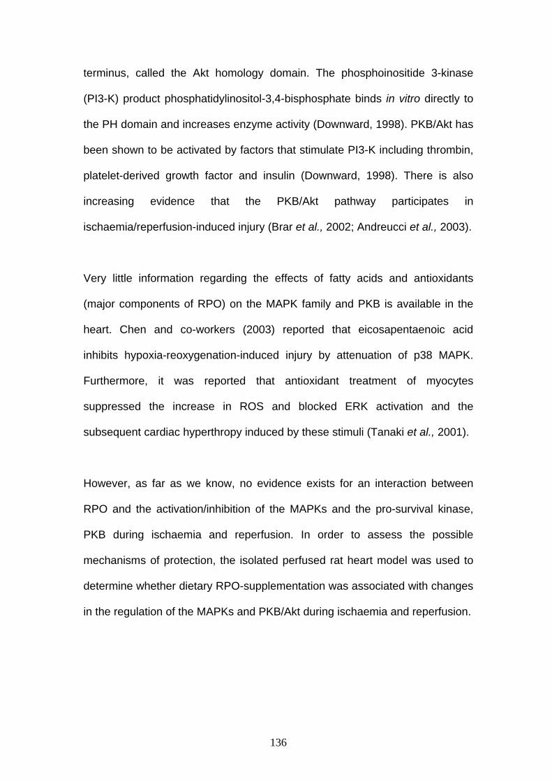
136
terminus, called the Akt homology domain. The phosphoinositide 3-kinase
(PI3-K) product phosphatidylinositol-3,4-bisphosphate binds in vitro directly to
the PH domain and increases enzyme activity (Downward, 1998). PKB/Akt has
been shown to be activated by factors that stimulate PI3-K including thrombin,
platelet-derived growth factor and insulin (Downward, 1998). There is also
increasing evidence that the PKB/Akt pathway participates in
ischaemia/reperfusion-induced injury (Brar et al., 2002; Andreucci et al., 2003).
Very little information regarding the effects of fatty acids and antioxidants
(major components of RPO) on the MAPK family and PKB is available in the
heart. Chen and co-workers (2003) reported that eicosapentaenoic acid
inhibits hypoxia-reoxygenation-induced injury by attenuation of p38 MAPK.
Furthermore, it was reported that antioxidant treatment of myocytes
suppressed the increase in ROS and blocked ERK activation and the
subsequent cardiac hyperthropy induced by these stimuli (Tanaki et al., 2001).
However, as far as we know, no evidence exists for an interaction between
RPO and the activation/inhibition of the MAPKs and the pro-survival kinase,
PKB during ischaemia and reperfusion. In order to assess the possible
mechanisms of protection, the isolated perfused rat heart model was used to
determine whether dietary RPO-supplementation was associated with changes
in the regulation of the MAPKs and PKB/Akt during ischaemia and reperfusion.
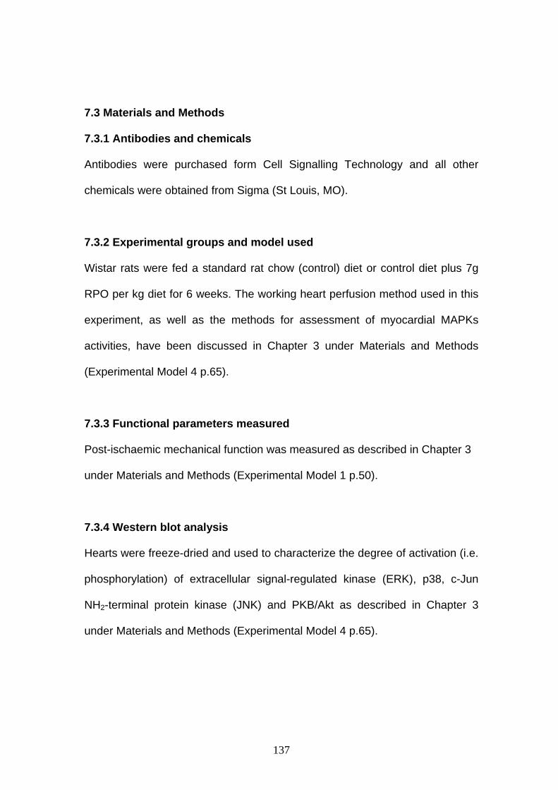
137
7.3 Materials and Methods 7.3.1 Antibodies and chemicals Antibodies were purchased form Cell Signalling Technology and all other
chemicals were obtained from Sigma (St Louis, MO).
7.3.2 Experimental groups and model used Wistar rats were fed a standard rat chow (control) diet or control diet plus 7g
RPO per kg diet for 6 weeks. The working heart perfusion method used in this
experiment, as well as the methods for assessment of myocardial MAPKs
activities, have been discussed in Chapter 3 under Materials and Methods
(Experimental Model 4 p.65).
7.3.3 Functional parameters measured
Post-ischaemic mechanical function was measured as described in Chapter 3
under Materials and Methods (Experimental Model 1 p.50).
7.3.4 Western blot analysis
Hearts were freeze-dried and used to characterize the degree of activation (i.e.
phosphorylation) of extracellular signal-regulated kinase (ERK), p38, c-Jun
NH2-terminal protein kinase (JNK) and PKB/Akt as described in Chapter 3
under Materials and Methods (Experimental Model 4 p.65).
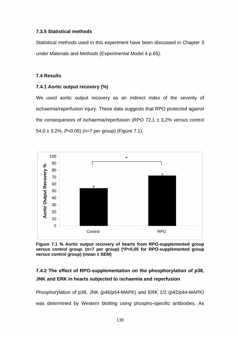
138
7.3.5 Statistical methods
Statistical methods used in this experiment have been discussed in Chapter 3
under Materials and Methods (Experimental Model 4 p.65).
7.4 Results 7.4.1 Aortic output recovery (%)
We used aortic output recovery as an indirect index of the severity of
ischaemia/reperfusion injury. These data suggests that RPO protected against
the consequences of ischaemia/reperfusion (RPO 72,1 ± 3,2% versus control
54,0 ± 3,2%, P<0.05) (n=7 per group) (Figure 7.1).
0
10
20
30
40
50
60
70
80
90
100
Control RPO
Aor
tic O
utpu
t Rec
over
y %
Figure 7.1 % Aortic output recovery of hearts from RPO-supplemented group versus control group. (n=7 per group) (*P<0,05 for RPO-supplemented group versus control group) (mean ± SEM)
7.4.2 The effect of RPO-supplementation on the phosphorylation of p38, JNK and ERK in hearts subjected to ischaemia and reperfusion Phosphorylation of p38, JNK (p46/p54-MAPK) and ERK 1/2 (p42/p44-MAPK)
was determined by Western blotting using phospho-specific antibodies. As
*
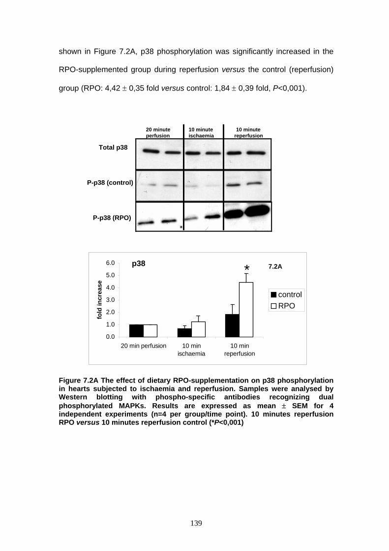
139
shown in Figure 7.2A, p38 phosphorylation was significantly increased in the
RPO-supplemented group during reperfusion versus the control (reperfusion)
group (RPO: 4,42 ± 0,35 fold versus control: 1,84 ± 0,39 fold, P<0,001).
0.0
1.0
2.0
3.0
4.0
5.0
6.0
20 min perfusion 10 minischaemia
10 minreperfusion
fold
incr
ease
controlRPO
Figure 7.2A The effect of dietary RPO-supplementation on p38 phosphorylation in hearts subjected to ischaemia and reperfusion. Samples were analysed by Western blotting with phospho-specific antibodies recognizing dual phosphorylated MAPKs. Results are expressed as mean ± SEM for 4 independent experiments (n=4 per group/time point). 10 minutes reperfusion RPO versus 10 minutes reperfusion control (*P<0,001)
Total p38
P-p38 (control)
P-p38 (RPO)
20 minute perfusion
10 minute ischaemia
10 minute reperfusion
* 7.2A p38

140
Ischaemia/reperfusion caused significant increases in both JNK54 and JNK46
phosphorylation of control group from 20 minutes perfusion to 10 minutes
reperfusion (from 1,0 ± 0 fold to 3,9 ± 0,23 fold, P<0,001 for JNK54 and 1,0 ± 0
to 6,83 ± 0,66 fold, P<0,001 for JNK46) (Figure 7.2B).
0123456789
20'pe
rf
10'isc
h
10're
perf
20'pe
rf
10'isc
h
10're
perf
fold
incr
ease
controlRPO
Figure 7.2B The effect of dietary RPO-supplementation on JNK phosphorylation in hearts subjected to ischaemia and reperfusion. Samples were analysed by Western blotting with phospho-specific antibodies recognizing dual phosphorylated MAPKs. Results are expressed as mean ± SEM for 4 independent experiments (n=4 per group/time point). JNK54: 10 minutes reperfusion RPO versus 10 minutes reperfusion control (**P<0,001). JNK 54 and 46: 10 minutes reperfusion control versus 20 minutes perfusion control (*P<0,001); 10 minutes reperfusion RPO versus 10 minutes reperfusion control (**P<0,001)
20 minute perfusion
10 minute ischaemia
10 minute reperfusion
Total JNK
P-JNK (control)
P-JNK (RPO)
JNK 54 JNK 46
JNK 54 JNK 46
**
*
**
7.2B
*
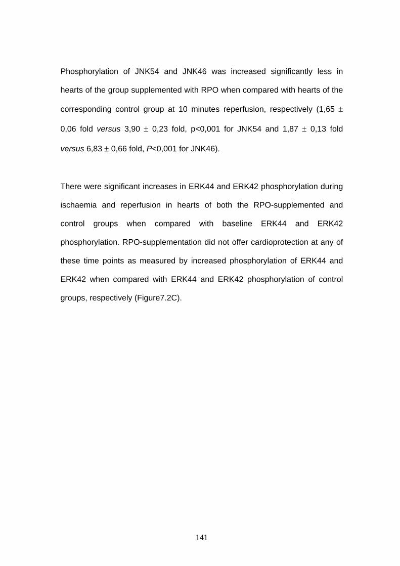
141
Phosphorylation of JNK54 and JNK46 was increased significantly less in
hearts of the group supplemented with RPO when compared with hearts of the
corresponding control group at 10 minutes reperfusion, respectively (1,65 ±
0,06 fold versus 3,90 ± 0,23 fold, p<0,001 for JNK54 and 1,87 ± 0,13 fold
versus 6,83 ± 0,66 fold, P<0,001 for JNK46).
There were significant increases in ERK44 and ERK42 phosphorylation during
ischaemia and reperfusion in hearts of both the RPO-supplemented and
control groups when compared with baseline ERK44 and ERK42
phosphorylation. RPO-supplementation did not offer cardioprotection at any of
these time points as measured by increased phosphorylation of ERK44 and
ERK42 when compared with ERK44 and ERK42 phosphorylation of control
groups, respectively (Figure7.2C).
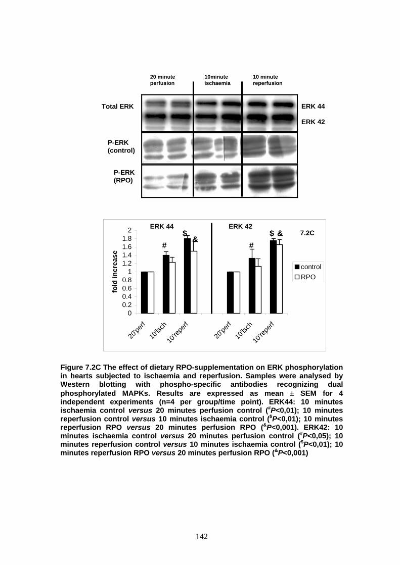
142
00.20.40.60.8
11.21.41.61.8
2
20'pe
rf
10'isc
h
10're
perf
20'pe
rf
10'isc
h
10're
perf
fold
incr
ease
controlRPO
Figure 7.2C The effect of dietary RPO-supplementation on ERK phosphorylation in hearts subjected to ischaemia and reperfusion. Samples were analysed by Western blotting with phospho-specific antibodies recognizing dual phosphorylated MAPKs. Results are expressed as mean ± SEM for 4 independent experiments (n=4 per group/time point). ERK44: 10 minutes ischaemia control versus 20 minutes perfusion control (#P<0,01); 10 minutes reperfusion control versus 10 minutes ischaemia control ($P<0,01); 10 minutes reperfusion RPO versus 20 minutes perfusion RPO (&P<0,001). ERK42: 10 minutes ischaemia control versus 20 minutes perfusion control (#P<0,05); 10 minutes reperfusion control versus 10 minutes ischaemia control ($P<0,01); 10 minutes reperfusion RPO versus 20 minutes perfusion RPO (&P<0,001)
20 minute perfusion
10minute ischaemia
10 minute reperfusion
Total ERK
P-ERK (control)
P-ERK (RPO)
ERK 44 ERK 42
ERK 44 ERK 42
# $
& #
$ & 7.2C

143
7.4.3 The effect of RPO-supplementation on the phosphorylation of PKB/Akt in hearts subjected to ischaemia and reperfusion Phosphorylation of PKB/Akt (Ser473) was determined by Western blotting using
phospho-specific antibodies. There was a significant increase in PKB/Akt
phosphorylation of hearts in the RPO-supplemented group compared to the
control group during reperfusion (4,03 ± 1,1 fold versus 1,03 ± 0,11 fold,
P<0,01) (Figure 7.3).
0.01.02.03.04.05.06.07.0
20'perf 10'isch 10'reperf
fold
incr
ease
controlRPO
Figure 7.3 The effect of dietary RPO-supplementation on PKB phosphorylation in hearts subjected to ischaemia and reperfusion. Samples were analysed by Western blotting with phospho-specific antibodies recognizing dual phosphorylated MAPKs. Results are expressed as mean ± SEM for 4 independent experiments (n=4 per group/time point). 10 minutes reperfusion RPO versus 10 minutes reperfusion control (*P<0,01)
20 minute 10 minute 10 minute perfusion ischaemia reperfusion
Total PKB
P-PKB Ser473 (control)
P-PKB Ser473 (RPO)
*PKB/Akt

144
7.4.4 The effect of RPO-supplementation on PARP cleavage and caspase-3 activation in hearts subjected to ischaemia and reperfusion The control group showed a significant increase in PARP cleavage during
reperfusion compared to PARP cleavage in the control perfusion group (2,1 ±
0,27 fold versus 1,0 ± 0 fold, P<0,01) (Figure 7.4A). However RPO-
supplementation significantly attenuated PARP cleavage (0,6 ± 0,17 fold for
RPO-group versus 2,1 ± 0,27 fold for control group, P<0,001) during
reperfusion.
0.00.51.01.52.02.53.0
20'perf 10'isch 10'reperf
fold
incr
ease
controlRPO
Figure 7.4A: The effect of dietary RPO-supplementation on PARP cleavage during ischaemia and reperfusion. Samples were analysed by Western blotting with antibodies recognizing cleaved PARP and caspase-3. Results are expressed as mean ± SEM for 4 independent experiments (n=4 per group/time point). PARP: 10 minutes reperfusion control versus 20 minutes perfusion control (#P<0,01); 10 minutes reperfusion RPO versus 10 minutes reperfusion control (*P<0,001).
#
*
7.4A
20 minute perfusion
10 minute ischaemia
10 minute reperfusion
Cleaved PARP (control)
Cleaved PARP (RPO)
PARP cleavage
145
Hearts of the control group showed a significant increase in caspase-3
activation during reperfusion when compared with caspase-3 activation of
hearts during perfusion (P<0,05). (Figure 7.4B).
0.00.51.01.52.02.53.0
20 minperfusion
10 minischaemia
10 minreperfusion
fold
incr
ease
controlRPO
Figure 7.4B: The effect of dietary RPO supplementation on caspase-3 activation during ischaemia and reperfusion. Samples were analysed by Western blotting with antibodies recognizing cleaved PARP and caspase-3. Results are expressed as means ± S.E.M. for 4 independent experiments (n=4 per group/per time point). Caspase-3: 10 minutes reperfusion control versus 20 minutes perfusion control (*P<0.05)
7.5 Discussion We have demonstrated that RPO-supplementation offered significant
protection against ischaemia/reperfusion-induced injury in the isolated
perfused working heart as reflected by improved functional recovery. Except
for our own, no other evidence exists for the role of RPO in functional recovery
after ischaemia/reperfusion induced injury (Esterhuyse et al., 2005). However,
20 minute perfusion
10 minute ischaemia
10 minute reperfusion
Cleaved caspase-3 (control)
Cleaved caspase-3 (RPO)
* 7.4B Caspase-3
146
some evidence does exist for the effect of some of the major components of
RPO on cardiac function. Meehan and co-workers (1994) demonstrated that
oleic acid improved functional recovery in ischaemic/reperfused hearts.
Serbinova and co-workers (1992) showed that RPO vitamin E was more
effective than tocopherols in protecting against ischaemia/reperfusion injury in
the isolated Langendorff perfused heart. Das and co-workers (in press) also
demonstrated that palm tocotrienol provided cardioprotection as proved by
reduction of ischaemia/reperfused-mediated increases in ventricular
dysfunction, ventricular arrhythmias and myocardial infarct size. Furthermore,
Bilgin-Karabulut and co-workers (2001) has showed that pre-treatment with a
combination of vitamin A and vitamin E offered protection against venous
ischaemia/reperfusion-induced injury, Interestingly, these vitamins were not
effective when used as single agents (Bilgin-Karabulut et al., 2001).
In response to ischaemia, cells activate various signal transduction pathways
which may be either harmful, or allow adaptation to this stressful environment.
Recent studies suggested that the MAPKs are important regulators of
apoptosis in response to myocardial ischaemia/reperfusion. Therefore, we
characterized the three major MAPK subfamily members that are activated
during ischaemia and reperfusion in our model and investigated the influence
of RPO on their phosphorylation status.
Dietary RPO-supplementation significantly increased the generic p38 isoform
phosphorylation during reperfusion (Figure 7.2A). Despite reports to the
contrary (Mackay and Mochly-Rosen, 2000; Marais et al., 2001), several
investigators support the concept that p38 activation protects the heart from
147
ischaemia/reperfusion-induced injury (Maulik et al., 1996; Weinbrenner et al.,
1997). These opposing results may be attributed to the different isoforms (α
and β) expressed in the heart (Saurin et al., 2000), which appear to mediate
opposing effects. The p38α-isoform is implicated in apoptosis, whereas p38β is
anti-apoptotic in rat cardiac myocytes. JNK phosphorylation (JNK54 and
JNK46) was significantly increased during reperfusion but was attenuated by
dietary RPO supplementation. JNK phosphorylation appears to be pro-
apoptotic in many cell types (Obata et al., 2000; Park et al., 2000), however
their exact role in regulating cell death is unclear. For example, Hreniuk and
co-workers (2001) found that inhibition of JNK46, but not JNK54, significantly
reduced reoxygenation-induced apoptosis. Wang and co-workers (1998), on
the other hand, reported that activation of JNK by transfection of cultured rat
neonatal cardiomyocytes with mitogen activated protein kinase kinase 7
(MKK7), an upstream activator of JNK, induced hypertrophy rather than
apoptosis. Although dietary RPO-supplementation had no effect on ERK
phosphorylation compared to the control group, the ERK cascade appears to
specifically mediate cell growth and survival signals. For instance, it has been
shown that inhibition of ERK enhances ischaemia/reperfusion-induced
apoptosis and that sustained activation of this kinase during simulated
ischaemia mediates adaptive cytoprotection in cultured neonatal
cardiomyocytes (Punn et al., 2000).
We also investigated the involvement of PKB/Akt in the cellular response to
dietary RPO supplementation. RPO was responsible for a significant increase
in PKB phosphorylation during reperfusion (Figure 7.3). This is in agreement
with results of Fujio and co-workers (2000) who showed that PKB/Akt
148
promotes survival of cardiomyocytes in vitro and protects against
ischaemia/reperfusion injury in the mouse heart. PKB is activated downstream
of PI-3-kinase by the phosphoinositide-dependent protein kinases PDK-1 and
PDK-2 (Anderson et al., 1998). PKB in turn phosphorylates a number of
downstream targets relevant to cell survival functions, including the pro-
apoptotic Bcl-2 family member BAD (Del Peso et al., 1997). Phosphorylation of
BAD on Ser136 by PKB inhibits its pro-apoptotic function, thus promoting cell
survival (Datta et al., 1997). Interestingly, BAD is not only a substrate for PKB,
but is also phosphorylated by the MAPK kinase MEK (Punn et al., 2000),
linking the classical Ras-MAPK pathway to cell survival.
Apoptosis has been consistently observed in cardiac myocytes after
reperfusion and may represent a direct mechanism by which myocytes are
damaged (Abe et al., 2000). Indeed, in our model, reperfusion injury also
resulted in cleavage of PARP to its proteolyzed products, a phenomenon well
known to result from caspase-3 activation. RPO-supplementation significantly
reduced PARP cleavage during reperfusion, and attenuated caspase-3
activation, although not significantly.
In summary, our results have shown that RPO-supplementation caused
increased phosphorylation of p38 and PKB and reduce phosphorylation of
JNK. Both increased PKB and p38 phosphorylation and the inhibition of JNK
phosphorylation may contribute to the protection of the cell against apoptosis.
The attenuation of PARP cleavage would in turn be expected to inhibit
apoptosis. Results presented in other studies (Bogoyevitch et al., 1996; Punn
149
et al., 2000), as well as our own (Engelbrecht et al., 2004), indicate that the
MAPKs are central regulators of reactive signalling in cardiac myocytes.
Dietary RPO-supplementation was associated with increased percentage
myocardial EPA after ischaemia in our perfusion model, which may effect
intracellular signalling cascades. The ability to directly manipulate MAPK
signalling has been shown to protect cardiomyocytes from
ischaemia/reperfusion-induced apoptosis/injury. This notion suggests that
members of the MAPK signalling cascade would be ideal targets for
pharmacological intervention to treat ischaemia/reperfusion injury. Therefore,
according to our results, a daily dosage RPO of 0,58 mg/kg suggest to be
beneficial to humans.
In the current study we have demonstrated for the first time that RPO might
exert its beneficial effects during reperfusion through increased PKB/Akt and
p38 phosphorylation and dephosphorylation of JNK, which might be associated
with inhibition of apoptosis and improved function. Thus, RPO might offer an
alternative, non-pharmacological strategy to protect the heart against
ischaemia/reperfusion-induced injury.
150
CHAPTER 8
CONCLUSION
To our knowledge no previous studies have investigated the effect of dietary
RPO-supplementation on cell signalling associated with cardioprotection
during ischaemia/reperfusion injury. In the current study we provide data of
dietary RPO-intervention on the signalling pathways that may be involved in
cardioprotection during ischaemia and reperfusion.
An in vitro working rat heart perfusion model was used to investigate the
effects of dietary RPO-supplementation on myocardial post-ischaemic
functional recovery and the mechanisms involved. Our results clearly indicated
that dietary RPO-supplementation of a standard rat chow diet protects against
the consequences of global ischaemia/reperfusion in the isolated perfused rat
heart as reflected by improved aortic output recovery. Based on our results, we
propose that the protective effect of RPO may be associated with either its
antioxidant characteristics, and/or changes in the phospholipid fatty acid
composition of the myocardium during ischaemia/reperfusion injury. We
hypothesize that the palm oil vitamin E antioxidant properties may contribute to
elevated cGMP and decreased cAMP concentrations early in ischaemia. This
may be the more prominent mechanism of cardioprotection in this model using
a standard rat chow diet. It is possible that cGMP may attenuate
ischaemia/reperfusion injury by inhibiting the cAMP induced increase in the
slow inward calcium current, thus leading to a decrease in ischaemic cytosolic
calcium concentrations.
We also investigated whether RPO-supplementation offers the same
protection against ischaemia/reperfusion injury when supplemented with a
151
high-cholesterol diet and tried to elucidate possible mechanisms involved in
this protection. Increased reperfusion aortic output recovery and rate pressure
product values with RPO-supplemention of a high-cholesterol diet were
associated with a decreased cAMP concentration during reperfusion. Although
suppression of cAMP during reperfusion could be involved in cardioprotection,
we were of the opinion that this was not the only mechanism of protection. In
addition, our results clearly demonstrated that the RPO-induced protection of a
high-cholesterol diet could not be linked with increased NO-cGMP signalling
and confirms our hypothesis that cGMP may not be the only mechanism of
protection. This following sequence of events should be considered: In the
presence of cholesterol, superoxide production is increased. Superoxide would
compete to re-direct the reaction towards lipid peroxidation, instead of cGMP
production. Our results indicate that neither SOD activity, nor LPO production
increased. We therefore speculate that RPO antioxidants acted as free radical
scavengers and could potentially improve NO availability. However, we have
no data to support this argument, but it does create an opportunity for future
investigations.
From our work it is also evident that most of the RPO-induced changes
occurred during the ischaemic period with the NO-cGMP pathway being a
major role player.
Recent studies suggested that the MAPK family, PKB/Akt and signal
transduction caspases are important regulators of apoptosis in response to
myocardial ischaemia/reperfusion. We demonstrated for the first time that
152
dietary RPO-supplementation protects the isolated perfused working rat heart
from ischaemia/reperfusion-induced apoptosis/injury through MAPK-, PKB/Akt-
and caspase dependent pathways during the reperfusion period. Dietary RPO-
supplementation exerts its beneficial effects through increased PKB/Akt and
p38 phosphorylation and dephosphorylation of JNK, all of which might be
associated with inhibition of apoptosis and improved functional recovery.
Dietary RPO-supplementation was associated with increased percentage
myocardial EPA after ischaemia in our perfusion model, which could be
involved in the modulation of MAPK enzyme activity.
In summary our results suggest that dietary RPO-supplementation offered
protection against ischaemia/reperfusion injury as reflected by improved aortic
output recovery. We could not demonstrate whether the fatty acids (although
little contribution to that of diet) or antioxidant content individually, or as a
combination, was responsible for these protective effects. Our results suggest
that hearts of cholesterol-fed animals were protected through a different
mechanism (may possibly include the antioxidant capacity of RPO). The
proposed mechanisms include RPO protection in ischaemia via the NO-
cGMPpathway and MAPK, PKB/Akt and caspase involvement during
reperfusion (Figure 8.1).
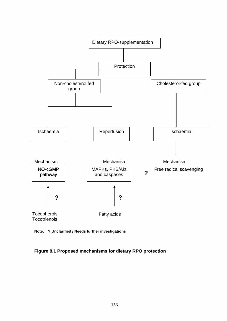
153
Mechanism Mechanism Mechanism
?
? ?
Note: ? Unclarified / Needs further investigations
Figure 8.1 Proposed mechanisms for dietary RPO protection
Dietary RPO-supplementation
NO-cGMP pathway
Non-cholesterol fed group
Cholesterol-fed group
Protection
Ischaemia Reperfusion Ischaemia
Free radical scavenging
Fatty acids Tocopherols Tocotrienols
NO-cGMP pathway
MAPKs, PKB/Akt and caspases

154
ADDENDUM
CONGRESSES A: International 1. International Palm Oil Congress (PIPOC 2003)
24-28 August 2003, Putrajaya Marriott Hotel, Putrayaya , Malaysia Paper: Dietary red palm oil supplementation improves post ischaemic
functional recovery in the isolated perfused rat heart. AJ Esterhuyse, EF du Toit, J van Rooyen.
2. 2004 ISHR World Congress Satellite: Cellular Injury in Ischaemia, Kruger Park, South Africa, 13-15 August 2004. Poster: NO-cGMP pathway involved in red palm oil mediated ischaemic
protection. Jacques van Rooyen, Johan Esterhuyse, Spinnler Benade,
Eugene du Toit. (Abstract accepted and published in CVSA).
3. Oils and fats International Congress, Putra World Trade Centre,
Kuala Lumpur, Malaysia, 29 September-2 October 2004 Invited Speaker: Red palm oil: Myth or Magic? The effect of the NO-
cGMP pathway. Van Rooyen J; Esterhuyse AJ; Du Toit EF.
B: National 1. 32ndAnnual Congress of the PSSA, September 12-15, 2004. Coffee
Bay,Transkei, South Africa. Plenary Speaker : Red palm oil: Myth or Magic. Jacques van Rooyen;
Johan Esterhuyse; Eugene F du Toit.
Poster: A role for dietary red palm oil induced cardioprotection in
isolated rat hearts. Esterhuyse AJ; Du Toit EF; Bester DJ; Benadé AJS; Van Rooyen J.
155
Poster: Cardiac function in isolated perfused rat heart is not negatively
affected by dietary red palm oil. Bester DJ; Van Rooyen J; Du Toit EF;
Benadé AJS; Esterhuyse AJ.
2. Academic Day, University of Stellenbosch, South Africa, 18 August 2004 Poster: Dietary red palm oil offers protection against
ischaemia/reperfusion injury. Esterhuyse AJ; Bester DJ; Van Rooyen
J; Du Toit EF.
PUBLICATIONS 1. Esterhuyse AJ; Du Toit EF; Van Rooyen J. Dietary red palm oil
supplementation protects against the consequences of global ischaemia
in the isolated perfused rat heart. Asia Pac J Clin Nutr (accepted 2004).
2. Esterhuyse AJ; Du Toit EF; Benadé AJS; Van Rooyen J. Dietary red
palm oil improves reperfusion cardiac function in the isolated perfused
rat heart of animals fed a high cholesterol diet. Prostagl, Leukot Essent Fatty Acids 2005; 72:153-161.
3. Anna-Mart Engelbrecht; Johan Esterhuyse; Eugene Du Toit;
Jacques van Rooyen. p38-MAPK and PKB/AKT, possible role players
in red palm oil induced protection of the isolated perfused rat heart. J Nutr Biochem (accepted 2005).
Submitted: 3. Johan Esterhuyse; Jacques van Rooyen; Hans Strijdom; Dirk
Bester; Eugene du Toit. Proposed mechanisms for red palm oil
induced cardioprotection in a hyperlipidaemic perfused rat heart model.
(2005).
156
REFERENCES
Abe J-I, Baines CP, Berk BC. Role of mitogen-activated protein kinase in
ischaemia and reperfusion injury. Circ Res 2000; 6: 607-609.
Abeywardena MY, Charnock JS. Dietary lipid modification of myocardial
eicosanoids following ischaemia and reperfusion in the rat. Lipids 1995;
30(12): 1151-1156.
Abeywardena MY, McLennan PL, Charnock JS. Changes in the myocardial
eicosanoid production following long-term dietary lipid supplementation in rats.
Am J Clin Nutr 1991; 53: 1039S-1041S.
Abudu N, Miller JJ, Attaelmannan M, Levinson SS. Vitamins in human
atherosclerosis with emphasis on vitamin C and vitamin E. Clinica Chimica
Acta 2004; 339:11-25.
Adderley SR, Fritzgerald DJ. Oxidative damage of cardiomyocyte is limited by
extracellular regulated kinases 1 and 2-mediated induction of cylooxygenase-
2. J Biol Chem 1999; 274: 5038-5046.
Anderson KE, Coadwell J, Stephens LR, Hawkins PT. Translocation of PDK-1
to the plasma membrane is important in allowing PDK-1 to activate protein
kinase B Curr Biol 1998; 8:684-691.
157
Andreucci M, Michael A, Kramers C, Park KM, Chen A, Matthaeus T,
Alessandrini A, Haq S, Force T, Bonventre JV. Renal ischaemia/reperfusion
and ATP depletion/repletion in LCC-PK(1) cells results in phosphorylation of
FKHR and FKHR1. Kidney Int 2003; 64: 1189-1198.
Aoki H, Kang PM, Hampe J, Yoshimura K, Noma T, Matsuzaki M, Izumos S.
Direct activation of mitochondrial apoptosis machinery by c-Jun N-terminal
kinase in adult cardiac myocytes. J Biol Chemistry 2002; 277(12): 10244-
10250.
Araki M, Tanaka M, Hasegawa K, Yokota R, Maeda T, Ishikawa M, Yabuuchi
Y, Sasayama S. Nitric oxide inhibition improved myocardial metabolism
independent of tissue perfusion during ischaemia but not during reperfusion. J
Mol Cell Cardiol 2000; 32(3): 375-84.
Azzi A, Ricciarelli R, Zing GJM. Non-antioxidant molecular functions of α-
tocopherol (vitamin E). FEBS Lett 2002; 519: 8-10.
Bagchi K, Puri S. Free radicals and antioxidants in health and disease. EMHJ
1998; 4(2): 350-360.
Barancik M, Htun P, Strohm C, Kilian S, Schaper W. Inhibition of the cardiac
p38-MAPK pathway by SB203580 delays ischaemic cell death. J Cardiovasc
Pharmacol 2000; 35: 474-483.
158
Beresewics A, Karwatowska-Prokopczuk E, Lewartowski B, Cedro-
Ceremuzynska K. A protective role of nitric oxide in isolated
ischaemic/reperfusion rat heart. Cardiovasc Res 1995; 30: 1001-1008.
Bilgin-Karabulut A, Ademoglu E, Aydin I, Ere M, Gokkusu C. Protective effects
of vitamins A and E pretreatment in venous ischaemia/reperfusion injury. J
Reconstr Microsurg 2001; 17: 425-429.
Bogoyevitch M. Signalling via stress activated mitogen-activated protein
kinases in the cardiovascular system. Cardiovasc Res 2000; 45: 826-842.
Bogoyevitch MA, Gillespie-Brown J, Ketterman AJ, Fuller SJ, Ben-Levy R,
Ashworth A, Marshall CJ, Sugden PH. Stimulation of the stress-activated
mitogen-activated protein kinase subfamilies in perfused heart: p38/RK
mitogen-activated protein kinases and c-Jun N-terminal kinases are activated
by ischaemia/reperfusion. Circ Res 1996; 79: 162-173.
Bolli R. Cardioprotective function of inducible nitric oxide synthase and role of
nitric oxide in myocardial ischaemia and preconditioning: an Overview of a
decade of research. J Mol Cell Cardiol 2001; 33: 1897-1918.
Bradford MM. A rapid and sensitive method for quantitation of microgram
quantities of protein utilizing the principle of protein-dye binding. Anal Biochem
1976; 71: 248-254.
159
Brar BK, Stephanou A, Knight R, Latchman DS. Activation of protein kinase B/
Akt by urocorton is essential for its ability to protect cardiac cells against
hypoxia/reoxygenation-induced cell death. J Mol Cell Cardiol 2002; 34: 483-
492.
Burton GW, Ingold KU. Auto-oxidation of biological molecules. The antioxidant
activity of vitamin E and related chain-breaking phenolic antioxidants in vitro. J
Am Chem Soc 1981; 103: 6472-6477.
Cameron IL, Munoz J, Barnes CJ. High dietary level of synthetic vitamin E on
lipid peroxidation, membrane fatty acid composition and cytotoxicity in breast
cancer xenograft and in mouse host tissue. Cancer Cell Int 2003; 3(1):181-
187.
Cardone MH, Salvesen GS, Widmann C, Johnson G, Frisch SM. The
regulation of anoikis: MEKK-1 activation requires cleavage by caspase. Cell
1997; 90: 315-323.
Chandrasekharan N. Changing concepts in lipid nutrition in health and
nutrition. Med J Malaysia 1999; 54(3): 408-428.
Charnock JS, Sundram K, Abeywardena MY, McLennan PL, Tan DT. Dietary
fats and oils in cardiac arrhythmia in rats. Am J Clin Nutr 1991; 53: 1047S-
1049S
160
Chen H, Li D, Roberts GJ, Saldeen T, Mehta JL. Eicosapentaenoic acid
inhibits hypoxia-reoxygenation-induced injury by attenuating upregulation of
MMP-1 in adult rat myocyte. Cardiovasc Res 2003; 59: 7-13.
Chow CK, Hong CB. Dietary vitamin E and selenium and toxicity of nitrite and
nitrate. Toxicology 2002; 180: 195-207.
Codex Alimentarius Vol. XI FAO/WHO Rome 1983; Introduction and pages
115-130.
Communal C, Colucci WS, Singh K. p38 Mitogen-activated protein kinase
pathway protects adult rat ventricular myocytes against beta-adrenergic
receptor-stimulated apoptosis: evidence for Gi-dependant activation. J Biol
Chem 2000; 275: 19395-19400.
Cottrell RC. Introduction: nutritional aspects of palm oil. Am. J Clin. Nutr 1991;
53: 989S-1009S.
Csont T, Balogh G, Csonka C, Boros I, Horvath I, Vigh L, Ferdinandy P.
Hyperlipidaemia induced by high cholesterol diet inhibits heat shock response
in rat hearts. Biochem Biophys Res Commun 2002; 290: 1535-1538.
Das S, Powell SR, Wang P, Divald A, Nesaretman K, Tosaki A, Cordis GA,
Maulik N, Das DK. Cardioprotection with palm tocotrienol; antioxidant activity
161
of tocotrienol is linked with its ability to stabilize proteasomes. Am J Physiol
Heart Circ Physiol. 2005; in press.
Datta SR, Dudek H, Tao X, Masters S, Fu H, Gutoh Y, Greenberg ME. Akt
phosphorylation of BAD couples survival signals to the cell-intrinsic death
machinery. Cell 1997; 91: 231-241.
Davis RJ. Signal transduction by the JNK group of MAP kinase. Cell 2000;
103: 239-252.
Del Peso L, Gonzales-Gracia M, Page C, Herrera R, Nunez G. Interleuken-3
induced phosphorylation of BAD through protein kinase AKT. Science 1997;
278: 687- 689.
Deliconstantinos G, Villiotou V, Stavrides JC. Modulation of particulate nitric
oxide activity and peroxynitrite synthesis in cholesterol enriched endothelial
cell membranes. Biochem Pharmacol 1995; 49(11): 1589-1600.
Denys A, Hichami A, Khan NA. Eicosapentaenoic acid and docosahexaenoic
acid modulate MAP kinase enzyme activity in human T-cells. Mol and Cell
Biochem 2002; 232; 143-148.
Depré C, Fierain L, Hue L. Activation of nitric oxide synthase by ischaemia in
the perfused heart. Cardiovasc Res 1996; 33: 82-87.
162
Diniz YS, Cicogna AC, Padovani CR, Santana LS, Faine LA, Novelli ELB.
Diets rich in saturated and polyunsaturated fatty acids: Metabolic shifting and
cardiac health. Nutr 2004; 20: 230-234.
Dougherty CJ, Kubasiak LA, Prentice H, Andreka P, Bishopric NH, Webster
KA. Activation of c-Jun N-terminal kinase promotes survival of cardiac
myocytes after oxidative stress. Biochem J 2002; 362: 561-571.
Dong C, Yang DD, Wysk M, Whitmarsh AJ, Xu J, Davis RJ, Flavell RA.
Defective T cell differentiation in the absence of Jnk1. Science (1998); 282:
2092-2095.
Downward J. Mechanisms and consequences of activation of protein kinase
B/Akt. Curr Opin Cell Biol 1998; 10: 262-267.
Droge W. Free radicals in the physiological control of cell function. Physiol Rev
2002; 82: 47-95.
Du Toit EF, Meiring J, Opie LH. Relation of cyclic nucleotide ratios to
ischaemic and reperfusion injury in nitric oxide–donor treated hearts. J
Cardiovasc Pharmacol 2001; 38: 529-538.
Du Toit EF, Muller CA, McCarthy J, Opie LH. Levosimendan: Effects of a
calcium sensitizer on function and arrhythmias and cyclic nucleotide levels
163
during ischaemia/reperfusion in the Langendorff-perfused guinea pig heart. J
Pharmacol Exp Ther 1999; 290: 505-514.
Earnshaw WC. Apoptosis: lessons from in vitro systems. Trends Cell Biol
1995; 5: 217-220.
Engelbrecht AM, Niesler C, Page C, Lochner A. p38 and JNK have distinct
regulatory functions on the development of apoptosis during simulated
ischaemia and reperfusion in neonatal cardiomyocytes. Basic Res Cardiol
2004; 99:308-314.
Engelbrecht AM, Niesler C, Page C, Lochner A. Long-chain polyunsaturated
fatty acids protect neonatal cardiomyocytes against apoptosis by upregulation
of the phosphatase, MKP-1. J Mol Cardiol 2004 in press.
Esposito LA, Melov S, Panov A, Cottrell BA, Wallace DC. Mitochondrial
disease in mouse results in increased oxidative stress. Proc Natl Acad Sci
USA 1999; 96: 4820-4825.
Esterhuyse AJ, du Toit EF, Benade AJS, van Rooyen J. Dietary red palm oil
improves reperfusion cardiac function in the isolated perfused rat heart of
animals fed a high cholesterol diet. Prostagland Leukot Essent Fatty Acids
2005; 72:153-161.

164
Esterhuyse AJ, Du Toit EF, Van Rooyen J. Dietary red palm oil
supplementation improves post ischaemic functional recovery in the isolated
perfused rat heart. Proceedings of Food Technology and Nutrition Conference.
Malaysian Palm Oil Board 2003; 192-201.
Fanger GR, Johnson NL, Johnson GL. MEK kinase are regulated by EGF and
selectively interact with Rac/Cdc42. EMBO J 1997; 16: 4961-4972.
Faine LA, Diniz YS, Almeida JA, Novelli ELB. Toxicity of ad libitum
overfeeding: effects on cardiac tissue. Food Chem Toxicol 2002; 3: 130.
Ferdinandy P, Schultz R. Review. Nitric oxide, superoxide, and peroxynitrite in
myocardial ischaemia-reperfusion injury and preconditioning. Br J Pharmacol
2003; 138: 532-543.
Ferdinandy P, Szilvassy Z, Horvath LI, Csont T, Csonka C, Nagy E,
Scentgyorgyi R, Nagy I, Koltai M, Dux L. Loss of pacing-induced
preconditioning in rat hearts: Role of nitric oxide and cholesterol-enriched diet.
J Mol Cell Cardiol 1997; 29: 3321-3333.
Ferdinandy P, Panas D, Schulz R. Peroxynitrite contributes to spontaneous
loss of cardiac efficiency in isolated working rat hearts. Am J Physiol 1999;
276: H1861-1867.

165
Fischer S, Weber PC. Prostaglandin I3 is formed in vivo in man after dietary
eicosapentaenoic acid. Nature 1984; 307; 165-168.
Folch J, Lees M, Sloane-Stanley GH. A simple method for the isolation and
purification of total lipids from animal tissues. J Biol Chem 1957; 226: 497-509.
Folkers K. Relevance of the biosynthesis of coenzyme Q10 and for the four
bases of DNA as a rationale for the molecular causes of cancer and therapy.
Biochem Biophys Res Commun 1996; 224(2): 358-361.
Fujio Y, Nguyen T, Wencker D, Kitsis RN, Walsh K. Akt promotes survival of
cardiomyocytes in vitro and protects against ischaemia/reperfusion in mouse
heart. Circulation 2000; 101: 660-667.
Gapor A, Ong ASH, Kato A, Watanabe H, Kawade T. Antioxidant activities of
palm vitamin E with special reference to tocotrienols. Elaeis 1989; 1: 63-67.
Garg ML, Sebokova E, Thompson ARB. Delta 6-desaturase activity in liver
microsomes of rats fed diets enriched with cholesterol and/or n-3 fatty acids.
Biochem J 1988; 249: 351-356.
Gartner A, Nasmyth K, Ammerer G. Signal transduction in Saccharomyces
cerevisiae requires tyrosine and threonine phosphorylation of FUS3 and KSS1.
Geenes D 1992; 7: 1280-1292.

166
Gilham B, Papachristodoulou DK, Thomas JH. Wills’ biochemical basis of
medicine .3rd edition 1997. p. 23-77.
Giraldez RR, Panda A, Xia Y, Sanders SP, Zweier JL. Decreased Nitric Oxide
Synthase activity caused impaired endothelium-dependent relaxation in the
post-ischaemic heart. Am Soc Biochem Mol Biol Inc 1997; 272(34): 21420-
21426.
Giricz Z, Csonka C, Onody A, Csont T, Ferdinandy P. Role of cholesterol-
enriched diet and the mevalonate pathway in cardiac nitric oxide synthesis.
Basic Res Cardiol 2003; 98: 304-310.
Goh SH, Choo YM, Ong ASH. Minor constituents of palm oil. J Am Oil Chem
Soc 1985; 62: 237-240.
Guthrie N, Chambers AF, Gapor A, Carroll KK. In vitro inhibition of proliferation
of receptor-positive MCF-7 human breast cancer cells by palm oil tocotrienols.
FASEB J 1995; 9: A988.
Han H, Lee JD, Bibbs L, Ulevitc RJ. A MAP kinase targeted by endotoxin and
hyperosmolarity in mammalial cells. Science 1994; 256: 808-811.
Hano O, Thompson-Gorman SL, Zweier JL, Lakatta EG. Coenzyme Q10
enhances cardiac functional and metabolic recovery and reduces Ca2+
167
overload during postischaemic reperfusion. Am J Physiol 1994; 266: H2174-
H2181.
Hare JM, Comerford ML. Role of nitric oxide in the regulation of myocardial
function. Prog. Lipid Res 1995; 38: 155-166.
Hart RW, Dixit R, Seng J, Turturro A, Leakey JEA, Feuers R, Duffy P,
Buffington C, Cowan G, Lewis S, Pipkin J, Li SY. Adaptive role of caloric intake
on the degenerative disease process. Toxicol Sci 1999; 52:3-12.
Heyden S. Polyunsaturated and monounsaturated fatty acids in the diet
prevent coronary heart disease via cholesterol reduction. Ann Nutr Metab
1994; 38 (3): 117. 122.
Hood RL. Tocotrienols and cholesterol metabolism. Ong ASH, Niki E, Packer
L, eds. Nutrition, Lipids, Health and Disease 1995; 96-103 AOCS Press
Champaign, IL.
Horribon DF. Gamma linoleic acid: An intermediate in fatty acid metabolism
with potential as an ethical pharmaceutical and as a food. Rev Contemp
Pharmacother 1990; 1: 1-45.
Hreniuk D, Garay M, Garrde W, Monia BP, McKay RA, Cioffi CL. Inhibition of
c-Jun N-terminal kinase 1, but not c-Jun N-terminal kinase 2, suppresses
168
apoptosis induced by ischaemia/reperfusion in rat cardiac myocytes. Mol
Pharmacol 2001; 59: 867-874.
Huot J, Houle F, Marceau F, Landry J. Oxidative stress-induced actin
reorganization mediated by the p38 mitogen-activated protein kinase/heat
shock protein 27 pathway in vascular endothelial cells. Circ Res 1997; 80(3):
383-392.
Illarion V, Murad T, Murad F. Protein nitration in cardiovascular diseases.
Pharmacol Rev 2002; 54: 619-634.
Ip YT, Davis RJ. Signal transduction by c-Jun N-terminal kinase (JNK): from
inflammation to development. Curr Opin Cell Biol 1998; 10: 205-219.
Jacobson MD, Burne JF, Raff MC. Programmed cell death and Bcl-2
protection in the absence of a nucleus. EMBO J 1994; 13: 1899-1910.
Jiang Y, Chen C, Li Z. Characterization of the structure and function of a new
mitogen-activated protein kinase (p38β). J Biol Chem 1996; 271: 920-926.
Jiang Y, Gram H, Zhao M. Characterization of the structure and function of the
fourth member of p38 group MAP kinase-p38γ. J Biol Chem 1997; 272: 122-
128.
169
Jump DB. Mini-review. The Biochemistry of 2-3 polyunsaturated fatty acids. J.
Biol. Chem 2002; 277(11): 8755-8758.
Kajstura JW, Cheng W, Reiss K, Clark WA, Sonneblick EH, Krajewski S, Reed
JC, Olivetti G, Anversa P. Apoptotic and necrotic myocyte cell deaths are
independent contributing variables of infarct size in rats. Lab Invest 1996; 9:
949-956.
Kamal-Eldin A, Appelqvist L. The chemistry and antioxidant properties of
tocopherols and tocotrienols. Lipids 1996; 31: 671-701.
Kamen B. A new dietary oil for the new millenium, red palm oil. Total Health
2000; 22(6): 62-63.
Kang JX, Leaf A. Prevention of fatal cardiac arrhythmias by polyunsaturated
fatty acids. Am J Clin Nutr 2000; 71(1): 202-207.
Kauffmann-Zeh A, Rodriguez-Visiana P, Ulrich E, Gilbert C, Coffer P,
Downward J, Evan G. Suppression of c-Myc-induced apoptosis by Ras
signalling through PI(3)K and PKB. Nature 1997; 385: 544-548.
Kaufmann SH. Induction of endonucleolytic DNA cleavage in human acute
myelogenous leukemia cells by etoposite, camptothecin and other cytotoxic
anticancer drugs:a cautionary note. Cancer Res 1998; 49: 5870-5878.
170
Kaufmann SH, Desnoyers S, Ottaviano Y, Davidson NE, Poirier GG. Specific
proteolytic cleavage of poly (ADP-ribose) polemerase an early marker of
chemotherapy-induced apoptosis. Cancer Res 1993; 53: 3976-3985.
Kennedy SG, Wagner AJ, Conzen SD, Jordan J, Bellacosa A, Tyhlis PN. The
P13-kinase Akt signalling pathway delivers an anti-apoptotic signal. Genes
Dev 1997; 11: 701-713.
Khor HT, Chieng DY, Ong KK. Tocotrienols inhibit HMG-CoA reductase activity
in the guinea pig. Nutr. Res 1995; 15: 537-544.
Kline K, Yu W, Sanders BG. Vitamin E: mechanisms of action as tumour cell
growth inhibitors. J Nutr 2001; 131(1): 161S-163S.
Kothakota S, Azuma T, Reinhard C, Klippel A, Tang J, Chu K, McGarry TJ,
Kirschner MW, Koths K, Kwiatkowski DJ, Williams LT. Caspase-3-generated
fragment of gelsolin: effector of morphological change in apoptosis. Science
1997; 278: 294-298.
Kyriakis JM, Avruch J. Sounding the alarm: protein kinase cascades activated
by stress and inflammation. J Biol Chem 1996; 40: 24313-24316.
Knight RJ, Buxton DB. Stimulation of c-Jun kinase and mitogen activated
protein kinase by ischaemia and reperfusion in the perfused rat heart. Biochem
Biophys Res Commun 1996; 218: 83-88.

171
Kojda G, Harrison D. Interactions between NO and reactive oxygen species:
pathophysiological importance in atherosclerosis, hypertension, diabetes and
heart failure. Cardiovasc Res 1999; 43: 562-571.
Kritchevsky D. Impact of red palm oil on human nutrition and health. Food Nutr
Bull 2000; 21(2): 182-188.
Kuan CY, Yang D, Samanta Roy DR, Davis RJ, Rakic P, Flavell RA. The Jnk1
and Jnk2 protein kinases are required for regional specific apoptosis during
early brain development. Neuron 1999; 22: 667-676.
Laderoute KR, Webster KA. Hypoxia/reoxygenation stimulates Jun kinase
activity through redox signalling in cardiac myocytes. Circ Res 1997; 80: 336-
344.
Lee JC, Laydon JT, McDonnel PC. A protein kinase involved in the regulation
of inflammatory cytokine biosynthesis. Nature 1994; 372: 739-746.
Leppa S, Saffrich R, Ansorge W, Bohmann D. Differential regulation of c-Jun
by ERK and JNK during PC12 cell differentiation. EMBO J 1998; 17: 4404-
4413.
172
Li Z, Jiang Y, Ulevitch RJ, Han J. The primary structure of p38 gamma: a new
member of p38 group of MAPK kinase. Biochem Biophys Res Commun 1996;
228: 334-340.
Li Y, Kang JX, Leaf A. Differential effects of various eicosanoids on the
production or prevention of arrhythmias in cultured neonatal rat cardiac
myocytes. Prostaglandins 1997; 54: 511-530.
Lin A. Activation of the JNK signalling pathway: breaking the break on
apoptosis. BioEssays 2003; 25: 17-24.
Lockwood K, Moesgaard S, Folkers K. Partial and complete regression of
breast cancer in patients in relation to dosage of coenzyme Q10. Biochem
Biophys Res Commun 1994; 1994(3): 1504-1508.
Lockwood K, Moesgaard S, Yamamoto T, Folkers K. Progress on therapy of
breast cancer with vitamin Q10 and the regression of metastases. Biochem
Biophys Res Commun 1995; 212(1):172-177.
Ma XL, Kumar S, Gao F, Louden CS, Lopez BL, Christopher TA, Wang CF,
Lee JC, Feuerstein C, Yue TL. Inhibition of p38 mitogen-activated protein
kinase decreases cardiomyocyte apoptosis and improves cardiac function after
myocardial ischaemia and reperfusion. Circulation 1999; 99: 1685-1691.
173
Mackay K, Mochly-Rosen D. Involvement of a p38 mitogen-activated protein
kinase phosphates in protecting neonatal rat cardiac myocytes from ischaemia.
J Mol Cell Cardiol 2000; 32: 1585-1588.
Mansour SJ, Matten WT, Herman AS, Candia JM, Rong S, Fakasawa K, Van
de Woude GF, Ahn NG. Transformation of mammalian cells by constitutively
active MAP kinase. Science 1994; 265: 966-970.
Marais E, Genade S, Huisamen B, Strijdom JG, Moolman JA, Lochner A.
Activation of p38 MAPK induced by a multi-cycle ischaemic preconditioning
protocol is associated with attenuated p38 MAPK activity during sustained
ischaemia and reperfusion. J Mol Cell Cardiol 2001; 33: 769-778.
Maulik N, Engelman DT, Watanabe M, Engelman RM, Maulik G, Cordis GA,
Das DK. Nitric oxide signalling in ischaemic heart. Cardiovasc Res 1995; 30:
593-601.
Maulik N, Watanabe M, Zu Y-L, Huang C-K, Cordis GA, Schley JA, Das DK.
Ischaemic preconditioning triggers the activation of MAP kinases and MAPKAP
kinase 2 in rat hearts. FEBS Lett 1996; 396: 233-237.
Mertens S, Craxton M, Goedert M. SAP kinase-3, a new member of the family
of mammalian stress-activated protein kinase. FEBS Lett 1996; 383: 273-276.

174
Mehta RS, Gunnett CA, Harris SR, Bunce OR, Hartle DK. High fish oil diet
increases oxidative stress potential in mammary gland of spontaneously
hypertensive rats. Clin Exp Pharmacol Physiol 1994; 21(11): 881-889.
Mirnikjoo B, Brown SE, Kim HFS, Marangell LB, Sweatt JD, Weeber EJ.
Protein kinase inhibition by omega-3 fatty acids. J Biol Chem 2001; 276:
10888-10896.
Mohan P, Brutsaert DL, Sys U. Myocardial performance is modulated by
interaction of cardiac endothelium derived nitric oxide and prostaglandins.
Cardiovasc Res 1995; 29: 637-640.
Moncada S, Higgs A. Mechanisms of disease. Review articles. N Engl J Med
1993; 329: 2002- 2011.
Nagendran B, Unnithan UR, Choo YM, Sundram K. Characteristics of red palm
oil, a carotene- and vitamin E-rich refined oil for food uses. Food Nutr Bull
2000; 21: 189-194.
Nair SSD, Leitch JW, Falconer J, Garg ML. Prevention of cardiac arrhythmia
by dietary (n-3) polyunsaturated fatty acids and their mechanism of action. J
Nutr 1997; 127: 383-393.
175
Nakano A, Baines CP, Kim SO, Pelech SL, Downey JM, Cohen MV, Critz SD.
Ischaemic preconditioning activates MAPKAP2 in the isolated rabbit heart:
evidence for involvement of p38 MAPK. Circ Res 2000; 86: 144-151.
Naseem SA, Kontos MC, Rao PS, Jesse RL, Hess ML, Kukreja RC. Sustained
inhibition of nitric oxide by N-nitro-L-arginine improves myocardial function
following ischaemia/reperfusion in isolated perfused rat heart. Mol Cell Cardiol
1995; 27:419-426.
Nemoto S, Xiang J, Huang S, Lin A. Induction of apoptosis by SB202190
through inhibition of p38β mitogen-activated protein kinase. J Biol Chem 1998;
273: 16415-16420.
Newaz MA, Yousefipour Z, Nawal N, Adeeb N. Nitric oxide synthase activity in
blood vessels of spontaneously hypersensitive rats: antioxidant protection by
gamma-tocotrienol. J Physiol Pharmacol 2003; 54(3): 319-327.
Nicholson DW, Thornberry NA. Caspases: killer proteases. Trends Biochem
Sci 1997; 22: 299-306.
Novelli ELB, Diniz YS, Almeida JA, Machado T, Proenca V, Tibirica T, Faine L,
Ribas BO. Toxic mechanism of nickel exposure on cardiac tissue. Toxic Subs
Mech 2000; 19: 177-187.

176
Novelli ELB, Fernandes AAH, Campos KE, Diniz YS, Almeida JA, Faine LA,
Ribas BO. The adverse effect of a high-energy dense diet on cardiac tissue. J
Nutr Environ Med 2002; 12: 287-294.
Obati T, Brown GE, Yaffe MB. MAP kinase pathways activated by stress: the
p38 MAPK pathway. Crit Care Med 2000; 28: N67-N77.
Omuro T, Yoshiyama M, Shimada T, Shimizu N, Kim S, Iwao H, Takeuchi K,
Yoshikawa J. Activation of mitogen-activated protein kinases in in vivo
ischaemia/reperfused myocardium in rats. J Mol Cell Cardiol 1999; 31: 1269-
1279.
Ong ASH, Goh SH. Palm oil: A healthful and cost-effective dietary component.
Food Nutr. Bull 2002; 23 (1): 11-22.
Onody A, Csonka C, Giricz Z, Ferdinandy P. Hyperlipidaemia induced by a
cholesterol-enriched diet leads to enhanced peroxynitrite formation in rat
hearts. Cardiovasc Res 2003; 58: 663-670.
Oo [Nee Geh] SL, Chang P, Chan KE. Toxicological and pharmacological
studies on palm vitee. Nutr Res 1992; 2(1): S217-S222.
Ooi CK, Choo YM, Yap CM, Ma AN. Refining of red palm oil. Elaeis 1996; 8:
20-28.
177
Pabla R, Bland-Ward P, Moore PK, Curtis MJ. An endogenous protectant
effect of cardiac cyclic GMP against reperfusion-induced ventricular fibrillation
in the rat heart. Br J Pharmacol 1995; 116(7): 2923-30.
Pabla R, Curtis J. Effects of NO modulation on cardiac arrhythmias in the
isolated rat heart. Circ Res 1995; 77: 984-992.
Park WH, Seol JG, Kim ES, Hyun JM, Jung CW, Lee CC, Binderup L, Koefer
HP, Kim BK, Lee YY. Induction of apoptosis by vitamin D3 analogue EB1089
in NCI-H929 myeloma cells via activation of caspase-3 and p38-MAPK. Br J
Haem 2000; 109: 576-583.
Pereira SL, Leonard AE, Mukerji P. Recent advances in the study of fatty acid
desaturases from animals and lower eukaryotes. Prostagland Leukot Essent
Fatty Acids 2003; 68: 97-106.
Pombo CM, Bonventre JV, Avruch J, Woodgett JR, Kyriakis JM, Force T. The
stress-activated protein kinases are major c-Jun amino-terminal kinase
activated by ischaemia and reperfusion. J Biol Chem 1994; 269: 26546-26551.
Portakel O, Ozkaya O, Erden IM. Coenzyme Q10 concentrations and
antioxidant status in tissues of breast cancer patients. Clin Biochem 2000;
33(4): 279-284.
178
Punn A, Mockridge JW, Farooqui S, Marber MS, Heads RJ. Sustained
activation of p42/p44 mitogen-activated protein kinase during recovery from
simulated ischaemia mediates adaptive cytoprotection in cardiomyocytes.
Biochem J 2000; 350: 891-899.
Puskas LG, Nagy ZB, Girics Z, Onody A, Csonka C, Kitajka K, Hackler L,
Zvara A, Ferdinandy P. Cholesterol diet-induced hyperlipidaemia influences
gene expression pattern of rat hearts: a DNA microarray study. FEBS Lett
2004; 562: 99-104.
Qureshi AA, Bradlow BA, Salser WA, Brace LD. Novel tocotrienols of rice bran
modulate cardiovascular disease risk parameters of hypercholesterolaemic
humans. Nutr Biochem 1997; 8: 290-298.
Ricciarelli R, Zing JM, Azzi A. Vitamin E: protective role of a Janus molecule.
FASEB J 2001; 15: 2314-2325.
Rimm EB, Stampfer MJ, Ascherio A, Giovannucci E, Colditz GA, Willett WC.
Vitamin E composition and the risk of coronary heart disease in men. N Engl J
Med 1993; 328: 1450-1456.
Robinson MJ, Cobb MH. Mitogen-activated protein kinase pathways. Curr Opin
Cell Biol 1997; 9: 180-186.
179
Rubbo H, Radi R, Trujillos M, Telleri R, Kaly-Anaraman B, Barnes S, Kirk M,
Freeman BA. Nitric oxide regulation of superoxide and peroxynitrite-dependent
lipid peroxidation. Formation of novel nitrogen-containing oxidized lipid
derivatives. J Biol Chem 1994; 269: 26066-26075.
Samavati L, Monic MM, Sanlioglu S, Buettner GR, Oberley LW, Hunninghake
GW. Mitochondrial KATP channel openers activate the ERK kinase by an
oxidant-dependent mechanism. Am J Physiol 2002; 283: C273-C281.
Saurin AT, Martin JC, Heads RJ, Foley C, Mockridge JW, Wright MJ, Wang Y,
Marber S. The role of differential activation of p38 mitogen activated protein
kinase in preconditioned ventricular myocytes. Faseb J 2000; 14: 2237-2246.
Schrauwen P, Westerterp KR. The role of high-fat and physical activity in the
regulation of body weight. Br J Nutr 2000; 84: 417427.
Schwenke DC. Does lack of tocopherols and tocotrienols put women at
increased risk of breast cancer? J Nutr Biochem 2002; 13(1): 2-20.
Serbinova E, Kagan V, Han D, Packer L. Free radical recycling and
intramembrane mobility in the antioxidant properties of alpha-tocopherol and
alpha-tocotrienol. Free Radical Biol Med 1991; 10: 263-275.
Serbinova E, Khwaja S, Catudioc J, Ericson J, Torres Z, Gapor A, Kagan V,
Packer L. Palm Oil Vitamin E protects against ischaemia/reperfusion injury in
180
the isolated perfused Langendorff heart. Nutr Res 1992; 12(Suppl1): S203-
S215.
Shinmuru K, Xuan Y-T, Tang X-L, Kodani E, Han H, Zhu Y, Bolli R. Inducible
nitric oxide synthase modulates cyclooxygenase-2 activity in the heart of
conscious rabbits during the late phase of ischaemic preconditioning.
Circulation 2000 (submitted).
Siess W, Roth P, Scherer B. Platelet membrane fatty acids,platelet
aggregation and thromboxane formation during a mackerel diet. Lancet 1988;
1: 441-444.
Siow TL, Kalmer GB, Sanheraj S, Tai G, Oh SS, Pelech SL. Identification of
two essential phosphorylated threonine residues in the catalytic domain of
MEKK 1. J Biol Chem 1997; 272: 7586-7594.
Smuts CM, Kruger M, Van Jaarsveld PJ, Fincham JE, Schall R, Van der
Merwe KJ, Benadé AJS. The influence of fish oil supplementation on plasma
lipoproteins and arterial lipids in vervet monkeys with established
atherosclerosis. Prostagl Leukot Essent Fatty Acids 1992; 47: 129-138.
Stampfer MJ, Hennekens CH, Manson JE, Colditz GA, Rosner B, Willet WC.
Vitamin E consumption and the risk of coronary heart disease in woman. N
Engl J Med 1993; 328: 1444-1449.
181
Steenbergen C. The role of p38 mitogen-activated protein kinase in myocardial
ischaemia/reperfusion injury; relationship to ischaemic preconditioning. Basic
Res Cardiol 2002; 97: 276-285.
Stephens NG, Parsons A, Schofield PM, Kelly F, Cheeseman K, Mitchinson
MJ. Randomized controlled trial of vitamin E in patients with coronary heart
disease. Cambridge heart antioxidant study (CHAOS). Lancet 1996; 347: 781-
786.
Strijdom H, Muller C, Lochner A. Direct intracellular nitric oxide detection in
isolated adult cardiomyocytes: flow cytometric analysis using the fluorescent
probe, diaminofluorescein. J Mol and Cell Cardiol 2004; 37(4): 897-902.
Sugden PH, Clerk A. “Stress-responsive” mitogen-activated protein kinases (c-
Jun N-terminal kinases and p38 mitogen-activated protein kinases) in the
myocardium. Cic Res 1998; 85: 1164-1172.
Sumii K, Sperelakis N. cGMP dependant protein kinase regulation of the L-
Type Ca2+ current in rat ventricular myocytes. Circ Res 1995; 77: 803-812.
Sundram K, Anisah I, Hayes KC, Jeyamalar R, Pathmanathan R. Trans fatty
acids adversely impact lipoprotein profiles relative to specific saturated fatty
acids in humans. J. Nutr 1997; 127: 514S-520S.

182
Sundram K, Basiron Y. Modulation of human lipids and lipoproteins by dietary
palm oil and palm olein: a review. Palm oil nutrition update 2. Retrieved:
February 11, 2004, from http://mpob.gov.my/nut_upd2.htm.
Sundram K, Sambanthamurthi R, Tan YA. Palm fruit chemistry. Asia Pac J Clin
Nutr 2003; 12(3): 355-362.
Suzuki YJ, Tsuchiya M, Wassall SR. Structural and dynamic membrane
properties of α tocopherol and α tocotrienol: implications to the molecular
mechanism of their antioxidant potency. Biochem 1993; 32: 10692-10699.
Szekeres L, Scilvassy Z, Ferdinandy P, Nagy I, Karcsu S, Csati S. Delayed
cardiac protection against harmful consequences of stress can be induced in
experimental atherosclerosis in rats. J Mol Cardiol 1997; 29: 1977-1983.
Szilvassy Z, Csont T, Pali T, Droy-Lefaix MT, Ferdinandy P. Nitric Oxide,
peroxynitrite and cGMP in atheroschlerosis-induced hypertension in rabbits:
Beneficial effect of Cicletanine. J Vasc Res 2001; 38: 39-46.
Takeishi Y, Abe J, Lee J-D Kawakatsu H, Walsh RA, Berk BC. Differential
regulation of p90 ribosomal S6 kinase and big mitogen-activated protein kinase
1 by ischaemia/reperfusion and oxidative stress in perfused guinea pig hearts.
Circ Res 1999; 85: 1164-1172.
183
Tanaka K, Honda M, Takabatake T. Redox regulation of MAPK pathways and
cardiac hypertrophy in adult rat cardiac myocytes. J Am Coll Cardiol. 2001; 37:
676-685.
Theriault A, Chao J, Wang QI, Adeli K. Tocotrienol: A review of its therapeutic
potential. Clin Biochem 1999; 32(5): 309-319.
Van Jaarsveld PJ, Smuts CM, Tichelaar HY, Kruger M, Benadé AJS. Effect of
palm oil on plasma lipoprotein concentrations and plasma low-density
lipoprotein composition in non-human primates. Int J Food Sci Nutr 2000; 51:
S21-S30.
Van Rooyen J, Swanevelder JC, Morgenthal JC, Benadé AJS. Diet can
manipulate the metabolism of EPA and GLA in erythrocyte membrane and
plasma. Prostagl Leukot Essent Fatty Acids 1998; 59(1): 27-38.
Venditti P, Masullo P, Di Meo S, Agnisola C. Protection against
ischaemic/reperfusion-induced oxidative stress by vitamin E treatment. Arch
Physiol Biochem 1999; 107(1): 27-34.
Wall J. Antioxidants in prevention of reperfusion damage of vascular
endothelium. Pharmacol 2000; 1: 67-71.

184
Wang XS, Diener K, Manthey CL. Molecular cloning and characterization of a
novel p38 mitogen-activated protein kinase. J Biol Chem 1997; 272: 23668-
23674.
Wang Y, Su B, Sah VP, Brown JH, Han J, Chien KR. Cardiac hyperthrophy
induced by mitogen-activated protein kinase 7, a specific activator for c-Jun
NH2- terminal kinase in ventricular muscle. J Biol Chem 1998; 273: 5423-5426.
Warnholtz A, Nickenig G, Schulz E, Macharzina R, Brasen JH, Skatchkov M,
Heitzer T, Stasch JP, Grienling KK, Harrison DG, Bohm M, Meinertz T, Munzel
T. Increased NADH-oxidase- mediated superoxide production in the early
stages of atherosclerosis: evidence for involvement of the renin-angiotensin
system. Circulation 1999; 99: 2027-2033.
Weinbrenner C, Liu GS, Cohen MV, Downey JM. Phosphorylation of tyrosine
182 of p38 mitogen-activated protein kinase correlates with the protection of
preconditioning in the rabbit heart. J Mol Cell Cardiol 1997; 29: 2383-2391.
Willet WC, Stampfer MJ, Manson JE, Colditz GA, Speizer FE, Rosner BA,
Sampson LA, Hennekens CH. Intake of trans fatty acids and risk of coronary
heart disease among women. Lancet 1993; 341: 581-585.
Williams MW, Taft CS, Ramnauth S, Zhao Z, Vinten-Johansen J. Endogenous
nitric oxide (NO) protects against ischaemia-reperfusion injury in the rabbit.
Cardiovasc Res 1995; 30: 79-86.
185
Wood R, Kubena K, O’Brian B, Tseng S, Martin G. Effect of butter, mono- and
polyunsaturated fatty acid rich butter, trans fatty acid margarine and zero trans
fatty acid margarine on serum lipids and lipoproteins in healthy men. J Lipid
Res 1993; 34:1-11.
Xie YW, Wolin MS. Role of nitric oxide and its interaction with superoxide in
the suppression of cardiac muscle mitochondrial respiration. Involvement in
response to hypoxia/reoxygenation. Circulation 1996; 94: 2580-2586.
Yokoyama H, Lingle DM, Crestanello JA, Kamelgard J, Kott BR, Momeni R.
Millili J, Mortensen SA, Whitman GJ. Coenzyme Q10 protects coronary
endothelium function from ischaemia/reperfusion injury via an antioxidant
effect. Surgery 1996; 120(2); 189-196.
Yoshida Y, Niki E, Noguchi N. Comparative study on the action of tocopherols
and tocotrienols as antioxidant: chemical and physical effects. Chem Phys
Lipids 2003; 123: 63-75.
Yue TL, Wang C, Gu JL, Ma XL, Kumar S, Lee JC, Feuerstein GZ, Thomas H,
Maleeff B, Ohlstein EH. Inhibition of extracellular signal-regulated kinase
enhances ischaemia/reoxygenated-induced apoptosis in cultured cardiac
myocytes and exaggerates reperfusion injury in isolated perfused heart. Circ
Res 2000; 86: 692-699.
186
Zechner D, Craig R, Hanford HS, McDonough PM, Sabbadini RA, Glembotski
CC. MKK6 activates myocardial cell NF-kβ and inhibits apoptosis in a p38
mitogen-activated protein kinase-dependent manner. J Biol Chem 1998; 273:
8232-8239.
Zou H, Henzel WJ, Liu X, Lutschig A, Wang X. Apaf-1, a human homologous
to C. elegans CED-4, participates in cytochrome c-dependent activation of
caspase-3. Cell 1997; 90: 405-413.
Zweier JL, Samouilov A, Kuppusamy P. Non-enzymatic nitric oxide synthesis
in biological systems. Biochim Biophys Acta 1999; 1411: 250-262.