dieulafoy’s lesions of the rectum: a rare cause of lower...
TRANSCRIPT

Massive hematochezia typically occurs in elderlypatients (average 60 years old) (1), carries a mortality
rate of up to 30% (2-5) and accounts for about 0.7% of alldischarges from hospital (6). Although there are many pos-sible causes of lower gastrointestinal bleeding (7), the eti-ology of massive lower gastrointestinal bleeding is muchmore limited. The most common cause of massive lower
gastrointestinal bleeding is diverticulosis, which accountsfor approximately 30% to 40% of all cases (7-13).Although arteriovenous malformations are responsible foronly 2% of all lower gastrointestinal bleeding (7), theyaccount for 15% to 30% of massive gastrointestinal hemor-rhage (8,9,11-19). Other causes of massive lower gastroin-testinal bleeding include (in order of decreasing frequency)
BRIEF COMMUNICATION
Dieulafoy’s lesions of the rectum:A rare cause of lower
gastrointestinal bleeding
Robert Enns MD FRCP
Division of Gastroenterology, Department of Medicine, St Paul’s Hospital, University of British Columbia, Vancouver, British ColumbiaCorrespondence: Dr Robert Enns, 300-1144 Burrard Street, Vancouver, British Columbia V6K 2A5. Telephone 604-688-7017,
fax 604-689-2004, e-mail [email protected] for publication June 23, 2000. Accepted November 10, 2000
R Enns. Dieulafoy’s lesions of the rectum: A rare cause oflower gastrointestinal bleeding. Can J Gastroenterol2001;15(8):541-544. Dieulafoy’s lesions located outside of thestomach are rare occurrences. Lesions found within the colontypically present with painless, massive hematochezia (ie,greater than 5 U). If they can be accurately located, endoscopictherapy in the form of adrenaline injection, sclerotherapy orcauterization appears to have long term success. The presentreport details the case of a 72-year-old man who presented withmassive hematochezia and who was discovered to have aDieulafoy’s lesion within the rectum. The lesion was located justdistal to a previous surgical anastomosis, and was successfullytreated with adrenaline and electrocautery. Colonic Dieulafoy’slesions are rare but should always be considered in the differen-tial diagnosis of massive hematochezia, because endoscopic ther-apy appears to result in complete cessation of bleeding.
Key Words: Bleeding; Colon; Dieulafoy
Lésions rectales de Dieulafoy : Rare cause desaignement gastro-intestinalRÉSUMÉ : Les lésions de Dieulafoy situées à l’extérieur de l’estomacsont des phénomènes rares. Les lésions observées au niveau du côlons’accompagnent généralement de selles sanglantes indolores (c.-à-d. plusde 5 U). S’il est possible de les localiser avec précision, le traitementendoscopique par injection d’adrénaline, sclérothérapie ou cautérisationsemble réussir à long terme. On présente ici le cas d’un homme de 72 ansqui a présenté des selles sanglantes importantes et chez qui l’on a déceléune lésion de Dieulafoy au niveau rectal. La lésion était située à la por-tion distale d’une anastomose chirurgicale antérieure et a été traitée avecsuccès par adrénaline et cautérisation. Les lésions de Dieulafoy du côlonsont rares, mais doivent toujours être envisagées dans le diagnostic dif-férentiel dans les cas de selles sanglantes importantes, parce que le traite-ment endoscopique semble entraîner un arrêt complet du saignement.
541Can J Gastroenterol Vol 15 No 8 August 2001
enns-dieu.qxd 26/01/2007 1:57 PM Page 541
cancer, polyps, inflammatory bowel disease and ischemia. Dieulafoy’s lesion, also known as ‘caliber-persistent artery
of the stomach’, was originally reported by Gallard (20) andsubsequently was described further by the French surgeonDieulafoy (21) in 1889. It is usually a gastric lesion found inthe proximal one-third of the stomach, near the eosopha-gogastric junction. Histologically, it is defined as a thick-walled arterial vessel surrounded by a very shallow ulcer (22).The presentation is relatively uniform, with patients present-ing with massive upper gastrointestinal hemorrhage (some-times recurrent) and melena. The lesion is uncommon (1%to 2% of upper gastrointestinal hemorrhages) (23-25) andsometimes difficult to locate endoscopically. Once located(usually in the body or fundus of the stomach), endoscopictherapy is the treatment of choice. Originally confined to thestomach, Dieulafoy’s lesions have now rarely been described
in the esophagus (23), small bowel and colon (26-29). Wedescribe a case of massive lower gastrointestinal hemorrhagesecondary to a rectal Dieulafoy’s lesion, which was treatedsuccessfully endoscopically.
CASE PRESENTATIONA 72-year-old white man presented to hospital with short-ness of breath. He had an extensive cardiac history, and hadbeen managed on amiodarone, nitroglycerin andfurosemide. Investigations on his shortness of breath wereextensive and included computerized tomography of hischest, bronchoscopy and subsequent open-lung biopsy.Bronchiolitis obliterans with organizing pneumonia andinterstitial fibrosis (likely secondary to amiodarone) wasdiagnosed. Intravenous steroids were administered with someimprovement in his respiratory parameters. Surgical history
Enns
Can J Gastroenterol Vol 15 No 8 August 2001542
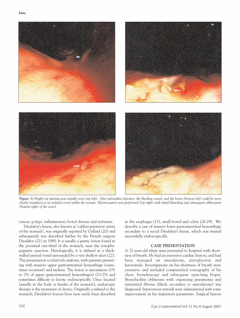
Figure 1) Bright red spurting was initially seen (top left). After adrenaline injection, the bleeding ceased, and the lesion (bottom left) could be moreclearly visualized as an isolated vessel within the rectum. Electrocautery was performed (top right) with initial blanching and subsequent obliteration(bottom right) of the vessel
enns-dieu.qxd 26/01/2007 1:58 PM Page 542
included a sigmoid colon resection for diverticular disease.His creatinine was also noted to be elevated to 205 µmol/L(normal less than 100 µmol/L), and a renal biopsy did notdemonstrate any evidence of vasculitis. Three weeks afteradmission, passage of bloody stools was noted. His hemo-globin decreased from 104 g/L to 92 g/L, but he did notdevelop any orthostatic changes. Although he was trans-fused with 2 U of blood that evening, his hemoglobin fellfurther the following day to 84 g/L. Although he had noupper gastrointestinal symptoms, an upper endoscopy wasperformed, which demonstrated superficial esophagealulcers. These were biopsied and proved to be herpes simplexesophagitis. Because no blood was visualized in the upperintestinal tract, a colonoscopy was performed immediatelyafter the upper endoscopy. This demonstrated dark bloodthroughout the colon (with the surgical anastamosis visibleat 20 cm from the anal verge) but no evidence of activebleeding. There was no blood within the terminal ileum.
Because there was no evidence of active bleeding, thepatient was managed supportively. The following day, rec-tal bleeding recurred; this time it appeared bright red incolour. A total of 8 U of packed red blood cells were trans-fused. A repeat unprepped colonoscopy was performed.Initially, visualization was challenging within the rectum,because bright red blood coated the entire region up to thesurgical anastamosis. Careful irrigation showed whatappeared to be a ‘spurting’ site of bleeding 5 cm distal tothe anastamosis (Figure 1, Top left). Three millilitres of1/10,000 adrenaline were injected into the bleedingregion, with ‘blanching’ of the mucosa and subsequent ces-sation of bleeding. The site was then localized and deter-mined to be Dieulafoy’s lesion of the rectum (Figure 1,Bottom left). Using a 10 French BICAP probe (bipolarprobe, Circon ACMI Corporation, USA) electrocoagula-tion (20 J, 5 s intervals, five applications), the lesion was
cauterized (Figure 1, Top right, Bottom right). Over thenext 12 months, there was no recurrence of bleeding.
DISCUSSION‘Exulceratio simplex’ was a term coined by Dieulafoy (21)to describe a superficial gastric mucosal lesion that hebelieved to be the initial stages of a gastric ulcer whose pro-gression was interrupted by the occurrence of bleeding.The ‘Dieulafoy’ lesion has now, however, been character-ized histologically as an unusually large artery coursing justbeneath the gastric mucosa (22). Once thought to be a rarecause of gastrointestinal hemorrhage, the widespread use ofemergency endoscopy has led to increasing numbers ofreports of this lesion in various parts of the gastrointestinaltract. Although usually considered an acquired abnormal-ity, a congenital etiology has been suggested by authorswho have discovered the lesion in patients as young as 20weeks old (30). The most common site of these lesionsremains the stomach, with most lesions located in the body(67%) and a smaller number (25%) in the fundus of thestomach (25).
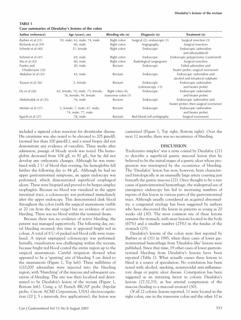
Dieulafoy’s lesions of the colon were first reported byBarbier et al (31) in 1985, when three cases of lower gas-trointestinal hemorrhage from ‘Dieulafoy-like’ lesions werepublished. Since that time, 19 other cases of lower gastroin-testinal bleeding from Dieulafoy’s lesions have beenreported (Table 1). What actually causes these lesions tobleed is a source of speculation. No correlation has beennoted with alcohol, smoking, nonsteroidal anti-inflamma-tory drugs or peptic ulcer disease. Constipation has beensuggested as an initiating factor in colonic Dieulafoy’slesions (27,32,33), as has arterial compression of themucosa (leading to a mucosal erosion) (30).
Of all 22 colonic lesions reported, 11 were located in theright colon, one in the transverse colon and the other 10 in
Dieulafoy’s lesions of the rectum
Can J Gastroenterol Vol 15 No 8 August 2001 543
TABLE 1Case summaries of Dieulafoy’s lesions of the colon
Author (reference) Age (years), sex Bleeding site (n) Diagnosis (n) Treatment (n)
Barbier et al (31) 59, male; 63, male; 74, male Right colon Surgical (2); endoscopic (1) Surgical resection (3)Richards et al (39) 60, male Right colon Angiography Surgical resectionSchmitt et al (40) 21, female Right colon Endoscopic Endoscopic (adrenaline
and athoxysklerol)Schmid et al (41) 69, male Right colon Endoscopic Endoscopic polypectomy (cauterized)Ma et al (42) 60, male Right colon Radiological (angiogram) Surgical resectionFranko and 20, male Rectum Endoscopic Failed adrenaline and
Chadavoyne (33) heater probe; surgical (oversewn)Abdulian et al (32) 43, male Rectum Endoscopic Endoscopic (adrenaline and
alcohol and tetradocyl sulphate)Tooson et al (36) 5, female Rectum Endoscopic Endoscopic (adrenaline
(colonoscopy ×3) and heater probe)Dy et al (26) 65, female; 70, male; 73, female; Right colon (4); Endoscopic Endoscopic (adrenaline
76, female; 94, female transverse colon (1) and heater probe)Abdelmalek et al (35) 76, male Rectum Endoscopic Endoscopic (adrenaline and
heater probe), then surgical (oversewn)Meister et al (27) 5, female; 7, male; 67, male; Rectum Endoscopic Endoscopic (adrenaline
74, male; 77, male and heater probe)Eguchi et al (37) 78, male Rectum Red blood cell scintigraphy Surgical (oversewn)
enns-dieu.qxd 26/01/2007 1:58 PM Page 543
the rectum. Seventeen of the 22 lesions were diagnosed byendoscopic means. Similar to Dieulafoy’s lesions of thestomach, approximately twice as many men were affected aswomen (15 versus seven) (34). The mean age was 58 yearsold. Eight lesions (three rectal, five right colon) were treatedsurgically (three oversewn, five right hemicolectomies). Theother 14 lesions were treated endoscopically with a combi-nation of adrenaline and heater probe (n=7); heater probealone (n=3); adrenaline and yttrium-argon-garnet laser(n=1); adrenaline and a sclerosing agent (n=2); and snarepolypectomy (n=1). Two patients treated endoscopicallysubsequently had recurrent bleeding. One had been treatedwith adrenaline injection, and a repeat endoscopic treat-ment with a sclerosing agent (alcohol and sodium tetradocylsulphate) successfully stopped the bleeding (32). Anotherhad a lesion at the hepatic flexure treated initially withheater probe coagulation and adrenaline. Repeat therapywith the same modalities was successful when he rebledthree days after the initial treatment (26). One of the lesionstreated surgically (oversewn) had previously failed endo-scopic therapy (33). The other two rectal lesions weretreated surgically (despite successful endoscopic treatmentin one) (35), presumably on the assumption that endoscopictherapy would not result in long term success in the cessa-tion of bleeding.
This case of a Dieulafoy’s lesion is the 11th discovered inthe rectum. All presented with hematochezia. Only twowere female (both five years old) (36). Overall, 10 of 11patients with rectal Dieulafoy’s lesions were diagnosedendoscopically (one by nuclear scintigraphy). Threepatients subsequently had surgical oversewing performed,but only one of the surgical cases had failed endoscopicmanagement (33). In another, surgical oversewing was per-formed intraoperatively when the lesion was discoveredwithin the rectum (37). The case presented is the only onetreated with bipolar electrocauterization. It is possible thatthe previous colonic surgery that our patient underwentmay have altered blood flow patterns and, theoretically,made a vulnerable vessel more likely to bleed. However,because other patients with Dieulafoy’s lesions of the rec-tum had not had prior surgical intervention of the colon,this theory of etiology is unsupported.
With the advent of therapeutic endoscopy, the manage-ment of gastric Dieulafoy’s lesions has been altered drasti-cally. In as late as 1986, surgical therapy was considered tobe the treatment of choice (34). A host of endoscopicmodalites (heater probe, injection sclerotherapy, monopo-lar electrocoagulation, bipolar electrocoagulation, band lig-ation, hemoclips [38] and laser photocoagulation) havebeen used successfully in the therapy of these lesions.Approximately 85% of patients managed endoscopicallyhave not had any evidence of rebleeding (25). This clearlyhas defined endoscopic management as the primary modal-ity of therapy in the stomach. Within the colon, however,the diagnosis can be even more challenging. Bright redblood may limit visibility and, as in several cases listedabove, has led to ‘blind’ hemicolectomies. Ideally, the site
of bleeding should be localized, and, if possible, endoscopictherapy should be initiated, because in most patients, it issuccessful. It appears that multiple endoscopic therapies canbe successful in treating Dieulafoy’s lesions in the colon;surgery should be reserved for those who fail endoscopictherapy. These cases further show that long term successwith endoscopic treatment is also expected and that surgi-cal intervention is usually not required.
Enns
Can J Gastroenterol Vol 15 No 8 August 2001544
REFERENCES1. Boley SJ, Sammartano R, Adams A, et al. On the nature and etiology
of vascular ectasias of the colon. Degenerative lesions of aging.Gastroenterology 1977;72:650-60.
2. Berner JS, Mauer K, Lewis BS. Push and sonde enteroscopy for thediagnosis of obscure gastrointestinal bleeding. Am J Gastroenterol1994;89:2139-42.
3. Howard TJ, Plaskon LA, Wiebke EA, et al. Nonocclusive mesentericischemia remains a diagnostic dilemma. Am J Surg 1996;171:405-8.
4. Lewis BS, Kornbluth A, Waye JD. Small bowel tumours: yield ofenteroscopy. Gut 1991;32:763-5.
5. Milewski PJ, Schofield PF. Massive colonic haemorrhage – the case forright hemicolectomy. Ann R Coll Surg Engl 1989;71:253-9.
6. Vernava AM, Moore BA, Longo, et al. Lower gastrointestinalbleeding. Dis Colon Rectum 1997;40:846-58.
7. Vernava AM, Longo WE, Virgo KS, et al. A nationwide study of theincidence and etiology of lower gastrointestinal bleeding. Surg ResCommun 1996;18:113-20.
8. Jensen DM, Machicado GA. Diagnosis and treatment of severehematochezia. The role of urgent colonoscopy after purge.Gastroenterology 1988;95:1569-74.
9. Rosen AM, Fleischer DE. Upper GI bleeding in the elderly: diagnosisand management. Geriatrics 1933;44:26-8.
10. Caos A, Benner KG, Manier J, et al. Colonoscopy after Golytelypreparation in acute rectal bleeding. J Clin Gastroenterol 1986;8:46-9.
11. Heer M, Ammann R, Buhler H. [Clinical significance of colonicangiodysplasias]. Schweiz Med Wochenschr 1984;114:1416-22.
12. Leitman IM, Paull DE, Shires GT. Evaluation and management ofmassive lower gastrointestinal hemorrhage. Ann Surg 1989;209:175-80.
13. Rossini FP, Ferrari A, Spandre M, et al. Emergency colonoscopy.World J Surg 1989;13:190-2.
14. Boley SJ, DiBiase A, Brandt LJ, Sammartano RJ. Lower intestinalbleeding in the elderly. Am J Surg 1979;137:57-64.
15. Wright HK. Massive colonic hemorrhage. Surg Clin North Am1980;60:1297-304.
16. Baum S, Athanasoulis CA, Waltman AC. Angiographic diagnosis andcontrol of large-bowel bleeding. Dis Colon Rectum 1974;17:447-53.
17. Colacchio TA, Forde KA, Patsos TJ, et al. Impact of moderndiagnostic methods on the management of active rectal bleeding. Ten year experience. Am J Surg 1982;143:607-10.
18. Forde KA. Colonoscopy in acute rectal bleeding. Gastrointest Endosc1981;27:219-20.
19. Jensen DM, Machicado GA. Colonoscopy for diagnosis and treatmentof severe lower gastrointestinal bleeding. Routine outcomes and costanalysis. Gastrointest Endosc Clin North Am 1997;7:477-98.
20. Gallard T. Aneurysmes miliaires de l’estomac donnant lieu a deshematemeses montelles. Bull Soc Med Paris 1884;1:84-91.
21. Dieulafoy G. Exulceratio simplex. L’intervention chirurgicale dans leshematemeses foudroyantes consecutives a l’exulceration simplex del’estomac. Bull Acad Med 1889;39:49-84.
22. Juler GL, Labitzke HG, Lamb R, et al. The pathogenesis of Dieulafoy’sgastric erosion. Am J Gastroenterol 1984;79:195-200.
23. Jaspersen D, Korner T, Schorr W, et al. Extragastric Dieulafoy’s diseaseas unusual source of intestinal bleeding. Esophageal visible vessel. Dig Dis Sci 1994;39:2558-60.
24. Pointner R, Schwab G, Konigsrainer A, et al. Endoscopic treatment ofDieulafoy’s disease. Gastroenterology 1988;94:563-6.
25. Reilly HF, al-Kawas FH. Dieulafoy’s lesion. Diagnosis andmanagement. Dig Dis Sci 1991;36:1702-7.
26. Dy NM, Gostout CJ, Balm RK. Bleeding from the endoscopically-identified Dieulafoy lesion of the proximal small intestine and colon.Am J Gastroenterol 1995;90:108-11.
enns-dieu.qxd 26/01/2007 1:58 PM Page 544
Dieulafoy’s lesions of the rectum
Can J Gastroenterol Vol 15 No 8 August 2001 545
27. Meister T, Varilek G, Marsanao, et al. Endoscopic management ofrectal Dieulafoy-like lesions: A case series and review of the literature.Gastrointest Endosc 1998;48:302-4.
28. Gadenstatter M, Wetscher G, Crookes PF, et al. Dieulafoy’s disease ofthe large and small bowel. J Clin Gastroenterol 1998;27:169-72.
29. Goins WA, Chatman DM, Kaviani MJ. Massive lower gastrointestinalbleeding due to ‘Dieulafoy’s vascular malformation’ of the jejunum:case report. J Natl Med Assoc 1995;87:766-70.
30. Rossi NP, Green EW, Pike JD. Massive bleeding of the uppergastrointestinal tract due to Dieulafoy’s erosion. Arch Surg1968;97:797-800.
31. Barbier P, Luder P, Triller J, et al. Colonic hemorrhage from a solitaryminute ulcer. Report of three cases. Gastroenterology 1985;88:1065-8.
32. Abdulian JD, Santoro MJ, Chen YK, Collen MJ. Dieulafoy-like lesionof the rectum presenting with exsanguinating hemorrhage: successfulendoscopic sclerotherapy. Am J Gastroenterol 1993;88:1939-41.
33. Franko E, Chadavoyne R. Massive rectal bleeding from a Dieulafoy’stype ulcer of the rectum: a review of this unusual disease. Am J Gastroenterol 1991;88:1939-41.
34. Veldhuyzen ZS, Bartelsman JF, Schipper ME, Tytgat GN. Recurrent massive haematemesis from Dieulafoy vascularmalformations – a review of 101 cases. Gut 1986;27:213-22.
35. Abdelmalek MF, Pockaj BA, Leighton JA. Rectal bleeding from amucous fistula secondary to a Dieulafoy’s lesion. J Clin Gastroenterol1997;24:259-61.
36. Tooson JD, Marsano LS, Gates LK Jr. Pediatric rectal Dieulafoy’slesion. Am J Gastroenterol 1995;90:2232-3.
37. Eguchi S, Maeda J, Taguchi H, et al. Massive gastrointestinal bleedingfrom a Dieulafoy-like lesion of the rectum. J Clin Gastroenterol 1997;24:262-3.
38. Parra-Blanco A, Takahashi H, Mendez JP, et al. Endoscopicmanagement of Dieulafoy lesions of the stomach: a case study of 26 patients. Endoscopy 1997;29:834-9.
39. Richards WO, Grove-Mahoney D, Williams LF. Hemorrhage from a Dieulafoy type ulcer of the colon: a new cause of lowergastrointestinal bleeding. Am Surg 1988;54:121-4.
40. Schmitt W, Lux G, Giedl J. Colonic haemorrhage from solitarysubmucosal vessels diagnosed by lower gastrointestinal Doppler-endoscopy. Endoscopy 1987;19:43-5.
41. Schmid K, Pointner R, Feichtinger J. Exulceratio simplex Dieulafoy ofthe colon – A case report. Endoscopy 1988;20:88-9.
42. Ma CK, Padda H, Pace EH, Szilagyi E. Submucosal arterialmalformation of the colon with massive hemorrhage. Report of a case.Dis Colon Rectum 1989;32:149-52.
enns-dieu.qxd 26/01/2007 1:58 PM Page 545

Submit your manuscripts athttp://www.hindawi.com
Stem CellsInternational
Hindawi Publishing Corporationhttp://www.hindawi.com Volume 2014
Hindawi Publishing Corporationhttp://www.hindawi.com Volume 2014
MEDIATORSINFLAMMATION
of
Hindawi Publishing Corporationhttp://www.hindawi.com Volume 2014
Behavioural Neurology
EndocrinologyInternational Journal of
Hindawi Publishing Corporationhttp://www.hindawi.com Volume 2014
Hindawi Publishing Corporationhttp://www.hindawi.com Volume 2014
Disease Markers
Hindawi Publishing Corporationhttp://www.hindawi.com Volume 2014
BioMed Research International
OncologyJournal of
Hindawi Publishing Corporationhttp://www.hindawi.com Volume 2014
Hindawi Publishing Corporationhttp://www.hindawi.com Volume 2014
Oxidative Medicine and Cellular Longevity
Hindawi Publishing Corporationhttp://www.hindawi.com Volume 2014
PPAR Research
The Scientific World JournalHindawi Publishing Corporation http://www.hindawi.com Volume 2014
Immunology ResearchHindawi Publishing Corporationhttp://www.hindawi.com Volume 2014
Journal of
ObesityJournal of
Hindawi Publishing Corporationhttp://www.hindawi.com Volume 2014
Hindawi Publishing Corporationhttp://www.hindawi.com Volume 2014
Computational and Mathematical Methods in Medicine
OphthalmologyJournal of
Hindawi Publishing Corporationhttp://www.hindawi.com Volume 2014
Diabetes ResearchJournal of
Hindawi Publishing Corporationhttp://www.hindawi.com Volume 2014
Hindawi Publishing Corporationhttp://www.hindawi.com Volume 2014
Research and TreatmentAIDS
Hindawi Publishing Corporationhttp://www.hindawi.com Volume 2014
Gastroenterology Research and Practice
Hindawi Publishing Corporationhttp://www.hindawi.com Volume 2014
Parkinson’s Disease
Evidence-Based Complementary and Alternative Medicine
Volume 2014Hindawi Publishing Corporationhttp://www.hindawi.com