different metastasis promotive potency of small g-proteins rala
TRANSCRIPT
PRIMARY RESEARCH Open Access
Different metastasis promotive potency of smallG-proteins RalA and RalB in in vivo hamstertumor modelVera A Rybko, Anna V Knizhnik, Andrei V Komelkov, Vasily N Aushev, Lyubov S Trukhanova andElena M Tchevkina*
Abstract
Background: Previously we have shown that oncogenic Ha-Ras stimulated in vivo metastasis through RalGEF-Ralsignaling. RalA and RalB are highly homologous small G proteins belonging to Ras superfamily. They can beactivated by Ras-RalGEF signaling pathway and influence cellular growth and survival, motility, vesicular transportand tumor progression in humans and in animal models. Here we first time compared the influence of RalA andRalB on tumorigenic, invasive and metastatic properties of RSV transformed hamster fibroblasts.
Methods: Retroviral vectors encoding activated forms or effector mutants of RalA or RalB proteins were introducedinto the low metastatic HET-SR cell line. Tumor growth and spontaneous metastatic activity (SMA) were evaluatedon immunocompetent hamsters after subcutaneous injection of cells. The biological properties of cells, includingproliferation, clonogenicity, migration and invasion were determined using MTT, wound healing, colony formationand Boyden chamber assays respectively. Protein expression and phosphorylation was detected by Westen blotanalysis. Extracellular proteinases activity was assessed by substrate-specific zymography.
Results: We have showed that although both Ral proteins stimulated SMA, RalB was more effective in metastasisstimulation in vivo as well as in potentiating of directed movement and invasion in vitro. Simultaneous expressionof active RalA and RalB didn’t give synergetic effect on metastasis formation. RalB activity decreased expression ofCaveolin-1, while active RalA stimulated MMP-1 and uPA proteolytic activity, as well as CD24 expression. Both Ralproteins were capable of Cyclin D1 upregulation, JNK1 kinase activation, and stimulation of colony growth andmotility. Among three main RalB effectors (RalBP1, exocyst complex and PLD1), PLD1 was essential for RalB-dependent metastasis stimulation.
Conclusions: Presented results are the first data on direct comparison of RalA and RalB impact as well as ofRalA/RalB simultaneous expression influence on in vivo cell metastatic activity. We showed that RalB activationsignificantly more than RalA stimulates SMA. This property correlates with the ability of RalB to stimulate in vitroinvasion and serum directed cell movement. We also found that RalB-PLD1 interaction is necessary for theacquisition of RalB-dependent high metastatic cell phenotype. These findings contribute to the identification ofmolecular mechanisms of metastasis and tumor progression.
Keywords: metastasis, Ral proteins, invasion, Ral effector mutants, tumor growth
* Correspondence: [email protected] of Oncogenes Regulation, Institute of Carcinogenesis, Russian N.N. Blokhin Cancer Research Center, Kashirskoye shosse 24, 115478, Moscow,Russia
Rybko et al. Cancer Cell International 2011, 11:22http://www.cancerci.com/content/11/1/22
© 2011 Rybko et al; licensee BioMed Central Ltd. This is an Open Access article distributed under the terms of the Creative CommonsAttribution License (http://creativecommons.org/licenses/by/2.0), which permits unrestricted use, distribution, and reproduction inany medium, provided the original work is properly cited.
BackgroundMetastatic spread of primary tumors is a major determi-nant of cancer-related death. Metastatic process involvesmultiple steps including local tumor cells dissemination,survival in blood circulation, arrest in vasculature, extra-vasation and growth in distant organs and tissues [1].Investigation of signaling pathways regulating metastasisand associated gene expression changes is an importantstep for designing therapeutic strategies.Small G proteins RalA and RalB belong to Ras super-
family [2] and are implicated in tumorigenesis, invasionand metastasis [3-7]. RalA and RalB share 82% aminoacid identity [8] and participate in numerous cellularprocesses such as endocytosis, exocytosis, actin reorga-nization and cell motility, proliferation and modulationof cancer-associated genes expression (for review see[9]). Like all GTPases Ral proteins cycle between activeGTP- and inactive GDP-bound states. Ral can bind toand regulate activity of various proteins including Ralbinding protein-1 (RalBP1, RLIP76) [10], phospholipaseD1 (PLD1) [11], filamin A [12], exocyst subunits Sec5and Exo84 [13]. Although RalA and RalB have almostidentical effector-binding domains, these two proteinsmay preferentially utilize different effectors [14].Ral proteins are activated by RalGEFs, some of which
are Ras effectors (i.e. RalGDS, Rgl1, Rgl2). OncogenicRas mutations were found in subset of human tumorsand cell lines. RalA and RalB were shown to be activatedin pancreatic cancers, aggressive malignancies with highfrequency of Ras mutations [15]. It was shown that RalAand RalB were necessary for acquisition of aggressivecellular phenotype in diverse models of tumor progres-sion. Ral proteins were capable to stimulate prostatecancer metastasis to bone. Suppression of RalB activityled to decrease of oncogenic Ras-mediated invasion invitro and reduced metastasis after prostate cancer cellsintracardiac injection [16]. Ral GTPases also mediatedprogression of bladder cancer in animal models [17].However, little emphasis has been made on comparisonof individual roles of RalA and RalB and their down-stream partners in tumor progression.Here we compared the influence of constitutively
active RalA and RalB expression on tumor progression.We showed that both active Ral proteins enhancedspontaneous metastatic activity (SMA) of HET-SR cells,however RalB was more potent in stimulation of lungcolonization, as well as in promotion of cell invasionand directed migration. SMA stimulating effect dependson N-terminus of RalB protein, which is known to becritical for RalB-PLD1 interaction. We also found RalA-dependent increase of extracellular matrix proteinasesactivity and RalA/B mediated metastasis-associated sig-naling cascades stimulation.
ResultsRalB is more potent in metastasis stimulation than RalAPreviously we have shown that introduction of oncogenicHa-Ras stimulated spontaneous metastatic activity ofRSV-transformed hamster embryo fibroblasts (HET-SRcell line) through activation of RalGDS signaling pathway.Overexpression of active RalA also enhanced lung metas-tasis formation in immunocompetent hamsters [18].Here we compared the ability of RalA and RalB to influ-
ence metastatic potential and associated properties oftumorigenic low-metastatic HET-SR cell line. For this pur-pose we generated stable cell lines HET-SR-RalA andHET-SR-RalB expressing active GTP-bound forms of Ralproteins (RalA G23V and RalB G23V) in retroviral vectors(pLXSN and pBabe-puro, respectively). Expression of exo-genous Ral proteins was confirmed by Western blot analy-sis of total cell populations selected on G-418 (HET-SR-RalA) or puromycin (HET-SR-RalB) (Figure 1a).For evaluation of SMA, 104 cells of HET-SR-RalA or
HET-SR-RalB in parallel with cell lines expressingempty vectors were subcutaneously injected into 10immunocompetent syngeneic animals. Two monthslater tumor-bearing hamsters were sacrificed; paraffin-embedded lungs were step-sectioned and stained withhematoxylin-eosin (Figure 1b). Metastatic lesions inlungs were counted microscopically. We found statisti-cally significant increase in metastatic nodules numberper animal (in comparison with empty vector-expressingcells) for both cell lines (Figure 1c). RalB-dependent sti-mulation of SMA was noticeably higher: cells expressingactive RalB formed 63.7 ± 19, while HET-SR-RalAformed 20 ± 4 lung nodules per animal in comparisonwith 7 ± 1.9 and 4 ± 2 nodules formed by cell linesbearing empty vectors, HET-SR-pBabe and HET-SR-pLXSN respectively. Therefore, RalB effect on metas-tases stimulation was 3 times more than RalA.
Simultaneous expression of active Ral proteins has noadditive effect on metastasisIt was previously shown that RalA and RalB proteinsmay have non-overlapping or even opposite influenceon several cellular properties [4,6,19-21]. At the sametime there were no data concerning effect of simulta-neous expression of active RalA and RalB on metastasis.To test whether Ral proteins cooperate in SMA stimula-tion we introduced active RalB in retroviral vectorpBabe-puro (or pBabe-puro vector alone as a control)into HET-SR-RalA cells. The expression of RalB inderived HET-SR-RalA-RalB cells was confirmed by wes-tern blot analysis (Figure 1d). SMA assay revealed thatHET-SR-RalA-RalB cell line formed 16.2 ± 1.3 lungmetastatic nodules. This value was comparable to con-trol HET-SR-RalA-pBabe cells (12.3 ± 3.2) and
Rybko et al. Cancer Cell International 2011, 11:22http://www.cancerci.com/content/11/1/22
Page 2 of 12
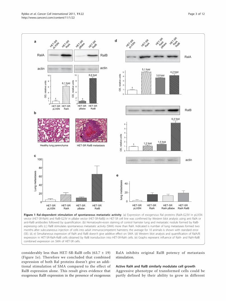
considerably less than HET-SR-RalB cells (63.7 ± 19)(Figure 1e). Therefore we concluded that combinedexpression of both Ral proteins doesn’t give an addi-tional stimulation of SMA compared to the effect ofRalB expression alone. This result gives evidence thatexogenous RalB expression in the presence of exogenous
RalA inhibits original RalB potency of metastasisstimulation.
Active RalA and RalB similarly modulate cell growthAggressive phenotype of transformed cells could bepartly defined by their ability to grow in different
a
RalA RalB
actin actin
HET-SR
pLXSN
HET-SR
RalA HET-SR
pBab
eHET-S
R
RalB
b
HET-SR RalB metastasisHealthy lung parenchyma
200 m
c
200 m
e
lung
met
asta
ses
HET-SR pLXSN
HET-SR RalA
HET-SR RalA pBabe
HET-SR RalA RalB
0
20
40
60
80
0
20
40
60
80
100
Lung
met
asta
ses
HET-SR pBabe
HET-SR pLXSN
HET-SR RalA
HET-SR RalB
0
2
4
6
8
10
OD
, rel
ativ
e un
its
HET-SR pLXSN
HET-SR RalA
0
2
4
6
8
10O
D, r
elat
ive
units
HET-SR pBabe
HET-SR RalB
11
6,1 fold
8,6 fold
11
actin
RalB
RalA
dHET-S
R
pLXSN
HET-SR
RalA HET-SR
RalA
pBab
e HET-SR
RalA
RalB
0
1
2
3
OD
, rel
ativ
e un
its
1
5,1 fold
3,8 fold4,2 fold
0
1
2
3
OD
, rel
ativ
e un
its
1 1,2 fold 1,5 fold
6,4 fold
4
5
4
5
6
Figure 1 Ral-dependent stimulation of spontaneous metastatic activity. (a) Expression of exogenous Ral proteins (RalA-G23V in pLXSNvector (HET-SR-RalA) and RalB-G23V in pBabe vector (HET-SR-RalB)) in HET-SR cell line was confirmed by Western blot analysis using anti RalA oranti-RalB antibodies followed by quantification. (b) Hematoxylin-eosin staining of control hamster lung and metastatic nodule formed by RalB-expressing cells (c.) RalB stimulates spontaneous metastatic activity (SMA) more than RalA. Indicated is number of lung metastases formed twomonths after subcutaneous injection of cells into adult immunocompetent hamsters; the average for 10 animals is shown with standard error(SE). (d, e) Simultaneous expression of RalA and RalB doesn’t give additive effect on SMA. (d) Western blot analysis and quantification of RalA/Bexpression in HET-SR-RalA-RalB cells obtained by RalB transduction into HET-SR-RalA cells. (e) Graphs represent influence of RalA- and RalA-RalBcombined expression on SMA of HET-SR cells.
Rybko et al. Cancer Cell International 2011, 11:22http://www.cancerci.com/content/11/1/22
Page 3 of 12
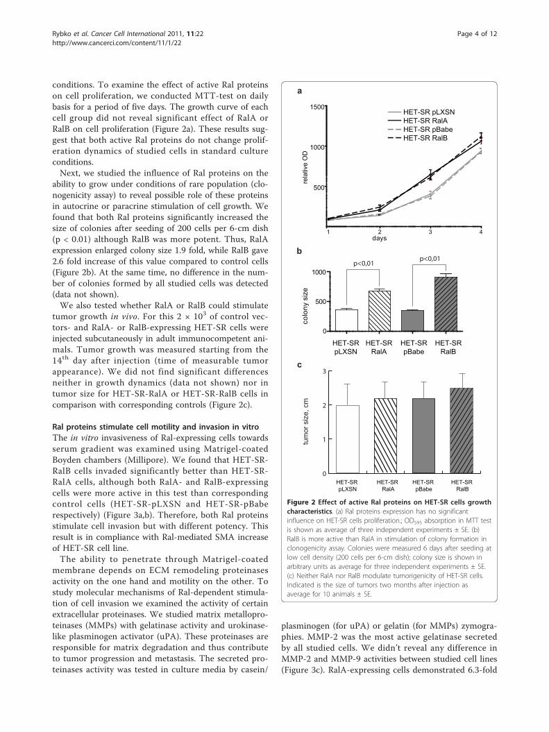
conditions. To examine the effect of active Ral proteinson cell proliferation, we conducted MTT-test on dailybasis for a period of five days. The growth curve of eachcell group did not reveal significant effect of RalA orRalB on cell proliferation (Figure 2a). These results sug-gest that both active Ral proteins do not change prolif-eration dynamics of studied cells in standard cultureconditions.Next, we studied the influence of Ral proteins on the
ability to grow under conditions of rare population (clo-nogenicity assay) to reveal possible role of these proteinsin autocrine or paracrine stimulation of cell growth. Wefound that both Ral proteins significantly increased thesize of colonies after seeding of 200 cells per 6-cm dish(p < 0.01) although RalB was more potent. Thus, RalAexpression enlarged colony size 1.9 fold, while RalB gave2.6 fold increase of this value compared to control cells(Figure 2b). At the same time, no difference in the num-ber of colonies formed by all studied cells was detected(data not shown).We also tested whether RalA or RalB could stimulate
tumor growth in vivo. For this 2 × 10³ of control vec-tors- and RalA- or RalB-expressing HET-SR cells wereinjected subcutaneously in adult immunocompetent ani-mals. Tumor growth was measured starting from the14th day after injection (time of measurable tumorappearance). We did not find significant differencesneither in growth dynamics (data not shown) nor intumor size for HET-SR-RalA or HET-SR-RalB cells incomparison with corresponding controls (Figure 2c).
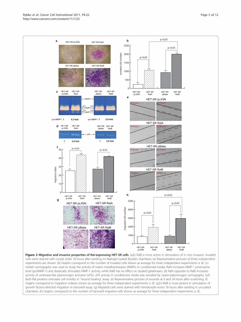
Ral proteins stimulate cell motility and invasion in vitroThe in vitro invasiveness of Ral-expressing cells towardsserum gradient was examined using Matrigel-coatedBoyden chambers (Millipore). We found that HET-SR-RalB cells invaded significantly better than HET-SR-RalA cells, although both RalA- and RalB-expressingcells were more active in this test than correspondingcontrol cells (HET-SR-pLXSN and HET-SR-pBaberespectively) (Figure 3a,b). Therefore, both Ral proteinsstimulate cell invasion but with different potency. Thisresult is in compliance with Ral-mediated SMA increaseof HET-SR cell line.The ability to penetrate through Matrigel-coated
membrane depends on ECM remodeling proteinasesactivity on the one hand and motility on the other. Tostudy molecular mechanisms of Ral-dependent stimula-tion of cell invasion we examined the activity of certainextracellular proteinases. We studied matrix metallopro-teinases (MMPs) with gelatinase activity and urokinase-like plasminogen activator (uPA). These proteinases areresponsible for matrix degradation and thus contributeto tumor progression and metastasis. The secreted pro-teinases activity was tested in culture media by casein/
plasminogen (for uPA) or gelatin (for MMPs) zymogra-phies. MMP-2 was the most active gelatinase secretedby all studied cells. We didn’t reveal any difference inMMP-2 and MMP-9 activities between studied cell lines(Figure 3c). RalA-expressing cells demonstrated 6.3-fold
a
21 3 4
500
1000
1500HET-SR pLXSNHET-SR RalAHET-SR pBabeHET-SR RalB
days
rela
tive
OD
b
HET-SRpLXSN
HET-SRpBabe
HET-SRRalA
HET-SRRalB
0
500
1000
colo
ny s
ize
p<0,01p<0,01
c
HET-SR pBabe
HET-SR pLXSN
HET-SR RalA
HET-SR RalB
0
1
2
3
tum
or s
ize,
cm
Figure 2 Effect of active Ral proteins on HET-SR cells growthcharacteristics. (a) Ral proteins expression has no significantinfluence on HET-SR cells proliferation.; OD595 absorption in MTT testis shown as average of three independent experiments ± SE. (b)RalB is more active than RalA in stimulation of colony formation inclonogenicity assay. Colonies were measured 6 days after seeding atlow cell density (200 cells per 6-cm dish); colony size is shown inarbitrary units as average for three independent experiments ± SE.(c) Neither RalA nor RalB modulate tumorigenicity of HET-SR cells.Indicated is the size of tumors two months after injection asaverage for 10 animals ± SE.
Rybko et al. Cancer Cell International 2011, 11:22http://www.cancerci.com/content/11/1/22
Page 4 of 12
HET-SR pBabe
HET-SR pLXSN HET-SR RalA
HET-SR RalB
a
e
HET-SR pBabe
HET-SR pLXSN0 hours 24 hours
0 hours 24 hours
HET-SR RalA
HET-SR RalB
0 hours 24 hours
0 hours 24 hours
p<0,05p<0,05f
HET-SR pBabe
HET-SR pLXSN
HET-SR RalA
HET-SR RalB
0
10
20
30
40
50
mig
ratio
n in
dex
HET-SR pLXSN
HET-SR pBabe
HET-SR RalA
HET-SR RalB
g
HET-SR pLXSN
HET-SR pBabe
HET-SR RalA
HET-SR RalB
c
MMP9
MMP2
MMP1
HET-SR pLXSN
HET-SR RalA
HET-SR pBabe
HET-SR RalB
d
uPA
p<0,05
p<0,05
b
HET-SR pBabe
HET-SR pLXSN
HET-SR RalA
HET-SR RalB
0
500
1000
1500
2000
2500
inva
ded
cell
num
ber
p<0,05
p<0,05
p<0,05
h
HET-SR pBabe
HET-SR pLXSN
HET-SR RalA
HET-SR RalB
0
1000
2000
3000
4000
5000
mig
rate
d ce
ll nu
mbe
r
p<0,05
1 2,8 fold 1 0,9 fold
1 6,3 fold 1 0,9 foldpro-MMP1: pro-MMP1:
pro pro
pro pro
Figure 3 Migrative and invasive properties of Ral-expressing HET-SR cells. (a,b) RalB is more active in stimulation of in vitro invasion. Invadedcells were stained with crystal violet 18 hours after seeding on Matrigel-coated Boyden chambers; (a) Representative pictures of three independentexperiments are shown. (b) Graphs correspond to the number of invaded cells shown as average for three independent experiments ± SE. (c)Gelatin zymography was used to study the activity of matrix metalloproteases (MMPs) in conditioned media. RalA increases MMP-1 proenzymelevel (proMMP-1) and drastically stimulates MMP-1 activity, while RalB has no effect on studied gelatinases. (d) RalA opposite to RalB increasesactivity of urokinase-like plasminogen activator (uPA). uPA activity in conditioned media was revealed by casein-plasminogen zymography. (e,f)Both Ral proteins stimulate cell motility in “wound healing” assay. (e) Representative pictures of wounds at 0 and 24 hours after scratching. (f)Graphs correspond to migration indexes shown as average for three independent experiments ± SE. (g,h) RalB is more potent in stimulation ofgrowth factors-directed migration in transwell assay. (g) Migrated cells were stained with hematoxylin-eosin 18 hours after seeding in uncoatedchambers. (h) Graphs correspond to the number of transwell migrated cells shown as average for three independent experiments ± SE.
Rybko et al. Cancer Cell International 2011, 11:22http://www.cancerci.com/content/11/1/22
Page 5 of 12
increase of MMP-1 proenzyme level compared to thecontrol HET-SR-pLXSN cells. In contrast, MMP-1 activ-ity in HET-SR-RalB cells remained at the same level asin the control HET-SR-pBabe. Moreover, active form ofMMP-1 was detected only in RalA expressing cells.Comparison of uPA activity in conditioned media alsorevealed its significant (2.8-fold) increase in RalA-expressing cells (Figure 3d). Therefore, RalB-associatedincrease of in vitro invasion is unlikely to be caused bydifferences in studied ECM proteinases activity.We also tested whether Ral-mediated changes in cell
motility could contribute to invasion stimulation. Cellswere subjected to in vitro “wound healing” assay. Asshown in Figures 3e,f, expression of active RalA andRalB lead to similar increase in “wound healing” effi-ciency (migration indexes for RalA and RalB were 44%and 42%, whereas migration indexes for controls were25% and 27% respectively).Results obtained on proteinases activity and wound
healing do not explain RalB-mediated increase of inva-siveness in Matrigel-coated chambers. This increasecould be determined by difference in efficiency ofserum-directed migration. We used migration throughuncoated porous inserts assay to check this possibility.We found that both RalA- and RalB-overexpressing cellsdemonstrated higher levels of transwell migration thancorresponding control cells. Moreover, RalB was signifi-cantly more active than RalA in promoting directedmigration (Figure 3g,h).Thereby, we conclude that the difference in Ral-
mediated stimulation of in vitro invasion definitely cor-related with SMA of studied cells and is most probablydetermined by the difference in their ability for chemo-tactic movement.
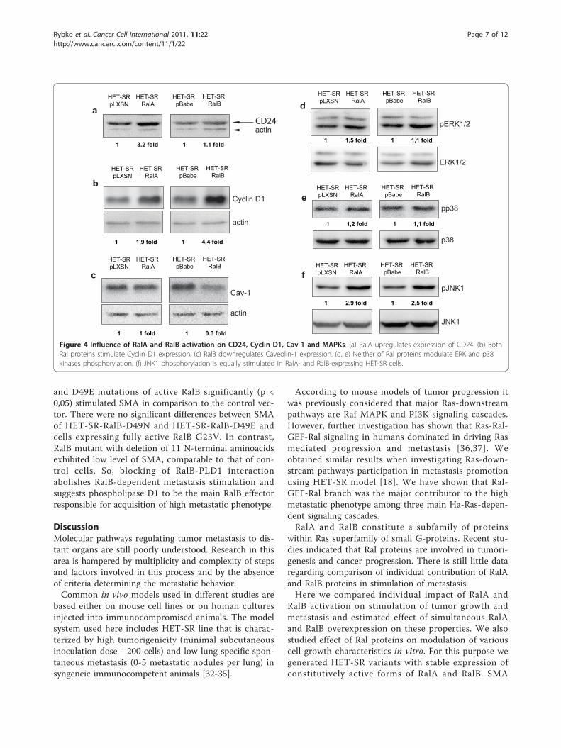
Ral proteins regulate CD24, Cav-1, Cyclin D1 and pJNK1protein expressionRal influence on intracellular signaling and proteinexpression has been intensively investigated. Here westudied some different branches of metastasis-associatedsignaling pathways, potentially regulated by Ral proteins(Figure 4).It was previously shown that RalA depletion downre-
gulated cell surface highly-glycosylated protein CD24expression in bladder cancer cell lines [22]. This mole-cule may regulate cell survival and proliferation as wellas tumor metastasis. CD24 has been proposed to be amarker of pancreatic cancer stem cells and may be asso-ciated with unfavorable prognosis [23]. To test whetherRal-mediated increase of SMA is associated withincrease of CD24 expression we estimated its level byWestern blotting. We revealed that overexpression ofactive RalA but not RalB upregulated CD24 expression(Figure 4a). This observation suggests that CD24 is
unlikely to be a marker of highly metastatic cell pheno-type in the studied experimental model.It was shown that RalB could increase the NF-�B-
dependent expression of Cyclin D1 [24]. Cyclin D1 func-tions as a well-known mitogenic mediator. Recent studiesrevealed that Cyclin D1 also acts as a motogen and pro-motes cell migration [25,26]. We tested Cyclin D1 levelin cells expressing active RalA and RalB and found thatboth Ral proteins stimulated expression of this protein incomparison to the control cells (Figure 4b). At the sametime RalB-dependent increase of CyclinD1 expressionwas 2.5 fold higher than in RalA expressing cells. There-fore, elevation of CyclinD1 level in both RalA and RalBexpressing cells correlates with stimulation of cell moti-lity. However, RalB influence on CyclinD1 is noticeablystronger than that of RalA, what correlates with morepronounced effect of RalB on SMA.Caveolin-1 (Cav-1) is a structural protein of caveolae,
a special type of lipid rafts that can modulate variousproteins activity e.g. Ras, Cyclin D1, Erk1/2, p38 andothers [27]. Cav-1 downregulation is associated withaggressiveness of certain tumors and cell lines and itwas previously shown that Cav-1 depletion downregu-lated RalA expression [28]. We found that overexpres-sion of active RalB (but not RalA) in HET-SR cells ledto more than 3 fold decrease of Cav-1 (Figure 4c).Therefore Cav-1 downregulation in studied model cor-relates with aggressive cell phenotype.We also studied the influence of Ral activation on
phosphorylation status of three main MAPK kinases:extracellular signal-regulated kinase (ERK1/2), c-Jun N-terminal kinase (JNK1), and p38 MAPK. We didn’t findsignificant changes neither in ERK1/2 nor in p38 kinasephosphorylation (Figure 4d,e). At the same time, JNK1was activated both in RalA- and RalB-overexpressingcells (Figure 4f).
Stimulation of metastasis by active RalB expressiondepends on RalB-PLD interactionIn order to study RalB-downstream signaling and toreveal RalB partners mainly responsible for metastasisstimulation, we tested the influence of RalB effectormutants on spontaneous metastatic activity of HET-SRcells. Sequences encoding three effector mutants ofactive RalB: D49N, D49E (effector loop mutants) andΔN11 (11 N-terminal amino-acids deleted) were clonedinto pBabe-puro retroviral vector and stably expressedin HET-SR cells. These three mutations impeded RalBinteraction with downstream partners RalBP1 [29], Sec5and Exo84 exocyst subunits [30] and PLD1 respectively[31]. Expression of RalB mutant proteins was confirmedby Western blot analysis of total cell lysates after selec-tion on puromycin (Figure 5a). In vivo analysis ofobtained cell lines (Figure 5b) revealed that both D49N
Rybko et al. Cancer Cell International 2011, 11:22http://www.cancerci.com/content/11/1/22
Page 6 of 12
and D49E mutations of active RalB significantly (p <0,05) stimulated SMA in comparison to the control vec-tor. There were no significant differences between SMAof HET-SR-RalB-D49N and HET-SR-RalB-D49E andcells expressing fully active RalB G23V. In contrast,RalB mutant with deletion of 11 N-terminal aminoacidsexhibited low level of SMA, comparable to that of con-trol cells. So, blocking of RalB-PLD1 interactionabolishes RalB-dependent metastasis stimulation andsuggests phospholipase D1 to be the main RalB effectorresponsible for acquisition of high metastatic phenotype.
DiscussionMolecular pathways regulating tumor metastasis to dis-tant organs are still poorly understood. Research in thisarea is hampered by multiplicity and complexity of stepsand factors involved in this process and by the absenceof criteria determining the metastatic behavior.Common in vivo models used in different studies are
based either on mouse cell lines or on human culturesinjected into immunocompromised animals. The modelsystem used here includes HET-SR line that is charac-terized by high tumorigenicity (minimal subcutaneousinoculation dose - 200 cells) and low lung specific spon-taneous metastasis (0-5 metastatic nodules per lung) insyngeneic immunocompetent animals [32-35].
According to mouse models of tumor progression itwas previously considered that major Ras-downstreampathways are Raf-MAPK and PI3K signaling cascades.However, further investigation has shown that Ras-Ral-GEF-Ral signaling in humans dominated in driving Rasmediated progression and metastasis [36,37]. Weobtained similar results when investigating Ras-down-stream pathways participation in metastasis promotionusing HET-SR model [18]. We have shown that Ral-GEF-Ral branch was the major contributor to the highmetastatic phenotype among three main Ha-Ras-depen-dent signaling cascades.RalA and RalB constitute a subfamily of proteins
within Ras superfamily of small G-proteins. Recent stu-dies indicated that Ral proteins are involved in tumori-genesis and cancer progression. There is still little dataregarding comparison of individual contribution of RalAand RalB proteins in stimulation of metastasis.Here we compared individual impact of RalA and
RalB activation on stimulation of tumor growth andmetastasis and estimated effect of simultaneous RalAand RalB overexpression on these properties. We alsostudied effect of Ral proteins on modulation of variouscell growth characteristics in vitro. For this purpose wegenerated HET-SR variants with stable expression ofconstitutively active forms of RalA and RalB. SMA
c
actin
Cav-1
fpJNK1
JNK1
d
pERK1/2
ERK1/2
epp38
p38
actin
aCD24
b
actin
Cyclin D1
HET-SRpLXSN
HET-SR RalA
HET-SR pBabe
HET-SR RalB
1 3,2 fold 1 1,1 fold
1 1,9 fold 1 4,4 fold
HET-SRpLXSN
HET-SR RalA
HET-SR pBabe
HET-SR RalB
HET-SRpLXSN
HET-SR RalA
HET-SR pBabe
HET-SR RalB
1 1 fold 1 0,3 fold
HET-SRpLXSN
HET-SR RalA
HET-SR pBabe
HET-SR RalB
HET-SRpLXSN
HET-SR RalA
HET-SR pBabe
HET-SR RalB
HET-SRpLXSN
HET-SR RalA
HET-SR pBabe
HET-SR RalB
1 1,5 fold 1 1,1 fold
1 1,2 fold 1 1,1 fold
1 2,9 fold 1 2,5 fold
Figure 4 Influence of RalA and RalB activation on CD24, Cyclin D1, Cav-1 and MAPKs. (a) RalA upregulates expression of CD24. (b) BothRal proteins stimulate Cyclin D1 expression. (c) RalB downregulates Caveolin-1 expression. (d, e) Neither of Ral proteins modulate ERK and p38kinases phosphorylation. (f) JNK1 phosphorylation is equally stimulated in RalA- and RalB-expressing HET-SR cells.
Rybko et al. Cancer Cell International 2011, 11:22http://www.cancerci.com/content/11/1/22
Page 7 of 12

analysis of Ral-expressing HET-SR cells after subcuta-neous injection revealed that both RalA and RalBincreased SMA but RalB was much more potent in sti-mulation of lung metastasis.These results are consistent with the idea that RalA
and RalB contribute to different aspects of tumorigen-esis. Earlier, it was suggested that RalA was essential foranchorage-independent growth of transformed cellswhile RalB was responsible for tumor cell-autonomoussurvival [6,14,19,20]. Data published later proposed themechanisms of RalB anti-apoptotic action through acti-vation of RalB/TBK1 signaling pathway [38,39]. More-over, Lim et al. showed that RalA knockdown reducedtumorigenic growth of transformed cells (pancreaticcancer cell lines) while RalB inhibition decreased inva-sion and experimental metastasis [15]. Noteworthy, inthis study metastatic activity was assessed after intrave-nous injection, thus reflecting later steps of cancer pro-gression, i.e.: ability of cells to survive in circulation andto form secondary focuses. Data presented here give evi-dence that RalB, significantly more than RalA, stimulates
formation of lung metastases after subcutaneousinjection.Further we searched for in vitro RalA- and RalB-
dependent alterations in cell properties associated withacquisition of aggressive in vivo phenotype. Study ofgrowth dynamics didn’t show significant changes in pro-liferation of RalA- and RalB-expressing HET-SR var-iants. At the same time both RalA and RalB stimulatedwound healing as well as Cyclin D1 expression. Theseresults are in concordance with suggested role of Ralproteins in cell motility stimulation [40].Clonogenicity analysis revealed significant Ral-depen-
dent increase in size but not in number of formed colo-nies. This effect was more pronounced in RalB than inRalA expressing cells. We can speculate that betteradaptation to growth under conditions of rare cell den-sity could reflect abilities to form micrometastases atdistant sites.Results on invasion through Matrigel-coated chambers
correlated with SMA of HET-SR-Ral derivatives: Ral-expressing cells were more invasive compared to corre-sponding controls. At the same time, HET-SR-RalB cellswere more aggressive than HET-SR-RalA. This resultcorresponds with mentioned above data on RalB deple-tion dependent decrease of invasion and experimentalmetastasis [15]. Invasion assay combines two processes:proteolytic degradation of matrigel barrier and chemo-tactic movement on serum gradient. Study of proteolyticactivity in conditioned media revealed uPA and MMP-1stimulation only by active RalA. Therefore, RalB depen-dent gain of invasion could not be explained by contri-bution of studied proteinases. At the same time wefound that RalB stimulated migration on serum gradientin uncoated chambers more than RalA. This resultshows that different invasion capacity demonstrated bystudied cells could be defined by chemotactic movementrather than by activity of studied proteases. It could be aresult of RalB-dependent changes in growth-factor- orchemokine-receptors regulation or downstream signal-ing [41].We also examined some Ral-associated proteins that
could serve as potential markers of high metastatic phe-notype. Several studies suggest that tumor cells arrivingin target organ may roll on activated endothelium beforebeing able to arrest and proliferate [42,43]. It was pre-viously shown that cell surface molecule CD24expressed on tumor cells can support rolling on P-selec-tin and thus CD24-P-selectin pathway may be an impor-tant element in recruiting tumor cells to target organs[15,44]. CD24 has been shown to be RalA-regulated in amodel of bladder cancer-derived cell lines [22]. We con-firmed that RalA, but not RalB, upregulated CD24expression which could contribute to RalA-mediatedincrease of SMA.
a
b
actin
RalB
01020304050607080
lung
met
asta
ses
HET-SR
pBab
e
HET-SR
pBab
eHET-S
R
RalB
HET-SR
RalB
HET-SR
HET-SR
HET-SR
HET-SR
HET-SR
RalB D
49E
HET-SR
RalB D
49E
90*
Figure 5 Among three main RalB effectors, PLD1 is essentialfor HET-SR-RalB high metastatic phenotype. (a) Western blotanalysis was used to confirm expression of exogenous RalB G23Veffector mutants: ΔN11, D49N and D49E (blocking interaction withPLD1, RalBP1 and exocyst complex respectively). (b) SMA test ofHET-SR cells expressing active RalB mutants. Activated RalB as wellas its effector domain mutants D49N and D49E give statisticallysignificant increase of SMA compared to the control (empty vectorexpressing cells) (p < 0,05), while deletion of N-terminus leads toabrogation of activated RalB-dependent SMA stimulation.
Rybko et al. Cancer Cell International 2011, 11:22http://www.cancerci.com/content/11/1/22
Page 8 of 12
Cav-1 draws attention as a potential platform for sig-nalosome assembly. Alterations of Cav-1 expression indifferent tumor types are associated with aggressivebehavior of cell lines and are proposed to be a prognos-tic marker for some human malignancies (for review see[27]). It was shown that Cav-1 could modulate activityand expression of several proteins including RalA [27].We revealed that expression of active RalB, opposite toRalA, lead to decreased Cav-1 expression. Thus, we pro-pose that a feedback loop can exist between Ral proteinsand Cav-1, giving additional level of complexity to thisbranch of signaling.We also checked whether Ral proteins could influence
MAPK signaling cascades in studied model. It was pre-viously shown that Ral proteins could potentiate JNK1and p38 activation [45-47]. Here we revealed that bothRalA and RalB activity induced JNK1 phosphorylation,but we didn’t find significant changes in ERK1/2 or p38activation.In order to define RalB downstream partners in
metastasis stimulation, we used effector loop mutantsthat abrogated interactions with certain effectors. Westudied the SMA-stimulating activity of three RalB effec-tor mutants blocking interactions with PLD1 (ΔN11),RalBP1 (D49N) and exocyst complex (D49E). RalBΔN11 mutant was the only one incapable to stimulatespontaneous metastasis. So, we propose that interactionwith PLD1 is crucial for RalB-dependent lung metas-tases formation. PLD1 is a well-known second-messen-ger producer that regulates membrane traffic,cytoskeletal reorganization and cell survival. Its activitywas elevated in some human tumors [48]. High levels ofPLD1 activity have been shown in T24 bladder andCalu-1 lung cancer cells that harbor mutations in H-Rasand K-Ras, respectively. The PLD1 activity in these cellsprovided a survival signal that prevented apoptosis inthe conditions of serum starvation [49,50]. PLD1 activitycan also regulate growth factor receptor endocytosis[51]. We suppose that RalB-PLD1 mediated influenceon growth factors signaling is important for RalB-depen-dent chemotactic movement and stimulation of SMA.Based on the assumption that RalA and RalB have
nonoverlapping functions, we checked the hypothesisthat simultaneous RalA and RalB activation may havecumulative effect on SMA. Surprisingly, we found thatRalB expression did not strengthen the RalA-mediatedincrease of SMA. Moreover, simultaneous expression ofboth Ral proteins resulted in less level of SMA com-pared to that of RalB alone expressing cells. That mightbe a result of RalA-PLD interaction which could seques-ter this effector from RalB. Future studies of RalA andRalB in carcinogenesis and the specificity of their inter-actions with effectors would hopefully open furtheropportunities for target drug development.
ConclusionResults presented here first time show that RalB activa-tion significantly more than RalA stimulates lung metas-tasis after subcutaneous injection of transformed cellsinto immunocompetent animals. This property corre-lates with the ability of RalB to stimulate in vitro inva-sion and directed chemotactic cell movement. We alsofound that among three main RalB effectors (RalBP1,exocyst complex and PLD1) interaction with PLD1 isessential for the acquisition of RalB-dependent highmetastatic cell phenotype. Besides, we hope to be thefirst to study the effect of simultaneous RalA/RalBexpression on cell metastatic potential. We also pre-sented here the data concerning effect of RalA and RalBon expression, phosphorylation status and activity ofvarious key proteins known to be involved in tumorprogression and metastasis. We suppose that our find-ings contribute to the identification of molecularmechanisms of metastasis and tumor progression.
MethodsCell cultures and plasmidsGP-293 line was purchased from Clontech; HET-SR(Rous sarcoma virus-transformed hamster embryo fibro-blasts) cell line was kindly provided by Dr G.I. Deichman[32], Carcinogenesis Institute, Moscow). All cell lineswere maintained in Dulbecco’s modified Eagle’s mediumwith 10% fetal bovine serum (FBS; PAA Laboratories) in37°C and 5% CO2 atmosphere. pRK5-RalA-G23V vectorand pSRa-RalB-G23V vectors (D49N, D49E, ΔN11) weregranted by Dr Jacques Camonis (Transduction du signalet oncogenèse, Institut Curie, France). RalA sequencewas cloned into pLXSN retroviral vector by EcoRI andXhoI sites; RalB encoding sequences were cloned inpBabe-puro retroviral vector by BamHI and SalI. All con-structs were verified by sequencing.
Production of stable cell linesGP-293 cells were cotransfected with retroviral vectorsand pVSVG (Clontech) using Lipofectamine 2000 (Invi-trogen) according to manufacturer’s protocol. 48 and 72hours after transfection, virus-containing media wasapplied to 50% confluent HET-SR cells in the presenceof 8 mcg/ml Polybrene (Sigma). Infected cells wereselected in 1.1 mg/ml G418-containing medium (Calbio-chem) for pLXSN-infected cells for 14 days and in 3.5mcg/ml puromycin-containing medium (Sigma) forpBabe-puro infected cells for 7 days.
Analysis of tumor growth and spontaneous metastaticactivity (SMA) in vivo2 × 104 cells in 0.5 ml of serum-free media wereinjected subcutaneously in adult (10 weeks old) Syrianhamsters (Mesocricetus auratus). Two months after
Rybko et al. Cancer Cell International 2011, 11:22http://www.cancerci.com/content/11/1/22
Page 9 of 12
injection, animals were sacrificed and lungs were collected.Lungs were fixed in alcoholic formalin (10% of formalinand 63% of ethanol). Paraffin-embedded tissues were step-sectioned and stained with hematoxylin-eosin. Metastatictumor nodules in the lungs were counted microscopically(72 sections per lung per hamster of ten hamsters pergroup). SMA test for each cell line was performed twice.Tumor growth was hand measured every 7 days.The animal experimental protocols were approved by
the Committee for Ethics of Animal Experimentationand the experiments were conducted in accordance withthe Guidelines for Animal Experiments in N.N. BlokhinCancer Research Center.
Preparation of conditioned media4 × 105 cells were seeded in 6-well plates in full med-ium. 18 hours later the medium was replaced with 1 mlof serum-free DMEM and 24 hours later the mediumwas centrifuged 10 minutes at 3000 g. The supernatantwas stored at -70°C and used for zymographic analysis.Gelatin zymography was performed using 8% SDS-
PAGE gels, containing 0.2% gelatin (AppliChem). Condi-tioned media samples were mixed 1:1 with zymographysample buffer (0.125 M Tris-HCl pH 6.8; 20% glycerol;4% SDS; 0.05% Bromophenol blue (Sigma)) and loadedto the gels. After electrophoresis gels were incubated 30minutes in 2.5% Triton X-100 at room temperature, 30minutes in collagenase activation buffer (50 mM Tris-HCl, pH 7.4, containing 6.6 mM CaCl2 and 200 mMNaCl and 0.2% Brij-35) at room temperature and 4hours in the same buffer at 37°C. After incubation gelswere stained with Coomassie Blue G-250 solution (20%EtOH; 0.08% Coomassie G-250 (Bio-Rad); 1.6% phos-phoric acid; 8% ammonium sulfate) overnight. Gelati-nases activity was visualized as distinct bands indicatingproteolysis of the substrate.Casein-plasminogen zymography was performed in
10% SDS- PAGE gels containing plasminogen (0.04 u/ml, Sigma) and a-casein (2 mg/ml, Fluka). Electrophore-tic separation of the conditioned media samples wasperformed as described for gelatin zymography. Gelswere incubated 30 minutes with Triton X-100 (2.5%) atroom temperature, 30 minutes in distilled water atroom temperature, and 4 hours in uPA activation buffer(25 mM Tris-HCl, pH 7.4, containing 3.3 mM CaCl2and 100 mM NaCl) at 37°C. Caseinolytic bands werevisualized after Coomassie Blue G-250 solution staining.
Western-blot analysis and antibodiesWestern blot analysis was proceeded as described pre-viously [18]. Following primary antibodies were used:anti-RalA (Upstate, Millipore), anti-RalB (Upstate, Milli-pore), anti-Cyclin D1 (Sigma), anti-Caveolin-1 (Sigma);anti-phospho-JNK1 (T183, Abcam), anti-JNK1 (Abcam),
anti-phospho-p38 (Y182 and T180, Abcam), anti-p38(Abcam), anti-ERK1/2 (Cell Signalling), anti-phospho-ERK1/2 (T202 and T204, Cell Signalling), anti-b-actin(Abcam); anti-CD24 (Chemicon). Images of obtainedblots were captured using Kodak GelLogic 2200 Imagingsystem and processed using Kodak Molecular ImagingSoftware SE ver. 5.0.1.27
Proliferation assayFor proliferation dynamics analysis 5 × 10³ cells wereseeded in triplicate on 96-well tissue culture plates.MTT-analysis was conducted daily. Cell proliferationwas analyzed using 3-(4,5-dimethylthiazole-2-yl)-2,5-diphenyl tetrazolium bromide (MTT, Sigma). In brief,0.5 mcg/ml MTT in media was added to every plate for1 hour, then cells were lysed using acidic isopropanoland OD data was measured at 595 nm using microplatereader Benchmark Plus, BioRad. Cell doubling time wascalculated and graphs were plotted using GraphPadPrizm software, ver. 5.02.
Wound Healing Assay3 × 105 cells were seeded on a 6-well plate and 24 hourslater “wounds” were scratched with a 1000-mcl pipettetip, washed with medium and photographed with a digi-tal camera DP71 using inverted microscope OlympusIX-51 (10 × objective lens). Matched pair-markedwound regions were photographed again after 24 hours.The width of the wound at the same position was mea-sured repeatedly by using ImageJ 1.42 I software (6 mea-sures per well and 3 wells per sample, 18 totalmeasurement points per cell line). Migration index wascalculated by following formula: % Migration = (thewidth of initial wound-the width of wound after 24 h) ×100/the width of initial wound.
In vitro invasion assayInvasive ability of cells was measured with a QCM CellInvasion Colorimetric Assay (Millipore) according to themanufacturer’s protocol. Briefly, cells (2 × 10⁵) in 0.5 mlof serum-free DMEM were seeded into the upper cham-ber with Matrigel-coated membrane. 0.75 ml of DMEMcontaining 10% fetal bovine serum was added into thelower chamber. After 18 hours incubating at 37°C, mem-branes were collected and noninvading cells wereremoved from the upper surface of the membrane usinga cotton swab. Membranes were stained with 0.1% crystalviolet, and photographed with digital camera DP71 usinginverted microscope Olympus IX-51 with 10 × objective.
Transwell Migration AssayCorning Costar Transwell plates (8 μm) were pretreatedaccording to the manufacturer’s protocol. Directed moti-lity assay was performed in uncoated chambers in
Rybko et al. Cancer Cell International 2011, 11:22http://www.cancerci.com/content/11/1/22
Page 10 of 12
similar conditions, as for in vitro invasion assay, but 1 ×10⁵ cells were seeded in the upper chambers. After incu-bating (18 hours at 37°C), membranes were collectedand noninvaded cells were removed from the upperchamber using cotton swab, stained with 0.1% crystalviolet and photographed with digital camera DP71 usinginverted microscope Olympus IX-51 with 10 × objective.
Clonogenicity Assay2 × 102 cells were seeded on 6-cm Petri dish. 6 dayslater formed colonies were fixed with ethanol andstained with crystal violet. Pictures of Petri dishes weretaken by compact camera and colonies number and sizewere measured using ImageJ software.
Statistical analysisAll cell culture experiments were held in triplicate.Graph data represent the mean ± standard error calcu-lated from indicated number of independent experi-ments. Differences between two groups were assessedusing Mann-Whitney U test. Simultaneous comparisonof three or more groups was performed by using Krus-kal-Wallis one-way analysis of variance followed Dunnspost-test to compare with control group, if it was neces-sary. Quantification of Western blot data was made byMolecular Imaging Research ver. 5.01 software byKodak. Results were analyzed and graphs built usingGraphPad Prizm ver. 5.02 by GraphPad Software.
AbbreviationsGEF: guanidine exchange factor; MAPK: mitogen-activated protein (MAP)kinases; MMP: matrix metalloproteinases; PLD1: phospholipase D1; SMA:spontaneous metastatic activity; uPA: urokinase-like plasminogen activator.
AcknowledgementsThe authors thank Dr. Galina Deichman (Cancer Research Center, Moscow,Russia) for granting HET-SR cells, Dr. Jacques Camonis (Transduction dusignal et oncogenèse, Institut Curie, France) for granting Ral expressingvectors and Dr. Armand Tavitian for fruitful discussions.
Authors’ contributionsThe original plan of the research was devised and written EMT and VAR EMTdesigned the study, supervised the experiments and discussed the resultsVAR manufactured cell derivates, performed molecular cloning, cell cultureexperiments, and western blot analyses. AVK performed gelatin and casein-plasminogen zymographies. AVK performed the MTT assay, wound assay,statistical analysis, performed all calculations and figures and finalized themanuscript. VNA performed tumor growth and SMA analyses, LST performedhistological verification and counting of lung metastases. All authors readand approved the manuscript.
Competing interestsThe authors declare that they have no competing interests.
Received: 8 February 2011 Accepted: 29 June 2011Published: 29 June 2011
References1. Leber MF, Efferth T: Molecular principles of cancer invasion and
metastasis (review). IntJOncol 2009, 34:881-895.
2. Chardin P, Tavitian A: The ral gene: a new ras related gene isolated bythe use of a synthetic probe. EMBO J 1986, 5:2203-2208.
3. Gildea JJ, Harding MA, Seraj MJ, Gulding KM, Theodorescu D: The role ofRal A in epidermal growth factor receptor-regulated cell motility. CancerRes 2002, 62:982-985.
4. Oxford G, Owens CR, Titus BJ, Foreman TL, Herlevsen MC, Smith SC,Theodorescu D: RalA and RalB: antagonistic relatives in cancer cellmigration. Cancer Res 2005, 65:7111-7120.
5. Feig LA, Urano T, Cantor S: Evidence for a Ras/Ral signaling cascade.Trends BiochemSci 1996, 21:438-441.
6. Chien Y, White MA: RAL GTPases are linchpin modulators of humantumour-cell proliferation and survival. EMBO Rep 2003, 4:800-806.
7. Smith SC, Theodorescu D: The Ral GTPase pathway in metastatic bladdercancer: key mediator and therapeutic target. Urol Oncol 2009, 27:42-47.
8. Chardin P, Tavitian A: Coding sequences of human ralA and ralB cDNAs.Nucleic Acids Res 1989, 17:4380.
9. Bodemann BO, White MA: Ral GTPases and cancer: linchpin support ofthe tumorigenic platform. NatRevCancer 2008, 8:133-140.
10. Ikeda M, Ishida O, Hinoi T, Kishida S, Kikuchi A: Identification andcharacterization of a novel protein interacting with Ral-bindingprotein 1, a putative effector protein of Ral. JBiolChem 1998,273:814-821.
11. Kim JH, Lee SD, Han JM, Lee TG, Kim Y, Park JB, Lambeth JD, Suh PG,Ryu SH: Activation of phospholipase D1 by direct interaction with ADP-ribosylation factor 1 and RalA. FEBS Lett 1998, 430:231-235.
12. Ohta Y, Suzuki N, Nakamura S, Hartwig JH, Stossel TP: The small GTPaseRalA targets filamin to induce filopodia. Proc Natl Acad Sci USA 1999,96:2122-2128.
13. Moskalenko S, Tong C, Rosse C, Mirey G, Formstecher E, Daviet L,Camonis J, White MA: Ral GTPases regulate exocyst assembly throughdual subunit interactions. JBiolChem 2003, 278:51743-51748.
14. Cascone I, Selimoglu R, Ozdemir C, Del NE, Yeaman C, White M, Camonis J:Distinct roles of RalA and RalB in the progression of cytokinesis aresupported by distinct RalGEFs. EMBO J 2008, 27:2375-2387.
15. Lim KH, O’Hayer K, Adam SJ, Kendall SD, Campbell PM, Der CJ, Counter CM:Divergent roles for RalA and RalB in malignant growth of humanpancreatic carcinoma cells. CurrBiol 2006, 16:2385-2394.
16. Yin J, Pollock C, Tracy K, Chock M, Martin P, Oberst M, Kelly K: Activation ofthe RalGEF/Ral pathway promotes prostate cancer metastasis to bone.MolCell Biol 2007, 27:7538-7550.
17. Smith SC, Oxford G, Baras AS, Owens C, Havaleshko D, Brautigan DL,Safo MK, Theodorescu D: Expression of ral GTPases, their effectors, andactivators in human bladder cancer. ClinCancer Res 2007, 13:3803-3813.
18. Tchevkina E, Agapova L, Dyakova N, Martinjuk A, Komelkov A, Tatosyan A:The small G-protein RalA stimulates metastasis of transformed cells.Oncogene 2005, 24:329-335.
19. Shipitsin M, Feig LA: RalA but not RalB enhances polarized delivery ofmembrane proteins to the basolateral surface of epithelial cells. MolCellBiol 2004, 24:5746-5756.
20. Camonis JH, White MA: Ral GTPases: corrupting the exocyst in cancercells. Trends Cell Biol 2005, 15:327-332.
21. Li G, Han L, Chou TC, Fujita Y, Arunachalam L, Xu A, Wong A, Chiew SK,Wan Q, Wang L, Sugita S: RalA and RalB function as the critical GTPsensors for GTP-dependent exocytosis. JNeurosci 2007, 27:190-202.
22. Smith SC, Oxford G, Wu Z, Nitz MD, Conaway M, Frierson HF, Hampton G,Theodorescu D: The metastasis-associated gene CD24 is regulated by RalGTPase and is a mediator of cell proliferation and survival in humancancer. Cancer Res 2006, 66:1917-1922.
23. Li C, Lee CJ, Simeone DM: Identification of human pancreatic cancerstem cells. Methods Mol Biol 2009, 568:161-173.
24. Henry DO, Moskalenko SA, Kaur KJ, Fu M, Pestell RG, Camonis JH,White MA: Ral GTPases contribute to regulation of cyclin D1 throughactivation of NF-kappaB. MolCell Biol 2000, 20:8084-8092.
25. Li Z, Wang C, Prendergast GC, Pestell RG: Cyclin D1 functions in cellmigration. Cell Cycle 2006, 5:2440-2442.
26. Li Z, Wang C, Jiao X, Lu Y, Fu M, Quong AA, Dye C, Yang J, Dai M, Ju X,Zhang X, Li A, Burbelo P, Stanley ER, Pestell RG: Cyclin D1 regulatescellular migration through the inhibition of thrombospondin 1 andROCK signaling. Mol Cell Biol 2006, 26:4240-4256.
27. Goetz JG, Lajoie P, Wiseman SM, Nabi IR: Caveolin-1 in tumor progression:the good, the bad and the ugly. Cancer Metastasis Rev 2008, 27:715-735.
Rybko et al. Cancer Cell International 2011, 11:22http://www.cancerci.com/content/11/1/22
Page 11 of 12

28. Sunaga N, Miyajima K, Suzuki M, Sato M, White MA, Ramirez RD, Shay JW,Gazdar AF, Minna JD: Different roles for caveolin-1 in the development ofnon-small cell lung cancer versus small cell lung cancer. Cancer Res 2004,64:4277-4285.
29. Bauer B, Mirey G, Vetter IR, Garcia-Ranea JA, Valencia A, Wittinghofer A,Camonis JH, Cool RH: Effector recognition by the small GTP-bindingproteins Ras and Ral. JBiolChem 1999, 274:17763-17770.
30. Moskalenko S, Henry DO, Rosse C, Mirey G, Camonis JH, White MA: Theexocyst is a Ral effector complex. NatCell Biol 2002, 4:66-72.
31. Jiang H, Luo JQ, Urano T, Frankel P, Lu Z, Foster DA, Feig LA: Involvementof Ral GTPase in v-Src-induced phospholipase D activation. Nature 1995,378:409-412.
32. Deichman GI, Kashleva HA, Kluchareva TE, Matveeva VA: Clustering ofdiscrete cell properties essential for tumorigenicity and metastasis. II.Studies of Syrian hamster embryo fibroblasts transformed by Roussarcoma virus. Int J Cancer 1989, 44:908-910.
33. Isachenko N, Dyakova N, Aushev V, Chepurnych T, Gurova K, Tatosyan A:High expression of shMDG1 gene is associated with low metastaticpotential of tumor cells. Oncogene 2006, 25:317-322.
34. Tatosyan A, Yatsula B, Shtutman M, Moinova E, Kaverina I, Musatkina E,Leskov K, Mizenina O, Zueva E, Calothy G, Dezelee P: Two novel variants ofthe v-src oncogene isolated from low and high metastatic RSV-transformed hamster cells. Virology 1996, 216:347-356.
35. Zueva E, Rubio LI, Duconge F, Tavitian B: Metastasis-focused cell-basedSELEX generates aptamers inhibiting cell migration and invasion. Int JCancer 2011, 128:797-804.
36. Hamad NM, Elconin JH, Karnoub AE, Bai W, Rich JN, Abraham RT, Der CJ,Counter CM: Distinct requirements for Ras oncogenesis in human versusmouse cells. Genes Dev 2002, 16:2045-2057.
37. Rangarajan A, Hong SJ, Gifford A, Weinberg RA: Species- and cell type-specific requirements for cellular transformation. Cancer Cell 2004,6:171-183.
38. Chien Y, Kim S, Bumeister R, Loo YM, Kwon SW, Johnson CL, Balakireva MG,Romeo Y, Kopelovich L, Gale M Jr, Yeaman C, Camonis JH, Zhao Y,White MA: RalB GTPase-mediated activation of the IkappaB family kinaseTBK1 couples innate immune signaling to tumor cell survival. Cell 2006,127:157-170.
39. Barbie DA, Tamayo P, Boehm JS, Kim SY, Moody SE, Dunn IF, Schinzel AC,Sandy P, Meylan E, Scholl C, Yeaman C, Camonis JH, Zhao Y, White MA:Systematic RNA interference reveals that oncogenic KRAS-driven cancersrequire TBK1. Nature 2009, 462:108-112.
40. Oxford G, Theodorescu D: The role of Ras superfamily proteins in bladdercancer progression. JUrol 2003, 170:1987-1993.
41. Nakashima S, Morinaka K, Koyama S, Ikeda M, Kishida M, Okawa K,Iwamatsu A, Kishida S, Kikuchi A: Small G protein Ral and its downstreammolecules regulate endocytosis of EGF and insulin receptors. EMBO J1999, 18:3629-3642.
42. Giavazzi R, Foppolo M, Dossi R, Remuzzi A: Rolling and adhesion ofhuman tumor cells on vascular endothelium under physiological flowconditions. JClinInvest 1993, 92:3038-3044.
43. Lafrenie RM, Buchanan MR, Orr FW: Adhesion molecules and their role incancer metastasis. Cell Biophys 1993, 23:3-89.
44. Aigner S, Ramos CL, Hafezi-Moghadam A, Lawrence MB, Friederichs J,Altevogt P, Ley K: CD24 mediates rolling of breast carcinoma cells on P-selectin. FASEB J 1998, 12:1241-1251.
45. Balakireva M, Rosse C, Langevin J, Chien YC, Gho M, Gonzy-Treboul G,Voegeling-Lemaire S, Aresta S, Lepesant JA, Bellaiche Y, White M, Camonis J:The Ral/exocyst effector complex counters c-Jun N-terminal kinase-dependent apoptosis in Drosophila melanogaster. MolCell Biol 2006,26:8953-8963.
46. de Ruiter ND, Wolthuis RM, van DH, Burgering BM, Bos JL: Ras-dependentregulation of c-Jun phosphorylation is mediated by the Ral guaninenucleotide exchange factor-Ral pathway. MolCell Biol 2000, 20:8480-8488.
47. Norman KL, Hirasawa K, Yang AD, Shields MA, Lee PW: Reovirus oncolysis:the Ras/RalGEF/p38 pathway dictates host cell permissiveness toreovirus infection. Proc Natl Acad Sci USA 2004, 101:11099-11104.
48. Foster DA, Xu L: Phospholipase D in cell proliferation and cancer.MolCancer Res 2003, 1:789-800.
49. Shi M, Zheng Y, Garcia A, Xu L, Foster DA: Phospholipase D provides asurvival signal in human cancer cells with activated H-Ras or K-Ras.Cancer Lett 2007, 258:268-275.
50. Zheng Y, Rodrik V, Toschi A, Shi M, Hui L, Shen Y, Foster DA:Phospholipase D couples survival and migration signals in stressresponse of human cancer cells. JBiolChem 2006, 281:15862-15868.
51. Shen Y, Xu L, Foster DA: Role for phospholipase D in receptor-mediatedendocytosis. MolCell Biol 2001, 21:595-602.
doi:10.1186/1475-2867-11-22Cite this article as: Rybko et al.: Different metastasis promotive potencyof small G-proteins RalA and RalB in in vivo hamster tumor model.Cancer Cell International 2011 11:22.
Submit your next manuscript to BioMed Centraland take full advantage of:
• Convenient online submission
• Thorough peer review
• No space constraints or color figure charges
• Immediate publication on acceptance
• Inclusion in PubMed, CAS, Scopus and Google Scholar
• Research which is freely available for redistribution
Submit your manuscript at www.biomedcentral.com/submit
Rybko et al. Cancer Cell International 2011, 11:22http://www.cancerci.com/content/11/1/22
Page 12 of 12