different polycomb group cbx family proteins associate ... · cbx proteins and complexes they...
TRANSCRIPT

Different polycomb group CBX family proteinsassociate with distinct regions of chromatinusing nonhomologous protein sequencesClaudius Vincenz* and Tom K. Kerppola*
Howard Hughes Medical Institute and Department of Biological Chemistry, University of Michigan Medical School, Ann Arbor, MI 48109-0650
Edited by Steven Henikoff, Fred Hutchinson Cancer Research Center, Seattle, WA, and approved August 22, 2008 (received for review June 2, 2008)
Polycomb group proteins are transcriptional repressors recruited tomany developmental control genes. The specificity of polycombgroup protein targeting is incompletely understood. Subunits ofpolycomb repressive complexes (PRC) are encoded by multigenefamilies in vertebrates. Five chromodomain-containing CBX familyproteins are thought to mediate chromatin association by PRC1complexes. We visualized the recruitment of CBX proteins tochromatin using bimolecular fluorescence complementation (BiFC)analysis, wherein fragments of fluorescent proteins fused to CBXfamily members and histone H3 form a fluorescent complex whenthe CBX proteins bind to nucleosomes. Different CBX family pro-teins associated with nucleosomes in different subnuclear regionsin both ES cells and fibroblasts. The total populations of most CBXproteins had distributions distinct from those of the chromatin-associated complexes, indicating that most of these CBX proteinswere not bound to nucleosomes. The conserved chromodomainand chromobox regions of CBX proteins were dispensable forchromatin association. The absence of H3 K27 trimethylation inEED null ES cells had minimal effects on chromatin association byCBX proteins. The BiFC complexes did not colocalize with anti-trimethyl-K27 immunofluorescence, with the exception of inactiveX. Metaphase spreads derived from stable cell lines with inducibleCBX fusion expression revealed reciprocal patterns of chromosomeassociation by CBX2 and CBX6 BiFC complexes. H3.2 purified fromCBX2–H3.2 BiFC complexes was enriched in trimethyl-K27, dimeth-yl-K4, and acetyl-K9 modifications. We conclude that different CBXproteins are recruited to distinct chromatin regions through non-conserved interactions, expanding the regulatory diversity of poly-comb group proteins.
bimolecular fluorescence complementation � embryonic stem cells �histone modification � polycomb group transcription factors �subnuclear localization
Polycomb group (PcG) proteins mediate the stable inheri-tance of cell states. They regulate the expression of numer-
ous genes that control the maintenance and differentiation ofvertebrate stem cells (1). Deletion of genes encoding PcGproteins in mouse results in the inability to establish ES cells (2)or impairment of embryonic or adult stem cell functions (3–6).The roles of PcG proteins in the maintenance of pluripotencysuggest that they constitute a cellular memory.
Biochemical characterization of PcG proteins has identifiedpolycomb repressive complexes 1 and 2 (PRC1 and PRC2) (7–9).PRC2 complexes have methyltransferase activity that trimethy-lates lysine-27 (K27) of histone H3 (10, 11). PRC1 complexescontain CBX proteins that can bind H3 trimethylated on K27 (9,12). These complementary activities have engendered a modelaccording to which H3 K27 trimethylation by PRC2 mediatesPRC1 recruitment.
Analysis of the binding targets of several PcG proteins hasidentified hundreds of genes that are co-occupied by PRC1 andPRC2 subunits (13, 14). A large proportion of these genes istrimethylated on H3 K27. A null mutation in any one of the genesencoding PRC2 components (EZH2, EED, Suz12) eliminates H3
K27 trimethylation, but these mutations produce different phe-notypes and have distinct effects on ES cells (2, 3, 6). Despite theabsence of detectable H3 K27 trimethylation in Suz12-deficientES cells, similar levels of CBX8 and Bmi-1 binding to most genesis observed (3). Several PRC1 components are recruited to theinactive X in EED null cells, and CBX2 is recruited to hetero-chromatin in male pronuclei lacking EZH2 (15, 16). Thus, inseveral cases PRC1 recruitment does not require PRC2 or H3K27 trimethylation.
PRC1 binding to nucleosomes is thought to be mediated atleast in part by the conserved chromodomain of CBX familyproteins (CBX2, CBX4, CBX6, CBX7, and CBX8) (17–21). Theisolated chromodomains of CBX family proteins can specificallyrecognize H3 trimethylated on K27, but some of them can alsobind peptides containing other modifications in vitro (21). It isnot known whether different CBX proteins target PRC1 com-plexes to the same or different genes or whether the regionsconserved among these proteins are required for chromatinassociation.
Live cell imaging enables visualization of molecular processesin their native environments. Bimolecular fluorescence comple-mentation (BiFC) is based on formation of a fluorescent com-plex when fragments of a fluorescent protein are broughttogether by an interaction between proteins fused to the frag-ments (22). This approach has been used to visualize histone H4binding by bromodomain proteins (23). We have adapted theBiFC assay to compare the subnuclear distributions of chroma-tin-associated CBX proteins and to investigate the roles ofconserved protein domains and H3 K27 trimethylation in nu-cleosome binding by CBX proteins in living cells.
ResultsPatterns of Chromatin Association by Different CBX Proteins. Thesubnuclear locations of nucleosome binding by different CBXproteins can provide information about differences in theirbinding specificities in the normal cellular environment. Wevisualized the chromatin-associated subpopulation of each CBXprotein using BiFC analysis. Each CBX family member was fusedto a fluorescent protein fragment, and histone H3 isoforms werefused to the complementary fragment. Nucleosome binding bythe CBX fusions was predicted to result in formation of fluo-rescent complexes. We compared the distributions of the chro-
Author contributions: C.V. and T.K.K. designed research; C.V. performed research; C.V. andT.K.K. contributed new reagents/analytic tools; C.V. and T.K.K. analyzed data; and C.V. andT.K.K. wrote the paper.
The authors declare no conflict of interest.
This article is a PNAS Direct Submission.
Freely available online through the PNAS open access option.
*To whom correspondence may be addressed. E-mail: [email protected] or [email protected].
This article contains supporting information online at www.pnas.org/cgi/content/full/0805317105/DCSupplemental.
© 2008 by The National Academy of Sciences of the USA
16572–16577 � PNAS � October 28, 2008 � vol. 105 � no. 43 www.pnas.org�cgi�doi�10.1073�pnas.0805317105
Dow
nloa
ded
by g
uest
on
Feb
ruar
y 23
, 202
0

matin-associated CBX proteins with the total population of eachCBX protein fused to YFP to determine whether the entirepopulation of these proteins was associated with chromatin. Allfusions were made at identical positions in the CBX proteins andat positions in H3 variants that were not predicted to alter theirfunctions [see supporting information (SI) Results]. We initiallyfocused on CBX protein interactions with H3.2 because it has thehighest level of K27 trimethylation (24). The distributions werecompared in mouse ES cells (PGK12.1) and in mouse embryofibroblasts (MEFs).
CBX2–H3.2 BiFC complexes were enriched in Hoechst-staining chromocenters, whereas the total population of CBX2fused to YFP was localized to foci that were not associated withchromocenters (Fig. 1 A vs. F and K vs. P). CBX6–H3.2 BiFCcomplexes as well as the total population of CBX6 fused to YFPwere nearly uniformly distributed in the nucleoplasm (Fig. 1 Cvs. H and M vs. R). BiFC complexes formed by CBX4, CBX7, andCBX8 with H3.2 had distinct distributions in different cell types.In most ES cells CBX4–, CBX7–, and CBX8–H3.2 BiFC com-plexes were enriched in chromocenters (Fig. 1 B, D, and E). InMEFs the same complexes were enriched in small foci and wereexcluded from regions encompassing chromocenters (Fig. 1 L, N,and O). The total populations of CBX4, CBX7, and CBX8 hadsimilar distributions in ES cells and MEFs that were generallydistinct from the distributions of BiFC complexes. CBX4 fusedto YFP was localized to small foci (Fig. 1 G and Q), whereas the
total populations of CBX7 and CBX8 fused to YFP were nearlyuniformly distributed in the nucleoplasm, with some exclusionfrom chromocenters (Fig. 1 I, J, S, and T). The distributions ofCBX proteins and complexes they formed on chromatin variedamong individual cells (Table S1). Nevertheless, the distribu-tions of BiFC complexes formed by each CBX protein with H3.2in a majority of cells were distinct both from one another as wellas from the distributions of the same proteins fused to YFP.
We tested whether BiFC complex formation required specificrecognition of H3 by each CBX protein by deletion of theN-terminal tail of H3. Deletion of the N-terminal tail preventsreplication-dependent assembly of H3.1 and H3.2 into chromatin(25) and eliminates most sites of posttranslational modification.BiFC complex formation by all CBX proteins was reduced by40%–80% according to quantification by flow cytometry (Fig.2). The BiFC complexes formed by CBX proteins with taillessH3.2 accumulated in regions that excluded Hoechst staining(Fig. S1). Similar regions were occasionally observed in cells thatexpressed high levels of CBX proteins with wild-type H3.2 (i.e.,Fig. 1K). This nonspecific BiFC complex formation did notinterfere with determination of the distributions of BiFC com-plexes formed by CBX protein binding to H3 assembled innucleosomes.
We examined the levels of CBX fusion protein expression andtheir target gene preferences and functions to determinewhether they were valid models for the endogenous CBX
A B C D E
F G H I J
K L M N O
P Q R S T
Fig. 1. Comparison of the distributions of chromatin-associated and total CBX proteins. (A–E) Distributions of chromatin-associated CBX proteins in ES cellsvisualized by BiFC analysis. BiFC complexes formed by the CBX proteins indicated above the images with H3.2 fused to complementary fluorescent proteinfragments (green) and Hoechst staining of DNA (red) were imaged in live cells. (F–J) Distributions of the total populations of CBX proteins fused to YFP (green)and Hoechst staining of DNA (red) imaged in ES cells. (K–O) Distributions of chromatin-associated CBX proteins in MEFs visualized by BiFC analysis as describedfor panels A–E. (P–T) Distributions of CBX proteins fused to YFP in MEFs imaged as described for panels F–J. The images shown represent the most frequentlyobserved distributions in each population. The diagrams (Right) represent the BiFC complexes and YFP fusions visualized in each row.
Vincenz and Kerppola PNAS � October 28, 2008 � vol. 105 � no. 43 � 16573
CELL
BIO
LOG
Y
Dow
nloa
ded
by g
uest
on
Feb
ruar
y 23
, 202
0

proteins. The CBX2, CBX6, and CBX7 fusions were expressedat levels comparable to or lower than those of the correspondingendogenous CBX proteins in ES cells (Fig. 2 and Fig. S2). TheCBX4 and CBX8 fusions were expressed at similar levels, butendogenous CBX4 and CBX8 were not detected in ES cells. InES cells that stably produced CBX2–H3.2 BiFC complexes (seebelow), the CBX2 fusions bound to genes recognized by endog-enous CBX2 (Fig. S3) and CBX proteins fused to Venus bindselectively to genes that are recognized by endogenous PcGproteins in ES cells (26). CBX4 and CBX6 fused to the GAL4DNA binding domain repressed an integrated reporter genecarrying GAL4 binding sites (Fig. S4). Taken together, theseresults suggest that the fusion proteins were valid probes forvisualization of CBX protein interactions in cells.
Effects of Regions Conserved Among CBX Proteins on Association withH3 and Ring1B. The distinct distributions of chromatin-associatedCBX proteins were seemingly at odds with the proposed con-served roles of the chromodomain and chromobox regions inchromatin association by CBX proteins. We quantified theeffects of mutations in these regions on BiFC complex formationby each CBX protein using flow cytometry. We initially exam-ined BiFC complex formation with H3.1 and the Ring1B subunitof PRC1 in COS1 cells (Fig. 2 Top) where the CBX mutants wereexpressed at comparable levels (Fig. S5 A–E). Most CBXproteins formed BiFC complexes with all H3 variants withsimilar efficiencies (Fig. S5F).
Deletion of the chromodomains of CBX2, CBX4, and CBX8reduced BiFC complex formation with H3.1 but had either noeffect or increased their association with Ring1B (Fig. 2 A, B, andE; �Chr). In contrast, deletion of the chromodomains of CBX6and CBX7 had little effect on BiFC complex formation withH3.1 but increased their association with Ring1B (Fig. 2 C and
D). More efficient complementation with Ring1B could be dueto elimination of competing interactions by these deletions. Thedivergent effects of chromodomain deletions on BiFC complexformation with H3.1 by different CBX proteins suggest that thechromodomain did not have a conserved role in chromatinassociation by CBX proteins.
A single amino acid substitution in the chromodomains of allCBX proteins reduced BiFC complex formation with H3.1 andeither reduced or had no effect on their association with Ring1B(Fig. 2 I16F and I17F). The general decrease in BiFC complexformation is consistent with the dominant effect of the corre-sponding mutation in Drosophila Pc on polycomb target geneexpression (27) and suggests that this mutation affected inter-actions with many partners.
Chromobox deletions in CBX2, CBX4, CBX6, and CBX7caused either no change or a small increase in BiFC complexformation with H3.1 (Fig. 2; �Box). Deletion of the chromoboxof CBX4 reduced BiFC complex formation with Ring1B, but thecorresponding deletions in CBX2, CBX6, and CBX7 had nosignificant effect on this interaction. Deletion of the CBX8chromobox reduced BiFC complex formation with both H3.1and Ring1B, possibly reflecting its lower level of expression (Fig.S5). The chromobox was therefore not necessary for interactionswith either H3.1 or Ring1B by the majority of CBX proteins.
To determine whether the chromodomain and chromobox hadredundant roles, we examined the effects of deleting bothregions. The combined deletion caused a small reduction inBiFC complex formation by CBX2 and CBX8 but had either noeffect or increased BiFC complex formation by CBX4, CBX6,and CBX7 with either H3.1 or Ring1B (Fig. 2; �Chr �Box).Consequently, neither the chromodomain nor the chromoboxwas essential for chromatin association or Ring1B binding byCBX proteins in COS1 cells.
A B C D E
Fig. 2. Quantitative BiFC analysis of the effects of conserved CBX protein domains and H3 K27 trimethylation on interactions with H3 and Ring1B. (A–E) Flowcytometry quantitation of the fluorescence intensities of BiFC complexes formed by the CBX proteins indicated at the top of each panel in COS1, ES, and EEDnull ES cells. The histograms show the fluorescence intensities of cells containing BiFC complexes formed by different CBX proteins with H3 (solid bars) or Ring1B(open bars). The mutations in CBX and H3 are indicated below each bar in the histograms. WT, wild type; �Tail, deletion of N-terminal tail of H3.1 or H3.2; I16F,I17F, point mutation in the CBX chromodomain; �Chr, deletion of the CBX chromodomain; �Box, deletion of the CBX chromobox; �Chr �Box, combined deletionof both CBX domains. Normalized fluorescence � BiFC fluorescence/CFP fluorescence. Corrected fluorescence � Normalized fluorescence/Relative expressionlevel. The relative expression levels were calculated separately for mutations in each CBX protein. The levels of exogenous (Exo) and endogenous (Endo) proteinexpression in ES cells and EED null ES cells were determined by Western blotting using mixtures of antibodies directed against GFP and the respective CBX proteins.CBX4 and CBX8 antibodies were not used because the endogenous proteins were not detected (Fig. S2).
16574 � www.pnas.org�cgi�doi�10.1073�pnas.0805317105 Vincenz and Kerppola
Dow
nloa
ded
by g
uest
on
Feb
ruar
y 23
, 202
0

Determinants of Chromatin Association by CBX Proteins in ES Cells.We quantified the effects of chromodomain and chromoboxmutations in ES cells where the CBX fusions were expressed atlevels comparable to those of endogenous CBX2, CBX6, andCBX7 (Fig. 2 Bottom). Almost all mutations had the same effectson BiFC complex formation with H3.2 in ES cells as with H3.1in COS1 cells. The only cell-type- or H3-variant-specific effectswere caused by chromobox deletions in CBX4 and CBX7 as wellas deletion of the chromodomain or chromobox of CBX6.
The dispensability of the chromodomain in chromatin asso-ciation by CBX proteins raised the question of whether K27trimethylation affected H3 binding by these proteins. We quan-tified BiFC complex formation in EED null ES cells that have nodetectable H3 K27 trimethylation (6). The intensities of BiFCsignals in EED null ES cells were comparable to those observedin wild-type ES cells for both wild-type and mutant CBXproteins. The only difference was an increase in BiFC complexformation caused by combined chromodomain and chromoboxdeletions in CBX7.
Comparison of BiFC Complex Distributions with Anti-trimethyl-K27Immunofluorescence. To examine whether BiFC complexesformed by CBX proteins were associated with regions of high H3K27 trimethylation, we compared the distributions of CBX–H3.2BiFC complexes with anti-trimethyl-K27 immunofluorescence.CBX2–H3.2 and CBX7–H3.2 but not CBX4–H3.2 BiFC com-plexes colocalized with a bright spot of anti-trimethyl-K27immunoreactivity corresponding to the inactive X (Fig. S6).Previously, CBX2, CBX6, CBX7, and CBX8 but not CBX4fusions were shown to be localized to the inactive X (21). Thus,BiFC analysis reported faithfully on CBX protein recruitment tothe inactive X. Apart from the inactive X and aggregatesassociated with overexpression, BiFC complexes did not colo-calize with anti-trimethyl-K27 immunoreactivity. Thus, CBXprotein binding on autosomes did not coincide with regions ofhigh anti-trimethyl-K27 immunoreactivity in MEFs or ES cells.
Distributions of Stably Expressed CBX–H3.2 BiFC Complexes in Inter-phase and on Metaphase Chromosomes. To control the timing andlevels of BiFC complex formation, we developed stable cell lines
A B C D
E F
Fig. 3. Distributions of CBX–H3.2 BiFC complexes during interphase and on metaphase chromosomes in stable ES cell lines. (A and C) Distributions of stablyexpressed CBX2–H3.2 and CBX6–H3.2 BiFC complexes, respectively (green), and Hoechst staining (red) during interphase. (B and D) Immunofluorescencedetection of endogenous CBX2 and CBX6, respectively, in fixed ES cells. The spots in the �CBX6 image represent background signal that was not competed byantigen. (E and F) Metaphase spreads from cell lines stably expressing CBX2–H3.2 and CBX6–H3.2 BiFC complexes (green) and Hoechst staining (red). The montageof individual chromosomes shows regions with exclusive BiFC (green arrowheads) and Hoechst (red arrowheads) signals, as well as overlapping (yellowarrowheads) signals. (Scale bars, 10 �m in whole spreads, 1 �m in individual chromosomes.)
Vincenz and Kerppola PNAS � October 28, 2008 � vol. 105 � no. 43 � 16575
CELL
BIO
LOG
Y
Dow
nloa
ded
by g
uest
on
Feb
ruar
y 23
, 202
0
that expressed the CBX2 and CBX6 fusions under the control of adoxycycline-inducible promoter and the H3.2 fusion constitutivelyat �1% of the level of total cellular H3. The time course of BiFCcomplex formation and disappearance closely followed changes inthe level of CBX2 fusion protein expression (Fig. S7). The distri-butions of the CBX2–H3.2 and CBX6–H3.2 BiFC complexes in thestable cell lines were similar to those observed in transientlyexpressing cells and remained stable over several weeks of expres-sion (compare Fig. 3 A and C with Fig. 1 A and C; Movie S1).Endogenous CBX2 was distributed in a granular pattern that wasexcluded from regions resembling nucleoli, whereas endogenousCBX6 was uniformly distributed in the nucleus (Fig. 3 B and D).
To determine whether BiFC complexes formed by differentCBX proteins were associated with different chromosomal re-gions, we examined their distributions on metaphase spreads.Both CBX2–H3.2 and CBX6–H3.2 BiFC complexes formeddiscrete banding patterns that were symmetrically disposed onsister chromatids. CBX2–H3.2 BiFC complexes were enriched indistinct foci on the chromosomes, overlapping regions of brightHoechst staining (Fig. 3E). In contrast, CBX6–H3.2 BiFCcomplexes were distributed over a larger proportion of mostchromosomes and were excluded from regions of bright Hoechststaining (Fig. 3F). The reciprocal patterns of CBX2 and CBX6BiFC complexes on metaphase chromosomes were concordantwith their distinct distributions in interphase nuclei.
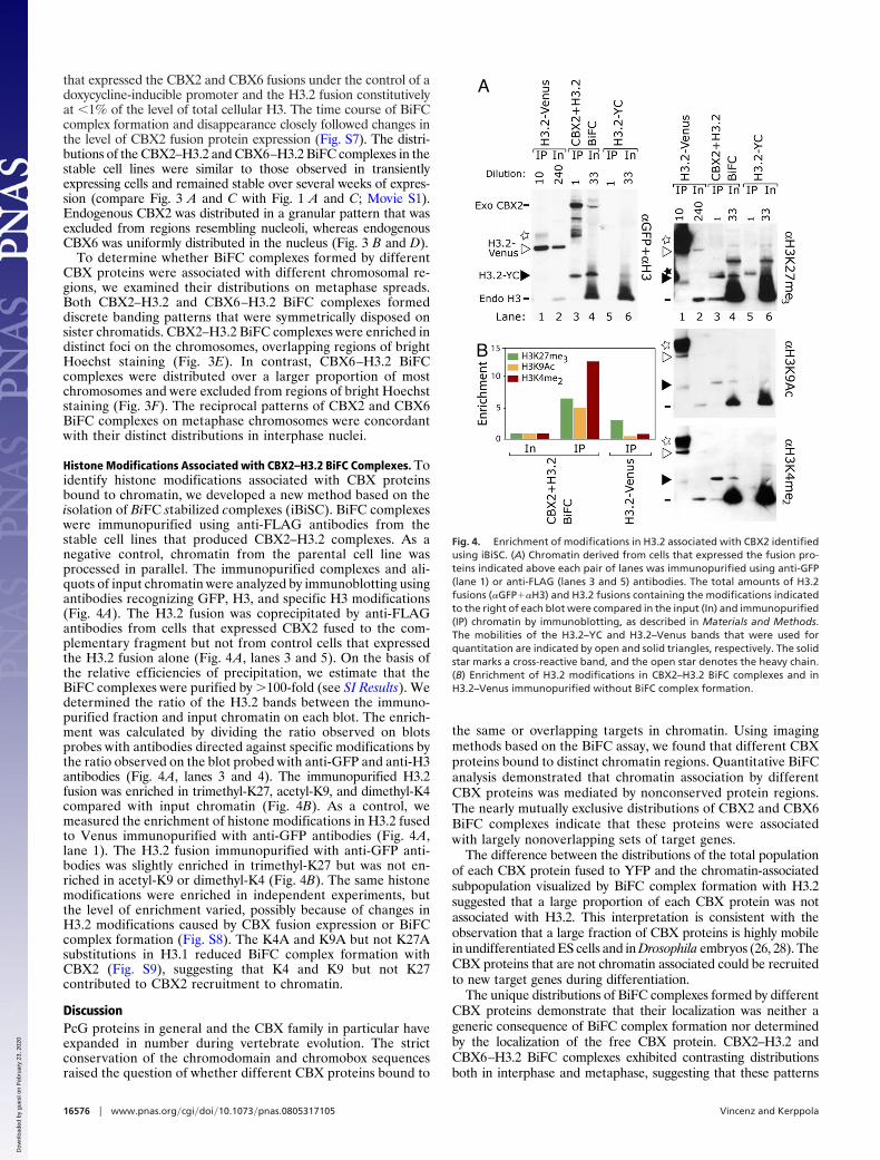
Histone Modifications Associated with CBX2–H3.2 BiFC Complexes. Toidentify histone modifications associated with CBX proteinsbound to chromatin, we developed a new method based on theisolation of BiFC stabilized complexes (iBiSC). BiFC complexeswere immunopurified using anti-FLAG antibodies from thestable cell lines that produced CBX2–H3.2 complexes. As anegative control, chromatin from the parental cell line wasprocessed in parallel. The immunopurified complexes and ali-quots of input chromatin were analyzed by immunoblotting usingantibodies recognizing GFP, H3, and specific H3 modifications(Fig. 4A). The H3.2 fusion was coprecipitated by anti-FLAGantibodies from cells that expressed CBX2 fused to the com-plementary fragment but not from control cells that expressedthe H3.2 fusion alone (Fig. 4A, lanes 3 and 5). On the basis ofthe relative efficiencies of precipitation, we estimate that theBiFC complexes were purified by �100-fold (see SI Results). Wedetermined the ratio of the H3.2 bands between the immuno-purified fraction and input chromatin on each blot. The enrich-ment was calculated by dividing the ratio observed on blotsprobes with antibodies directed against specific modifications bythe ratio observed on the blot probed with anti-GFP and anti-H3antibodies (Fig. 4A, lanes 3 and 4). The immunopurified H3.2fusion was enriched in trimethyl-K27, acetyl-K9, and dimethyl-K4compared with input chromatin (Fig. 4B). As a control, wemeasured the enrichment of histone modifications in H3.2 fusedto Venus immunopurified with anti-GFP antibodies (Fig. 4A,lane 1). The H3.2 fusion immunopurified with anti-GFP anti-bodies was slightly enriched in trimethyl-K27 but was not en-riched in acetyl-K9 or dimethyl-K4 (Fig. 4B). The same histonemodifications were enriched in independent experiments, butthe level of enrichment varied, possibly because of changes inH3.2 modifications caused by CBX fusion expression or BiFCcomplex formation (Fig. S8). The K4A and K9A but not K27Asubstitutions in H3.1 reduced BiFC complex formation withCBX2 (Fig. S9), suggesting that K4 and K9 but not K27contributed to CBX2 recruitment to chromatin.
DiscussionPcG proteins in general and the CBX family in particular haveexpanded in number during vertebrate evolution. The strictconservation of the chromodomain and chromobox sequencesraised the question of whether different CBX proteins bound to
the same or overlapping targets in chromatin. Using imagingmethods based on the BiFC assay, we found that different CBXproteins bound to distinct chromatin regions. Quantitative BiFCanalysis demonstrated that chromatin association by differentCBX proteins was mediated by nonconserved protein regions.The nearly mutually exclusive distributions of CBX2 and CBX6BiFC complexes indicate that these proteins were associatedwith largely nonoverlapping sets of target genes.
The difference between the distributions of the total populationof each CBX protein fused to YFP and the chromatin-associatedsubpopulation visualized by BiFC complex formation with H3.2suggested that a large proportion of each CBX protein was notassociated with H3.2. This interpretation is consistent with theobservation that a large fraction of CBX proteins is highly mobilein undifferentiated ES cells and in Drosophila embryos (26, 28). TheCBX proteins that are not chromatin associated could be recruitedto new target genes during differentiation.
The unique distributions of BiFC complexes formed by differentCBX proteins demonstrate that their localization was neither ageneric consequence of BiFC complex formation nor determinedby the localization of the free CBX protein. CBX2–H3.2 andCBX6–H3.2 BiFC complexes exhibited contrasting distributionsboth in interphase and metaphase, suggesting that these patterns
A
B
Fig. 4. Enrichment of modifications in H3.2 associated with CBX2 identifiedusing iBiSC. (A) Chromatin derived from cells that expressed the fusion pro-teins indicated above each pair of lanes was immunopurified using anti-GFP(lane 1) or anti-FLAG (lanes 3 and 5) antibodies. The total amounts of H3.2fusions (�GFP��H3) and H3.2 fusions containing the modifications indicatedto the right of each blot were compared in the input (In) and immunopurified(IP) chromatin by immunoblotting, as described in Materials and Methods.The mobilities of the H3.2–YC and H3.2–Venus bands that were used forquantitation are indicated by open and solid triangles, respectively. The solidstar marks a cross-reactive band, and the open star denotes the heavy chain.(B) Enrichment of H3.2 modifications in CBX2–H3.2 BiFC complexes and inH3.2–Venus immunopurified without BiFC complex formation.
16576 � www.pnas.org�cgi�doi�10.1073�pnas.0805317105 Vincenz and Kerppola
Dow
nloa
ded
by g
uest
on
Feb
ruar
y 23
, 202
0

reflected interactions with target loci that had distinct subnucleardistributions independent of BiFC complex formation.
The role of H3 K27 trimethylation in PRC1 recruitment iswidely assumed despite accumulating genetic data demonstrat-ing PRC1 functions independent of PRC2 activity (3, 15, 16).Our experiments demonstrated that neither the chromodomainsnor H3 K27 trimethylation were required for chromatin associ-ation by CBX proteins in cells, and chromatin-associated CBXproteins did not colocalize with H3 K27 trimethylation outsidethe inactive X. The BiFC data do not exclude the possibility thatK27 methylation stabilizes CBX protein association with chro-matin. These results suggest that PRC1 recruitment in theabsence of K27 trimethylation is not limited to special cases, suchas the inactive X and the male pronucleus (3, 15, 16).
The visualization of CBX–H3.2 BiFC complexes on meta-phase chromosomes provides a unique genome-wide view ofCBX protein binding. Metaphase chromosomes are more con-densed than polytene chromosomes in Drosophila salivaryglands, but they can be analyzed in any dividing cell type, andchanges in complex distributions in response to stimuli anddifferentiation can be examined. Cytogenetic imaging of BiFCcomplexes provides high-content information that enables com-parison of the genome-wide occupancy of chromatin bindingproteins in single cells.
The identification of histone modifications that are associatedwith a particular regulatory protein complex in cells has beendifficult. The iBiSC approach enabled detection of H3.2 modi-fications that were enriched in CBX2–H3.2 BiFC complexes.The roles of these modifications in CBX2 recruitment or func-tions are not clear, but ‘‘bivalent domain’’ H3 trimethyl-K4 andH3 trimethyl-K27 modifications correlate with PcG complexbinding, and H3 acetyl-K9 modifications are altered in embryoslacking PRC1 components (13, 29). Further studies of thetemporal and causal relationships between these modificationsand CBX2 binding are important.
The unique patterns of chromatin association by differentCBX proteins both in interphase nuclei and on metaphasechromosomes suggest that these proteins bind to nonredundant
sets of target genes. Although the biologic functions of severalCBX proteins have been studied in different experimentalsystems, no comparative studies of their functions have beenperformed in the same cell type. Our results provide newmotivation for investigation of the mechanisms of selective CBXprotein recruitment.
Materials and MethodsPlasmids, Cell Lines, and Imaging. The construction of plasmids encoding CBX,H3, and Ring1B fusions, the cell lines, transfection protocols, fluorescencemicroscopy, flow cytometry, and immunologic methods are described in detailin SI Materials and Methods and Tables S2 and S3.
Derivation of Stable ES Cell Lines and Preparation of Metaphase Spreads. ES celllines with integrated expression constructs encoding CBX and H3.2 fused tocomplementary fluorescent protein fragments were established by two suc-cessive rounds of transfection. First, a cell line was established that expressedthe H3.2 fusion constitutively. Second, CBX2 or CBX6 fused to the comple-mentary fluorescent protein fragment under the control of a doxycycline-regulated promoter was introduced together with the doxycycline-responsivetransactivator. Positive clones were identified by Western blotting. Meta-phase spreads were prepared according to classic protocols (30) but withoutmethanol acetic acid fixation to preserve BiFC complex fluorescence, as de-scribed in detail in SI Materials and Methods.
iBiSC and Determination of the Enrichment of Histone Modifications. iBiSC wasperformed under the same conditions as ChIP analysis but without cross-linking (13). The immunoprecipitated and input chromatin were analyzed byWestern blotting using antibodies recognizing specific modifications (�Mod)and total H3.2 (�Total). The enrichment of histone modifications was calcu-lated using Eq. 1.
Enrichment �
H3.2�YCIP�Mod
H3.2�YCIn�Mod
H3.2�YCIP�Total
H3.2�YCIn�Total
[1]
ACKNOWLEDGMENTS. We thank Caroline Becker, Zunair Mahmood, andJ’aime Manion for capable assistance; Terry Magnuson for providing EED nullES cells; and members of the Kerppola laboratory, in particular Xiaojun Ren,for constructive criticisms and valuable suggestions.
1. Sparmann A, van Lohuizen M (2006) Polycomb silencers control cell fate, developmentand cancer. Nat Rev Cancer 6:846–856.
2. O’Carroll D, et al. (2001) The polycomb-group gene Ezh2 is required for early mousedevelopment. Mol Cell Biol 21:4330–4336.
3. Pasini D, Bracken AP, Hansen JB, Capillo M, Helin K (2007) The polycomb group proteinSuz12 is required for embryonic stem cell differentiation. Mol Cell Biol 27:3769–3779.
4. Voncken JW, et al. (2003) Rnf2 (Ring1b) deficiency causes gastrulation arrest and cellcycle inhibition. Proc Natl Acad Sci USA 100:2468–2473.
5. Park IK, et al. (2003) Bmi-1 is required for maintenance of adult self-renewing haema-topoietic stem cells. Nature 423:302–305.
6. Montgomery ND, et al. (2005) The murine polycomb group protein Eed is required forglobal histone H3 lysine-27 methylation. Curr Biol 15:942–947.
7. Shao Z, et al. (1999) Stabilization of chromatin structure by PRC1, a Polycomb complex.Cell 98:37–46.
8. Levine SS, et al. (2002) The core of the polycomb repressive complex is compositionallyand functionally conserved in flies and humans. Mol Cell Biol 22:6070–6078.
9. Kuzmichev A, Nishioka K, Erdjument-Bromage H, Tempst P, Reinberg D (2002) Histonemethyltransferase activity associated with a human multiprotein complex containingthe Enhancer of Zeste protein. Genes Dev 16:2893–2905.
10. Muller J, et al. (2002) Histone methyltransferase activity of a Drosophila Polycombgroup repressor complex. Cell 111:197–208.
11. Czermin B, et al. (2002) Drosophila enhancer of Zeste/ESC complexes have a histone H3methyltransferase activity that marks chromosomal Polycomb sites. Cell 111:185–196.
12. Cao R, et al. (2002) Role of histone H3 lysine 27 methylation in Polycomb-groupsilencing. Science 298:1039–1043.
13. Boyer LA, et al. (2006) Polycomb complexes repress developmental regulators inmurine embryonic stem cells. Nature 441:349–353.
14. Bracken AP, Dietrich N, Pasini D, Hansen KH, Helin K (2006) Genome-wide mapping ofPolycomb target genes unravels their roles in cell fate transitions. Genes Dev 20:1123–1136.
15. Schoeftner S, et al. (2006) Recruitment of PRC1 function at the initiation of X inacti-vation independent of PRC2 and silencing. EMBO J 25:3110–3122.
16. Puschendorf M, et al. (2008) PRC1 and Suv39h specify parental asymmetry at consti-tutive heterochromatin in early mouse embryos. Nat Genet 40:411–420.
17. Pearce JJ, Singh PB, Gaunt SJ (1992) The mouse has a Polycomb-like chromobox gene.Development 114:921–929.
18. Alkema MJ, et al. (1997) MPc2, a new murine homolog of the Drosophila polycombprotein is a member of the mouse polycomb transcriptional repressor complex. J MolBiol 273:993–1003.
19. Bardos JI, Saurin AJ, Tissot C, Duprez E, Freemont PS (2000) HPC3 is a new humanpolycomb orthologue that interacts and associates with RING1 and Bmi1 and hastranscriptional repression properties. J Biol Chem 275:28785–28792.
20. Gil J, Bernard D, Martinez D, Beach D (2004) Polycomb CBX7 has a unifying role incellular lifespan. Nat Cell Biol 6:67–72.
21. Bernstein E, et al. (2006) Mouse polycomb proteins bind differentially to methylatedhistone H3 and RNA and are enriched in facultative heterochromatin. Mol Cell Biol26:2560–2569.
22. Hu CD, Chinenov Y, Kerppola TK (2002) Visualization of interactions among bZIP andRel family proteins in living cells using bimolecular fluorescence complementation.Mol Cell 9:789–798.
23. Kanno T, et al. (2004) Selective recognition of acetylated histones by bromodomainproteins visualized in living cells. Mol Cell 13:33–43.
24. Hake SB, et al. (2006) Expression patterns and post-translational modifications asso-ciated with mammalian histone H3 variants. J Biol Chem 281:559–568.
25. Ahmad K, Henikoff S (2002) The histone variant H3.3 marks active chromatin byreplication-independent nucleosome assembly. Mol Cell 9:1191–1200.
26. Ren X, Vincenz C, Kerppola TK (2008) Changes in the distributions and dynamics ofpolycomb repressive complexes during embryonic stem cell differentiation. Mol CellBiol 28:2884–2895.
27. Hagstrom K, Muller M, Schedl P (1997) A Polycomb and GAGA dependent silencer adjoinsthe Fab-7 boundary in the Drosophila bithorax complex. Genetics 146:1365–1380.
28. Ficz G, Heintzmann R, Arndt-Jovin DJ (2005) Polycomb group protein complexesexchange rapidly in living Drosophila. Development 132:3963–3976.
29. Fujimura Y, et al. (2006) Distinct roles of Polycomb group gene products in transcrip-tionally repressed and active domains of Hoxb8. Development 133:2371–2381.
30. Tjio JH, Levan A (1956) The chromosome number of man. Hereditas 42:1–6.
Vincenz and Kerppola PNAS � October 28, 2008 � vol. 105 � no. 43 � 16577
CELL
BIO
LOG
Y
Dow
nloa
ded
by g
uest
on
Feb
ruar
y 23
, 202
0