different roles for the axin interactions with the samp
TRANSCRIPT
Different Roles for the Axin Interactions with the SAMPversus the Second Twenty Amino Acid Repeat ofAdenomatous Polyposis ColiJean Schneikert1, Jan Gustav Ruppert1, Jurgen Behrens1*, Eva Maria Wenzel2¤
1 Nikolaus-Fiebiger-Center for Molecular Medicine, University of Erlangen-Nurnberg, Erlangen, Germany, 2 Centre for Cancer Biomedicine, Faculty of Medicine, University
of Oslo, Oslo, Norway
Abstract
Wnt signalling is prevented by the proteosomal degradation of b-catenin, which occurs in a destruction complex containingadenomatous polyposis coli (APC), APC-like (APCL), Axin and Axin2. Truncating mutations of the APC gene result in theconstitutive stabilisation of b-catenin and the initiation of colon cancer, although tumour cells tolerate the expression ofwild-type APCL. Using the colocalisation of overexpressed Axin, APC and APCL constructs as a readout of interaction, wefound that Axin interacted with the second twenty amino acid repeat (20R2) of APC and APCL. This interaction involved adomain adjacent to the C-terminal DIX domain of Axin. We identified serine residues within the 20R2 of APCL that wereinvolved in Axin colocalisation, the phosphorylation of truncated APCL and the down-regulation of b-catenin. Our resultsindicated that Axin, but not Axin2, displaced APC, but not APCL, from the cytoskeleton and stimulated its incorporation intobright cytoplasmic dots that others have recognised as b-catenin destruction complexes. The SAMP repeats in APC interactwith the N-terminal RGS domain of Axin. Our data showed that a short domain containing the first SAMP repeat intruncated APC was required to stimulate Axin oligomerisation. This was independent of Axin colocalisation with 20R2. Ourdata also suggested that the RGS domain exerted an internal inhibitory constraint on Axin oligomerisation. Considering ourdata and those from others, we discuss a working model whereby b-catenin phosphorylation involves Axin and the 20R2 ofAPC or APCL and further processing of phospho-b-catenin occurs upon the oligomerisation of Axin that is induced bybinding the SAMP repeats in APC.
Citation: Schneikert J, Ruppert JG, Behrens J, Wenzel EM (2014) Different Roles for the Axin Interactions with the SAMP versus the Second Twenty Amino AcidRepeat of Adenomatous Polyposis Coli. PLoS ONE 9(4): e94413. doi:10.1371/journal.pone.0094413
Editor: Cara Gottardi, Northwestern University Feinberg School of Medicine, United States of America
Received June 25, 2013; Accepted March 16, 2014; Published April 10, 2014
Copyright: � 2014 Schneikert et al. This is an open-access article distributed under the terms of the Creative Commons Attribution License, which permitsunrestricted use, distribution, and reproduction in any medium, provided the original author and source are credited.
Funding: This work was supported by a grant from the Deutsche Forschungsgemeinschaft to JS [SCHN 1189/1-2]. The authors acknowledge support byDeutsche Forschungsgemeinschaft and Friedrich-Alexander-Universitat Erlangen-Nurnberg (FAU) within the funding programme Open Access Publishing. Thefunders had no role in study design, data collection and analysis, decision to publish, or preparation of the manuscript.
Competing Interests: The authors have declared that no competing interests exist.
* E-mail: [email protected]
¤ Current address: Department of Biochemistry, Institute for Cancer Research, Oslo University Hospital, Oslo, Norway
Introduction
Homeostasis of the colonic epithelium requires the proliferation
of the stem cells located at the bottom of the crypts, the subsequent
expansion of the daughter cell population, the differentiation and
migration of these cells toward the surface and ultimately apoptosis
and release into the lumen [1]. These processes are partly
coordinated by Wnt family growth factors that encourage cell
proliferation. This increase in cell proliferation occurs upon the
accumulation of the transcription factor b-catenin [2,3], which
controls a genetic program at the origin of cell proliferation. In the
absence of Wnt stimulation, b-catenin is targeted for degradation
in a destruction complex consisting of the tumour suppressor APC
bound to Axin or Axin2, which interacts with casein kinase 1a(CK1a) and glycogen synthase kinase 3b (GSK3b) [4,5]. The
phosphorylation of b-catenin catalysed by these kinases generates
a signal for the subsequent ubiquitination of phosphorylated b-
catenin, followed by proteasomal degradation. Several models
have been proposed to concatenate the currently available data
[6–9]. The most recent model opposes the classical view of Wnt-
elicited disruption of the destruction complex based on evidence
that the destruction complex remains essentially intact upon Wnt
stimulation [9–11].
The mechanistic steps that involve APC are not completely
understood. APC is a large protein of 2843 amino acids (fig. 1)
[12,13]. The long N-terminal region contains two dimerisation
domains [14,15] that bracket the armadillo repeat domain, which
is followed by b-catenin-binding sites termed the 15 [16–18] and
20 [19] amino acid repeats (15R and 20R, respectively); the 20Rs
are intermingled with the b-catenin inhibitory domain (CiD)
[8,20] and the SAMP repeats that are Axin/Axin2-binding sites
[21,22]. The C-terminus of APC is involved in microtubule
dynamics, and it is not known whether this region plays a role in b-
catenin degradation [23]. The 15R bind to b-catenin and are
important for targeting b-catenin for degradation [24]. The 20R
are heterogeneous; the 20R1 and 20R3 bind to b-catenin with
differing affinities [18], but only the 20R3 has been implicated in
b-catenin degradation [24,25]. The role of the 20R1 remains
unknown [24]. Paradoxically, the 20R2 cannot bind to b-catenin
[18]. The CiD likely constitutes an interaction surface for a
component crucial for b-catenin degradation [8,20].
PLOS ONE | www.plosone.org 1 April 2014 | Volume 9 | Issue 4 | e94413

APC has received particular attention because truncating
mutations have been identified in approximately 80% of colorectal
tumours [26]. The mutations tend to cluster in the middle of the
open reading frame; this region has therefore been termed the
mutation cluster region (MCR) [27]. This clustering reflects
simultaneous positive and negative selection processes [28]. On
one hand, the mutations remove the SAMP repeats, a nuclear
export signal [29], the 20R3 [30] and the CiD [20], all domains
known to contribute to b-catenin degradation. As a consequence,
these mutations stabilise b-catenin and shift the homeostatic balance
toward cell proliferation and the initiation of tumourigenesis. On
the other hand, truncating mutations almost always occur after the
first 15R [24,31]. The resulting fragment may have multiple roles
because the armadillo repeat domain binds to several partners [32–
36]. The strong selection for the presence of the first 15R in
truncated APC suggests that it serves an important purpose.
Besides APC, colorectal cancer cells also express the paralogue
APC-like (APCL) [37–39]. APC and APCL display similar
topological organisation and share most of the functional domains,
with the exception of the 15R region, the second dimerisation
domain and the C-terminus (fig. 1). APCL targets b-catenin for
destruction upon ectopic expression [37–38], and it is puzzling
that sequence alterations in APCL have yet to be reported in colon
cancer.
Axin has been proposed to be the limiting component of the b-
catenin destruction complex because it is present at extremely low
concentrations in Xenopus extracts [40]. The evidence that APC
and Axin or Axin2 interact via the SAMP repeats stems from
studies that analysed the contribution of the SAMP repeats to b-
catenin degradation using APC constructs lacking approximately
the N-terminal thousand amino acids including the 15R [25], or
containing point mutations in the 15R that abolish b-catenin
binding [24]. Based on these results, it was thought that Axin and
Axin2 catalyse b-catenin degradation by binding to the SAMP
repeats of APC. However, this is only partly satisfying because the
ectopic expression of Axin or Axin2 in colorectal cancer cell lines
expressing APC that is truncated before the SAMP repeats results
in efficient b-catenin destruction [21,41,42]. Furthermore, ectopic
expression of truncated APC lacking all the SAMP repeats also
elicits efficient b-catenin degradation [20]. Both results create a
paradox as they do not reveal any dependent relationship between
Axin and its ability to bind to the SAMP repeats of APC to elicit b-
catenin degradation.
Axin oligomerises through its DIX domain (fig. 1); this
oligomerisation is observed as bright cytoplasmic spots upon
ectopic Axin expression [43–46]. Interestingly, ectopically ex-
pressed Axin lacking the DIX domain exhibits a diffuse
intracellular distribution and results in the accumulation of
phosphorylated b-catenin that does not undergo subsequent
degradation [43]. This indicates that Axin oligomers represent
the fully active destruction complex. It has also been shown that
APC is essential for destruction complex assembly, that APC
provokes Axin oligomerisation and that Wnt stimulation leads to
Dishevelled activation, which oligomerises with Axin and prevents
it from oligomerising with APC [45,46]. In addition, it has been
demonstrated that the destruction complex catalyses the ubiqui-
tination of phosphorylated b-catenin; this ubiquitination event is
inhibited by Wnt stimulation [9]. Together, these data suggest that
Axin binding to APC results in Axin oligomerisation that is
associated with the ubiquitination of phosphorylated b-catenin; all
of these steps are inhibited by Wnt stimulation as a result of
Dishevelled activation. However, this model does not convey how
b-catenin phosphorylation is related to the APC/Axin interaction
despite observations that both proteins individually stimulate b-
catenin phosphorylation [43,47]. In addition, this model does not
provide an answer to the contradictory observations that the
Axin/APC interaction is either not perturbed [9] or is affected
[48,49] by Wnt stimulation.
The study presented here aimed to solve this paradox by
providing evidence that Axin performs a different function when
bound to the second twenty amino acid repeat (20R2) of APC than
when bound to the SAMP repeats of APC.
Results
APC and APCL differentially influence the intracellularlocalisation of Axin
We analysed the interaction of Axin with both APC and APCL
in colocalisation experiments using N-terminal tagged constructs
(fig. 1) that were transiently transfected into colorectal cancer cell
Figure 1. Schematic representation of human APC and APCLand rat Axin. Functional modules are shown, including the twodimerisation domains of APC (DIM), the armadillo repeat domain (Arm),the 15 (15RA to D) and 20 (20R1 to 7) amino acid repeats that functionas b-catenin binding sites, the b-catenin inhibitory domain (CiD)involved in b-catenin degradation and the SAMP repeats that representAxin or Axin2 binding sites. The mutation cluster region (MCR) containsmost of the APC truncating mutations that have been observed incolon cancer. The RGS (SAMP-binding region), DIX (oligomerisation), b-catenin and GSK3b domains in Axin are indicated. The numbers indicatethe amino acid positions. The numbers with arrows correspond to thesize of the different constructs analysed in this study, which were fusedat the N-terminus to YFP (APC and APCL) or myc (Axin).doi:10.1371/journal.pone.0094413.g001
Interaction of Axin with the 20R2 of APC and APCL
PLOS ONE | www.plosone.org 2 April 2014 | Volume 9 | Issue 4 | e94413

lines. We used three different Axin expression constructs, mouse
flag-Axin (fAxin), rat myc-Axin (mycAxin) and human YFP-Axin
(yAxin). When the three constructs were transiently expressed in
DLD1 or SW480 cells, we observed two intracellular localisation
patterns for all three constructs, a diffuse distribution and a dotty
cytoplasmic distribution (fig. S1A); the proportion of these
patterns varied between constructs and cell lines (fig. S1B). The
dotty pattern is commonly observed upon ectopic expression of
Axin [50,51] and has also been described for endogenous Axin
[52,53]. The dots are oligomers [44] that form spontaneously as a
function of increasing Axin concentration [45]. The different
proportions of the two localisation patterns in a given cell line may
therefore reflect differences in the expression levels of the three
constructs based on the species of origin of the construct and/or
the influence of the tag on Axin oligomerisation. For a given
construct, the different proportion of Axin localisation between
two cell lines may reflect different expression levels resulting from
different transfection efficiencies. We established that the trans-
fection efficiency is better in SW480 cells than in DLD1 cells
[unpublished data]. Meanwhile, the careful inspection of any cell
Figure 2. The RGS domain of Axin is not necessary for colocalisation with APC or APCL. DLD1 cells were transiently transfected on day 1with the indicated APC (A) or APCL (B) constructs or with the indicated N-terminal myc-tagged Axin constructs, either individually or in combination.The cells were stained on day 3 with an anti-myc antibody and Hoechst dye. Cells transfected with yAPCL alone displayed dotty and fibre-likeexpression patterns, with yAPC alone exhibited a fibre-like expression pattern, with myc-Axin alone displayed diffuse and dotty expression patternsand with myc-Axin(298–832) alone exhibited only a dotty expression pattern. Coexpression of myc-Axin and APC or APCL resulted in colocalisation,which was also observed with when the RGS domain (298–832) was deleted from Axin. The percentages indicate the proportion of differentlocalisation patterns in the transfected cells. The imaging parameters were identical for the myc constructs; the yAPCL signal was more intense thanthe yAPC signal. Bar, 10 mM.doi:10.1371/journal.pone.0094413.g002
Interaction of Axin with the 20R2 of APC and APCL
PLOS ONE | www.plosone.org 3 April 2014 | Volume 9 | Issue 4 | e94413

line transfected with any construct revealed that many cells
expressed higher levels of diffuse Axin and other cells exclusively
displayed discrete dots. This indicates that additional factors
influence Axin oligomerisation.
When individually expressed in DLD1 cells, YFP-labelled APC
(yAPC) predominantly exhibited a fibre-like pattern (fig. 2A),
likely corresponding to colocalisation with microtubules, as has
been previously described [54,55]. We confirmed the fortuitous
observation that myc-Axin was diffusely localised in the cytoplasm
of most transfected DLD1 cells (approximately 80%). Co-
expression of myc-Axin and yAPC resulted in two predominant
observations, either the recruitment of myc-Axin to fibre-bound
yAPC (41% of myc-Axin- and yAPC-positive cells) or the re-
localisation of yAPC and myc-Axin to bright spots (59% of myc-
Axin- and yAPC-positive cells). Thus, yAPC and myc-Axin
mutually stimulate the recruitment of one another into dots that
represent the b-catenin destruction complex, or the degradasome
[43–46]. When individually expressed in DLD1 cells, YFP-labelled
APCL (yAPCL) displayed a fibre-like pattern in approximately
two-thirds of the transfected cells (fig. 2B), likely corresponding to
colocalisation with microtubules and/or actin fibres, as has been
previously described [39]. In the remaining one-third of the
transfected cells, yAPCL localised to bright punctuate structures
surrounding the nucleus. Cells exhibiting both types of localisation
were rare (approximately 10% of the transfected cells) and were
counted as the dotty type. In contrast to yAPC, the addition of
myc-Axin neither increased the proportion of cells containing
yAPCL dots nor decreased the fraction displaying fibre-bound
yAPCL. Axin was no longer diffusely localised and instead
colocalised with both types of yAPCL, which may also have been
interacting with endogenous Axin. Notably, there was no
correlation between the different intracellular localisation patterns
and the expression level of each construct: we commonly identified
numerous cells with high levels of diffuse myc-Axin next to cells
with few and discrete myc-Axin dots as well as the inverse. These
observations were also common to yAPCL and the other
constructs in all the subsequent experiments. We concluded that
myc-Axin and yAPC mutually stimulated the incorporation of one
another into dots, whereas yAPCL imposed its own intracellular
localisation pattern on myc-Axin.
The N-terminus of Axin, which contains the RGS domain,may inhibit oligomerisation
The well-known interaction between Axin and APC occurs via
the N-terminal RGS domain and the SAMP repeats (fig. 1).
Deletion of the RGS domain (myc-Axin(298–832); fig. 2) resulted
in a transition from a diffuse localisation pattern to a fully
penetrant dotty cytoplasmic pattern. This probably reflected Axin
oligomerisation because these dots disappeared upon deletion of
the DIX domain that mediates oligomerisation (see below). We
concluded that the N-terminus of Axin, which contains the RGS
domain, may have inhibited Axin oligomerisation.
The colocalisation of Axin with APC or APCL isindependent of the RGS
Surprisingly, the co-expression of yAPC and myc-Axin(298–
832) (fig. 2) resulted in the partial elimination of myc-Axin(298–
832) dots and in the recruitment of myc-Axin(298–832) to fibre-
bound yAPC. Similarly, myc-Axin(298–832) colocalised with
yAPCL, despite lacking the RGS domain. We concluded that
Axin may contain a second interaction domain for both APC and
APCL that is independent of the RGS-SAMP interface.
yAPCL1728 dots are not Golgi vesiclesIn a first step toward identifying the domains involved in this
putative novel Axin-APC(L) interaction, we utilised yAPCL
constructs progressively deleted from the C-terminus (fig. 1).
These constructs localised exclusively as cytoplasmic dots when
transiently expressed in the DLD1 colorectal cancer cell line
(fig. 3). Fibre localisation was lost upon deleting the C-terminus,
which likely contains a microtubule-binding site based on analogy
to the C-terminus of APC [23,39]. The punctuate structures were
typically restricted to the vicinity of the nucleus and may have
represented Golgi vesicles, as has been previously described [39].
To investigate this possibility, SW480 cells transiently expressing
yAPC1728 were probed with antibodies against the Golgi markers
GM130 [56], giantin [57] and TGN46 [58] (fig. S2). Although
displaying a very similar appearance as yAPCL1728, none of the
Golgi markers colocalised with yAPCL1728. Therefore, we
concluded that the yAPCL1728 dots were not Golgi vesicles.
The 20R2 of truncated APCL is necessary for Axincolocalisation
In contrast to myc-Axin, N-terminal flag-tagged Axin (flag-
Axin) was expressed as cytoplasmic dots in about half of the
transfected cells (fig. 3A, S1). Co-expression of yAPCL1728 and
flag-Axin resulted in colocalisation of the two proteins (fig. 3A).
Deletion of both SAMP repeats (yAPCL1179 or yAPCL1257
[unpublished data]) did not affect APCL/Axin colocalisation,
suggesting the presence of an Axin-binding site in APCL that does
not require the SAMP repeats. Importantly, deletion of the second
20 amino acid repeat (20R2) eliminated the colocalisation
(compare yAPCL1179 with yAPCL1147, fig. 3A). When individ-
ually expressed, flag-Axin displayed a diffuse cytoplasmic localisa-
tion in approximately half of the cell population (fig. 3A, S1). The
diffuse flag-Axin pattern was retained only upon co-expression of
APCL constructs lacking the 20R2. Similar results were obtained
using myc-Axin instead of flag-Axin [unpublished data]. We
concluded that Axin colocalisation with truncated APCL required
the 20R2.
Axin2 colocalises with the N-terminus of APCLWe performed similar experiments with N-terminal flag-tagged
Axin2 (fAxin2), which exhibited a diffuse cytoplasmic distribution
in DLD1 cells upon ectopic expression (fig. 3B). Surprisingly,
APCL constructs as short as yAPCL730 (fig. 1) colocalised with
flag-Axin2. Therefore, flag-Axin2 colocalised specifically with the
N-terminus of APCL, whereas Axin colocalisation with APCL was
lost when the 20R2 was deleted.
Amino acids 508-712 of Axin are required forcolocalisation with APCL truncated before the SAMPrepeats
We also performed the converse experiment, using yAPCL1179
truncated shortly after the 20R2 and deletion mutants of myc-
Axin, to identify a putative second APCL-binding site in Axin that
was independent of the RGS domain (fig. 4). We first confirmed
that myc-Axin and myc-Axin(298–832), which lacks the RGS
domain, colocalised with yAPCL1179 (fig. 4). Surprisingly,
further N-terminal deletions in myc-Axin (myc-Axin(508–832))
resulted in a fibre-like localisation pattern of Axin when
individually expressed, but the colocalisation with yAPCL1179
in dots remained. The colocalisation was lost upon deletion of
residues 508 to 712 (myc-Axin(713–832)). When individually
expressed, myc-Axin(713–832) was diffusely distributed through-
out the cell. The RGS domain alone (myc-Axin(1–229)) did not
Interaction of Axin with the 20R2 of APC and APCL
PLOS ONE | www.plosone.org 4 April 2014 | Volume 9 | Issue 4 | e94413

Figure 3. The 20R2 of APCL is required for Axin colocalisation in the absence of the SAMP repeats. DLD1 cells were transientlytransfected on day 1 with the indicated APCL constructs or the indicated N-terminal flag-tagged Axin (A) or Axin2 (B) constructs, either individually orin combination. The cells were stained on day 3 with an anti-flag antibody and Hoechst dye. When expressed alone, truncated yAPCL displayed adotty localisation pattern, flag-Axin exhibited a diffuse or dotty expression pattern and fAxin2 demonstrated diffuse localisation. A, APCL constructslacking the 20R2 (i.e., 1147 and shorter) did not colocalise with fAxin (c, d), whereas colocalisation occurred in the presence of the 20R2 (yAPCL1179
Interaction of Axin with the 20R2 of APC and APCL
PLOS ONE | www.plosone.org 5 April 2014 | Volume 9 | Issue 4 | e94413

colocalise with yAPCL1179, whereas deletions of either the DIX
domain (myc-Axin(1–713)) or the DIX and RGS domains (myc-
Axin(298–713)) did not affect Axin colocalisation with
yAPCL1179 (fig. 4). We concluded that Axin colocalisation with
yAPCL1179 depended on amino acids 508 to 712 of Axin, thereby
suggesting the presence of an APCL-binding site that was
independent of the RGS domain.
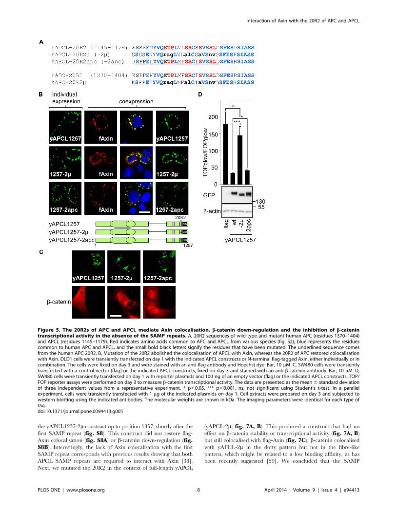
Conserved residues within the 20R2 are required fortruncated APCL to colocalise with Axin
To provide additional evidence that Axin binds to the 20R2 of
APCL, we compared the 20R2 sequences of APC and APCL from
different species (fig. S3). We identified eight amino acids
common to all inspected 20R2 sequences. These eight residues
were mutated in the context of yAPCL1257 (yAPCL1257-2m,
fig. 5A). Co-expression of yAPCL1257-2m and flag-Axin in DLD1
cells revealed that the mutations abolished the colocalisation of
these two proteins (fig. 5B). We concluded that the conserved
residues within the 20R2 were required for truncated APCL to
colocalise with Axin. Next, we removed the 1090 N-terminal
residues from yAPCL1257 and yAPCL1257-2m to create two
constructs, yAPCL(1091–1257) and yAPCL(1091–1257)-2m,
which exhibited a diffuse cytoplasmic distribution (fig. S4). The
expression of yAPCL(1091–1257) dissolved the flag-Axin dots,
whereas the expression of yAPCL(1091–1257)-2m did not affect
these dots. Together, we concluded that the conserved residues
within the 20R2 were required for truncated APCL to colocalise
with Axin, that the N-terminal residues of APCL were not
required and that colocalisation with the 20R2 inhibited Axin
oligomerisation.
The 20R2 of APC can replace that of APCL to enabletruncated APCL to colocalise with Axin
To determine whether the 20R2 of APC also colocalised with
Axin, we exchanged the 20R2 of APCL with the corresponding
sequence of its paralogue (yAPCL1257-2apc, fig. 5A). The 20R2
of APC could replace that of APCL to enable colocalisation with
flag-Axin (fig. 5B).
The 20R2 of truncated APCL is required for b-catenindown-regulation and can be replaced with the 20R2 ofAPC
We analysed the functional consequences of mutating the 20R2
of APCL or replacing it with that of APC. Immunofluorescence
experiments were performed in SW480 cells, in which b-catenin is
easily detectable, enabling its down-regulation upon the expression
of either APC or APCL to be observed. Mutating the 20R2 of
APCL in the context of the active yAPCL1257 (yAPCL1257-2m)
abolished b-catenin down-regulation and led to the colocalisation
of APCL with b-catenin (fig. 5C). The 20R2 of APC
(yAPCL1257-2apc) could substitute for that of APCL (fig. 5C).
yAPCL1257 and yAPCL1257-2apc, but not yAPCL1257-2m,
inhibited the TOP reporter that measures b-catenin transcrip-
tional activity (fig. 5D). We concluded that b-catenin degradation
by truncated APCL correlated with Axin colocalisation to the
20R2 in the absence of the SAMP repeats.
Identifying the residues in the 20R2 that are involved inphosphorylating truncated APCL
The immunoprecipitation of yAPCL(1091–1257) followed by
western blotting revealed a pattern of three bands (fig. 6A, B).
Alkaline phosphatase treatment of the immunoprecipitate elimi-
nated the upper band and strongly decreased the intensity of the
middle band, thereby indicating that these bands represent
phosphorylation events. The lower band, the intensity of which
increased upon alkaline phosphatase treatment, therefore corre-
sponded to the unphosphorylated form of yAPCL(1091–1257). To
investigate the possibility that the 20R2 might be phosphorylated,
we created additional constructs in the context of yAPCL(1091–
1257) in which we introduced the L1168V mutation (construct 1)
and individually mutated each conserved and potential phosphor-
ylatable Ser/Thr residue within the 20R2 (constructs 2–5)
(fig. 6A). The L1168V mutation was an unavoidable consequence
of the cloning strategy, but it did not influence the phosphorylation
pattern (fig. 6B). The mutation of Thr1155 to Ala (construct 2)
also did not affect the phosphorylation pattern. Each Ser residue
was individually mutated on the background of the LV mutation;
S1164 (construct 4) was a strong determinant of phosphorylation,
and S1160 and S1167 (constructs 3 and 5, respectively) were
weaker determinants of phosphorylation (fig. 6B), as evidenced by
the relative reduction in the intensity of the upper band. The
differences between the Ser residues might be explained by the
disruption of a phosphorylation site (S1164, construct 4) and the
alteration of a kinase recognition site (S1160 and S1167, constructs
3 and 5, respectively). Notably, the intensity of the middle band
did not decrease in the mutants, indicating the presence of a
phosphorylation site independent of the four conserved Ser/Thr
residues.
The residues of the 20R2 that are involved inphosphorylation are also involved in the down-regulation of b-catenin mediated by truncated APCL andthe colocalisation of Axin with truncated APCL
We next analysed the functional consequences of individually
mutating each of the four conserved Ser/Thr residues in the
context of yAPCL1257-L1168V (fig. 6C–F). We preliminary
established that the L1168V mutation did not affect the ability of
the ectopically expressed protein to inhibit b-catenin transcrip-
tional activity (fig. S5). The intracellular localisation of all the
constructs was similar (fig. 6F). TOP/FOP reporter assays
(fig. 6C) and b-catenin staining (fig. 6D, S6) in transiently
transfected SW480 cells indicated that each Ser/Thr residue was
required to prevent the accumulation of b-catenin and to inhibit
its transcriptional activity. Remarkably, the T1155A mutation
almost abolished these activities of yAPCL1257. These results
qualitatively correlated with the ability of these ectopically
expressed proteins to colocalise with Axin (fig. 6E, S7), as the
mutation of each Ser/Thr residue resulted in a partial loss of
colocalisation with flag-Axin. One notable quantitative discrepan-
cy occurred with yAPCL1257-L1168V-T1155A, which coloca-
lised with flag-Axin in a greater proportion of cells than expected
based on the b-catenin staining and reporter assays. We
hypothesised that T1155 might be involved in other functions in
addition to contributing to Axin colocalisation. Together, we
concluded that the down-regulation of b-catenin mediated by
truncated APCL as well as the Axin/APCL colocalisation
and yAPCL1728) (a, b). B, Axin2 interacted with the N-terminus of APC. The percentages indicate the proportion of different localisation patterns inthe transfected cells. The imaging parameters were identical for each type of tag. Bar, 10 mM.doi:10.1371/journal.pone.0094413.g003
Interaction of Axin with the 20R2 of APC and APCL
PLOS ONE | www.plosone.org 6 April 2014 | Volume 9 | Issue 4 | e94413

depended on the presence of the conserved T1155 residue. The
dependence on the conserved Ser residues in the 20R2 suggests a
phosphorylation-dependent regulation of truncated APCL im-
pacting on b-catenin activity and Axin colocalisation.
The SAMP repeats of APCL and APC do not compensatefor the inactivation of the 20R2 in terms of down-regulating b-catenin
APCL contains two identifiable SAMP repeats that may
contribute to b-catenin degradation and compensate for the
deleterious effect of mutating the 20R2. Therefore, we extended
Figure 4. Amino acids 508–713 of Axin are required for colocalisation with truncated APCL. DLD1 cells were transiently transfected onday 1 with wild-type or mutant N-terminal myc-tagged Axin, either individually or in combination with the yAPCL1179 expression vector. The cellswere fixed on day 3 and were stained with an anti-myc antibody and Hoechst dye. The presence of amino acids 508–713 in the myc-Axin deletionmutants resulted in colocalisation with yAPCL1179. Where applicable, the percentages indicate the proportion of different localisation patterns in thetransfected cells. The imaging parameters were identical for each type of tag. Bar, 10 mM.doi:10.1371/journal.pone.0094413.g004
Interaction of Axin with the 20R2 of APC and APCL
PLOS ONE | www.plosone.org 7 April 2014 | Volume 9 | Issue 4 | e94413
the yAPCL1257-2m construct up to position 1357, shortly after the
first SAMP repeat (fig. S8). This construct did not restore flag-
Axin colocalisation (fig. S8A) or b-catenin down-regulation (fig.S8B). Interestingly, the lack of Axin colocalisation with the first
SAMP repeat corresponds with previous results showing that both
APCL SAMP repeats are required to interact with Axin [38].
Next, we mutated the 20R2 in the context of full-length yAPCL
(yAPCL-2m, fig. 7A, B). This produced a construct that had no
effect on b-catenin stability or transcriptional activity (fig. 7A, B)
but still colocalised with flag-Axin (fig. 7C). b-catenin colocalised
with yAPCL-2m in the dotty pattern but not in the fibre-like
pattern, which might be related to a low binding affinity, as has
been recently suggested [59]. We concluded that the SAMP
Figure 5. The 20R2s of APC and APCL mediate Axin colocalisation, b-catenin down-regulation and the inhibition of b-catenintranscriptional activity in the absence of the SAMP repeats. A, 20R2 sequences of wild-type and mutant human APC (residues 1370–1404)and APCL (residues 1145–1179). Red indicates amino acids common to APC and APCL from various species (fig. S2), blue represents the residuescommon to human APC and APCL, and the small bold black letters signify the residues that have been mutated. The underlined sequence comesfrom the human APC 20R2. B, Mutation of the 20R2 abolished the colocalisation of APCL with Axin, whereas the 20R2 of APC restored colocalisationwith Axin. DLD1 cells were transiently transfected on day 1 with the indicated APCL constructs or N-terminal flag-tagged Axin, either individually or incombination. The cells were fixed on day 3 and were stained with an anti-flag antibody and Hoechst dye. Bar, 10 mM. C, SW480 cells were transientlytransfected with a control vector (flag) or the indicated APCL constructs, fixed on day 3 and stained with an anti-b-catenin antibody. Bar, 10 mM. D,SW480 cells were transiently transfected on day 1 with reporter plasmids and 100 ng of an empty vector (flag) or the indicated APCL constructs. TOP/FOP reporter assays were performed on day 3 to measure b-catenin transcriptional activity. The data are presented as the mean 6 standard deviationof three independent values from a representative experiment. * p,0.05, *** p,0.001, ns, not significant using Student’s t-test. In a parallelexperiment, cells were transiently transfected with 1 mg of the indicated plasmids on day 1. Cell extracts were prepared on day 3 and subjected towestern blotting using the indicated antibodies. The molecular weights are shown in kDa. The imaging parameters were identical for each type oftag.doi:10.1371/journal.pone.0094413.g005
Interaction of Axin with the 20R2 of APC and APCL
PLOS ONE | www.plosone.org 8 April 2014 | Volume 9 | Issue 4 | e94413

repeats and the other 20Rs of APCL cannot compensate for the
inactivation of the 20R2 in terms of down-regulating b-catenin.
APC has three SAMP repeats (fig. 1) that bind to Axin, and a
role in b-catenin degradation has been demonstrated for the first
one [24,25]. Therefore, the same eight point mutations that
inactivated APCL were introduced in the context of yAPC1641
(fig. 1) to create yAPC1641-2m (fig. S9). In contrast to yAPC1641,
yAPCL1641-2m did not down-regulate b-catenin (fig. S9A) and did
not affect b-catenin transcriptional activity when expressed at low
levels (fig. S9B). Interestingly, yAPC1641-2m excluded b-catenin
from the nucleus (fig. S9A). This correlated with the observation
that high levels of yAPC1641-2m inhibited b-catenin transcriptional
activity (fig. S9B). This might be explained by either sequestration
[60,61] or extremely efficient nuclear export [29] linked to APC.
Next, we mutated the 20R2 in the context of full-length yAPC
(yAPC-2m, fig. 8). In contrast to full-length wild-type APC, yAPC-
2m was unable to target b-catenin for degradation but did sequester
b-catenin outside the nucleus (fig. 8A). This second observation
Figure 6. The residues of the 20R2 required for APCL phosphorylation are also required to inhibit b-catenin transcriptional activity,to target b-catenin for degradation and to interact with Axin. A, The sequences of the wild-type and mutant 20R2 from APCL (residues 1145–1179). The constructs are numbered in B–E. Bold capital letters indicate residues common to APC and APCL from various species (fig. S2), and smallbold letters signify residues that have been mutated. B, Amino acids within the 20R2 that were involved in the phosphorylation of truncated APCL.293T cells were transiently transfected on day 1 with the indicated yAPCL(1091–1257) constructs. On day 3, cell extracts were subjected toimmunoprecipitation using an anti-GFP antibody followed by alkaline phosphatase treatment (CiP) and western blotting using the anti-GFP antibody.The arrow indicates the phosphorylated APCL fragments that were affected by mutation, and the arrowhead points to non-phosphorylated APCLconstructs. C, SW480 cells were transiently transfected on day 1 with reporter plasmids and 100 ng of an empty vector (flag) or the indicatedyAPCL1257 constructs. TOP/FOP reporter assays were performed on day 3 to measure b-catenin transcriptional activity. The data are presented as themean 6 standard deviation of three independent values from a representative experiment. * p,0.05 compared with mutant number 1, Student’s t-test. In a parallel experiment, cells were transiently transfected with 1 mg of the indicated plasmids on day 1. Cell extracts were prepared on day 3 andwere subjected to western blotting using the indicated antibodies. The molecular weights are shown in kDa. D, SW480 cells were transientlytransfected with a control vector (flag) or the indicated yAPCL1257 constructs, fixed on day 3 and stained with an anti-b-catenin antibody. Thepercentages indicate the number of b-catenin-positive cells (n = 100), and the data are presented as the mean 6 standard deviation of threeindependent experiments. * p,0.003 compared with mutant number 1, Student’s t-test. E, SW480 cells were transiently transfected with theindicated yAPCL1257 constructs and N-terminal flag-tagged Axin on day 1. The cells were fixed on day 3 and were stained with an anti-flag antibodyand Hoechst dye. The percentages indicate the number of cells without colocalisation (n = 100), and the data are presented as the mean 6 standarddeviation of three independent experiments. * p,0.009 compared with mutant number 1, Student’s t-test. F, Intracellular localisation of the indicatedyAPCL1257 constructs transfected as described in (D). Bar, 10 mM.doi:10.1371/journal.pone.0094413.g006
Interaction of Axin with the 20R2 of APC and APCL
PLOS ONE | www.plosone.org 9 April 2014 | Volume 9 | Issue 4 | e94413

likely explains the residual activity measured in the TOP/FOP
reporter assay (fig. 8B). yAPC-2m colocalised with flag-Axin
(fig. 8C). We concluded that the 20R2 of APC was necessary to
down-regulate b-catenin and that the SAMP repeats and the other
20Rs of APC cannot compensate for 20R2 inactivation in the
down-regulation of b-catenin.
The first SAMP repeat in APC promotes Axinoligomerisation
To understand the role of the first SAMP repeat in APC, we
performed colocalisation experiments in DLD1 cells using the
yAPC1641 and myc-Axin constructs (fig. 9). yAPC1641 exhibited
a diffuse cytoplasmic distribution when expressed alone. However,
Figure 7. The 20R2 of APCL is required to inhibit b-catenin transcriptional activity (A) and to down-regulate b-catenin (B) but is notnecessary to recruit Axin (C) in the presence of SAMP repeats. A, SW480 cells were transiently transfected on day 1 with reporter plasmidsand 100 ng of empty vector (flag) or the indicated APCL constructs. yAPCL-2m contains the mutations shown in figure 4A. TOP/FOP reporter assayswere performed on day 3 to measure b-catenin transcriptional activity. The data are presented as the mean 6 standard deviation of threeindependent values from a representative experiment. * p,0.004 compared with flag, Student’s t-test. ns, not significant. In a parallel experiment,cells were transiently transfected with 1 mg of the indicated plasmids on day 1. Cell extracts were prepared on day 3 and were subjected to westernblotting using the indicated antibodies. The molecular weights are presented in kDa. B, SW480 cells were transiently transfected with control vector(flag) or the indicated APCL constructs, fixed on day 3 and stained with an anti-b-catenin antibody. Bar, 10 mM. C, DLD1 cells were transientlytransfected on day 1 with the indicated APCL constructs or N-terminal flag-tagged Axin, either individually or in combination. The cells were fixed onday 3 and were stained with an anti-flag antibody. Where applicable, the percentages indicate the proportion of different localisation patterns in thetransfected cells. The imaging parameters were identical for each type of tag. Bar, 10 mM.doi:10.1371/journal.pone.0094413.g007
Interaction of Axin with the 20R2 of APC and APCL
PLOS ONE | www.plosone.org 10 April 2014 | Volume 9 | Issue 4 | e94413

co-expressing yAPC1641 and myc-Axin resulted in the formation
of bright cytoplasmic dots containing yAPC1641 and myc-Axin in
nearly all the co-transfected cells. Dot formation depended on the
presence of a short domain containing the SAMP repeat in APC
(compare yAPC1641 and yAPC1519) and the DIX domain of
Axin, which is known to mediate its oligomerisation (compare
myc-Axin and myc-Axin(1–713)) [46]. We concluded that
truncated APC promoted Axin oligomerisation, likely via the
binding of Axin to the SAMP repeat.
APC-mediated Axin oligomerisation is independent ofthe 20R2
To investigate the influence of the 20R2 on dot formation, we
repeated the colocalisation experiments and compared yAPC1641
with yAPC1641-2m (fig. 9) and yAPC with yAPC-2m (fig. 8C,S10). Inactivating the 20R2 did not affect the efficiency of dot
formation with Axin. We concluded that APC-stimulated Axin
oligomerisation was independent of the 20R2.
Axin, but not Axin2, stimulates co-oligomerisation withAPC, but not APCL
We discovered that myc-Axin (fig. 2, S10) and flag-Axin
(fig. 8C) delocalised yAPC from the cytoskeleton fibres and
stimulated the incorporation of yAPC into dots. This effect was not
observed with yAPCL (fig. 2, 7C). Furthermore, flag-Axin2 was
unable to delocalise yAPC or yAPCL from the cytoskeleton
(fig. 10). Thus, only Axin specifically delocalised APC from the
cytoskeleton fibres.
Discussion
The core destruction complex that targets b-catenin for
degradation integrates APC and Axin in a process that catalyses
the phosphorylation, ubiquitination and proteolysis of b-catenin.
The mechanistic details are not completely understood. The
results presented here (summarised in Tables 1 and 2) reveal an
unexpected view of the molecular machinery that controls the fate
of b-catenin.
Role of the 20R2The 20R2 cannot bind to b-catenin [18,30]. Rather, we provide
evidence that the 20R2 of both APC and APCL interacts with
Axin. This conclusion is based on the results of colocalisation
experiments using ectopically expressed constructs. Our attempts
to strengthen our observations with co-immunoprecipitation
experiments were not successful. These interactions possibly occur
with low affinity and/or are highly dynamic, similar to the
previously described Dishevelled and Axin oligomers [44–46].
Meanwhile, the various and minor modifications that we
introduced into the 20R2 of different APCL constructs led to
relatively important effects on Axin/APCL colocalisation, strongly
arguing against the unspecific aggregation of the two proteins.
What we interpreted as an Axin/APCL interaction through the
20R2 was observed with ectopically expressed constructs. This
interaction awaits confirmation at the endogenous level, which
might be difficult due to the two SAMP repeats that also bind to
Axin when present together [38]. Similarly, the interaction
between Axin and the 20R2 of APC from colon cancer cells
(truncated before the SAMP repeats) might be concealed by the
presence of an already known Axin-binding site located before the
20R2 [8,41].
Mutating the 20R2 clearly decreased or eliminated b-catenin
down-regulation by APC or APCL, even in the presence of SAMP
repeats, which are the classical Axin-binding sites in APC [21].
The 20R2 in Drosophila APC constitutes an important determi-
nant of b-catenin degradation [8]. Thus, the 20R2 is necessary to
down-regulate b-catenin, whereas the SAMP repeats are not
sufficient to down-regulate b-catenin when the 20R2-dependent
interaction with Axin is abolished. A previous publication reported
that Axin-mediated b-catenin phosphorylation depends on APC
when Axin cannot interact with b-catenin [62]. Another report
showed that an internal APC fragment extending from the
beginning of the 15R up to the end of the 20R3, but not a
fragment interrupted shortly before the 20R2, stimulated b-
Figure 8. The 20R2 of APC is required to inhibit b-catenintranscriptional activity (A) and to down-regulate b-catenin (B)but was not necessary to recruit Axin in the presence of SAMPrepeats (C). A, SW480 cells were transiently transfected with controlvector (flag) or the indicated APC constructs, fixed on day 3 and stainedwith an anti-b-catenin antibody. Bar, 10 mM. C, DLD1 cells weretransiently transfected on day 1 with the indicated APC constructs or N-terminal flag-tagged Axin, either individually or in combination. Thecells were fixed on day 3 and were stained with an anti-flag antibody.Bar, 10 mM. B, SW480 cells were transiently transfected on day 1 withreporter plasmids and 100 ng of empty vector (flag) or the indicatedAPC constructs. yAPC-2m contains the mutations illustrated in figure 4A.TOP/FOP reporter assays were performed on day 3 to measure b-catenin transcriptional activity. The data are presented as the mean 6standard deviation of three independent values from a representativeexperiment. * p,0.0001 compared with yAPC, Student’s t-test. In aparallel experiment, cells were transiently transfected with 1 mg of theindicated plasmids on day 1. Cell extracts were prepared on day 3 andwere subjected to western blotting using the indicated antibodies. Themolecular weights are presented in kDa. C, DLD1 cells were transientlytransfected on day 1 with the indicated APC constructs or N-terminalflag-tagged Axin, either individually or in combination. The cells werefixed on day 3 and were stained with an anti-flag antibody. Whereapplicable, the percentages indicate the proportion of differentlocalisation patterns in the transfected cells. The imaging parameterswere identical for each type of tag. Bar, 10 mM.doi:10.1371/journal.pone.0094413.g008
Interaction of Axin with the 20R2 of APC and APCL
PLOS ONE | www.plosone.org 11 April 2014 | Volume 9 | Issue 4 | e94413

catenin phosphorylation [47]. In conjunction with our results,
these results suggest that b-catenin phosphorylation requires
20R2-bound Axin.
Our results indicate that Axin does not colocalise with either the
first or third 20R of APCL and that neither APC nor APCL
contain another 20R that could functionally complement 20R2
inactivation. Comparing all the 20R sequences from APC and
APCL (fig. S11) revealed that 20R7 through 20R12 share the
four residues that we identified as being important for Axin
binding and b-catenin degradation. Moreover, the 20R1 of APC is
very similar to the 20R2, suggesting that most of the specificity
determinants for Axin binding are provided by sequences outside
the original short boundaries of the 20R2. Indeed, a comparison
after extending in the N- and C-terminal directions revealed that
the extended regions were more conserved in the 20R2s of APC
and APCL than in any other homologous 20R pair. The stronger
selective pressure on the 20R2 is likely related to the conservation
of the elements that create a specific Axin-binding site.
Role of the SAMP repeatsThe SAMP repeats of APC cannot complement 20R2
inactivation, but they are important for inhibiting tumour
development [63,64] and are crucial for b-catenin degradation
under specific conditions (see below) [24,25]. Our results indicate
that a short domain containing the first SAMP repeat in APC is
necessary to initiate Axin recruitment into cytoplasmic dots. This
recruitment also requires the DIX domain of Axin, which
mediates Axin oligomerisation [44,46]. The SAMP repeat binds
to the RGS domain of Axin [21]. We found that myc-Axin
exhibits diffuse cytoplasmic localisation in most of the transfected
cells when it is expressed alone, but removing the N-terminal
region containing the RGS domain elicits dot formation, which we
interpret as oligomerisation because the formation of this pattern
requires the DIX domain. These data suggest that the RGS
domain inhibits Axin oligomerisation. It follows that one role of
the SAMP repeat may be to release the inhibitory constraint
exerted by the RGS on Axin oligomerisation. Our results
correspond with a recent report demonstrating that stably
transfected full-length APC promotes Axin oligomerisation, an
event that does not occur when the RGS domain contains point
Figure 9. The first SAMP repeat of truncated APC is required for Axin oligomerisation, whereas the 20R2 is not necessary. DLD1 cellswere transiently transfected on day 1 with the indicated wild-type or mutant APC constructs and N-terminal myc-tagged Axin, either individually or incombination. yAPC1641-2m contains the mutations presented in figure 4A. The cells were fixed on day 3 and were stained with an anti-myc or an anti-flag antibody. Where applicable, the percentages indicate the proportion of different localisation patterns in the transfected cells. The imagingparameters were identical for each type of tag. Bar, 10 mM.doi:10.1371/journal.pone.0094413.g009
Interaction of Axin with the 20R2 of APC and APCL
PLOS ONE | www.plosone.org 12 April 2014 | Volume 9 | Issue 4 | e94413

mutations that abolish the interaction with the SAMP repeats [45].
Thus, the role of the SAMP repeat is intimately linked to Axin
oligomerisation. It was reported [43] that ectopically expressed
Axin lacking the DIX domain was fully competent at catalysing b-
catenin phosphorylation, but the subsequent degradation of b-
catenin was blocked. Thus, Axin oligomerisation correlates with
the degradation of phospho-b-catenin. Therefore, we propose that
Axin oligomerisation promoted by the RGS-SAMP interaction
might be necessary to catalyse the degradation of phospho-b-
catenin under endogenous conditions. In line, another report
mentioned that Axin lacking its RGS domain, and therefore more
prone to spontaneous oligomerisation, was more active at targeting
b-catenin for degradation than full-length Axin [41].
A new model for the destruction complexThe recognition of two different types of Axin binding sites on
APC that are apparently associated with two different functions
suggests a new model for the destruction complex (fig. 11).
Accordingly, Axin bound to the 20R2 catalyses b-catenin
phosphorylation, whereas Axin bound to the SAMP repeat
catalyses the subsequent degradation of phosphorylated b-catenin
by promoting Axin oligomerisation.
This model may help explain previously published and
apparently contradictory data. On the one hand, immunoprecip-
itation experiments using an antibody against Axin have shown
that the b-catenin destruction complex remains essentially intact
Figure 10. Stimulation of co-oligomerisation occurs only with the Axin/APC pair. DLD1 cells were transiently transfected on day 1 with full-length APC or APCL constructs or N-terminal flag-tagged Axin or Axin2, either individually or in combination. The cells were fixed on day 3 and werestained with an anti-flag antibody. Where applicable, the percentages indicate the proportion of different localisation patterns in the transfected cells.The imaging parameters were identical for the flag tag; the yAPCL signal was more intense than the yAPC signal. Bar, 10 mM.doi:10.1371/journal.pone.0094413.g010
Interaction of Axin with the 20R2 of APC and APCL
PLOS ONE | www.plosone.org 13 April 2014 | Volume 9 | Issue 4 | e94413

upon Wnt stimulation (9). On the other hand, using an antibody
against APC, it has been observed that Wnt induces the partial
dissociation of APC and Axin [48,49]. The discrepancies between
the two reports might be explained by the disruption of only one
type of APC-Axin interaction, either the 20R2-Axin interaction or
the SAMP-Axin interaction, which might have been revealed with
an APC antibody but not an Axin antibody. The observations that
Wnt stimulation inhibits b-catenin degradation without affecting
phosphorylation [9] and that an internal APC fragment lacking
the SAMP repeats but retaining the 20R2 stimulates b-catenin
phosphorylation [47] are consistent with the hypothesis that the
partial dissociation of the APC-Axin complex in response to Wnt
corresponds with the specific disruption of the RGS-SAMP
interaction. Alternatively, contradictory data indicate that Wnt
stimulation inhibits b-catenin phosphorylation but not its subse-
quent processing [65], suggesting a partial disruption of the Axin/
APC complex at the level of the 20R2 but not the SAMP repeats.
The mechanism of action of truncated APCThe SAMP repeats are not necessary for targeting b-catenin for
degradation when truncated APC retaining the CiD is ectopically
expressed [20]. This raises the question of how the degradation of
phospho-b-catenin is achieved under these circumstances. Several
observations suggest that the SAMP repeats cooperate with the
15R. First, the SAMP repeats are essential for b-catenin
degradation when b-catenin binding to the 15Rs has been
abolished either by deletion [25] or the introduction of point
mutations [24]. Second, ectopic expression experiments demon-
strated that the loss of the 15R was offset by the presence of a
SAMP repeat but not by the presence of the 20R1 and/or the
20R3, and vice versa [24]. Third, it has been shown that phospho-
b-catenin prefers to bind the 15R rather than the 20R1 [59].
Together, these observations suggest that both the 15R and the
SAMP repeats of APC collaborate in the same function. It follows
that the 15R may contribute to the degradation of phosphorylated
b-catenin and may provide the partial activity of truncated APC in
colon cancer cells, as has been discussed recently [59].
APCL remains wild-type in colon cancer cellsOur data provide a hint at the solution for another conflicting
situation in colon cancer cells. b-catenin is stabilised as a
consequence of mutations that truncate APC before the first
SAMP repeat, despite the expression of APCL [38,39,59], Axin
and Axin2 [66]. Several functional differences between APC and
APCL were recently described [59]. In the present study, we add
to these differences by reporting that Axin and APC mutually
Table 1. Summary of the colocalisation and b-catenin experiments.
Construct Colocalisation with:
Axin Axin2 b-catenin b-catenin degradation Reduction in TOP/FOP
yAPCL + + +
yAPCL-2m + + 2 2
yAPCL1728 +
yAPCL1357-2m 2
yAPCL1257 + + +
yAPCL1257-2m 2 + 2 2
yAPCL1257-2apc + + +
yAPCL1179 +
yAPCL1147 2
yAPCL730 2 +
yAPC + + +
yAPC-2m + 2 +/2
yAPC1641 + + +
yAPC1641-2m + 2 +/2
doi:10.1371/journal.pone.0094413.t001
Table 2. Summary of the co-oligomerisation experiments.
Construct Stimulated co-oligomerisation with:
Axin Axin2
yAPC + 2
yAPC-2m +
yAPC1641 +
yAPC1641-2m +
yAPC1519 2
yAPCL 2 2
doi:10.1371/journal.pone.0094413.t002
Interaction of Axin with the 20R2 of APC and APCL
PLOS ONE | www.plosone.org 14 April 2014 | Volume 9 | Issue 4 | e94413

stimulate co-oligomerisation in punctuate structures (fig. 10),
which have been proposed by others to represent fully active b-
catenin destruction complexes [43,45,46], whereas APCL recruits
Axin and Axin2 to the cytoskeleton. The origin of this difference is
unclear, but the data suggest that APCL differs profoundly from
APC in the way it interacts with Axin through the SAMP repeats;
this different interaction mode renders APCL an inefficient
partner for the degradation of phospho-b-catenin. This may
explain why APCL does not need to be mutated in colon cancer.
Notably, the ectopic expression of APCL leads to b-catenin
degradation. However, this is apparently related to the depen-
dency of APCL on endogenous truncated APC to provide the 15R
[59].
As a conclusion, the data presented here and elsewhere
converge on a working model that attributes b-catenin phosphor-
ylation to Axin bound to the 20R2 of either APC or APCL and the
degradation of phosphorylated b-catenin to the oligomerisation of
Axin bound to the SAMP repeats of APC.
Material and Methods
CellsHuman embryonic kidney cells expressing SV40 large T
antigen (HEK293T) and SW480 and DLD1 colorectal cancer
cell lines were obtained from American Type Culture Collection
(ATCC) and were maintained in DMEM (PAA Laboratories,
Colbe, Germany) supplemented with 10% foetal calf serum
(Perbio Laboratories, Frankfurt am Main, Germany), 1% penicil-
lin and 1% streptomycin (PAA Laboratories). The APC mutations
in the cell lines are described in [28], and the resulting truncated
APC protein products are reported in [67].
AntibodiesThe following primary antibodies were used in this study: anti-
GFP (Roche, Mannheim, Germany), anti-b-actin (Santa Cruz
Biotechnologies, Heidelberg, Germany), sheep anti-TGN46 (Ser-
otec, Dusseldorf, Germany), mouse-anti-GM130 (BD Transduc-
tion Laboratories, Heidelberg, Germany), rabbit anti-Giantin
(Covance, Munich, Germany), rabbit anti-flag and anti-b-catenin
H102 (Sigma-Aldrich, Taufkirchen, Germany) and anti-myc (Cell
Signaling, Danvers, MA, USA). Secondary antibodies coupled to
horseradish peroxidase or Cy3 were from Dianova, Hamburg,
Germany.
PlasmidsThe plasmids expressing the YFP-APC and YFP-APCL fusion
proteins were constructed using standard molecular biology
methods. The plasmid sequences are available upon request.
pcDNAflag [21] was used as a control vector. Human APCL was
obtained from H. Nakagawa and Y. Nakamura [37]. N-terminal
myc-tagged rat Axin and mutants thereof were kind gifts from T.
Kadoya and A. Kikuchi [68]. N-terminal flag-tagged mouse Axin
was from H. Clevers, and N-terminal flag-tagged mouse Axin2 has
been described elsewhere [21].
Plasmid transfectionPlasmids were transfected into cells overnight using 5 ml of
polyethylenimine (1 mg/ml) per mg of DNA. For DLD1 and
SW480 cells, 2 mg of total plasmid DNA was transiently
transfected into 250,000 cells in 35 mm dishes. For HEK293T
cells, 10 mg of total plasmid DNA was transiently transfected into
5,000,000 cells in 85 mm dishes.
TOP/FOP reporter assays [69]The TOPglow reporter consists of a tandem repeat of four
TCF/LEF1 binding sites inserted in front of a TATA box [70]
that drive luciferase expression in a b-catenin-dependent manner.
In the FOPglow reporter, the four binding sites are mutated to
abolish TCF/LEF1 binding. pUHD16.1, which expresses b-
galactosidase, was used as an internal control to correct for
variations in transfection efficiency and was transiently transfected
with the FOPglow or TOPglow plasmids at an equimolar ratio
(300 ng each). The transcriptional activity, which was measured
48 h post-transfection, was defined as the ratio of the TOPglow to
the FOPglow luciferase values corrected by the b-galactosidase
values.
Cell extractsTriton-X100 extracts were prepared in a 20 mM Tris-HCl,
pH 7.4, buffer containing 150 mM NaCl, 5 mM EDTA, 1%
Triton-X100, 1 mM DTT and 1 mM PMSF.
ImmunoprecipitationsImmunoprecipitations were performed using Triton-X100
extracts containing 50 mM NaF. The immunoprecipitates were
washed in a 50 mM Tris-HCl, pH 8, buffer containing 150 mM
NaCl, 5 mM EDTA and 1% Triton-X100.
Figure 11. Working model. The interactions between the 20R2 of APC and amino acids 508-713 of Axin and between the SAMP repeat of APC andthe RGS domain of Axin are shown with double arrows. We attribute putative functions to these interactions. See figure 1 for details.doi:10.1371/journal.pone.0094413.g011
Interaction of Axin with the 20R2 of APC and APCL
PLOS ONE | www.plosone.org 15 April 2014 | Volume 9 | Issue 4 | e94413

Western blottingThe blots were developed using Western LightningTM chemi-
luminescence reagents (Perkin Elmer Life Sciences, Boston, MA),
and the signals were detected using a LAS-3000-Fuji camera
(Raytest, Straubenhardt, Germany).
ImmunofluorescenceImmunofluorescence was performed as previously described
[71].
Supporting Information
Figure S1 The intracellular localisation of Axin is eitherdiffuse or dotty. DLD1 and SW480 cells were transiently
transfected on day 1 with N-terminal tagged expression constructs
(mouse flag-Axin, rat myc-Axin or human YFP-Axin). The cells
were fixed on day 3 and were stained with an anti-flag or an anti-
myc antibody. Bar, 10 mM. A, Representative images of Axin
intracellular localisation. The imaging parameters were identical
for each type of tag. B, Quantification of the proportion of cells
exhibiting a diffuse versus a dotty pattern. The percentages
indicate the number of cells with dots (n = 100), and the data are
presented as the mean 6 standard deviation of three independent
experiments.
(PDF)
Figure S2 The APCL dots are not Golgi vesicles. SW480
cells were transiently transfected on day 1 with yAPCL1728. The
cells were fixed on day 3 and were stained with the indicated
antibodies. Bar, 10 mM.
(PDF)
Figure S3 Comparison of the 20R2 sequences in APCand APCL from different species. Residues common to all
20R2 sequences are highlighted in red.
(PDF)
Figure S4 The 20R2 from a short internal APCLfragment is required to inhibit Axin oligomerisation.DLD1 cells were transiently transfected on day 1 with the N-
terminal YFP-labelled APCL constructs or N-terminal flag-tagged
Axin, either individually or in combination. The cells were fixed
on day 3 and were stained with an anti-flag antibody and Hoechst
dye. Where applicable, the percentages indicate the proportion of
different localisation patterns observed in the transfected cells. The
imaging parameters were identical for each type of tag. Bar,
10 mM.
(PDF)
Figure S5 The L1168V mutation in the 20R2 does notaffect the ability of truncated APCL to inhibit b-catenintranscriptional activity. SW480 cells were transiently trans-
fected on day 1 with reporter plasmids and 100 ng of an empty
vector (flag) or the indicated N-terminal YFP-tagged APCL
constructs. TOP/FOP reporter assays were performed on day 3
to measure b-catenin transcriptional activity (see Material and
Methods). The data are presented as the mean 6 standard
deviation of three independent values from a representative
experiment. In a parallel experiment, the cells were transiently
transfected with 1 mg of the indicated plasmids on day 1. Cell
extracts were prepared on day 3 and were subjected to western
blotting using the indicated antibodies.
(PDF)
Figure S6 Representative cells with or without b-catenindown-regulation upon expression of wild-type or mutantyAPCL1257-LV to illustrate the results presented in
figure 6C. SW480 cells were transiently transfected with the
indicated APCL constructs, fixed on day 3 and stained with an
anti-b-catenin antibody and Hoechst dye. A schematic of the
mutants is presented in figure 6A. The imaging parameters were
identical for each type of tag and antibody. Bar, 10 mM.
(PDF)
Figure S7 Representative cells with or without Axincolocalisation upon co-expression of wild-type or mutantyAPCL1257-LV to illustrate the results presented infigure 6D. SW480 cells were transiently transfected on day 1
with the indicated yAPCL1257 constructs and N-terminal flag-
tagged Axin. A schematic of the yAPCL1257 mutants is presented
in figure 6A. The cells were fixed on day 3 and were stained with
an anti-flag antibody and Hoechst dye. The imaging parameters
were identical for each type of tag. Bar, 10 mM.
(PDF)
Figure S8 The first SAMP repeat of APCL restoresneither Axin colocalisation (A)or b-catenin degradation(B) after mutation of the 20R2. A, DLD1 cells were
transiently transfected on day 1 with the indicated N-terminal
YFP-labelled APCL constructs or N-terminal flag-tagged Axin,
either individually or in combination. The cells were fixed on day
3 and were stained with an anti-flag antibody and Hoechst dye. B,
SW480 cells were transiently transfected with the indicated APCL
constructs, fixed on day 3 and stained with an anti-b-catenin
antibody and Hoechst dye. The imaging parameters were identical
for each type of tag. Bar, 10 mM.
(PDF)
Figure S9 The 20R2 of APC truncated after the firstSAMP repeat is required to target b-catenin for degra-dation (A) and to inhibit its transcriptional activity (B).A, SW480 cells were transiently transfected with a control vector
(flag) or the indicated APCL constructs, fixed on day 3 and stained
with an anti-b-catenin antibody and Hoechst dye. B, SW480 cells
were transiently transfected on day 1 with reporter plasmids and
100 ng of an empty vector (flag) or the indicated N-terminal YFP-
tagged APC constructs (see fig. 1, 3A). TOP/FOP reporter assays
were performed on day 3 to measure b-catenin transcriptional
activity (see Material and Methods). The data are presented as the
mean 6 standard deviation of three independent values from a
representative experiment. In a parallel experiment, cells were
transiently transfected with 1 mg of the indicated plasmids on day
1. Cell extracts were prepared on day 3 and were subjected to
western blotting using the indicated antibodies. yAPC1641-2mcontains the mutations shown in figure 5A. Bar, 10 mM.
(PDF)
Figure S10 The 20R2 of full-length APC is not necessaryto promote Axin co-oligomerisation. DLD1 cells were
transiently transfected on day 1 with the indicated N-terminal
YFP-tagged APC constructs or N-terminal myc-tagged Axin,
either individually or in combination. The cells were fixed on day
3 and were stained with an anti-myc antibody and Hoechst dye.
yAPC-2m contains the mutations shown in figure 5A. Where
applicable, the percentages indicate the proportion of different
localisation patterns observed in the transfected cells. The imaging
parameters were identical for each type of tag. Bar, 10 mM.
(PDF)
Figure S11 Pairwise comparison of the 20Rs of APC andAPCL. Red indicates amino acids common to the 20R2s of APC
and APCL from various species (fig. S3), red and blue represent
the residues common to the 20R2s of human APC and APCL and
the bold black letters indicate the amino acids common within
Interaction of Axin with the 20R2 of APC and APCL
PLOS ONE | www.plosone.org 16 April 2014 | Volume 9 | Issue 4 | e94413

each homologous 20R pair. The asterisks indicate residues from
the 20R2 of APCL that are important for Axin binding and b-
catenin degradation. Bar, 10 mM.
(PDF)
Acknowledgments
We thank H. Clevers, T. Kadoya, A. Kikuchi, H. Nakagawa and Y.
Nakamura for providing plasmids and G. Daum for technical assistance.
Author Contributions
Conceived and designed the experiments: JS. Performed the experiments:
JS JR EW. Analyzed the data: JS JR JB EW. Contributed reagents/
materials/analysis tools: JS JB EW. Wrote the paper: JS JR JB EW.
References
1. Clevers H, Nusse R (2012) Wnt/beta-catenin signaling and disease. Cell 149:1192–1205.
2. Behrens J, von Kries JP, Kuhl M, Bruhn L, Wedlich D, et al. (1996) Functional
interaction of beta-catenin with the transcription factor LEF-1. Nature 382:638–642.
3. Molenaar M, van de Wetering M, Oosterwegel M, Peterson-Maduro J, Godsave
S, et al. (1996) XTcf-3 transcription factor mediates beta-catenin-induced axis
formation in Xenopus embryos. Cell 86: 391–399.
4. Ikeda S, Kishida S, Yamamoto H, Murai H, Koyama S, et al. (1998) Axin, anegative regulator of the Wnt signaling pathway, forms a complex with GSK-
3beta and beta-catenin and promotes GSK-3beta-dependent phosphorylation ofbeta-catenin. EMBO J 17: 1371–1384.
5. MacDonald BT, Tamai K, He X (2009) Wnt/beta-catenin signaling:
components, mechanisms, and diseases. Dev Cell 17: 9–26.
6. Ha NC, Tonozuka T, Stamos JL, Choi HJ, Weis WI (2004) Mechanism ofphosphorylation-dependent binding of APC to beta-catenin and its role in beta-
catenin degradation. Mol Cell 15: 511–521.
7. Xing Y, Clements WK, Le Trong I, Hinds TR, Stenkamp R, et al. (2004)
Crystal structure of a beta-catenin/APC complex reveals a critical role for APCphosphorylation in APC function. Mol Cell 15: 523–533.
8. Roberts DM, Pronobis MI, Poulton JS, Waldmann JD, Stephenson EM, et al.
(2011) Deconstructing the beta-catenin destruction complex: mechanistic rolesfor the tumor suppressor APC in regulating Wnt signaling. Mol Biol Cell 22:
1845–1863.
9. Li VS, Ng SS, Boersema PJ, Low TY, Karthaus WR, et al. (2012) Wnt signalingthrough inhibition of b-catenin degradation in an intact Axin1 complex. Cell
149: 1245–1256.
10. Yokoyama N, Yin D, Malbo CC (2007) Abundance, complexation, andtrafficking of Wnt/beta-catenin signaling elements in response to Wnt3a. J Mol
Signal: 2, e11.
11. Hilger M, Mann M (2012) Triple SILAC to determine stimulus specific
interactions in the Wnt pathway. J Proteome Res 11: 982–994.
12. Groden J, Thliveris A, Samowitz W, Carlson M, Gelbert L, et al. (1991)Identification and characterization of the familial adenomatous polyposis coli
gene. Cell 66: 589–600.
13. Schneikert J, Behrens J (2007) The canonical Wnt signalling pathway and itsAPC partner in colon cancer development. Gut 56: 417–425.
14. Day CL, Alber T (2000) Crystal structure of the amino-terminal coiled-coil
domain of the APC tumor suppressor. J Mol Biol 301: 147–156.
15. Li Z, Kroboth K, Newton IP, Nathke IS (2008) Novel self-association of theAPC molecule affects APC clusters and cell migration. J Cell Sci 121: 1916–
1925.
16. Su LK, Vogelstein B, Kinzler KW (1993) Association of the APC tumorsuppressor protein with catenins. Science 262: 1734–1737.
17. Eklof Spink K, Fridman SG, Weis WI (2001). Molecular mechanisms of beta-
catenin recognition by adenomatous polyposis coli revealed by the structure of
an APC-beta-catenin complex. EMBO J 20: 6203–6212.
18. Liu J, Xing Y, Hinds TR, Zheng J, Xu W (2006) The third 20 amino acid repeatis the tightest binding site of APC for beta-catenin. J Mol Biol 360: 133–144.
19. Rubinfeld B, Souza B, Albert I, Muller O, Chamberlain SH, et al. (1993)
Association of the APC gene product with beta-catenin. Science 262: 1731–1734.
20. Kohler EM, Chandra SH, Behrens J, Schneikert J (2009) Beta-catenin
degradation mediated by the CID domain of APC provides a model for theselection of APC mutations in colorectal, desmoid and duodenal tumours. Hum
Mol Genet 18: 213–226.
21. Behrens J, Jerchow BA, Wurtele M, Grimm J, Asbrand C, et al. (1998)Functional interaction of an Axin homolog, conductin, with beta-catenin, APC,
and GSK3beta. Science 280: 596–599.
22. Spink KE, Polakis P, Weis WI (2000) Structural basis of the Axin-adenomatous
polyposis coli interaction. EMBO J 19: 2270–2279.
23. McCartney B, Nathke IS (2008) Cell regulation by the Apc protein Apc asmaster regulator of epithelia. Curr Opin Cell Biol 20: 186–193.
24. Kohler EM, Brauburger K, Behrens J, Schneikert J (2010) Contribution of the
15 amino acid repeats of truncated APC to beta-catenin degradation andselection of APC mutations in colorectal tumours from FAP patients. Oncogene
29: 1663–1671.
25. Rubinfeld B, Albert I, Porfiri E, Munemitsu S, Polakis P (1997) Loss of beta-catenin regulation by the APC tumor suppressor protein correlates with loss of
structure due to common somatic mutations of the gene. Cancer Res 57: 4624–
4630.
26. Muzny DM, Bainbridge MN, Chang K, Dinh HH, Drummond JA, et al. (2012)
Comprehensive molecular characterization of human colon and rectal cancer.
Nature 487: 330–337.
27. Miyoshi Y, Nagase H, Ando H, Horii A, Ichii S, et al. (1992) Somatic mutations
of the APC gene in colorectal tumors: mutation cluster region in the APC gene.
Hum Mol Genet 1: 229–233.
28. Rowan AJ, Lamlum H, Ilyas M, Wheeler J, Straub J, et al. (2000) APC
mutations in sporadic colorectal tumors: A mutational ‘‘hotspot’’ and
interdependence of the ‘‘two hits’’. Proc Natl Acad Sci USA 97: 3352–3357.
29. Rosin-Arbesfeld R, Cliffe A, Brabletz T, Bienz M (2003) Nuclear export of the
APC tumour suppressor controls beta-catenin function in transcription. EMBO J
22: 1101–1113.
30. Kohler EM, Derungs A, Daum G, Behrens J, Schneikert J (2008) Functional
definition of the mutation cluster region of adenomatous polyposis coli in
colorectal tumours. Hum Mol Genet 17: 1978–1987.
31. Schneikert J, Brauburger K, Behrens J (2011) APC mutations in colorectal
tumours from FAP patients are selected for CtBP-mediated oligomerisation of
truncated APC. Hum Mol Genet 20: 3554–3564.
32. Kawasaki Y, Senda T, Ishidate T, Koyama R, Morishita T, et al. (2000) Asef, a
link between the tumor suppressor APC and G-protein signaling. Science 289:
1194–1197.
33. Jimbo T, Kawasaki Y, Koyama R, Sato R, Takada S, et al. (2002) Identification
of a link between the tumour suppressor APC and the kinesin superfamily. Nat
Cell Biol 4: 323–327.
34. Watanabe T, Wang S, Noritake J, Sato K, Fukata M, et al. (2004) Interaction
with IQGAP1 links APC to Rac1, Cdc42, and actin filaments during cell
polarization and migration. Dev Cell 7: 871–883.
35. Major MB, Camp ND, Berndt JD, Yi X, Goldenberg SJ, et al. (2007) Wilms
tumor suppressor WTX negatively regulates Wnt/beta-catenin signaling.
Science 316: 1043–1046.
36. Grohmann A, Tanneberger K, Alzner A, Schneikert J, Behrens J (2007)
AMER1 regulates the distribution of the tumor suppressor APC between
microtubules and the plasma membrane. J. Cell Sci 120: 3738–3747.
37. Nakagawa H, Murata Y, Koyama K, Fujiyama A, Miyoshi Y, et al. (1998)
Identification of a brain-specific APC homologue, APCL, and its interaction
with beta-catenin. Cancer Res 58: 5176–5181.
38. van Es JH, Kirkpatrick C, van de Wetering M, Molenaar M, Miles A, et al.
(1999) Identification of APC2, a homologue of the adenomatous polyposis coli
tumour suppressor. Curr Biol 9: 105–108.
39. Jarrett CR, Blancato J, Cao T, Bressette DS, Cepeda M, et al. (2001) Human
APC2 localisation and allelic imbalance. Cancer Res 61: 7978–7984.
40. Lee E, Salic A, Kruger R, Heinrich R, Kirschner MW (2003) The roles of APC
and Axin derived from experimental and theoretical analysis of the Wnt
pathway. PLoS Biol 1: e10.
41. Hart MJ, de los Santos R, Albert IN, Rubinfeld B, Polakis P (1998)
Downregulation of beta-catenin by human Axin and its association with the
APC tumor suppressor, beta-catenin and GSK3 beta. Curr Biol 8: 573–581.
42. Nakamura T, Hamada F, Ishidate T, Anai K, Kawahara K, et al. (1998) Axin,
an inhibitor of the Wnt signalling pathway interacts with b-catenin, GSK-3b and
APC and reduces the b-catenin level. Genes Cells 3: 395–403.
43. Faux MC, Coates JL, Catimel B, Cody S, Clayton AH, et al. (2008) Recruitment
of adenomatous polyposis coli and beta-catenin to Axin-puncta. Oncogene 27:
5808–5820.
44. Schwarz-Romond T, Fiedler M, Shibata N, Butler PJ, Kikuchi A, et al. (2007)
The DIX domain of Dishevelled confers Wnt signaling by dynamic
oligomerisation. Nat Struct Mol Biol 14: 484–492.
45. Mendoza-Topaz C, Mieszczanek J, Bienz M (2011) The Adenomatous polyposis
coli tumour suppressor is essential for Axin complex assembly and function and
opposes Axin’s interaction with Dishevelled. Open Biol 1: e110013.
46. Fiedler M, Mendoza-Topaz C, Rutherford TJ, Mieszczanek J, Bienz M (2011)
Dishevelled interacts with the DIX domain oligomerisation interface of Axin to
interfere with its function in down-regulating b-catenin. Proc Natl Acad Sci USA
108: 1937–1942.
47. Yang J, Zhang W, Evans PM, Chen X, He X et al. (2006) Adenomatous
polyposis coli (APC) differentially regulates beta-catenin phosphorylation and
ubiquitination in colon cancer cells. J Biol Chem 281: 17751–17757.
Interaction of Axin with the 20R2 of APC and APCL
PLOS ONE | www.plosone.org 17 April 2014 | Volume 9 | Issue 4 | e94413

48. Valvezan AJ, Zhang F, Diehl JA, Klein PS (2012) Adenomatous polyposis coli
(APC) regulates multiple signaling pathways by enhancing glycogen synthasekinase-3 (GSK-3) activity. J Biol Chem 287: 3823–3832.
49. Tran H, Polakis P (2012) Reversible Modification of Adenomatous Polyposis
Coli (APC) with K63-linked Polyubiquitin Regulates the Assembly and Activityof the b-Catenin Destruction Complex. J Biol Chem 287: 28552–28563.
50. Fagotto F, Jho E, Zeng L, Kurth T, Joos T, et al. (1999) Domains of Axininvolved in protein-protein interactions, Wnt pathway inhibition, and Intracel-
lular localisation. J Cell Biol 145: 741–756.
51. Smalley MJ, Sara E, Paterson H, Naylor S, Cook D, et al. (1999) Interaction ofAxin and Dvl-2 proteins regulates Dvl-2-stimulated TCF-dependent transcrip-
tion. EMBO J 18: 2823–2835.52. Levina E, Oren M, Ben-Ze’evA (2004) Downregulation of betacatenin by p53
involves changes in the rate of beta-catenin phosphorylation and Axin dynamics.Oncogene 23: 4444–4453.
53. Wiechens N, Heinle K, Englmeier L, Schohl A, Fagotto F (2004) Nucleo-
cytoplasmic shuttling of Axin, a negative regulator of the Wnt-beta-cateninPathway. J Biol Chem 279: 5263–5267.
54. Munemitsu S, Souza B, Muller O, Albert I, Rubinfeld B, et al. (1994) The APCgene product associates with microtubules in vivo and promotes their assembly
in vitro. Cancer Res 54: 3676–3681.
55. Mogensuen MM, Tucker JB, Mackie JB, Prescott AR, Nathke IS (2002) Theadenomatous polyposis coli protein unambiguously localises to microtubule plus
ends and is involved in establishing parallel arrays of microtubule bundles inhighly polarized epithelial cells. J Cell Biol 157: 1041–1048.
56. Nakamura N, Rabouille C, Watson R, Nilsson T, Hui N, et al. (1995)Characterization of a cis-Golgi matrix protein, GM130. J Cell Biol. 131: 1715–
26.
57. Linstedt AD, Hauri HP (1993) Giantin, a novel conserved Golgi membraneprotein containing a cytoplasmic domain of at least 350 kDa. Mol Biol Cell 4:
679–93.58. Prescott AR, Lucocq JM, James J, Lister JM, Ponnambalam S (1997) Distinct
compartmentalization of TGN46 and beta 1,4-galactosyltransferase in HeLa
cells. Eur J Cell Biol 72: 238–46.59. Schneikert J, Vijaya Chandra SH, Ruppert JG, Ray S, Wenzel EM et al. (2013)
Functional comparison of human Adenomatous Polyposis Coli (APC) and APC-Like in targeting beta-catenin for degradation. PloS ONE, in press.
60. Krieghoff E, Behrens J, Mayr B (2006) Nucleo-cytoplasmic distribution of beta-
catenin is regulated by retention. J Cell Sci 119: 1453–1463.61. Schneikert J, Grohmann A, Behrens J (2007) Truncated APC regulates the
transcriptional activity of beta-catenin in a cell cycle dependent manner. Hum
Mol Genet 16: 199–209.62. Hinoi T, Yamamoto H, Kishida M, Takada S, Kishida S et al. (2000) Complex
formation of adenomatous polyposis coli gene product and axin facilitatesglycogen synthase kinase-3 beta-dependent phosphorylation of beta-catenin and
down-regulates beta-catenin. J Biol Chem 275: 34399–34406.
63. Smits R, Kielman MF, Breukel C, Zurcher C, Neufeld K, et al. (1999)Apc1638T: a mouse model delineating critical domains of the adenomatous
polyposis coli protein involved in tumorigenesis and development. Genes Dev13: 1309–1321.
64. Gaspar C, Franken P, Molenaar L, Breukel C, van der Valk M, et al. (2009) Atargeted constitutive mutation in the APC tumor suppressor gene underlies
mammary but not intestinal tumorigenesis. PLoS Genet 5: e1000547.
65. Hernandez AR, Klein AM, Kirschner MW (2012) Kinetic responses of b-catenin specify the sites of Wnt control. Science 338:1337–1340.
66. Lustig B, Jerchow B, Sachs M, Weiler S, Pietsch T, et al. (2002) Negativefeedback loop of Wnt signaling through upregulation of conductin/Axin2 in
colorectal and liver tumors. Mol Cell Biol 22: 1184–1193.
67. Vijaya-Chandra SH, Wacker I, Appelt UK, Behrens J, Schneikert J (2012) Acommon role for various human truncated adenomatous polyposis coli isoforms
in the control of Beta-catenin activity and cell proliferation. PLoS One 7:e34479.
68. Kadoya T, Kishida S, Fukui A, Hinoi T, Michiue T, et al. (2000) Inhibition ofWnt signaling pathway by a novel Axin-binding protein. J Biol Chem 275:
37030–37037.
69. van de Wetering M, Cavallo R, Dooijes D, van Beest M, van Es J, et al. (1997)Armadillo coactivates transcription driven by the product of the Drosophila
segment polarity gene dTCF. Cell 88: 789–799.70. Staal FJ, Noort MM, Strous GJ, Clevers HC (2002) Wnt signals are transmitted
through N-terminally dephosphorylated beta-catenin. EMBO Rep 3: 63–68.
71. Behrens J, Birchmeier W, Goodman SL, Imhof BA (1985) Dissociation ofMadin-Darby canine kidney epithelial cells by the monoclonal antibody anti-
arc-1: mechanistic aspects and identification of the antigen as a componentrelated to uvomorulin. J Cell Biol 101: 1307–1315.
Interaction of Axin with the 20R2 of APC and APCL
PLOS ONE | www.plosone.org 18 April 2014 | Volume 9 | Issue 4 | e94413