differentbrainnetworkactivationsinducedbymodulationand...
TRANSCRIPT

Hindawi Publishing CorporationEvidence-Based Complementary and Alternative MedicineVolume 2011, Article ID 951258, 8 pagesdoi:10.1155/2011/951258
Research Article
Different Brain Network Activations Induced by Modulation andNonmodulation Laser Acupuncture
Chang-Wei Hsieh,1 Jih-Huah Wu,2 Chao-Hsien Hsieh,3, 4 Qwa-Fun Wang,5
and Jyh-Horng Chen3
1 Department of Photonic and Communication Engineering, Asia University, Taichung 41354, Taiwan2 Department of Biomedical Engineering, Ming Chuan University, Taoyuan 33347, Taiwan3 Interdisciplinary MRI/MRS Laboratory, Department of Electrical Engineering, National Taiwan University, Taipei 10617, Taiwan4 Neurobiology and Cognitive Science Center, National Taiwan University, Taipei 10617, Taiwan5 School of Post-Baccalaureate Chinese Medicine, China Medical University, Taichung 40402, Taiwan
Correspondence should be addressed to Chao-Hsien Hsieh, [email protected] and Jyh-Horng Chen, [email protected]
Received 29 April 2010; Accepted 1 July 2010
Copyright © 2011 Chang-Wei Hsieh et al. This is an open access article distributed under the Creative Commons AttributionLicense, which permits unrestricted use, distribution, and reproduction in any medium, provided the original work is properlycited.
The aim of this study is to compare the distinct cerebral activation with continued wave (CW) and 10 Hz-modulated wave(MW) stimulation during low-level laser acupuncture. Functional magnetic resonance imaging (fMRI) studies were performedto investigate the possible mechanism during laser acupuncture stimulation at the left foot’s yongquan (K1) acupoint. There are12 healthy right-handed volunteers for each type of laser stimulation (10-Hz-Modulated wave: 8 males and 4 females; continuedwave: 9 males and 3 females). The analysis of multisubjects in this experiment was applied by random-effect (RFX) analysis. InCW groups, significant activations were found within the inferior parietal lobule, the primary somatosensory cortex, and theprecuneus of left parietal lobe. Medial and superior frontal gyrus of left frontal lobe were also aroused. In MW groups, significantactivations were found within the primary motor cortex and middle temporal gyrus of left hemisphere and bilateral cuneus.Placebo stimulation did not show any activation. Most activation areas were involved in the functions of memory, attention,and self-consciousness. The results showed the cerebral hemodynamic responses of two laser acupuncture stimulation modes andimplied that its mechanism was not only based upon afferent sensory information processing, but that it also had the hemodynamicproperty altered during external stimulation.
1. Introduction
Acupuncture, one of the oldest medical treatments, wasconsidered an ancient Chinese method to cure disease andreduce pain. However, an archaeological report suggestedanother origin. The tattoo locations on the Stone Agebody found in the Alps correspond closely with Chineseacupuncture points and furthermore correspond to pointsused to treat lumbar and leg-joint arthritis and abdominaldisorders [1]. Even so, after the passing of thousands of years,acupuncture-related treatments are not well developed inEurope. In ancient China, acupuncture is not only a folktherapy, but also incorporates the metaphysical theory of“Qi,” a supposed vital energy that runs through hypothesized
channels, called “meridians.” In recent decades, the inter-national community’s related programs and researches havebeen increasing, resulting in the traditional techniquesgradually gaining acceptance worldwide. Current researchesconcerning acupuncture can be classified into three kinds:the first is to revalidate the effect of specific acupuncturepoints; the second is to understand pain processing; the thirdis to modulate the pain with acupuncture [2].
Functional magnetic resonance imaging (fMRI), whichcan be used to observe the response of the human brainwith the advantages of noninvasiveness and nonradiation,has become an important scientific tool, in particular forthose senior human cognitive neuropsychological researchesthat cannot be done on animals. Functional MRI could show

2 Evidence-Based Complementary and Alternative Medicine
the signal change while the hemoglobin status is changing.Hemoglobin is diamagnetic when oxygenated but param-agnetic when deoxygenated. The magnetic resonance (MR)signal of blood is slightly different depending on the levelof oxygenation. Therefore, fMRI research uses the propertyof blood-oxygen-level dependent (BOLD) as the method fordetermining where activities occurred in the brain as theresult of various experiences. Functional MRI is an effectivetool to observe the human brain’s response of acupuncturestimulation. The application of the fMRI technique foracupuncture study has been used since the mid-1990s [3],and two important types of acupuncture experiments—needle and electrical stimulations—are performed [4–11].
Studies using fMRI have also investigated how acupunc-ture modulates well-characterized pain stimuli, nonpainsomatosensation, and even resting brain function [2]. Astime has passed, the technology used to stimulate acupunc-ture points has progressed from stone to metal and morerecently to electroacupuncture; the most recent technologicdevelopment has been the introduction of laser acupuncture,defined as the stimulation of traditional acupuncture pointswith low-intensity, nonthermal laser irradiation. Inspectingits therapeutic efficacy, Whittaker [12] considers the depth oflaser energy transmission the most important determinant.Among multiplicity stimulation methods, using low-levellaser therapy (LLLT) is the easiest way to design a pureplacebo acupuncture experiment in which subjects were notto distinguish placebo acupuncture from verum acupuncture[13–16].
Many papers investigate the biological models ofacupuncture. As Shang [17] proposed, the mechanism ofacupuncture may concern the nervous system, the cir-culatory system and other physiological systems. Dhondet al. [18] recently found that following verum, but notsham, acupuncture, there was increased resting functionalconnectivity between specific brain areas and the defaultmode network (DMN), a network of brain regions moreactive during a nontask processing state. Somehow, thosestudies pertaining to resting functional connectivity havea key idea similar to the meridian system of acupuncturein traditional Chinese medicine. The so-called functionalconnectivity is to observe that these brain regions are thoughtto possess dynamic, synchronized oscillations. Meridiansystems comprise collected acupoints having meridianswith like properties. A recent study from Wu et al. [19]indicates that functional connectivity has distinct frequency-specific features in the resting-state fMRI signal within thesefunctional networks. When Ulett and Han et al. explain themechanisms of acupuncture, they also focus on the differentfrequencies that stimulation produces with the release of dif-ferent neuropetides [20, 21]. Furthermore, both Zhang et al.[22] and Napadow et al. [11] using fMRI to observe the brainnetwork activation were mediated by electroacupuncture atdifferent frequencies. The resting state of brain functionalconnectivity has the distinct frequency-specific features, withthe similarity that the different frequencies of EA stimulationcreate different brain-area activation.
Siedentopf et al. [13, 15], using fMRI, observe thebrain activations ipsilateral to the body side to which laser
acupuncture is applied. They infer that the acupunctureis not solely based upon the processing of afferent neuralsomatosensory information. The other evidence shows thatthe low-level laser irradiation can promote local blood flow,and is the most possible path of the therapeutic mechanism[12, 16, 23]. Thus, using low-level laser as the stimulationsource of acupuncture has the benefit of focusing thephysiological systems involved in this study.
Despite many published studies concerning LLLT, nowork has been done comparing different modulated stimu-lation on the same acupoint in one experiment. This workhas been investigated with EA [11, 20–22]. This paper aimsto inspect whether laser with a mechanism distinct fromelectrical stimulation would produce a similar phenomenon.According to the ancient TCM book, Nan Jing (Classic ofDifficulties), the acupoint, K1 (Yongquan), is one of theWell (Jing) points. In clinical practice, stimulation K1 hasthe effects of curing insomnia, poor memory and mania,and so forth. The acupoint K1 is an important acupoint forhumans, but less attention has been given to it in recentresearches. From Whittaker’s review of 30 laser acupuncturearticles [12], modulated and nonmodulated treatments areoften used alternately in these related studies. Accordingto Han’s report [21], opioid peptides and opioid receptorsinvolved in analgesia are elicited by electroacupuncture ofdifferent frequencies. In low frequency (less than decadeHz), enkephalins release more efficiency; on the contrary inhigh frequency (up above decade Hz), dynorphins releasemore efficiency. At 10 Hz, the intermediate frequency, thereis a mediate secretion at both of those neuropeptides.Further, Lee’s study of 10 Hz external stimulation by soundor flash light can easily induce the somatic sensory so-called deqi in human beings [24]. Deqi is a big issue foracupuncture treatment of TCM. That is why we comparethe 10-Hz-modulated Laser therapy with continued one.Therefore, we use LLLT to stimulate K1 (Yongquan) acupointand observe the related human activations of the humanbrain.
2. Method and Materials
There are 12 healthy right-handed volunteers for eachtype of laser stimulation (10-Hz-modulated wave, MW: 8males and 4 females; continued wave, CW: 9 males and3 females), aged 27.9 ± 5.8 (mean ± S.D.) years for theMW pattern, and aged 28.6 ± 6.5 (mean ± S.D.) years forthe CW pattern. No subject had a history of psychiatricor neurological disorders. Subjects were told they wouldexperience laser acupuncture, but they were not told whetherit was at verum or placebo laser stimulation for these twolaser stimulation types. The acupuncture point, Yongquan(Kidney 1, K1), in this study was on the left foot. To avoidconditioning and long-lasting effects of laser acupuncture,placebo laser acupuncture was always executed before theverum experiment. Placebo acupuncture was used to controlartificially induced brain activity caused by the setup of theexperiment and to exclude any effects of anticipation. Thesubjects were told that the placebo and laser stimulation

Evidence-Based Complementary and Alternative Medicine 3
0 15 30 45 60 75 90Scan number
R: restA: acupuncture
R A R A R A
R: rest
Figure 1: Laser acupuncture paradigm design. The paradigm wasblock design with two conditions (Laser irradiation: A; Laser off: R)and each block was lasted 1-minute (15 scans). Total scan time was6 minutes (90 scans).
would be presented in a random order. Subjects were not ableto recognize placebo acupuncture or verum acupuncturebecause the verum stimulation and the dummy stimulationdiffered only by switching the laser light on or off. And theywere taught to lie easily with their hands on the abdomen,to close their eyes, and not to engage in any specific mentalactivity. The light in the scan room was turned off during thefMRI measurement, and there was no acoustic stimulationbesides the scanner noise. Each one was asked to providewritten consent, and the study protocol was approved by theResearch Ethics Committee of National Taiwan UniversityHospital.
Experiments were performed on a Bruker MEDSPEC3T system (Bruker, Ettlingen, Germany) with a quadraturehead coil. Foam padding and a flexible belt fastened on thesubject’s head were used to restrain head motions duringall image acquisitions. Images were acquired using gradient-echo echo planar imaging (EPI) with field of view 240 mm× 240 mm, matrix size of 64 × 64, 5 mm slice thicknesswith no gap, TE of 30 ms, and TR of 4 seconds. We usedlow-level laser diode that operates with continued wave laserbeam (30 mW output, 808 nm wavelength, and 1 cm radiusprofile). In the configuration of this experiment setup, weused the GRASS S44 Stimulator (GRASS TECHNOLOGIES,USA) at settings of 5 V and 10 Hz 50% duty cycle. Thestimulator was outside the MR scan room and the signalpassed through the filter panel to isolate the noise fromoutdoor of MR scan room to control the laser modulestimulating the acupoint K1 on the sole of left foot.
The fMRI scanning consisted of two runs for MWand CW stimulation types: one run applying placebostimulation (no stimulation laser diodes turned off) andanother run with laser stimulation. The order of the tworuns and the switching on/off of the laser were unknownto the participant. The laser stimulation module was set upbefore the first run and kept in exactly the same positionfor all runs. During the laser stimulation, the laser lightwas alternately switched on and off in a typical fMRIexperimental design block. For the duration of 15 fMRIscans, the laser light was alternately switched on (conditionA) and off (condition R), this being controlled by hand.The measurement on a single scan took 4 seconds. A totalof 90 scans were acquired (duration: 6 minutes, conditionsequence: RARARA, Figure 1). Three dummy scans wereacquired previous to the experimental time series to allowfor magnetic saturation.
The fMRI data were processed with Statistical ParametricMapping software (SPM2, http://www.fil.ion.ucl.ac.uk/spm/software/spm2/). After realignment, the images were nor-malized to the Montreal Neurological Institute (MNI) spaceand then smoothed spatially using an 8 mm × 8 mm ×8 mm Gaussian kernel. The smoothed data were analyzedvoxel by voxel for group analysis. Statistical inferences weredrawn on the basis of the general linear model as it wasimplemented in SPM. Linear contrasts were calculated forthe comparisons between conditions. Second level groupanalysis was executed with a random-effects model acrossall experimental sessions for each condition. We designedsix contrasts for analyses as follows: CW versus REST;MW versus REST; Placebo versus REST; (CW-REST) versus(Placebo-REST); (MW-REST) versus (Placebo-REST); (CW-REST) versus (MW-REST). And the conjunction analysiswas performed for CW and MW conditions. To achievesignificant activation at higher level comparisons betweenthese conditions, we used a lower threshold of P < .002uncorrected with 20 contiguous voxels for the comparisonof (MW-REST) versus (Placebo-REST), and others wereapplied at a threshold of P < .01 uncorrected with 20contiguous voxels.
2.1. Statistical Analysis. Processing of fMRI data was doneusing Statistical Parametric Mapping 2 (SPM2, The WelcomeDepartment of Cognitive Neurology, University College Lon-don, UK), and the preprocessing were realignment, coreg-istration, normalization of Talairach space, and smoothnesswith FWHM of 8 mm.
In this experiment, we are concerned with makingstatistical inferences from functional magnetic resonanceimaging studies involving multisubject. The majority ofearly studies in neuroimaging combined data from multiplesubjects using a “Fixed-Effects” (FFX) approach.
This methodology only takes into account the within-subject variability. It is used to report results as case studiesand shows the “average effect in the group.” If we want tomake formal inferences about the population from whichthe subjects are drawn, we have to use the Random-Effect(RFX) analysis. In neuroimaging, RFX is implemented usingthe computationally efficient summary-statistic approach. Inthe first level, fix the model for each subject using differentGeneral Linear Models (GLMs) for each subject. Then, definethe effect of interest for each subject with a contrast vector.Finally, feed the contrast images into each second levelGLM that implements a one-sample t-test. The analysis ofmultisubjects in this experiment was applied RFX analysis(uncorrected, P < .006).
3. Results
The group analyses for the laser stimulation versus restor placebo show the same activation areas, and there areno significant activations for placebo versus rest becausestimulation laser diodes were turned off during placebocondition. (C. M. Siedentopf et al.) The result of con-junction analysis for CW and MW conditions showed nosignificant activation. There are no significant deactivations

4 Evidence-Based Complementary and Alternative Medicine
Figure 2: Significant activations for continued wave stimulationincluded the inferior parietal lobule, the primary somatosensorycortex, and the precuneus of left parietal lobe. Medial and Superiorfrontal gyrus of left frontal lobe.
for all group analyses. Thus, the group analyses werejust displayed (CW-REST) versus (Placebo-REST), (MW-REST) versus (Placebo-REST) and (CW-REST) versus (MW-REST), respectively (see Tables 1–3).
3.1. Comparison of (CW-REST) and (Placebo-REST). Signif-icant activations for continued wave stimulation were foundwithin the inferior parietal lobule (IPL) of left parietal lobe,the left postcentral gyrus (BA 1, 2), the left precuneus (BA 7),the medial frontal gyrus of left parietal lobe, and left superiorfrontal gyrus (BA 6) (Table 1, Figure 2).
3.2. Comparison of (MW-REST) and (Placebo-REST). Signif-icant activations for modulated wave stimulation were foundwithin the precentral gyrus of left frontal lobe, the rightinferior frontal gyrus, the left postcentral gyrus (BA 3), themiddle temporal gyrus of left temporal lobe, the left cuneus(BA 18), and the right cuneus (Table 2, Figure 3).
3.3. Comparison of (CW-REST) and (MW-REST). Signifi-cant activations for continued wave stimulation versus mod-ulated wave stimulation were found within the left inferiorparietal lobule (IPL) (BA 40) and the left supramarginalgyrus (BA 40) (Table 3, Figure 4).
4. Discussion
Our study is the first report to compare the brain’shemodynamic response of MW with those of CW LLLT.This experimental study was designed to examine whetherdifferent laser acupuncture stimulation modes with thesame acupoint arouse distinct brain activations. The resultsindicate that even if we stimulated the same acupointwith the same laser beam but modified it with a different
Figure 3: Significant activations for modulated wave stimulationincluded the primary motor cortex and middle temporal gyrus ofleft hemisphere and bilateral cuneus.
Figure 4: Significant activations for continued wave stimulationversus modulated wave stimulation included the left inferiorparietal lobule (IPL) (BA 40) and the left supramarginal gyrus (BA40).
stimulation mode, the human brain functional MRI revealeddistinguishing activation.
Most articles pertaining to acupuncture researches [4–11,22, 25] are focused on the pain release and brain activationoccurring in the relevant area. In our experiment design, westimulated the K1 acupoint. The results of our study showedmost activation areas were related to attention and memorytasks, and are described as follows.
4.1. Activation Areas Related with Attention. First, the acti-vation areas involved with the attention mechanism aresuperior frontal gyrus and inferior frontal gyrus. Rushworthet al. [26] report that superior frontal gyrus is involved inthe selection of action sets. The present results demonstrate

Evidence-Based Complementary and Alternative Medicine 5
Table 1: Activation clusters of continued-wave laser stimulation versus placebo group resulta.
Laser stimulation type Anatomical Locations P-value T-value Cluster sizeb MNI-coordinates of max value
x y z
Continued wave
L Parietal Lobe, Inferior Parietal Lobule .001 4.37 144 −48 −46 44
L Parietal Lobe, Postcentral Gyrus, BA 1 .001 4.26 123 −42 −34 66
L Parietal Lobe, Postcentral Gyrus, BA 2
L Parietal Lobe, Precuneus, BA 7 .002 3.63 98 −12 −62 46
L Frontal Lobe, Medial Frontal Gyrus .002 3.59 173 −6 −8 56
L Frontal Lobe, Superior Frontal Gyrus, BA 6aActivation areas of random effects analysis for clusters which surpassed a threshold of P < .01 uncorrected, and degrees of freedom = 11.bEach voxel size = 2∗ 2∗ 2 mm3, and spatial extent threshold >20 voxels.
Table 2: Activation clusters of 10 Hz-modulation laser stimulation versus placebo group resulta.
Laser stimulation type Anatomical Locations P-value T-value Cluster sizeb MNI-coordinates of max value
x y z
Modulated wave
L Frontal Lobe, Precentral Gyrus .000 6.45 89 −60 −4 32
L Parietal Lobe, Postcentral Gyrus, BA 3
L Temporal Lobe, Middle Temporal Gyrus .000 5.52 25 −36 −76 18
L Occipital Lobe, Cuneus, BA 18 .000 5.28 66 −18 −80 22
R Occipital Lobe, Cuneus .000 4.49 31 24 −86 16aActivation areas of random effects analysis for clusters which surpassed a threshold of P < .002 uncorrected, and degrees of freedom = 11.bEach voxel size = 2∗ 2∗ 2 mm3, and spatial extent threshold >20 voxels.
that the right inferior frontal gyrus is significantly activatedduring CW stimulation. The same region is involved inattention mechanisms [27] and response selection [28]. Asmall number of functional imaging studies have reportedhigher-order decision-making activity in inferior parietallobe [29].
4.2. Activation Areas Related with Memory Tasks. Second,some activation regions presented in this study are concernedwith memory tasks, such as parahippocampal gyrus (BA 19),inferior parietal lobule (BA 40), posterior parietal cortex,precuneus [30], and middle frontal gyrus [31]. Wagner etal. [30] point out that parietal cortex emphasizes space-based attention and motor intention, and propose thatparietal cortex, including inferior parietal lobule precuneusand postcentral gyrus of parietal lobe, contributes to episodicmemory retrial. Similarly, McCarthy et al. [31] also find thatthe spatial working memory task preferentially activated themiddle frontal gyrus in the right hemisphere.
4.3. The Integrated Tasks Concerned with Precuneus. As theresult shown, precuneus is one of the brain-activation areasby K1 acupoint stimulated in our findings. Recently, the dis-coveries of functional imaging suggest a central role for theprecuneus in the wide spectrum of highly integrated tasks,including visuospatial imagery, episodic memory retrieval,and self-processing operations. Furthermore, precuneus andthe surrounding posteromedial area display the highestresting metabolic rates and are characterized by transientdecreases in the tonic activity during engagement in non-self-referential goal-directed actions (default-mode of brainfunction). Therefore, it has recently been proposed that
precuneus is involved in the interwoven network of theneural correlates of self-consciousness, and is engaged in self-related mental representations during rest. This hypothesisis consistent with the selective hypometabolism in theposteromedial cortex reported in a wide range of alteredconscious states, such as sleep, drug-induced anesthesia, andvegetative states [32].
To summarize, stimulating K1 could arouse the memory,attention, and the self-consciousness-related active area ofthe brain. This may provide evidence for revalidating thetheory of traditional Chinese medicine. Based upon theancient TCM book, Nan Jing (Classic of Difficulties), theacupoint, K1 (Yongquan), is one of the Well (Jing) points.At this point, the channel of acupuncture is at its mostsuperficial and thin state. Thus, it has a particular dynamiceffect when needled. The energy is at its most unstable statehere, so that it can be easily and readily influenced andchanged. According to chapter 68 of the Nan Jing, the Wellpoints are used to treat irritability, mental restlessness, andanxiety. The Well points have a particularly strong effect onthe mental state [33].
4.4. Acupuncture for Schizophrenia. Acupuncture has beenused in China to treat mental health disorders, includingschizophrenia, for more than 2000 years. As we know,schizophrenia is a psychotic disorder that alters patients’cognitive function, particularly affecting episodic memoryand attention capacities [34]. According to our findings,LLLT on K1 acupoint could arouse brain activation relativeto memory and attention capacities. Following this finding,we continued to explore whether the activation areas wereconcerned with schizophrenia. Shenton et al. [35] review
6 Evidence-Based Complementary and Alternative Medicine
Table 3: Activation clusters of continued-wave versus 10 Hz-modulation laser stimulations group resula.
Laser stimulation type Anatomical locations P-value T-value Cluster sizeb MNI-coordinates of max value
x y z
Continued wave versus L Parietal Lobe, Inferior Parietal Lobule, BA 40 .004 3.27 47 −46 −46 44
modulated wave L Parietal Lobe, Supramarginal Gyrus, BA 40aActivation areas of random effects analysis for clusters which surpassed a threshold of P < .01 uncorrected, and degrees of freedom = 11.bEach voxel size = 2∗ 2∗ 2 mm3, and spatial extent threshold >20 voxels.
Low level laser acupuncture
fMRI activations in brain
CW stimulation
MW stimulation
Modulation or non-modulation
That is the question
K1 acupoint
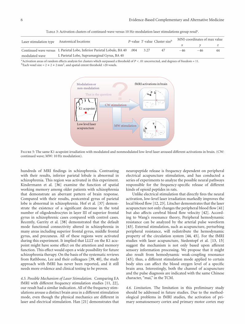
Figure 5: The same K1 acupoint irradiation with modulated and nonmodulated low-level laser aroused different activations in brain. (CW:continued wave; MW: 10 Hz modulation).
hundreds of MRI findings in schizophrenia. Contrastingwith their results, inferior parietal lobule is abnormal inschizophrenia. This region was activated in this experiment.Kindermann et al. [36] examine the function of spatialworking memory among older patients with schizophreniathat demonstrate an aberrant pattern of brain response.Compared with their results, postcentral gyrus of parietallobe is abnormal in schizophrenia. Hof et al. [37] demon-strate the existence of a significant decrease in the totalnumber of oligodendrocytes in layer III of superior frontalgyrus in schizophrenic cases compared with control cases.Recently, Garrity et al. [38] demonstrated that the defaultmode functional connectivity altered in schizophrenia inmany areas including superior frontal gyrus, middle frontalgyrus, and precuneus. All of these regions were activatedduring this experiment. It implied that LLLT on the K1 acu-point might have some effect on the attention and memoryfunction. This effect would open a wide possibility for futureschizophrenia therapy. On the basis of the systematic reviewsfrom Rathbone, Lee and their colleagues [39, 40], the studyapproach with fMRI has never been reported, and it stillneeds more evidence and clinical testing to be proven.
4.5. Possible Mechanism of Laser Stimulation. Comparing EAfMRI with different frequency stimulation studies [11, 22],our result had a similar indication. All of the frequency stim-ulations arouse a distinct brain area in a different stimulationmode, even though the physical mechanics are different inlaser and electrical stimulation. Han [21] demonstrates that
neuropeptide release is frequency dependent on peripheralelectrical acupuncture stimulation, and has conducted aseries of experiments to analyze the possible neural pathwaysresponsible for the frequency-specific release of differentkinds of opioid peptides in rats.
Unlike electrical stimulation that directly fires the neuralactivation, low-level laser irradiation markedly improves thelocal blood flow [12, 23]. Litscher demonstrates that the laseracupuncture not only changes the peripheral blood flow [41]but also affects cerebral blood flow velocity [42]. Accord-ing to Wang’s resonance theory, Peripheral hemodynamicresistance can be analyzed by the arterial pulse waveform[43]. External stimulation, such as acupuncture, perturbingperipheral resistance, will redistribute the hemodynamicproperty of the circulation system [44, 45]. For the fMRIstudies with laser acupuncture, Siedentopf et al. [13, 15]suggest the mechanism is not only based upon afferentsensory information processing. We propose that it mightalso result from hemodynamic weak-coupling resonance[43]; thus, a different stimulation mode applied to certainbody sites can affect the blood oxygen level of a specificbrain area. Interestingly, both the channel of acupunctureand the pulse diagnosis are indicated with the same Chinesecharacter, “mai,” in the TCM.
4.6. Limitation. The limitation in this preliminary studyshould be addressed in future studies. Due to the method-ological problems in fMRI studies, the activation of pri-mary somatosensory cortex and primary motor cortex may
Evidence-Based Complementary and Alternative Medicine 7
result from the spontaneous fluctuation of resting stateduring 1-munite block. According to the investigation offrequency specificity in resting fMRI [19], 1-munite blocklength (0.008 Hz) should be much less functional connec-tivity in sensorimotor system than 30 s-block (0.016 Hz).Even though it had been considered the impact of rest-ing state activity [46], primary somatosensory cortex andprimary motor cortex were still aroused in this experi-ment.
5. Conclusion
This study presented the functional brain mapping with thetwo stimulation modes of LLLT applied on the K1 acupoint.Attention- and memory-task-related brain regions wereactivated during LLLT on the K1 acupoint. Different brainregions rearranged the blood oxygen levels during differentstimulation modes, even though the LLLT was applied tothe same acupoint (Figure 5). The results imply that themechanism of acupuncture is not only based upon afferentsensory information processing, but also relates to thehemodynamic property altered during external stimulation.
Acknowledgments
The authors are grateful to the National Science Councilof the Republic of China for financially supporting thisresearch under Contracts NSC 97-2321-B-002-044 and NSC98-2627-B-002-022. They appreciate Dr. Der-Yow Chen (theLaboratory of Functional and Molecular Imaging, NIH,USA) for his suggestions on the statistical analysis. Andthey also thank the Instrumentation Center, National TaiwanUniversity, for MRI experiments.
References
[1] L. Dorfer, M. Moser, F. Bahr et al., “A medical report from thestone age?” Lancet, vol. 354, no. 9183, pp. 1023–1025, 1999.
[2] G. T. Lewith, P. J. White, and J. Pariente, “Investigatingacupuncture using brain imaging techniques: the currentstate of play,” Evidence-Based Complementary and AlternativeMedicine, vol. 2, no. 3, pp. 315–319, 2005.
[3] Z. H. Cho, S. C. Chung, J. P. Jones et al., “New findings ofthe correlation between acupoints and corresponding braincortices using functional MRI,” Proceedings of the NationalAcademy of Sciences of the United States of America, vol. 95, no.5, pp. 2670–2673, 1998.
[4] K. K. S. Hui, J. Liu, N. Makris et al., “Acupuncture modulatesthe limbic system and subcortical gray structures of thehuman brain: evidence from fMRI studies in normal subjects,”Human Brain Mapping, vol. 9, no. 1, pp. 13–25, 2000.
[5] M.-T. Wu, J.-M. Sheen, K.-H. Chuang et al., “Neuronalspecificity of acupuncture response: a fMRI study withelectroacupuncture,” NeuroImage, vol. 16, no. 4, pp. 1028–1037, 2002.
[6] C. M. Siedentopf, S. M. Golaszewski, F. M. Mottaghy, C.C. Ruff, S. Felber, and A. Schlager, “Functional magneticresonance imaging detects activation of the visual associationcortex during laser acupuncture of the foot in humans,”Neuroscience Letters, vol. 327, no. 1, pp. 53–56, 2002.
[7] J. Kong, L. Ma, R. L. Gollub et al., “A pilot study of functionalmagnetic resonance imaging of the brain during manual andelectroacupuncture stimulation of acupuncture point (LI-4Hegu) in normal subjects reveals differential brain activationbetween methods,” Journal of Alternative and ComplementaryMedicine, vol. 8, no. 4, pp. 411–419, 2002.
[8] S.-S. Yoo, E.-K. Teh, R. A. Blinder, and F. A. Jolesz, “Mod-ulation of cerebellar activities by acupuncture stimulation:evidence from fMRI study,” NeuroImage, vol. 22, no. 2, pp.932–940, 2004.
[9] K. K. S. Hui, J. Liu, O. Marina et al., “The integratedresponse of the human cerebro-cerebellar and limbic systemsto acupuncture stimulation at ST 36 as evidenced by fMRI,”NeuroImage, vol. 27, no. 3, pp. 479–496, 2005.
[10] J. Kong, R. L. Gollub, I. S. Rosman et al., “Brain activityassociated with expectancy-enhanced placebo analgesia asmeasured by functional magnetic resonance imaging,” Journalof Neuroscience, vol. 26, no. 2, pp. 381–388, 2006.
[11] V. Napadow, N. Makris, J. Liu, N. W. Kettner, K. K. Kwong,and K. K. S. Hui, “Effects of electroacupuncture versus manualacupuncture on the human brain as measured by fMRI,”Human Brain Mapping, vol. 24, no. 3, pp. 193–205, 2005.
[12] P. Whittaker, “Laser acupuncture: past, present, and future,”Lasers in Medical Science, vol. 19, no. 2, pp. 69–80, 2004.
[13] C. M. Siedentopf, S. M. Golaszewski, F. M. Mottaghy, C.C. Ruff, S. Felber, and A. Schlager, “Functional magneticresonance imaging detects activation of the visual associationcortex during laser acupuncture of the foot in humans,”Neuroscience Letters, vol. 327, no. 1, pp. 53–56, 2002.
[14] G. Litscher, D. Rachbauer, S. Ropele et al., “Acupuncture usinglaser needles modulates brain function: first evidence fromfunctional transcranial Doppler sonography and functionalmagnetic resonance imaging,” Lasers in Medical Science, vol.19, no. 1, pp. 6–11, 2004.
[15] C. M. Siedentopf, F. Koppelstaetter, I. A. Haala et al., “Laseracupuncture induced specific cerebral cortical and subcorticalactivations in humans,” Lasers in Medical Science, vol. 20, no.2, pp. 68–73, 2005.
[16] C. M. Siedentopf, A. Ischebeck, I. A. Haala et al., “Neuralcorrelates of transmeatal cochlear laser (TCL) stimulation inhealthy human subjects,” Neuroscience Letters, vol. 411, no. 3,pp. 189–193, 2007.
[17] C. Shang, “Prospective tests on biological models ofacupuncture,” Evidence-Based Complementary and AlternativeMedicine, vol. 6, no. 1, pp. 31–39, 2009.
[18] R. P. Dhond, C. Yeh, K. Park, N. Kettner, and V. Napadow,“Acupuncture modulates resting state connectivity in defaultand sensorimotor brain networks,” Pain, vol. 136, no. 3, pp.407–418, 2008.
[19] C. W. Wu, H. Gu, H. Lu, E. A. Stein, J.-H. Chen, and Y. Yang,“Frequency specificity of functional connectivity in brainnetworks,” NeuroImage, vol. 42, no. 3, pp. 1047–1055, 2008.
[20] G. A. Ulett, S. Han, and J.-S. Han, “Electroacupuncture:mechanisms and clinical application,” Biological Psychiatry,vol. 44, no. 2, pp. 129–138, 1998.
[21] J.-S. Han, “Acupuncture: neuropeptide release produced byelectrical stimulation of different frequencies,” Trends inNeurosciences, vol. 26, no. 1, pp. 17–22, 2003.
[22] W.-T. Zhang, Z. Jin, G.-H. Cui et al., “Relations betweenbrain network activation and analgesic effect induced by lowvs. high frequency electrical acupoint stimulation in differentsubjects: a functional magnetic resonance imaging study,”Brain Research, vol. 982, no. 2, pp. 168–178, 2003.

8 Evidence-Based Complementary and Alternative Medicine
[23] M. Komori, K. Takada, Y. Tomizawa et al., “Microcirculatoryresponses to acupuncture stimulation and phototherapy,”Anesthesia and Analgesia, vol. 108, no. 2, pp. 635–640, 2009.
[24] S. C. Lee, “The generation of ‘chi’ by stimulation methods,”Bulletin of the College of Engineering, vol. 46, pp. 117–125, 1989(Chinese).
[25] J. Kong, L. Ma, R. L. Gollub et al., “A pilot study of functionalmagnetic resonance imaging of the brain during manual andelectroacupuncture stimulation of acupuncture point (LI-4Hegu) in normal subjects reveals differential brain activationbetween methods,” Journal of Alternative and ComplementaryMedicine, vol. 8, no. 4, pp. 411–419, 2002.
[26] M. F. S. Rushworth, M. E. Walton, S. W. Kennerley, and D. M.Bannerman, “Action sets and decisions in the medial frontalcortex,” Trends in Cognitive Sciences, vol. 8, no. 9, pp. 410–417,2004.
[27] J. V. Pardo, P. T. Fox, and M. E. Raichle, “Localization of ahuman system for sustained attention by positron emissiontomography,” Nature, vol. 349, no. 6304, pp. 61–64, 1991.
[28] I. H. Jenkins, D. J. Brooks, P. D. Nixon, R. S. J. Frackowiak,and R. E. Passingham, “Motor sequence learning: a study withpositron emission tomography,” Journal of Neuroscience, vol.14, no. 6, pp. 3775–3790, 1994.
[29] T. J. Vickery and Y. V. Jiang, “Inferior parietal lobule supportsdecision making under uncertainty in humans,” CerebralCortex, vol. 19, no. 4, pp. 916–925, 2009.
[30] A. D. Wagner, B. J. Shannon, I. Kahn, and R. L. Buckner,“Parietal lobe contributions to episodic memory retrieval,”Trends in Cognitive Sciences, vol. 9, no. 9, pp. 445–453, 2005.
[31] G. McCarthy, A. Puce, R. T. Constable, J. H. Krystal, J. C.Gore, and P. Goldman-Rakic, “Activation of human prefrontalcortex during spatial and nonspatial working memory tasksmeasured by functional MRI,” Cerebral Cortex, vol. 6, no. 4,pp. 600–611, 1996.
[32] A. E. Cavanna and M. R. Trimble, “The precuneus: a review ofits functional anatomy and behavioural correlates,” Brain, vol.129, no. 3, pp. 564–583, 2006.
[33] G. Maciocia, The Channels of Acupuncture: Clinical Use of theSecondary Channels and Eight Extraordinary Vessels, ChurchillLivingstone: Elsevier, Oxford, UK, 2006.
[34] G. Kuperberg and S. Heckers, “Schizophrenia and cognitivefunction,” Current Opinion in Neurobiology, vol. 10, no. 2, pp.205–210, 2000.
[35] M. E. Shenton, C. C. Dickey, M. Frumin, and R. W. McCarley,“A review of MRI findings in schizophrenia,” SchizophreniaResearch, vol. 49, no. 1-2, pp. 1–52, 2001.
[36] S. S. Kindermann, G. G. Brown, L. E. Zorrilla, R. K. Olsen, andD. V. Jeste, “Spatial working memory among middle-aged andolder patients with schizophrenia and volunteers using fMRI,”Schizophrenia Research, vol. 68, no. 2-3, pp. 203–216, 2004.
[37] P. R. Hof, V. Haroutunian, V. L. Friedrich Jr. et al., “Loss andaltered spatial distribution of oligodendrocytes in the superiorfrontal gyrus in schizophrenia,” Biological Psychiatry, vol. 53,no. 12, pp. 1075–1085, 2003.
[38] A. G. Garrity, G. D. Pearlson, K. McKiernan, D. Lloyd, K. A.Kiehl, and V. D. Calhoun, “Aberrant “default mode” functionalconnectivity in schizophrenia,” American Journal of Psychiatry,vol. 164, no. 3, pp. 450–457, 2007.
[39] M. S. Lee, B.-C. Shin, P. Ronan, and E. Ernst, “Acupuncturefor schizophrenia: a systematic review and meta-analysis,”International Journal of Clinical Practice, vol. 63, no. 11, pp.1622–1633, 2009.
[40] J. Rathbone and J. Xia, “Acupuncture for schizophrenia,”Cochrane Database of Systematic Reviews, no. 4, Article IDCD005475, 2005.
[41] G. Litscher, “Ten years evidence-based High-tech acupunc-turea short review of peripherally measured effects,” Evidence-Based Complementary and Alternative Medicine, vol. 6, no. 2,pp. 153–158, 2009.
[42] G. Litscher, “Ten years evidence-based high-techacupuncture—a short review of centrally measured effects(part II),” Evidence-Based Complementary and AlternativeMedicine, vol. 6, no. 3, pp. 305–314, 2009.
[43] Y. Y. L. Wang, S. L. Chang, Y. E. Wu, T. L. Hsu, and W. K. Wang,“Resonance: the missing phenomenon in hemodynamics,”Circulation Research, vol. 69, no. 1, pp. 246–249, 1991.
[44] W. K. Wang, T. L. Hsu, H. C. Chang, and Y. Y. Wang, “Effectof acupuncture at Tsu San Li (St-36) on the pulse spectrum,”The American Journal of Chinese Medicine, vol. 23, no. 2, pp.121–130, 1995.
[45] W. K. Wang, T. L. Hsu, H. C. Chang, and Y. Y. L. Wang, “Effectof acupuncture at Tai-Tsih (k-3) on the pulse spectrum,”American Journal of Chinese Medicine, vol. 24, no. 3-4, pp.305–313, 1996.
[46] F. Beissner and C. Henke, “Methodological problems in fMRIstudies on acupuncture: a critical review with special emphasison visual and auditory cortex activations,” Evidence-BasedComplementary and Alternative Medicine. In press.

Submit your manuscripts athttp://www.hindawi.com
Stem CellsInternational
Hindawi Publishing Corporationhttp://www.hindawi.com Volume 2014
Hindawi Publishing Corporationhttp://www.hindawi.com Volume 2014
MEDIATORSINFLAMMATION
of
Hindawi Publishing Corporationhttp://www.hindawi.com Volume 2014
Behavioural Neurology
EndocrinologyInternational Journal of
Hindawi Publishing Corporationhttp://www.hindawi.com Volume 2014
Hindawi Publishing Corporationhttp://www.hindawi.com Volume 2014
Disease Markers
Hindawi Publishing Corporationhttp://www.hindawi.com Volume 2014
BioMed Research International
OncologyJournal of
Hindawi Publishing Corporationhttp://www.hindawi.com Volume 2014
Hindawi Publishing Corporationhttp://www.hindawi.com Volume 2014
Oxidative Medicine and Cellular Longevity
Hindawi Publishing Corporationhttp://www.hindawi.com Volume 2014
PPAR Research
The Scientific World JournalHindawi Publishing Corporation http://www.hindawi.com Volume 2014
Immunology ResearchHindawi Publishing Corporationhttp://www.hindawi.com Volume 2014
Journal of
ObesityJournal of
Hindawi Publishing Corporationhttp://www.hindawi.com Volume 2014
Hindawi Publishing Corporationhttp://www.hindawi.com Volume 2014
Computational and Mathematical Methods in Medicine
OphthalmologyJournal of
Hindawi Publishing Corporationhttp://www.hindawi.com Volume 2014
Diabetes ResearchJournal of
Hindawi Publishing Corporationhttp://www.hindawi.com Volume 2014
Hindawi Publishing Corporationhttp://www.hindawi.com Volume 2014
Research and TreatmentAIDS
Hindawi Publishing Corporationhttp://www.hindawi.com Volume 2014
Gastroenterology Research and Practice
Hindawi Publishing Corporationhttp://www.hindawi.com Volume 2014
Parkinson’s Disease
Evidence-Based Complementary and Alternative Medicine
Volume 2014Hindawi Publishing Corporationhttp://www.hindawi.com