differential effect of tea on long-term synaptic ...bmsr.usc.edu/files/2012/09/375.pdf ·...
TRANSCRIPT

Neurobiology of Learning and Memory 76, 375–387 (2001)
doi:10.1006/nlme.2001.4032, available online at http://www.idealibrary.com on
Differential Effect of TEA on Long-Term SynapticModification in Hippocampal CA1 and Dentate
Gyrus in vitro
Dong Song,*,‡,§ Xiaping Xie,*,‡,§ Zhuo Wang,†,‡,§ and Theodore W. Berger*,‡,§
*Department of Biomedical Engineering, †Department of Biological Sciences, ‡Program in Neuroscience,§Center for Neural Engineering, University of Southern California, Los Angeles, California 90089
The effectiveness of tetraethylammonium (TEA) and high-frequency stimulation(HFS) in inducing long-term synaptic modification is compared in CA1 and dentategyrus (DG) in vitro. High-frequency stimulation induces long-term potentiation(LTP) at synapses of both perforant path-DG granule cell and Schaffer collateral-CA1 pyramidal cell pathways. By contrast, TEA (25 mM) induces long-term depres-sion in DG while inducing LTP in CA1. The mechanisms underlying the differentialeffect of TEA in CA1 and DG were investigated. It was observed that T-type voltage-dependent calcium channel (VDCC) blocker, Ni2+ (50 mM), partially blocked TEA-induced LTP in CA1. A complete blockade of the TEA-induced LTP occurred whenNi2+ was applied together with the NMDA receptor antagonist, D-APV. TheL-type VDCC blocker, nifidipine (20 mM), had no effect on CA1 TEA-inducedLTP. In DG of the same slice, TEA actually induced long-term depression (LTD)instead of LTP, an effect that was blocked by D-APV. Neither T-type nor L-typeVDCC blockade could prevent this LTD. When the calcium concentration in theperfusion medium was increased, TEA induced a weak LTP in DG that was blockedby Ni2+. During exposure to TEA, the magnitude of field EPSPs was increased inboth CA1 and DG, but the increase was substantially greater in CA1. Tetraethylam-monium application also was associated with a large, late EPSP component in CA1that persisted even after severing the connections between CA3 and CA1. All ofthe TEA effects in CA1, however, were dramatically reduced by Ni2+. The resultsof this study indicate that TEA indirectly acts via both T-type VDCCs and NMDAreceptors in CA1 and, as a consequence, induces LTP. By contrast, TEA indirectlyacts via only NMDA receptors in DG and results in LTD. The results raise thepossibility of a major synaptic difference in the density and/or distribution ofT-type VDCCs and NMDA receptors in CA1 and DG of the rat hippocampus.q 2001 Academic Press
This research was supported by the National Institute of Mental Health (MH51722 and MH00343) and theOffice of Naval Research (N00014-98-1-0259).
Address correspondence and reprint requests to Dong Song, 403 HNB, University Park, University of SouthernCalifornia, Los Angeles, CA 90089. Fax: (213) 740-5687. E-mail: [email protected].
375 1074-7427/01 $35.00Copyright q 2001 by Academic Press
All rights of reproduction in any form reserved.
376 SONG ET AL.
Key Words: LTP; LTD; hippocampus; TEA; NMDA receptor; voltage-dependentcalcium channel.
INTRODUCTION
Various intracellular concentrations of calcium, [Ca2+]i, play a determinant role ininducing either long-term potentiation (LTP) (Jahr & Stevens, 1987; Lynch, Larson, Kelso,Barrionuevo, & Schottler, 1983; Malenka, 1991; Mayer & Westbrook, 1987) or long-termdepression (LTD) (Christofi, Novicky, & Bindman, 1991; Dudek & Bear, 1993; Hirsch &Crepel, 1992) of glutamatergic synapses. Namely, large increases in [Ca2+]i lead to theinduction of LTP, whereas small increases in [Ca2+]i result in LTD (Lisman, 1989). Theincreased intracellular calcium needed for the induction of LTP or LTD can be introducedvia multiple calcium-conducting channels. The induction of NMDA receptor-dependentLTP/LTD requires calcium influx into the postsynaptic region via the activated NMDAreceptor/channel. High-frequency stimulation (HFS) can induce LTP in both CA1 anddentate gyrus (DG) regions through activation of NMDA receptors (Bliss & Collingridge,1993). It is also possible to induce an LTP that is NMDA receptor independent by derivingcalcium from other sources. One of the sources is voltage-dependent calcium channels(VDCCs) (Grover & Teyler, 1990; Miyakawa et al., 1992). Bath application of the K+
channel blocker, tetraethylammonium (TEA), for 7 to 10 min could induce a novel typeof LTP (TEA LTP) in CA1 region (Aniksztejn & Ben-Ari, 1991). The induction of TEALTP was first shown to be NMDA receptor independent because the NMDA receptorantagonist, D-APV, failed to block the induction of TEA LTP (Aniksztejn & Ben-Ari,1991). Although others later reported that TEA LTP also partially depended on theactivation of the NMDA receptors (Huber, Mauk, & Kelly, 1995), all studies have agreedthat the critical induction event for TEA LTP was calcium influx via ionic channels otherthan NMDA receptors/channels, for example, L-type VDCC (Aniksztejn & Ben-Ari, 1991;Huang & Malenka, 1993). In contrast to the multiple studies of CA1, the effect of TEAin the DG region has not been investigated intensively. The only report available suggeststhat TEA also induced LTP in DG and that the TEA LTP was T-type VDCC dependent(Coogan, O’Leary, & O’Connor, 1999). Until now, no studies have been conducted tocompare TEA effect on these two regions in the same in vitro slice.
In this study, we first conducted a series of experiments in which field excitatorypostsynaptic potentials (EPSPs) were recorded simultaneously from DG and CA1 of asingle slice. Differential effects of TEA both on the induction of LTP and on the profileof EPSPs in the two regions were found and investigated. Our results demonstrate thatthe induction of TEA LTP in CA1 occurs via both NMDA-dependent and NMDA-independent mechanisms. The T-type VDCC is the main mediator for the NMDA-indepen-dent component in CA1. For the DG, by contrast, TEA could induce neither NMDA-dependent nor NMDA-independent LTP but instead induced an NMDA-dependent LTD(TEA LTD).
METHODS
Hippocampal slices were prepared from adult male Sprague–Dawley rats (200–250g). Animals first were anesthetized with 5% halothane, then were decapitated, and the
TEA AND LONG-TERM SYNAPTIC MODIFICATION 377
hippocampi then were rapidly dissected. Both hippocampi were sectioned into blockswhile being washed with cold oxygenated medium, and slices of tissue (400 micronsthick) then were cut perpendicular to the longitudinal axis using a vibratome. Slices wereincubated with medium consisting of 128 mM NaCl, 5 mM KCl, 1.25 mM NaH2PO4,26 mM NaHCO3, 10 mM glucose, 2 mM CaCl2, and 2 mM MgSO4, aerated with 95%O2/5% CO2. Hippocampal slices were maintained at 328C throughout the entire experi-ments. During the recording session, slices were transferred to the recording chamber andperfused at flow rates of 4 to 6 ml/min; the perfusion medium was changed to include 1mM MgSO4 and 100 mM picrotoxin (Sigma). Bipolar nichrome stimulating electrodeswere placed in the medial perforant path and Schaffer collateral to orthodromically activatedentate granule cells and CA1 pyramidal cells, respectively. A cut was made between theCA3 and CA1 regions to prevent epileptiform activity in the CA3 region from affectingthe recording in CA1. During the application of TEA (25 mM, Sigma), the concentrationof NaCl was changed correspondingly to maintain constant osmolarity of the perfusionmedium. D-APV (50 mM, Tocris), nickel chloride (50 mM, Aldrich), or nifidipine (20mM, Sigma) was added to the perfusion medium when used. Nifidipine was made dailyas a 10-mM stock in dimethyl sulfoxide (DMSO), stored in dark, and diluted to 20 mMfinal concentration in the perfusion medium immediately prior to application. Applicationof the DMSO (0.05%) alone had no effect on the LTP and EPSPs. Field EPSPs of theDG granule cell and/or CA1 pyramidal cell were evoked at a frequency of 0.1 Hz by 0.1ms duration impulses (intensity: 0.04–0.12 mA) and were recorded with microelectrodes.The extracellular field EPSPs of CA1 were recorded in stratum radiatum, and the extracellu-lar field EPSPs of DG were recorded in the middle third of the molecular layer. Theextracellular recording pipettes were filled with 2 M NaCl (resistance: 1–2 MV). Theintracellular recording pipettes were filled with 3 M potassium acetate (impedance: 100MV). High-frequency stimulation consisted of four stimulation trains separated by 5-sintervals. Each train had 10 impulses at a frequency of 100 Hz and was delivered at thesame intensity as that used to evoke baseline responses. All evoked responses wereamplified, digitized, and stored using a PC. Data from different slices were combined bynormalizing amplitudes of EPSPs relative to the average response amplitudes measuredduring the control period. Student’s t test was used for statistical comparisons.
RESULTS
TEA Induces Robust LTP in CA1 but Weak LTD in DG
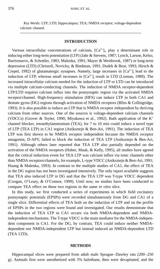
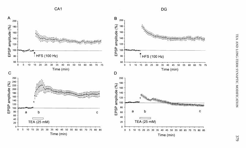
The effectiveness of TEA and high-frequency stimulation in inducing synaptic modifi-cation was compared for CA1 and dentate gyrus by recording simultaneously from bothsites of a single slice. High-frequency stimulation induced robust LTP in both CA1 andDG (Figs. 1A and 1B). Tetraethylammonium (25 mM) also induced robust LTP in CA1that lasted at least 60 min (Fig. 1C). The amplitude of TEA LTP in CA1, measured50 to 60 min following 10 min of TEA application, was 74 6 13% of baseline ( p ,
.001, n 5 9). This TEA LTP did not significantly occlude the HFS LTP (data not shown).In contrast to the effects of TEA in CA1, a weak but significant LTD was induced in DG30 to 60 min following TEA application (Fig. 1D). The amplitude of this LTD was212 6 3% of baseline 50 to 60 min after TEA application ( p , .01, n 5 14).
378 SONG ET AL.
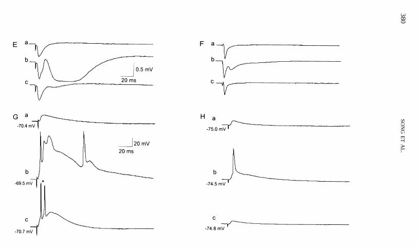
Another marked differential effect of TEA, which might be the cause for the abovedifferential effect on the synaptic modification, was observed on the EPSP profile duringTEA application. In CA1, a large late component in field EPSPs that lasted . 100 mswas observed with a 15-ms latency (Fig. 1E(b)). In DG, a short, rapidly decayed latecomponent in field EPSPs was observed with a 15-ms latency (Fig. 1F(b)).
Intracellular recording showed that there was no significant change in resting membranepotentials before, during, or after TEA application. Bursts of action potentials wereobserved in CA1 pyramidal cells (Fig. 1G) but not in DG granule cells (Fig. 1H). Consistentwith the field EPSP results, TEA induced potentiation of the intracellular EPSPs in CA1(n 5 3), but depression in DG measured 60 min after termination of TEA application(n 5 4).
L-Type VDCC Blocker Nifidipine Does Not Block the TEA LTP/LTD in CA1/DG
The role of L-type VDCCs in the induction of TEA LTP/LTD was investigated byincluding nifidipine (20 mM) in the perfusion medium. Results showed that nifidipinedid not block the TEA LTP in CA1 or TEA LTD in DG. The amplitude of TEA LTP inCA1, measured at 50 to 60 min after TEA application, was 61 6 9% of baseline ( p .
.10, n 5 5) (Fig. 2A). In the same slice, the amplitude of TEA LTD in DG, measured50 to 60 min after TEA application, was 29 6 4% of baseline ( p . .10, n 5 4) (Fig. 2B).
D-APV Partially Blocks the TEA LTP in CA1 and Completely Blocks the TEA LTDin DG
To determine the contribution of NMDA receptors to the induction of TEA LTP/LTD,D-APV (50 mM) was applied to the bath. Recordings showed that D-APV did not alterthe baseline amplitude of EPSPs in either CA1 or DG. However, D-APV partially blockedthe induction of TEA LTP (29 6 6%, p , .001, n 5 5) (Fig. 2C). In the same slice, theTEA LTD in DG was blocked completely (1 6 6%, p , .05, n 5 5) (Fig. 2D).
Ni2+ Partially Blocks the TEA LTP in CA1, and a Combination of Ni2+ and D-APVStrongly Blocks the TEA LTP in CA1
The role of T-type VDCCs in the induction of TEA LTP in CA1 was investigated. Ni2+
(50 mM), a T-type VDCC blocker, did not alter the amplitude of the baseline EPSPs. But
FIG. 1. Comparison of HFS- and TEA-induced synaptic modification in CA1 and DG. (A) HFS (100 Hz)induced LTP in CA1. (B) HFS (100 Hz) induced LTP in DG. (C) TEA (25 mM) induced LTP in CA1. (D)TEA (25 mM) failed to induce LTP in DG. A 10-min bath application of 25 mM TEA resulted in a weak butsignificant LTD in DG. In all of the following graphs: (a) before TEA application; (b) during TEA application;(c) 60 min after wash-out of TEA. (E) Representative field EPSPs recorded from stratum radiatum in CA1.Note that TEA resulted in a large late component in addition to enhancing the fast component of EPSPs. (F)Representative field EPSPs recorded from the middle third of the molecular layer in DG. (G) Representativeintracellular EPSPs recorded from a pyramidal cell in CA1. Note that membrane potentials were barely changedduring TEA application in both pyramidal and granule cells. Bursts of action potentials were present in theCA1 pyramidal cell (*) but not in the DG granule cell. (H) Representative intracellular EPSPs recorded froma granule cell in DG.
TE
AA
ND
LO
NG
-TE
RM
SYN
APT
ICM
OD
IFICA
TIO
N379
380SO
NG
ET
AL
.

TE
AA
ND
LO
NG
-TE
RM
SYN
APT
ICM
OD
IFICA
TIO
N381

382SO
NG
ET
AL
.
FIG. 2. The induction of TEA LTP in CA1 is both NMDA receptor and T-type VDCC dependent, whereas TEA LTD in DG is only NMDA dependent. (A) L-type VDCC blocker, nifidipine (20 mM) did not block the TEA LTP in CA1 (open circles). In all graphs, closed circles represent the TEA LTP/LTD under controlcondition. (B) Nifidipine (20 mM) did not block the TEA LTD in DG (open circles). (C) D-APV (50 mM) partially blocked the TEA LTP in CA1 (open circles). (D)D-APV (50 mM) blocked the TEA LTD in DG (open circles). (E) Ni2+ (50 mM) partially blocked the TEA LTP in CA1 (open circles). A combined application ofNi2+ (50 mM) and D-APV (50 mM) strongly blocked the TEA LTP in CA1 (closed triangles). (F) Ni2+ (50 mM) did not block the TEA LTD in DG (open circles).
TEA AND LONG-TERM SYNAPTIC MODIFICATION 383
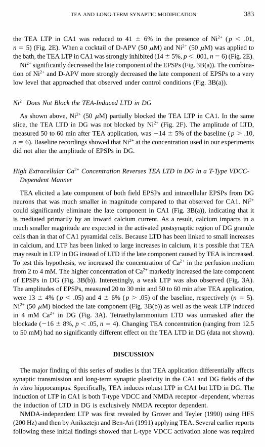
the TEA LTP in CA1 was reduced to 41 6 6% in the presence of Ni2+ ( p , .01,n 5 5) (Fig. 2E). When a cocktail of D-APV (50 mM) and Ni2+ (50 mM) was applied tothe bath, the TEA LTP in CA1 was strongly inhibited (14 6 5%, p , .001, n 5 6) (Fig. 2E).
Ni2+ significantly decreased the late component of the EPSPs (Fig. 3B(a)). The combina-tion of Ni2+ and D-APV more strongly decreased the late component of EPSPs to a verylow level that approached that observed under control conditions (Fig. 3B(a)).
Ni2+ Does Not Block the TEA-Induced LTD in DG
As shown above, Ni2+ (50 mM) partially blocked the TEA LTP in CA1. In the sameslice, the TEA LTD in DG was not blocked by Ni2+ (Fig. 2F). The amplitude of LTD,measured 50 to 60 min after TEA application, was 214 6 5% of the baseline ( p . .10,n 5 6). Baseline recordings showed that Ni2+ at the concentration used in our experimentsdid not alter the amplitude of EPSPs in DG.
High Extracellular Ca2+ Concentration Reverses TEA LTD in DG in a T-Type VDCC-Dependent Manner
TEA elicited a late component of both field EPSPs and intracellular EPSPs from DGneurons that was much smaller in magnitude compared to that observed for CA1. Ni2+
could significantly eliminate the late component in CA1 (Fig. 3B(a)), indicating that itis mediated primarily by an inward calcium current. As a result, calcium impacts in amuch smaller magnitude are expected in the activated postsynaptic region of DG granulecells than in that of CA1 pyramidal cells. Because LTD has been linked to small increasesin calcium, and LTP has been linked to large increases in calcium, it is possible that TEAmay result in LTP in DG instead of LTD if the late component caused by TEA is increased.To test this hypothesis, we increased the concentration of Ca2+ in the perfusion mediumfrom 2 to 4 mM. The higher concentration of Ca2+ markedly increased the late componentof EPSPs in DG (Fig. 3B(b)). Interestingly, a weak LTP was also observed (Fig. 3A).The amplitudes of EPSPs, measured 20 to 30 min and 50 to 60 min after TEA application,were 13 6 4% ( p , .05) and 4 6 6% ( p . .05) of the baseline, respectively (n 5 5).Ni2+ (50 mM) blocked the late component (Fig. 3B(b)) as well as the weak LTP inducedin 4 mM Ca2+ in DG (Fig. 3A). Tetraethylammonium LTD was unmasked after theblockade (216 6 8%, p , .05, n 5 4). Changing TEA concentration (ranging from 12.5to 50 mM) had no significantly different effect on the TEA LTD in DG (data not shown).
DISCUSSION
The major finding of this series of studies is that TEA application differentially affectssynaptic transmission and long-term synaptic plasticity in the CA1 and DG fields of thein vitro hippocampus. Specifically, TEA induces robust LTP in CA1 but LTD in DG. Theinduction of LTP in CA1 is both T-type VDCC and NMDA receptor -dependent, whereasthe induction of LTD in DG is exclusively NMDA receptor dependent.
NMDA-independent LTP was first revealed by Grover and Teyler (1990) using HFS(200 Hz) and then by Aniksztejn and Ben-Ari (1991) applying TEA. Several earlier reportsfollowing these initial findings showed that L-type VDCC activation alone was required
384SO
NG
ET
AL
.
FIG. 3. The TEA-elicited late component in DG is increased by raising extracellular Ca2+ concentration, resulting in the reversal of TEA LTD. The late componentin both CA1 and DG is mediated chiefly by T-type VDCC. (A) In medium with doubled Ca2+ concentration (4 mM), TEA induced weak LTP in DG (closed circles).This LTP was reversed by Ni2+ (50 mM) application (open circles). (B(a)) EPSP waveforms during the TEA application in CA1. In both panels B(a) and B(b), EPSPswere first averaged and normalized within single slices and then averaged across different slices (n 5 9). Note that the large late component was partially blockedby Ni2+ (50 mM) application (medium line) and strongly blocked by the combined application (thin line) of Ni2+ (50 mM) and D-APV (50 mM), compared to the latecomponent under control condition (thick line). (B(b)) EPSP waveforms during the TEA application in DG (n 5 14). Note that in 4 mM Ca2+ medium (medium line),the late component increased significantly (*) compared to the late component in 2 mM Ca2+ medium (thick line). This increase was blocked by Ni2+ (50 mM)application (thin line).
TEA AND LONG-TERM SYNAPTIC MODIFICATION 385
for the induction of this type of LTP in CA1 (Huang & Malenka, 1993). Subsequently,it was shown that TEA could induce LTP via both VDCC- and NMDA receptor-mediatedmechanisms (Huber et al., 1995). However, there is still controversy concerning theidentity of the VDCC channel type involved. Consistent with an earlier report (Hanse &Gustafsson, 1994), we found that the VDCC component was not blocked by the L-typeVDCC blocker nifidipine. We report here that it instead is blocked by Ni2+, indicatingthat T-type VDCCs are more likely to be involved in the induction process. The involvementof the T-type VDCC rather than other types of VDCCs in the induction of TEA LTPreceives further support from the comparison between the properties of the T-type VDCCand those of other types of VDCCs. For example, the voltage range in which the T-typeVDCC is activated (i.e., from 250 to 170 mV peaked at 0 mV) is substantially lowerthan that of the other types of VDCCs such as the L-type, which has an activation rangebetween 220 and 170 mV peaked at 130 mV (Bean, 1985). In fact, because of thevoltage activation property of the T-type VDCC, sub-threshold EPSPs in dendrites areable to open the T-type VDCC and induce a local increase in intracellular calcium (Mageeet al., 1995). Tetraethylammonium has been recently shown to be able to induce LTP inDG (Coogan et al., 1999). In their study, however, Coogan et al. (1999) used youngerrats, ,70–150 g, which would correspond to ,25 to 40 days old, whereas we conductedour experiments using rats more than 2 months old. Therefore, age may be a factor indetermining whether TEA induces LTP or LTD in DG such as down-regulation of T-typeVDCC during this restricted developmental period. We did observe that a weak LTP couldbe induced by TEA application when the concentration of calcium in the perfusion mediumwas raised to 4 mM.
Several mechanisms, either singularly or in combination, may be responsible for thedifferential cell region-specific effect of TEA in eliciting the enhanced late EPSP compo-nent and in inducing LTP/LTD. The first involves factors that govern the magnitude ofcalcium currents. It is known that the resting membrane potential of DG granule cells invitro is more hyperpolarized than that of CA1 pyramidal cells (Schwartzkroin, 1975;Staley, Otis, & Mody, 1992). Consequently, the same level of depolarization applied toDG and CA1 cell fields will result in lower levels of VDCC and NMDA receptor/channelactivation in DG compared to CA1. Thus, TEA application would be expected to resultin lower levels of calcium influx in DG relative to CA1. Long-term depression inducedin DG has been demonstrated (Thiels, Xie, Yeckel, Barrionuevo, & Berger, 1996; Xie,Berger, & Barrionuevo, 1992) to be associated with lower levels of depolarization com-pared to those that induce LTP. Based on these previous findings, we expected that TEAwould induce LTP in DG if it was able to introduce greater calcium influx. To test thispossibility, we raised calcium concentration in the perfusion medium and found that TEAcould indeed induce LTP in DG in the presence of higher levels of extracellular calcium.However, the magnitude of that late EPSP component and LTP was still small in absoluteterms and much smaller than that observed in CA1. In addition, this TEA-induced LTPcould be prevented by the VDCC blocker Ni2+ alone, in contrast to TEA-induced LTP inCA1 that could be prevented by either NMDA receptor antagonism or VDCC channelblockade (Huber et al., 1995). On the other hand, it has been widely documented thatHFS can reliably induce LTP in both DG and CA1 pathways. Taken together, it seemsthat the difference in resting membrane potential between the cell types alone cannoteasily explain the differential effects of TEA in these two subregions of hippocampus.
386 SONG ET AL.
The second set of mechanisms that could account for the differential effect of TEA inCA1 and DG involves factors that control the effective intracellular calcium concentration.It has been reported that calbindin-D28k, a calcium binding protein, is much more abundantin dentate granule cells than in CA1 pyramidal cells (Sloviter, 1989). Thus, even if calciuminflux into the two different cell types were equivalent, the effective intracellular calciumconcentration might be sufficiently lower in granule cells, compared to CA1 pyramidalcells, to account for the induction of LTD rather than LTP in DG. Although this set ofmechanisms might account for the differential effect on synaptic plasticity, it cannotexplain the differential effect of TEA on the calcium influx, that is, the difference in themagnitudes of the late EPSP components in CA1 and DG.
The third set of mechanisms to be considered involves potential differences in thedensity and/or distribution of NMDA and VDCC channels in CA1 pyramidal and dentategranule cell membranes. During TEA application, a large late EPSP component appearedin CA1 that could be substantially blocked by Ni2+. The late EPSP component in CA1cannot possibly result from intensified recurrent activities in CA3 given that we havesevered the connections between CA3 and CA1 during slice preparation. It also cannotresult from increased fast EPSP component alone given that increasing the magnitude ofEPSPs by raising stimulus intensity could not elicit the late component (data not shown).Therefore, the large late component is specifically due to the effect of TEA. Assumingthat the density of T-type VDCC is higher in the postsynaptic membrane of the CA1pyramidal cell than in that of the DG granule cell, the differential effect of TEA on thelate EPSP component can be explained as follows. In the presence of TEA, the inwardcalcium current via T-type VDCC is so large for the CA1 pyramidal cell that it becomesself-sustaining by counteracting the repolarization process, whereas the inward calciumcurrent in the DG granule cell is not large enough to counteract the repolarization processand, hence, quickly wanes. This may account for why the late component was muchsmaller in magnitude in DG. Because the repolariztion process relies on outward potassiumcurrents, it is possible that the late EPSP component might reflect different potassiumchannel properties between these two cell types. Our results, however, do not support thispossibility, given that changing TEA concentration (ranging from 12.5 to 50 mM) hadno significantly different effect on the late component, whereas increasing extracellularcalcium concentration could enlarge the magnitude of the late EPSP component in DG,resulting in a weak LTP. In summary, our results and the above analyses favor the thirdset of mechanisms (i.e., different density/distribution of VDCC and possibly NMDA) asthe main factor that underlies the differential effects of TEA found in this study.
The differential effects of TEA found in this study strongly suggest that the plasticcapacity of the perforant path–granule cell pathway is inherently weaker—in terms ofthe dynamic range, number of induction mechanisms, and so on—than that of the Schaffercollateral–pyramidal cell pathway. The proposed difference in distribution/density ofVDCC and NMDA, if confirmed by further studies, and its differential consequencerevealed by TEA application in our studies may have important implications in understand-ing the functional significance of each node of the trisynaptic loop in hippocampus.
REFERENCES
Aniksztejn, L., & Ben-Ari, Y. (1991). Novel form of long-term potentiation produced by a K+ channel blockerin the hippocampus. Nature, 349, 67–69.
TEA AND LONG-TERM SYNAPTIC MODIFICATION 387
Bean, B. P. (1985). Two kinds of calcium channels in canine atrial cells. Differences in kinetics, selectivity,and pharmacology. Journal of General Physiology, 86, 1–30.
Bliss, T. V., & Collingridge, G. L. (1993). A synaptic model of memory: Long-term potentiation in thehippocampus. Nature, 361, 31–39.
Christofi, G., Novicky, A. V., & Bindman, L. J. (1991). The postsynaptic induction of long-term depression(LTD) of synaptic transmission in isolated rat hippocampal slices requires extracellular calcium. Journalof Physiology, 438, 257.
Coogan, A. N., O’Leary, D. M., & O’Connor, J. J. (1999). P42/44 MAP kinase inhibitor PD98059 attenuatesmultiple forms of synaptic plasticity in rat dentate gyrus in vitro. Journal of Neurophysiology, 81, 103–110.
Dudek, S. M., & Bear, M. F. (1993). Bidirectional long-term modification of synaptic effectiveness in the adultand immature hippocampus. Journal of Neuroscience, 13, 2910–2918.
Grover, L. M., & Teyler, T. J. (1990). Two components of long-term potentiation induced by different patternsof afferent activation. Nature, 347, 477–479.
Hanse, E., & Gustafsson, B. (1994). TEA elicits two distinct potentiations of synaptic transmission in the CA1region of the hippocampal slice. Journal of Neuroscience, 14, 5028–5034.
Hirsch, J. C., & Crepel, F. (1992). Postsynaptic calcium is necessary for the induction of LTP and LTD ofmonosynaptic EPSPs in prefrontal neurons. An in vitro study in the rat. Synapse, 10, 173–175.
Huang, Y. Y., & Malenka, R. C. (1993). Examination of TEA-induced synaptic enhancement in area CA1 ofthe hippocampus: The role of voltage-dependent Ca2+ channels in the induction of LTP. Journal of Neurosci-ence, 13, 568–576.
Huber, K. M., Mauk, M. D., & Kelly, P. T. (1995). Distinct LTP induction mechanisms: Contribution of NMDAreceptors and voltage-dependent calcium channels. Journal of Neurophysiology, 73, 270–279.
Jahr, C. E., & Stevens, C. F. (1987). Glutamate activates multiple single channel conductances in hippocampalneurons. Nature, 325, 522–525.
Lisman, J. (1989). A mechanism for the Hebb and the anti-Hebb processes underlying learning and memory.Proceedings of the National Academy of Sciences (USA), 86, 9574–9578.
Lynch, G., Larson, J., Kelso, S., Barrionuevo, G., & Schottler, F. (1983). Intracellular injections of EGTA blockinduction of hippocampal long-term potentiation. Nature, 305, 719–721.
Magee, J. C., Christofi, G., Miyakawa, H., Christie, B., Lasser-Ross, N., & Johnston, D. (1995). Subthresholdsynaptic activation of voltage-gated Ca2+ channels mediates a localized Ca2+ influx into the dendrites ofhippocampal pyramidal neurons. Journal of Neurophysiology, 74, 1335–1342.
Malenka, R. C. (1991). The role of postsynaptic calcium in the induction of long-term potentiation. MolecularNeurobiology, 5, 289–295.
Mayer, M. L., & Westbrook, G. L. (1987). Permeation and block of N-methyl-D-aspartic acid receptor channelsby divalent cations in mouse cultured central neurons. Journal of Physiology, 394, 501–527.
Miyakawa, H., Ross, W. N., Jaffe, D., Callaway, J. C., Lasser-Ross, N., Lisman, J. E., & Johnston, D. (1992).Synaptically activated increases in Ca2+ concentration in hippocampal CA1 pyramidal cells are primarilydue to voltage-gated Ca2+ channels. Neuron, 9, 1163–1173.
Schwartzkroin, P. A. (1975). Characteristics of CA1 neurons recorded intracellularly in the hippocampal in vitroslice preparation. Brain Research, 85, 423–436.
Sloviter, R. S. (1989). Calcium-binding protein (calbindin-D28k) and parvalbumin immunocytochemistry: Local-ization in the rat hippocampus with specific reference to the selective vulnerability of hippocampal neuronsto seizure activity. Journal of Comparative Neurology, 280, 183–196.
Staley, K., Otis, T., & Mody, I. (1992). Membrane properties of dentate granule cells: Comparison of sharpmicroelectrode and whole-cell recording. Journal of Neurophysiology, 67, 1346–1358.
Thiels, E., Xie, X., Yeckel, M. F., Barrionuevo, G., & Berger, T. W. (1996). NMDA receptor-dependent LTDin different subfields of hippocampus in vivo and in vitro. Hippocampus, 6, 43–51.
Xie, X., Berger, T. W., & Barrionuevo, G. (1992). Isolated NMDA receptor-mediated synaptic responses expressboth LTP and LTD. Journal of Neurophysiology, 67, 1009–1013.