differential effects of the ahr on immunoglobulin gene
TRANSCRIPT
Wright State University Wright State University
CORE Scholar CORE Scholar
Browse all Theses and Dissertations Theses and Dissertations
2015
Differential Effects of The AhR on Immunoglobulin Gene Differential Effects of The AhR on Immunoglobulin Gene
Expression in Human B Cells Expression in Human B Cells
Naga Lakshmi Kaulini Burra Wright State University
Follow this and additional works at: https://corescholar.libraries.wright.edu/etd_all
Part of the Pharmacology, Toxicology and Environmental Health Commons
Repository Citation Repository Citation Burra, Naga Lakshmi Kaulini, "Differential Effects of The AhR on Immunoglobulin Gene Expression in Human B Cells" (2015). Browse all Theses and Dissertations. 1330. https://corescholar.libraries.wright.edu/etd_all/1330
This Thesis is brought to you for free and open access by the Theses and Dissertations at CORE Scholar. It has been accepted for inclusion in Browse all Theses and Dissertations by an authorized administrator of CORE Scholar. For more information, please contact [email protected].
DIFFERENTIAL EFFECTS OF THE AhR ON IMMUNOGLOBULIN
GENE EXPRESSION IN HUMAN B CELLS
A thesis submitted in partial fulfillments
of the requirements for the degree of
Master of Science
By
NAGA LAKSHMI KAULINI BURRA
B.S., Osmania University, 2011
2015
Wright State University
WRIGHT STATE UNIVERSITY
GRADUATE SCHOOL
August 26th, 2015
I HEREBY RECOMMEND THAT THE THESIS PREPARED UNDER MY SUPERVISION
BY Naga Lakshmi Kaulini Burra ENTITLED Differential Effects of The AhR on
Immunoglobulin Gene Expression in Human B Cells BE ACCEPTED IN PARTIAL
FULFILLMENT OF THE REQUIREMENTS FOR THE DEGREE OF Master of Science.
Courtney E.W. Sulentic, Ph.D.
Thesis Director
Jeffrey B Travers, M.D., Ph.D.
Chair, Department of
Pharmacology and Toxicology
Committee on Final Examination
Courtney E W Sulentic, Ph.D.
Nancy J Bigley, Ph.D.
David R Cool, Ph.D.
Mauricio Di Fulvio, Ph.D.
Robert E. W. Fyffe, Ph.D.
Vice President for Research
and Dean of the Graduate
School
iii
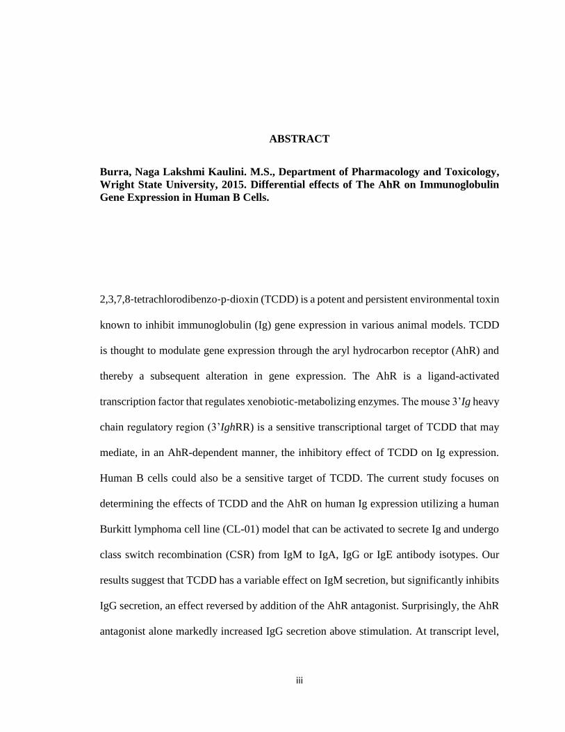
ABSTRACT
Burra, Naga Lakshmi Kaulini. M.S., Department of Pharmacology and Toxicology,
Wright State University, 2015. Differential effects of The AhR on Immunoglobulin
Gene Expression in Human B Cells.
2,3,7,8‐tetrachlorodibenzo‐p‐dioxin (TCDD) is a potent and persistent environmental toxin
known to inhibit immunoglobulin (Ig) gene expression in various animal models. TCDD
is thought to modulate gene expression through the aryl hydrocarbon receptor (AhR) and
thereby a subsequent alteration in gene expression. The AhR is a ligand-activated
transcription factor that regulates xenobiotic-metabolizing enzymes. The mouse 3’Ig heavy
chain regulatory region (3’IghRR) is a sensitive transcriptional target of TCDD that may
mediate, in an AhR-dependent manner, the inhibitory effect of TCDD on Ig expression.
Human B cells could also be a sensitive target of TCDD. The current study focuses on
determining the effects of TCDD and the AhR on human Ig expression utilizing a human
Burkitt lymphoma cell line (CL-01) model that can be activated to secrete Ig and undergo
class switch recombination (CSR) from IgM to IgA, IgG or IgE antibody isotypes. Our
results suggest that TCDD has a variable effect on IgM secretion, but significantly inhibits
IgG secretion, an effect reversed by addition of the AhR antagonist. Surprisingly, the AhR
antagonist alone markedly increased IgG secretion above stimulation. At transcript level,
iv
TCDD has variable effects on μ IGH functional transcripts but significantly inhibits γ1-4
germline/functional transcripts and Cε germline transcripts. Additionally, CD40L and IL-
4 stimulation induced de novo synthesis of Cε germline transcripts, a precursor to CSR.
However, α1-2 germline/functional transcripts increased in response to TCDD. Notably,
TCDD and stimulation had no effect on CYP1A1 expression. Additionally, in CL-01 cells,
we recently discovered SNPs in Exon-10 of the AhR, which encodes the transactivation
domain that regulates expression of other genes but does not affect ligand binding. The
AhR is heterozygous with one non-functional transactivation domain. Results also indicate
that a small proportion of the cells have undergone spontaneous class switch to all of the γ
and α isotypes rather than being induced to CSR.
v
TABLE OF CONTENTS
Page
I. INTRODUCTION…………………………………………………………….1
2,3,7,8-tetrachlorodibenzo-p-dioxin (TCDD)….……...…………………..1
The Aryl Hydrocarbon Receptor Signaling Pathway.…..………………....4
The Immune System and its functions…….………………………………7
B cells and antibody production ..……...…………………………………8
TCDD-induced imunological defects.....…….…………………….…….14
TCDD, AhR and B cells...……………..…….…………………….…….14
The AhR antagonist ...…………….…..…….…………………….…….16
Immunoglobulin Heavy Chain 3’ Regulatory Region (3’ IGHRR) ……18
The Mouse 3’ IghRR ..………………………………………...………...18
The Human 3’ IGHRR ………………………………………………..…19
Germline transcription and CSR..……….…………………..…………...23
Hypothesis and Objectives……………..……….…………..…………...26
II. MATERIALS AND METHODS…………………………...…….…………27
Chemicals and Reagents……………………………...……….…………27
Cell Line Model and Cell Culture Conditions.……………….………….27
Sandwich Enzyme-Linked Immunosorbent Assay (ELISA)...…….…….28
RNA isolation…………..…………….……..………………….………..28
cDNA synthesis, Real Time and Reverse Transcription PCR…………...29
vi
Statistical analysis of data……………………………………..…………34
III. RESULTS……………………………………………………………………35
IV. DISCUSSION AND CONCLUSIONS…….……..……..…………………..58
V. BIBLIOGRAPHY…………………………………………………………...69
vii
LIST OF FIGURES
Figure 1: Chemical structure of 2,3,7,8-tetrachlorodibenzo-p-dioxin (TCDD)…….3
Figure 2: The AhR signaling pathway………………………………………….…...6
Figure 3: Structure of an immunoglobulin (Ig)………………………………….….12
Figure 4: The AhR antagonist pathway...………………....…………………….….17
Figure 5: IGH gene loci of human and mouse ..………………..…………………..21
Figure 6: Differences between mouse and human hs1,2 enhancer ...……….………22
Figure 7: Class switch recombination ..…………………………………..……......25
Figure 8: Differential effects of TCDD on IgM and IgG secretion………………...36
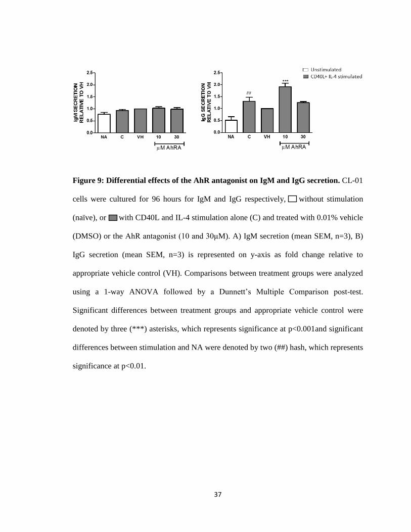
Figure 9: Differential effects of the AhR antagonist on IgM and IgG secretion…..37
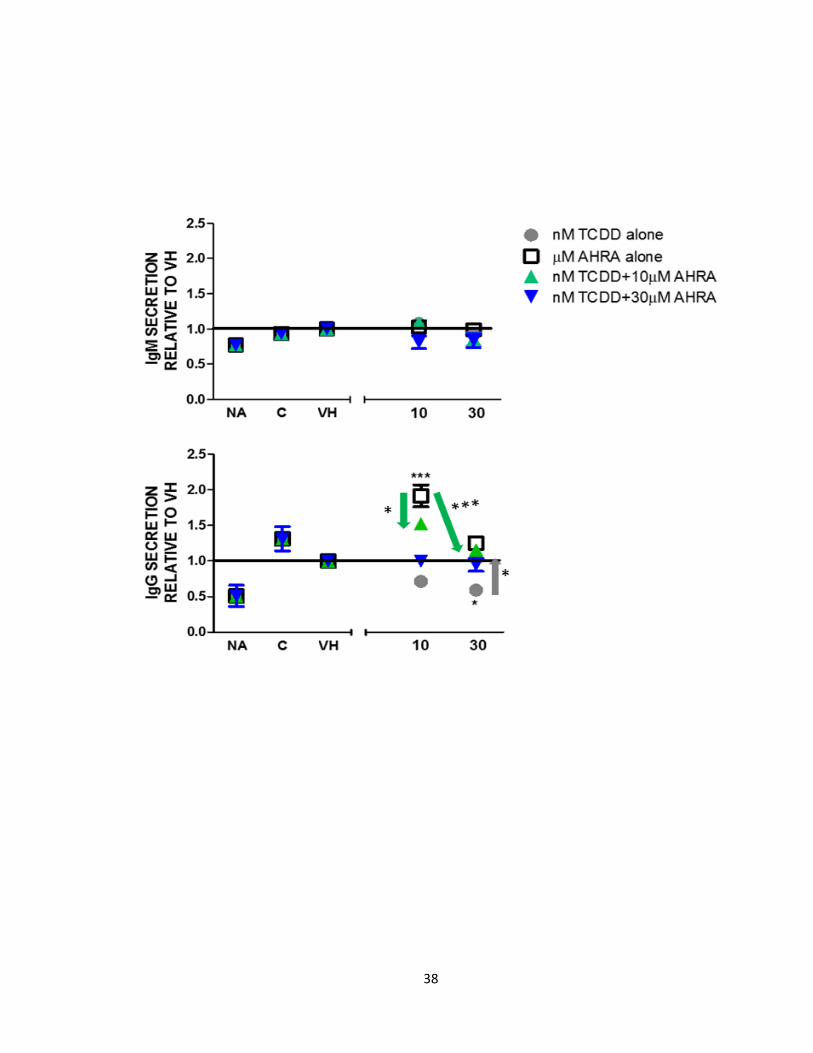
Figure 10: The AhR antagonist and TCDD antagonize each other’s effects on IgG
secretion but have no effect on IgM secretion …..………………….......38
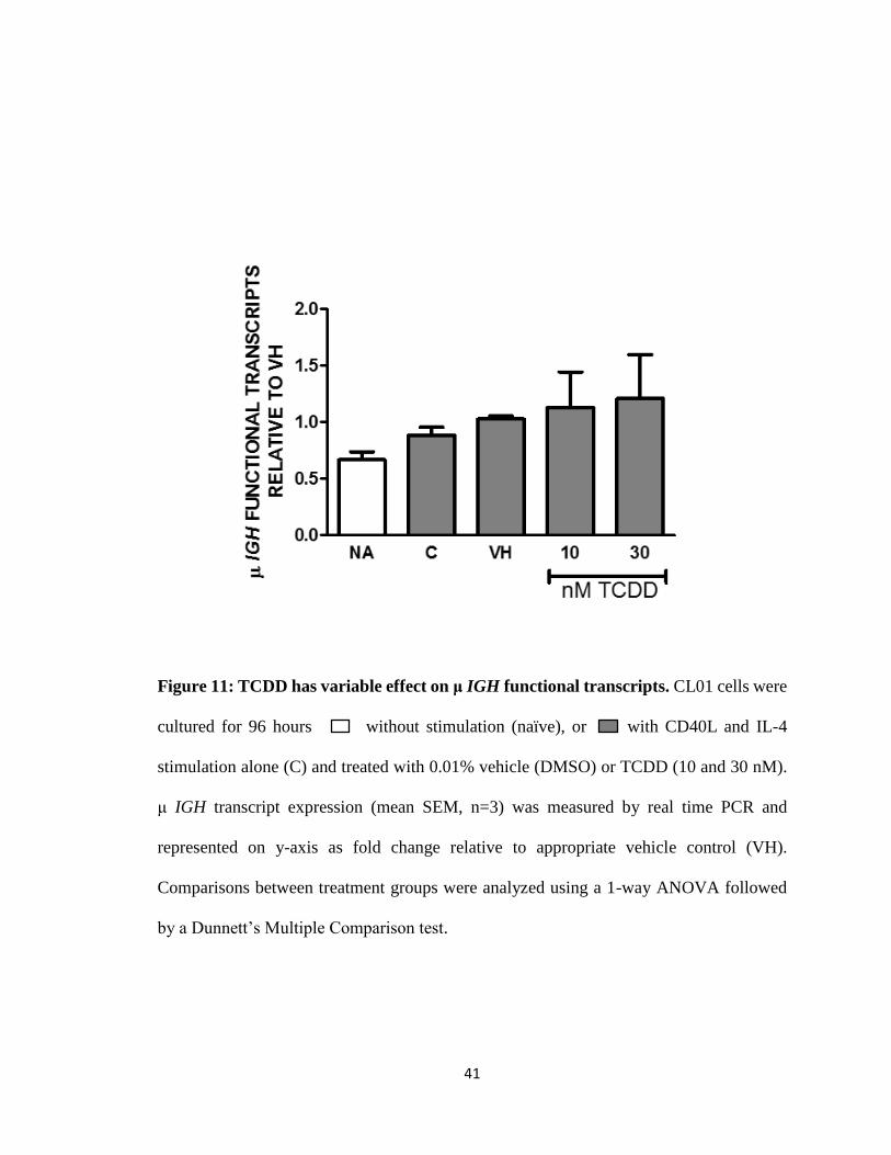
Figure 11: TCDD has a variable effect on μ IGH functional transcripts..……….….41
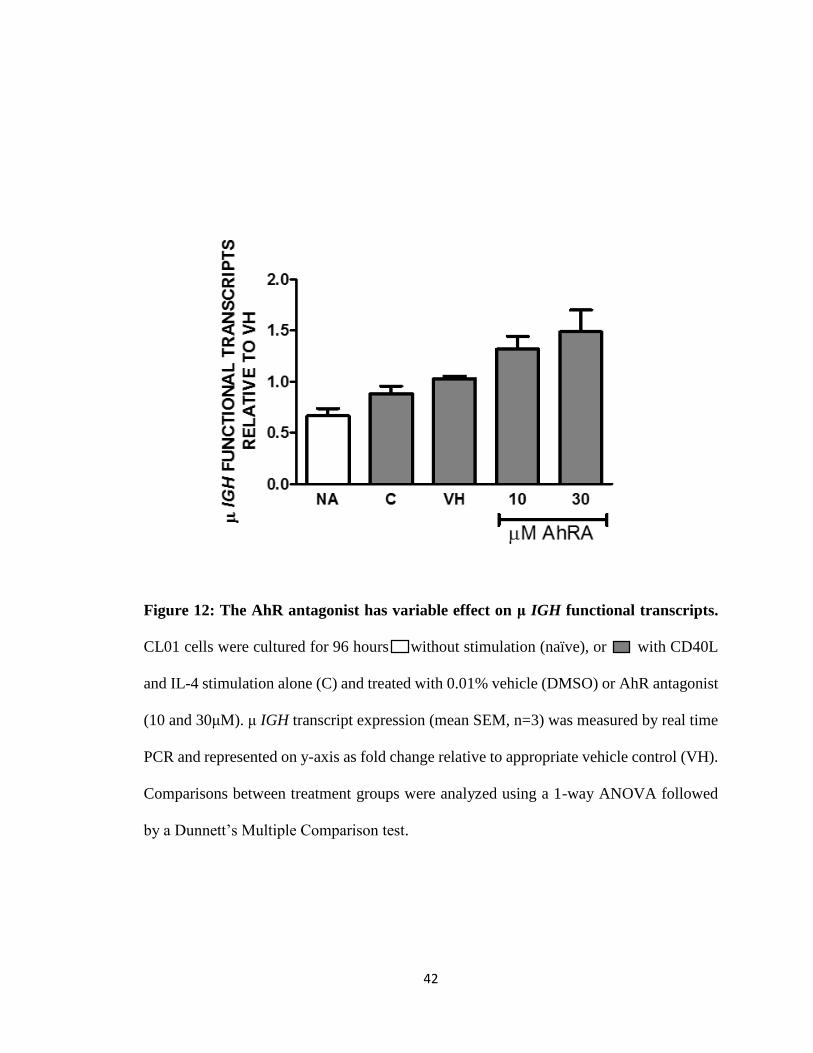
Figure 12: The AhR antagonist has a variable effect on μ IGH functional
transcripts………………………………………………………………...42
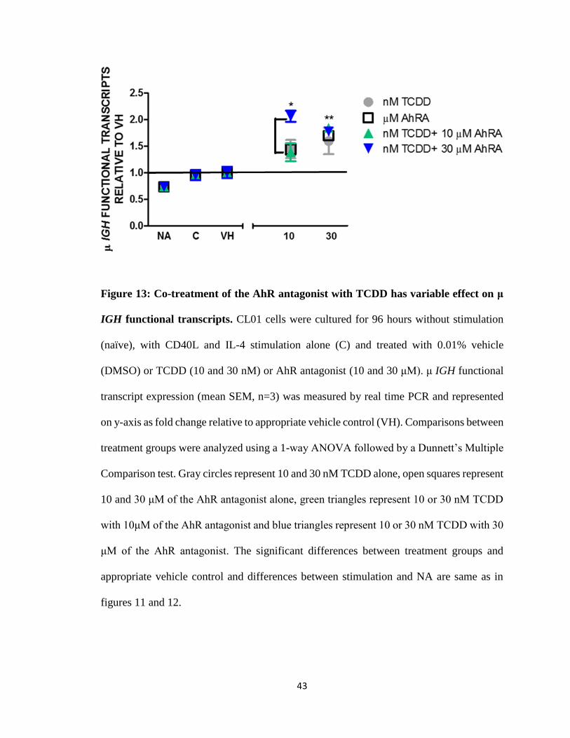
Figure 13: Co-treatment of the AhR antagonist with TCDD has a variable effect on μ
IGH functional transcripts………………………………………………43
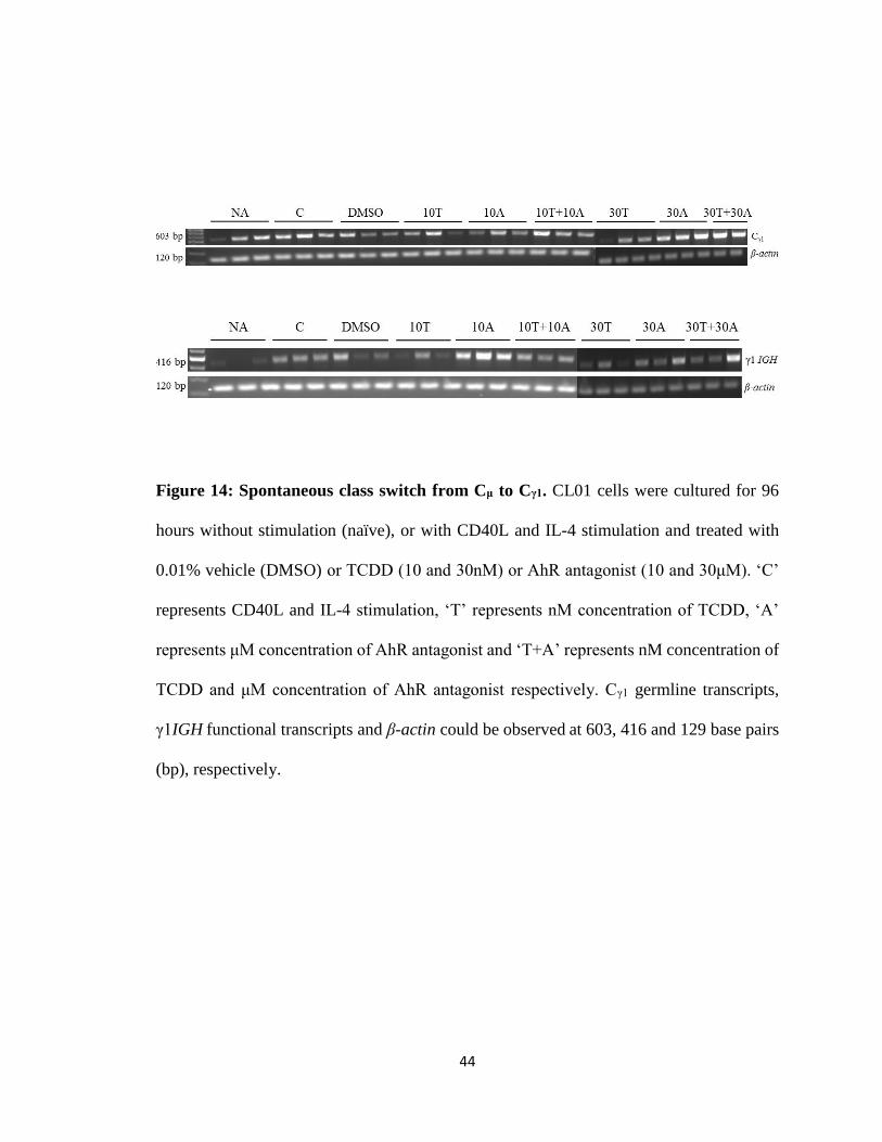
Figure 14: Spontaneous class switch from Cμ toCγ1………………………………..44
viii
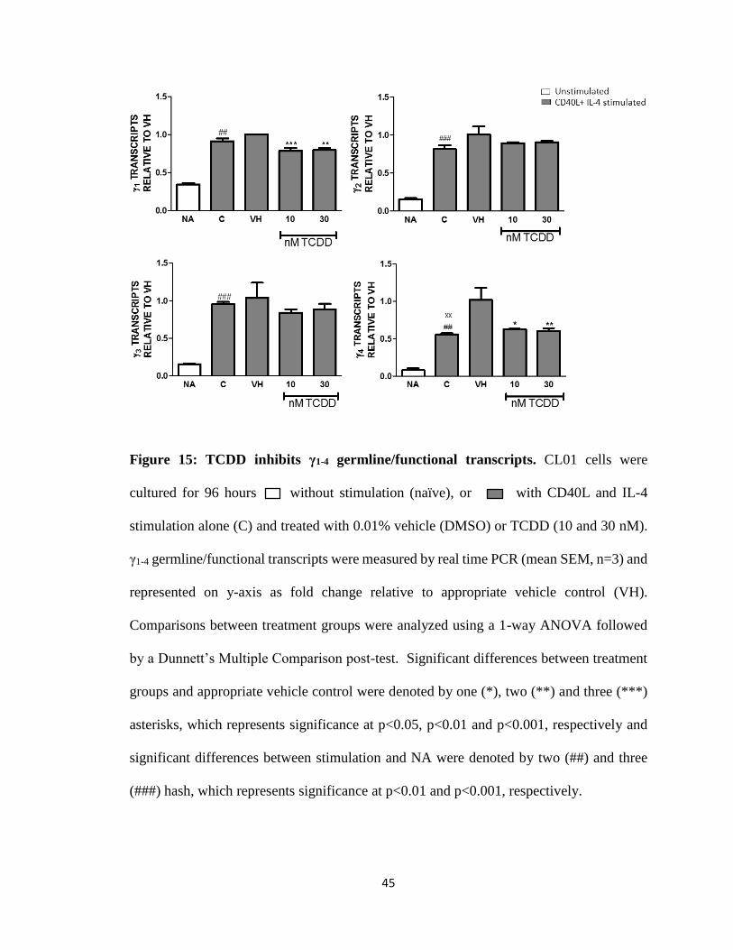
Figure 15: TCDD inhibits γ1-4 germline/functional transcripts……………………...45
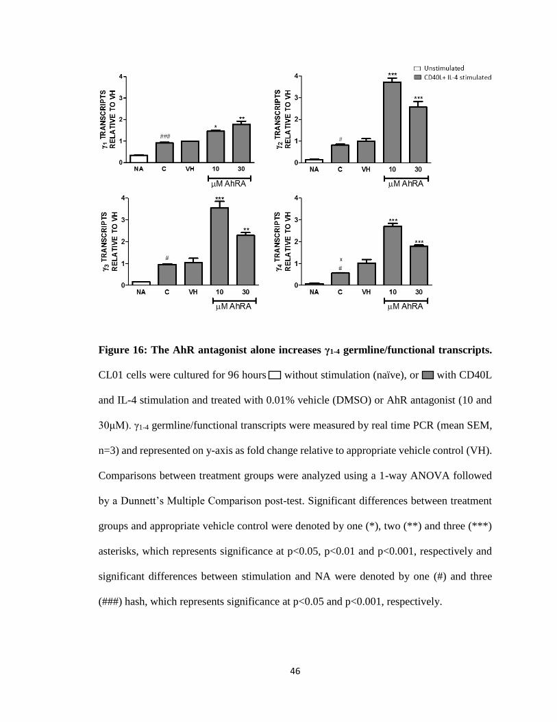
Figure 16: The AhR antagonist alone increases γ1-4 germline/functional
transcripts…………………………………………………..……………46
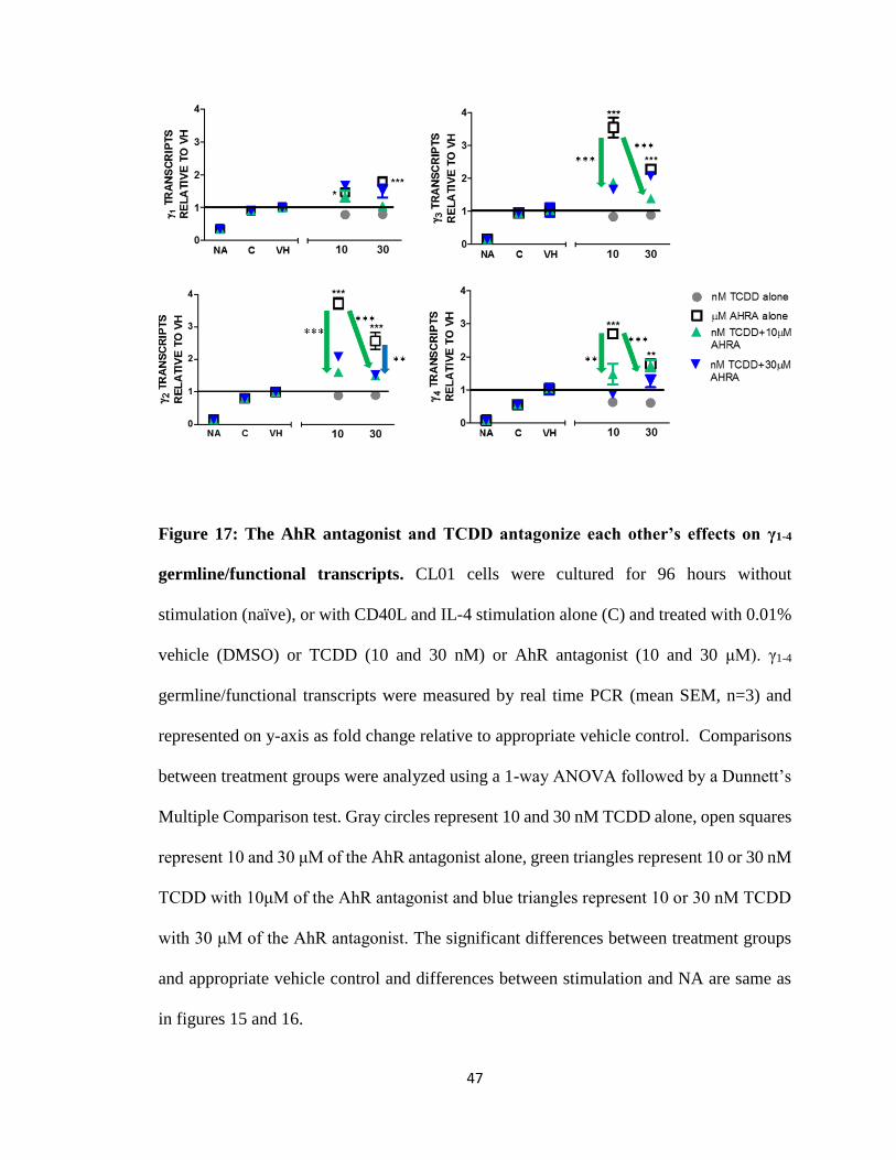
Figure 17: The AhR antagonist and TCDD antagonize each other’s effects on γ1-4
germline/functional transcripts………………………………………….47
Figure 18: Spontaneous class switch from Cμ to Cα1………………………….….....49
Figure 19: TCDD reverses the inhibition of stimulation in α germline/functional
transcripts…………………………………………………………….….50
Figure 20: The AhR antagonist inhibits α germline/functional transcripts……...….51
Figure 21: TCDD reverses the inhibition of α germline/functional transcripts by the
AhR antagonist………………………………………………………….52
Figure 22: TCDD inhibits stimulation-induced de novo Cε germline transcripts….54
Figure 23: The AhR antagonist alone increases Cε germline transcripts…………..55
Figure 24: The AhR antagonist reverses the inhibition of Cε germline
transcripts by TCDD…………………………………………………...56
Figure 25: Lack of CYP1A1 induction by TCDD………………………………….57
Figure 26: The human AhR gene with polymorphism sites………………………..64
ix
Figure 27: Location of heterozygous SNPs in the transactivation domain of the
human AhR……………………………………………………………..65
x
LIST OF TABLES
Table 1: Forward and reverse primers used in this study…………………………...…32
Table 2: PCR cycling conditions and expected product sizes…………………………33
xi
ACKNOWLEDGEMENTS
I am profoundly grateful to Dr. Courtney Sulentic for giving me an opportunity to
work in her lab. She has been a great friend, mentor and wonderful support and
encouraged me throughout the time it took me to complete my thesis work. I am
also grateful to the members of my thesis committee Dr. Nancy Bigley, Dr. David
Cool and Dr. Mauricio Di Fulvio for their generously given time and expertise to
better my work. I would like to specially thank my lab members Andrew Snyder,
Bassam Kashgari and Zahra Alfaheeda. I will cherish the memories I shared with
them and also their valuable friendship. I would also like to thank my other lab
members Siham Abdulla, Nicole Pastingel, Gabriel Crabb, Abdullah Freiwan and
Brooke Johnson for their support during my thesis work. I might not remember
everything I had done one day, but will remember all the lab meetings, SOT
meeting, fun-filled lab lunches, Cincinnati Escape Room, Dick’s Last Resort
(particularly), friendship, lab work at ungodly hours, and all the laughter and this
would not have been possible without Dr. Sulentic! I would also like to thank the
Department of Pharmacology and Toxicology for giving me this wonderful
opportunity to my pursue higher education, the staff of Pharmacology and
Toxicology, my family, and friends back home (The Zanys) and also at Wright
State University.
xii
DEDICATION
I dedicate my thesis work to my parents, Vijaya Lakshmi and Sesha Sai Burra, my
brother, Nrupendra Sai Burra and my grandmother, Srilakshmi Devasena Burra, for
their unconditional love, encouragement and support throughout my life. Thank
you very much, I wouldn’t have been here this day without you all. I hope I made
you proud.
1
I INTRODUCTION
2,3,7,8-tetrachlorodibenzo-p-dioxin (TCDD)
Dioxin(s) belong to a class of chemicals very similar in structure and toxic in nature.
They include polyhalogenated aromatic hydrocarbons (PAH) like polychlorinated dibenzo-
p-dioxins (PCDD), polychlorinated dibenzofurans (PCDF) and polychlorinated biphenyls
(PCB) (Mandal 2005). Since all of them exhibit hydrophobic properties and long half-lives,
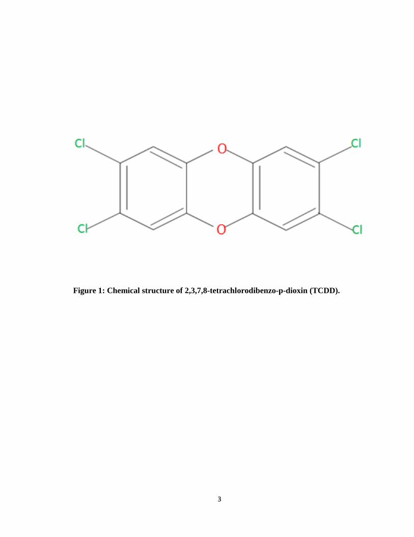
dioxins tend to bio-accumulate and persist as environmental contaminants. 2, 3, 7, 8-
Tetrachlorodibenzo-p-dioxin (TCDD) (Fig. 1) is the most studied PCDD as it is the most
potent and toxic amongst all dioxins.
TCDD is a planar molecule (C12H4Cl4O2), colorless and odorless at room
temperature. TCDD is not produced intentionally but as by-product of many industrial
processes such as paper and textile bleaching manufacture of chlorophenoxy herbicides
and pesticides, and metal smelting (McGregor et al. 1998). In fact, TCDD is produced
during the combustion of substances in the presence of chlorine, for example backyard
burning, forest fires, improper disposal of medical waste (Schecter et al. 2001). General
exposure to TCDD could be through soil, dust and/or smoke by inhalation or consumption
(Mandal 2005; Marinkovic et al. 2010). During the Vietnam War (1961-1971), TCDD was
discovered to be a contaminant in the herbicide Agent Orange, which was sprayed on
foliage to deliberately expose Vietnamese soldiers (Schecter et al. 2006). In Seveso, Italy
1976, several kilograms of TCDD were released from a pressure tank and inadvertently
2
thousands of inhabitants were exposed to TCDD (Mandal 2005). In Vienna 1997, two
women were exposed to the highest ever concentration of TCDD at their workplace
144ng/g of fat (Geusau et al. 1999). In 2004, Ukrainian President Viktor Yushchenko was
exposed to TCDD after a failed assassination attempt. TCDD was present at a
concentration of as high as 108 ng/g of fat (Sorg et al. 2009). In vivo animal studies have
shown that exposure to TCDD affects brain function, reproduction, hormone signaling and
immunity (Mandal 2005). Besides, TCDD has been shown to promote tumor growth and
is a classified human carcinogen (McGregor et al. 1998).
3
Figure 1: Chemical structure of 2,3,7,8-tetrachlorodibenzo-p-dioxin (TCDD).
4
The Aryl Hydrocarbon Receptor Signaling Pathway
The aryl hydrocarbon receptor (AhR) is a ligand-activated transcription factor
belonging to the basic-helix-loop-helix/Per-ARNT-Sim family (PAS), encoded by the Ahr
gene. AhR, in its inactivated state in the cytosol is coupled with several proteins: two heat-
shock protein 90 molecules (HSP90), co-chaperone p23, and the hepatitis B virus X-
associated protein, which is an AhR-interacting protein (previously known as XAP2) (Abel
and Haarmann-Stemmann 2010; Nebert and Karp 2008). XAP2 plays an essential role in
AhR activation, binds to the AhR and HSP90, while p23 directly binds to HSP90 (Endler,
Chen, and Shibasaki 2014; Meyer and Perdew 1999). The AhR is known to play an
important role in xenobiotic metabolism and also the gene expression of many drug
metabolizing enzymes that belong to the cytochrome p450 family. Amongst them, the
induction of CYP1A1 is used as a biomarker to study the AhR signaling pathway (Hu et al.
2007; Hansen et al. 2014). Additionally, the AhR is well-known to cross-talk with steroid
receptors like the estrogen and androgen receptors and alter their gene expression (Wormke
et al. 2003), suggesting that the AhR may have regulatory roles beyond drug metabolism.
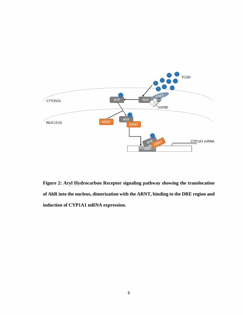
AhR is best characterized by studying its ligand TCDD (Fig. 1). The AhR undergoes a
conformational change upon binding to TCDD and dissociates from its protective cytosolic
protein complex, translocates into the nucleus to form a heterodimer with the AhR nuclear
translocator (ARNT). The AhR-ARNT heterodimer interacts with chromatin remodeling
factors and histone acetyltransferases to induce specific chromatin changes
(Schnekenburger, Peng, and Puga 2007) to bind to the dioxin responsive element (DRE;
5’-TNGCGTG-3’). DRE is present within the promoter or enhancer regions of target genes
and consequently alters gene expression (Fig. 2) (Abel and Haarmann-Stemmann 2010;
5
Landers and Bunce 1991). When the ligand is no longer present to activate the AhR, it
undergoes negative feedback inhibition by the AhR repressor (Mimura and Fujii-Kuriyama
2003) or released into the cytosol and degraded by the proteasome pathway (Pollenz 2002).
The AhR is expressed in liver, lungs, skin, gastrointestinal tract and other tissues
(Carlstedt-Duke 1979). Apart from its conventional role in metabolizing environmental
ligands, the AhR is involved in development, cellular oxidation/antioxidation, epidermal
barrier function (Noakes 2015), cell survival and proliferation, and in the regulation of
immune system (Vogel et al. 2014). Natural or artificial chemicals, pharmaceuticals, and
dietary constituents have also been identified as AhR ligands (e.g. aromatic hyrdrocarbons,
omeprazole, carbaryl, flavonoids, indolocarbazoles, etc.).
The human AhR has relatively low affinity for TCDD compared to mouse and
requires 10-fold higher concentrations of TCDD to induce a particular effect (Connor and
Aylward 2006; Okey, Riddick, and Harper 1994). This could explain why animals are more
sensitive to TCDD as compared to humans. Moreover, single nucleotide polymorphisms
(SNPs) have been described in both mouse and human AhR genes. The human and mouse
AhR proteins share 58% amino acid sequence in the C-terminal, which contains the
transactivation domain (Flaveny et al. 2008). SNPs in the transactivation domain of the
human AhR impairs the ability of the AhR to induce CYP1A1 gene expression in response
to TCDD (Wong, Okey, and Harper 2001). Interestingly, SNPs located in the ligand
binding domain of the human AhR did not affect ligand-induced CYP1A1 expression. SNPs
occurring in Exon-10 of the human AhR gene, which encodes the transactivation domain
involved in gene regulation, does not impact the ligand binding affinity (Harper et al.
2002).
6
Figure 2: Aryl Hydrocarbon Receptor signaling pathway showing the translocation
of AhR into the nucleus, dimerization with the ARNT, binding to the DRE region and
induction of CYP1A1 mRNA expression.
7
The Immune System and its functions
The human body has a built-in, intricate and interesting protection mechanism
against microbes including bacteria, viruses, fungi and other pathogens that invade our
body. The body’s defense mechanism is multi-layered and the first line of defense, the
innate immune system, produces an immediate but non-specific and antigen-independent
response at the site of infection. It includes physical barriers (skin and epithelial surfaces
lining the gut and lung, and chemical barriers like saliva and tears), phagocytes
(neutrophils, macrophages), dendritic cells, natural killer cells and also circulating plasma
proteins. The innate immune system is activated when it recognizes particular types of
molecules present on the pathogens that are not present in the host. Even if the body has
never been exposed to a particular kind of pathogen, the pathogen-associated molecules
stimulate the inflammatory responses. The innate immune system is the body’s most
dominant defense mechanism (Litman, Cannon, and Dishaw 2005). If the innate immune
system is unsuccessful in clearing the pathogen from the body, then the adaptive immune
system is activated by components of the innate immune system, is activated. The adaptive
immune system improves recognition of the pathogen by retaining it as immunological
memory even after the pathogen has been eliminated from the body, resulting in specificity
of the response to that pathogen (Pancer and Cooper 2006). This immunological memory
produces a more robust immune responses upon future exposure to the same pathogen.
Any foreign substance that is able to elicit an adaptive immune response is called an antigen
(antibody generator).
Adaptive immunity is divided into cell-mediated and humoral immunity. Cell-
mediated immunity is principally orchestrated by T cells by releasing cytokines in response
8
to pathogens. Humoral immunity, on the other hand is primarily mediated by B cells which
release antibodies (secreted form of immunoglobulins) in response to pathogens. The
secreted antibodies circulate in the bloodstream and other body fluids, binding specifically
to antigens that stimulated their production in the first place. Antibody binding neutralizes
and destroys the antigens by involving the components of the innate immune system such
as phagocytes, to ingest them. In cell-mediated immunity, some activated T cells (T helper
cells) produce cytokines that help direct the immune response and activate B cells (Brian
1988; Grewal and Flavell 1998) or some T cells (T cytotoxic cells) produce powerful
enzymes that induce the death of pathogen-infected cells, thereby successfully clearing the
pathogen from the body.
B Cells and Antibody production
B cells are the major effectors of the humoral immune response. B cells are
continuously produced in the bone marrow and as immature B cells, they circulate through
blood and lymph and compete for survival signals in the secondary lymphoid tissue until
they are activated by recognition of specific antigen. Immature naïve B cells have receptors
for antigens that are present in the plasma membrane. The B-cell receptor (BCR) is a
transmembrane protein present on the surface of B cells with a unique antigen binding site.
Each B-cell receptor has a membrane-bound immunoglobulin of one isotype (IgM/IgD).
After leaving the bone marrow, immature naïve B cells also start producing IgD molecules
on their surface that have the same antigen binding site as the IgM. B cells mature when
they co-expresses both IgM and IgD on their surface. In response to antigenic challenge
and interaction with helper T cells, B cells are activated to differentiate into plasma or
memory B cells. B cells are also activated in a T-cell independent manner through toll-like
9
receptors and BCR crosslinking. Plasma cells produce huge amounts of antibodies
continuously in response to antigens, whereas memory B cells help induce a more robust
and quicker immune response to antigens upon re-exposure (Brian 1988). A primary
immune response, mediated by IgM, occurs when immune cells encounter antigen for the
first time. Mature B cells with IgM on their surface differentiate to produce memory cells
and plasma cells. A secondary immune response is elicited upon re-exposure to the same
antigen, resulting in high-affinity antibodies such as IgG to eliminate the pathogen from
the body.
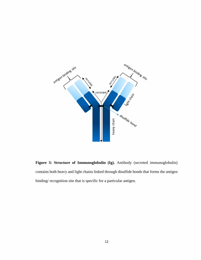
The structural unit of an antibody consists of four chains, of which two are
identical light (L) chains (each containing about 220 amino acids) and two identical heavy
(H) chains (each containing about 440 amino acids). Heavy and light chain of the antibody
are held together by covalent disulfide bonds (Fig. 3). Heavy and light chains together form
two identical antigen binding sites at the tip of each arm of the molecule. Because of two
binding sites, an antibody can be bivalent. Both heavy and light chains have a variable
sequence at their N-terminal ends and a constant sequence at their C-terminal ends. The N-
terminal ends of both heavy and light chains link up to form the antigen-binding site and
variability in their amino acid sequences results in the diversity of antigen binding sites. In
mammals, there are five different classes or isotypes of antibodies (IgA, IgD, IgE, IgG and
IgM), each encoded by its own constant region (α, δ, ε, γ and μ) within the immunoglobulin
heavy chain gene, respectively. Also, there are subclasses of IgG and IgA immunoglobulins
in humans, i.e., four IgG subclasses-IgG1, IgG2, IgG3, and IgG4 with γ1, γ2, γ3 and γ4 heavy
chain genes, respectively and two IgA subclasses- IgA1 and IgA2 with α1 and α2 heavy
chain genes, respectively. Differences in heavy chains gives different conformation to the
10
hinge and tail regions of antibodies, so that each class (and subclass) has a characteristic
property of its own. The two types of light chains are kappa (κ) and lambda (λ), which have
no significant difference in structure. Both isotypes of the light chain can be associated
with any of the heavy chain isotypes, but in an individual B cell the two light chains and
the two heavy chains will always be identical. In IgA1 for instance, the two light chains
could either be κ or λ with two α1 heavy chains but not one λ light chain and one κ light
chain with two α1 heavy chains.
Properties and functions of different antibody classes
IgM-
IgM with the heavy chain μ, is the first class of antibody to be made during the development
of a B cell and also the first antibody secreted upon B-cell activation and differentiation
into antibody-secreting cells or plasma cells. IgM is present as a monomer on the surface
of the B cell or as a pentamer when secreted. Pentameric IgM antibodies are linked together
by a J chain, having a total of 10 antigen binding sites. IgM eliminates pathogens during
the early stages of a humoral immune response. Mature B cells with IgM on their surface
can class switch to different antibody isotypes like IgA, IgG and IgE through DNA
recombination mechanisms involving somatic hypermutation and class switch
recombination, depending on the type of the antigen the cells encounter.
IgD-
IgD is secreted in very small amounts and is mostly attached to the surface of
immature and mature naive the B cells. Surface IgD appears to function as a co-receptor
along with IgM. IgD has also been shown to activate cells basophils and mast cells, which
secrete antimicrobial factors (Chen et al. 2009).
11
IgG-
IgG is a monomer (on the surface of a B cell and when secreted). There are four
subclasses of IgG (IgG1-4) in humans and is the major class of immunoglobulin present in
the blood. It is produced in large quantities during secondary immune responses and is the
only class of antibody capable of crossing the placental barrier to give passive immunity
to the fetus (Saji et al. 1999).
IgA-
IgA is the main class of antibody present in secretions like saliva, tears, milk, and
respiratory and intestinal secretions that guard entrances in the body. IgA is a monomer on
the surface of a B cell and both monomer and dimer in secretions.
IgE-
IgE protects against parasitic infections but is also responsible for binding to
allergens and triggering the release of histamine from mast cells and basophils. It is
involved in the symptoms of allergies like hay fever, asthma and hives. IgE is a monomer
on the surface of the B cell and also when secreted.
12
Figure 3: Structure of Immunoglobulin (Ig). Antibody (secreted immunoglobulin)
contains both heavy and light chains linked through disulfide bonds that forms the antigen
binding/ recognition site that is specific for a particular antigen.
13
The gene segments that encode the light chain variable region are the variable (V)
and the joining (J) segments. The heavy chain variable region has the diversity (D) region
along with the variable (V) and joining (J) segments. In V(D)J recombination, a somatic
assembly of the variable, diversity and joining gene segments gives the variable region
exons of the antigen receptors (Borghesi and Milcarek 2006; Market and Papavasiliou
2003). V(D)J recombination occurs during the developmental stages of the lymphocyte
(Mombaerts et al. 1992) and requires recombination associated genes (RAG-1 and RAG-
2) (van Gent et al. 1995; Oettinger et al. 1990). The end point of V(D)J recombination is
the surface expression of IgM in developing B cells. When B cells come into contact with
the antigen, they are capable of undergoing two additional forms of genetic alterations
(somatic hypermutation and class switch recombination) that enhance the response to its
cognate antigen by the antigen-specific B cell. During somatic hypermutation (SHM), high
rates of mutations are introduced into the germline DNA sequences of the assembled exons
of the immunoglobulin heavy and light chain variable regions. And it is through this
process that B-cell receptors express increased affinity for a particular antigen, a necessary
step for clonal selection. In the heavy chain of the immunoglobulin gene, class switch
recombination (CSR) occurs adjoining a rearranged variable region exon and downstream
CH (i.e. Cγ, Cα or Cε) exon through deletion of the intervening germline DNA. This allows
for the expression of an antibody with the same antigen-binding specificity but with an
altered CH effector function (Stavnezer and Kang 2008).
14
TCDD-induced immunological defects
The immune system has been identified as a sensitive and early target of TCDD.
Even at low levels of TCDD exposure (acute and chronic), rodents display innate and
acquired immune-related disturbances (Kerkvliet 2009, 1995; Kerkvliet, Shepherd, and
Baecher-Steppan 2002; Roman, Pollenz, and Peterson 1998; Vos, De Heer, and Van
Loveren 1997). Upon TCDD exposure, T cell suppression has been observed through
induction of Treg cells (Quintana 2013; Quintana et al. 2008). Also, TCDD is known to
directly target both mouse and human B cells (Lu et al. 2010; Sulentic and Kaminski 2011).
Compared to laboratory animals, humans may be less sensitive to TCDD due to its lower
affinity for AhR (Okey, Riddick, and Harper 1994).
TCDD, AhR and B Cells
In mouse B cells, TCDD affects B-cell maturation, activation, differentiation and
gene regulation (Lu et al. 2011; Lu et al. 2010; Sulentic and Kaminski 2011). Previous
reports in mouse primary and B-cell lines have shown that TCDD inhibits IgM secretion
when stimulated with LPS and treated with varying concentrations (0.03-30 nM TCDD)
(Marcus, Holsapple, and Kaminski 1998; Sulentic, Holsapple, and Kaminski 1998). In
primary human B cells, the effect of TCDD on IgM secretion in CD40L plus IL-4
stimulated cells was inhibitory (Lu et al. 2010). Previous studies have shown that TCDD
inhibited LPS-stimulated IgM secretion in the AhR-expressing mouse B-cell line
CH12.LX. Further, there was no inhibition in LPS-stimulated IgM secretion in the AhR-
deficient BCL-1 cells, suggesting a functional role of the AhR in TCDD-induced inhibition
of IgM (Sulentic, Holsapple, and Kaminski 1998, 2000). Additionally, it has been shown
that AhR and ARNT heterodimer binds to DRE binding sites within a 3’ transcriptional
15
regulatory region of the Ig heavy chain gene, which is involved in antibody secretion and
in Ig heavy chain gene expression (Sulentic, Holsapple, and Kaminski 2000). Also, there
was no TCDD-dependent AhR binding to DRE in the hs1,2 and hs4 enhancers in AhR-
deficient BCL-1 cells (Sulentic, Holsapple, and Kaminski 2000). Another study has
confirmed reversal of TCDD-mediated inhibition of IgA secretion in a mouse cell line by
an AhR antagonist or by shRNA-mediated AhR knock down (Wourms and Sulentic 2015).
This further suggests that AhR is involved in TCDD-induced inhibition of Ig secretion in
mouse B cells.
In addition, TCDD suppressed IgM secretion in human primary B cells in 9 among
12 donors, 2 donors had no effect on IgM secretion even at a dose as high as 100 nM. In
fact, 1 donor showed enhancement of IgM secretion (Lu et al. 2010). Further, TCDD
suppressed human primary B-cell differentiation by impairing B-cell activation through
suppression of activation markers such as CD80, CD86 and CD69 (Lu et al. 2011).
However, TCDD did not alter the levels of IgG, IgA or IgM from tonsillar mature B cells
of atopic patients, but enhanced IgE levels. In non-atopic patients, TCDD did not affect
IgE levels, suggesting that TCDD could aggravate allergic diseases (Kimata 2003).
Although the inhibitory effect of TCDD on Ig secretion in mouse B cells is established,
TCDD-mediated suppression of human B cells is still unclear.
16
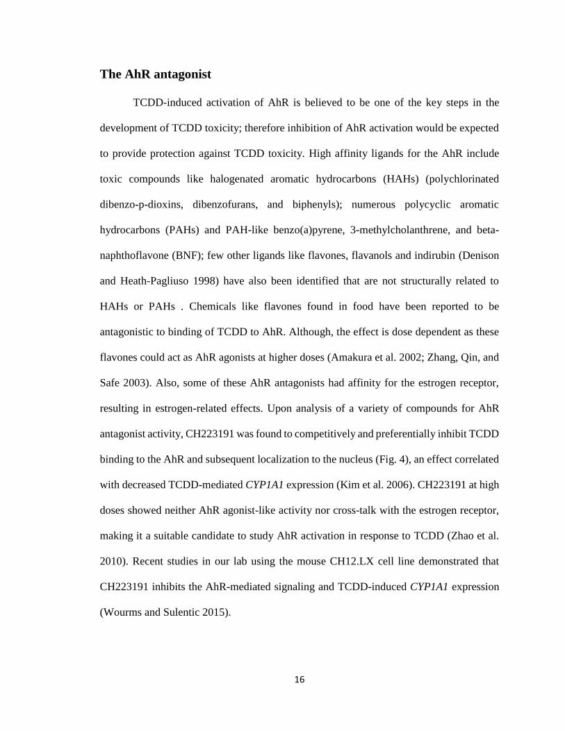
The AhR antagonist
TCDD-induced activation of AhR is believed to be one of the key steps in the
development of TCDD toxicity; therefore inhibition of AhR activation would be expected
to provide protection against TCDD toxicity. High affinity ligands for the AhR include
toxic compounds like halogenated aromatic hydrocarbons (HAHs) (polychlorinated
dibenzo-p-dioxins, dibenzofurans, and biphenyls); numerous polycyclic aromatic
hydrocarbons (PAHs) and PAH-like benzo(a)pyrene, 3-methylcholanthrene, and beta-
naphthoflavone (BNF); few other ligands like flavones, flavanols and indirubin (Denison
and Heath-Pagliuso 1998) have also been identified that are not structurally related to
HAHs or PAHs . Chemicals like flavones found in food have been reported to be
antagonistic to binding of TCDD to AhR. Although, the effect is dose dependent as these
flavones could act as AhR agonists at higher doses (Amakura et al. 2002; Zhang, Qin, and
Safe 2003). Also, some of these AhR antagonists had affinity for the estrogen receptor,
resulting in estrogen-related effects. Upon analysis of a variety of compounds for AhR
antagonist activity, CH223191 was found to competitively and preferentially inhibit TCDD
binding to the AhR and subsequent localization to the nucleus (Fig. 4), an effect correlated
with decreased TCDD-mediated CYP1A1 expression (Kim et al. 2006). CH223191 at high
doses showed neither AhR agonist-like activity nor cross-talk with the estrogen receptor,
making it a suitable candidate to study AhR activation in response to TCDD (Zhao et al.
2010). Recent studies in our lab using the mouse CH12.LX cell line demonstrated that
CH223191 inhibits the AhR-mediated signaling and TCDD-induced CYP1A1 expression
(Wourms and Sulentic 2015).
17
Figure 4: Inhibition of AhR translocation in to the nucleus by AhR antagonist.
18
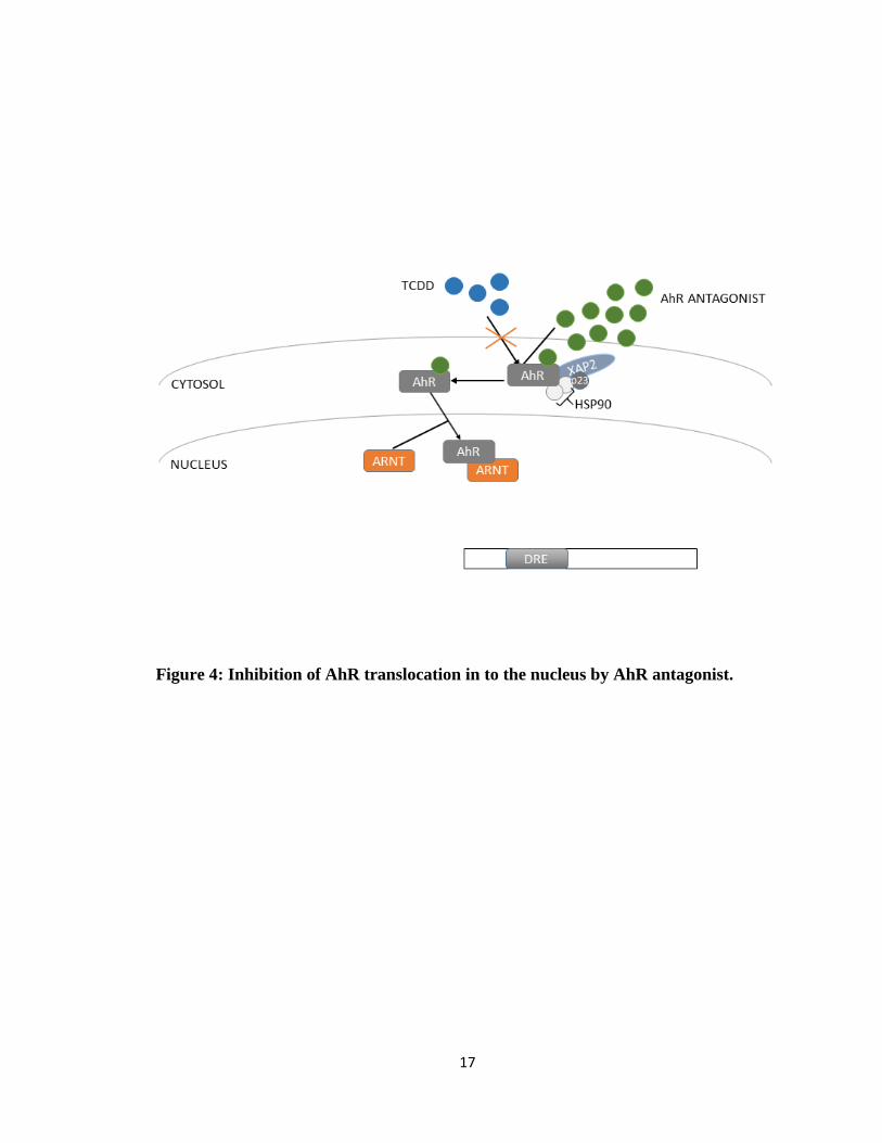
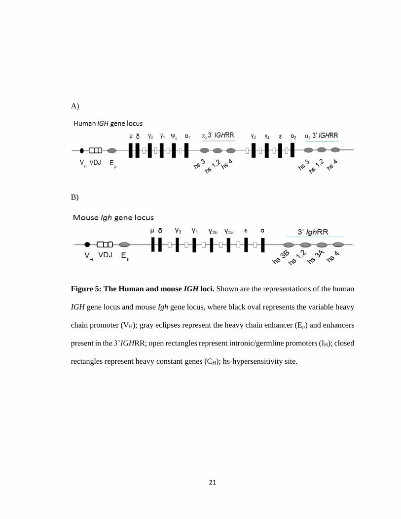
Immunoglobulin Heavy Chain 3’ Regulatory Region (3’IGHRR)
The human immunoglobulin heavy chain (IGH) gene and its counterpart, the mouse
immunoglobulin heavy chain (Igh) gene are located on chromosome 14 and 12,
respectively. They encode the heavy chains of antibodies via transcription-dependent DNA
remodeling events like VDJ recombination, CSR and SHM (Pinaud et al. 2011). The
rearranged human IGH after antigen stimulation consists of the VH promoter (variable
heavy chain promoter), the VDJ region, the Eμ (heavy chain intronic enhancer), the heavy
chain constant region genes for μ, δ, γ3, γ1, Ψε and α1, respectively, followed by the α1
3’IGHRR. The second set of heavy chain genes include the γ2, γ4, ε and α2, followed by
the α2 3’IGHRR (Mills et al. 1997; Sepulveda, Emelyanov, and Birshtein 2004; Sepulveda
et al. 2005) in humans. The two 3’IGHRR are termed α1 and α2 because they are present
downstream to the α2 and α2 heavy chain genes respectively (Fig. 5A). In mouse, the heavy
chain constant region genes are μ, δ, γ3, γ1, γ2b, γ2a, ε and α respectively, followed by the
mouse 3’IghRR (Madisen and Groudine 1994) (Fig. 5B). The VH promoter, present
upstream of the variable region, regulates the transcription of the IGH locus and gene
expression, the intronic enhancer Eμ is responsible for efficient V and DJ region
recombination in B-cell progenitors and Igh expression in mature B cells, and also the
expression of Cμ (Perlot et al. 2005).
The Mouse 3’IghRR
Murine 3’IghRR contains at least four DNA hypersensitive sites (hs)- hs3b, hs1,2,
hs3a and hs4. The hs1,2 enhancer is transcriptionally active in mature B cells and plasma
cells; both hs3a and hs3b enhancers have no activity in pre-B cells, B cells, or plasma cells
(Madisen and Groudine 1994; Matthias and Baltimore 1993; Saleque, Singh, and Birshtein
19
1999; Chauveau, Pinaud, and Cogne 1998) and the hs4 enhancer appears to function
throughout B-cell development, mainly being active in pre-B cells and plasma cells
(Madisen and Groudine 1994). Maximum effect was observed when all four enhancers
were together in all stages of B-cell development. There are several transcription factor
binding sites present within the 3’IghRR enhancers that include DRE, NF-κB, Octamer
(OCT), NF-αP, and AP-1/Ets, Pax5 that regulate 3’IghRR activity (Fig. 6). In the
CH12.LX, mouse B-cell line, TCDD was shown to induce AhR/ARNT-DRE binding
within the hs1,2 and hs4 enhancers of the 3’IghRR, as well as inhibition of 3’IghRR
activity. Additionally, TCDD-induced inhibition of μ heavy chain expression and IgM
secretion was observed to be AhR dependent (Sulentic, Holsapple, and Kaminski 1998,
2000). Reversal of TCDD-mediated inhibition of IgA secretion was confirmed in a mouse
cell line by an AhR antagonist or via shRNA-mediated AhR knock down (Wourms and
Sulentic 2015). Additionally, the murine 3’IghRR is sensitive to a variety of AhR and non-
AhR agonists (Henseler, 2009). (Henseler, Romer, and Sulentic 2009)
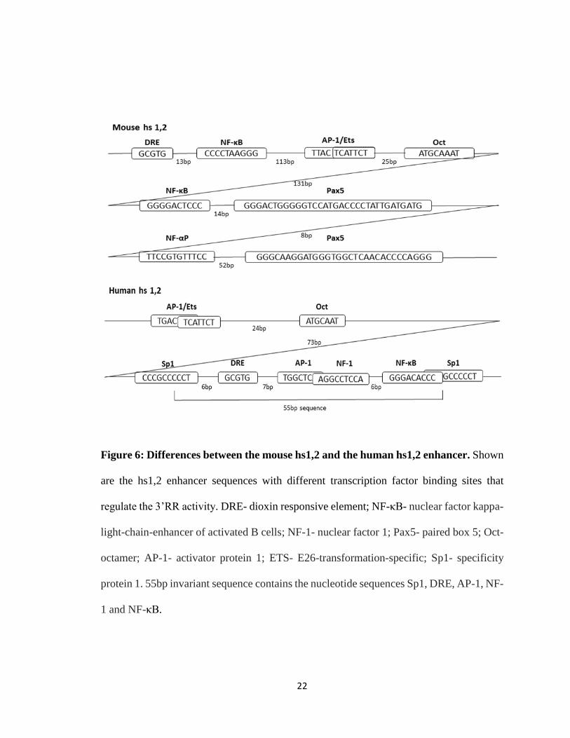
The Human 3’IGHRR
The human 3’IGHRR is comprised of three enhancer elements (hs3, hs1,2 and hs4),
which are 74%, 90%, and 76% homologous respectively to enhancers in the mouse
3’IghRR. Also, the human hs1,2 and hs4 lack the Pax5 binding sites that are significant to
the murine 3’IghRR activity. Additionally, the human α1 hs1,2 enhancer has a polymorphic
region called the invariant sequence (IS) of approximately 55 bp that is not observed in the
mouse hs1,2 enhancer (Fig. 6). The invariant sequence can be repeated one (α1A), two (α1B),
three (α1C) or four (α1D) times and alter the transcriptional activity (Fernando et al. 2012;
Denizot et al. 2001). The invariant sequence has several transcription factor binding sites
20
like AP-1, NF1, NF-κB and also a DRE site similar to the functional DRE found in the
mouse hs1,2 enhancer (Fernando et al. 2012; Chen and Birshtein 1997; Denizot et al. 2001)
and an increase in the number of 55 bp repeats may result in an increase in sensitivity to
TCDD. As the mouse 3’IghRR is involved in class switch recombination and Ig expression,
it is assumed that the human 3’IGHRR is also involved in CSR and Ig expression.
However, there are no studies to confirm this and there are differences between the mouse
3’IghRR and human 3’IGHRR. Additionally, varied responses to TCDD have been
demonstrated on hs1,2 enhancer activity between mouse and human genes. Contrary to the
observed TCDD-induced inhibition of the 3’IghRR and hs1,2 activities, TCDD activates
the human hs1,2 enhancer (Fernando et al. 2012).
Polymorphisms in the human hs1,2 enhancer appear to be involved in several
immune disorders namely plaque psoriasis, psoriatic arthritis, rheumatoid arthritis, coeliac
disease, dermatitis herpetiformis, systemic sclerosis lupus, and IgA nephropathy. Of the
different hs1,2 alleles, increase in the frequency of the α1B allele is shown to significantly
worsen the frequency of the above mentioned diseases (Aupetit et al. 2000; Cianci et al.
2008; Frezza et al. 2004; Giambra et al. 2009; Tolusso et al. 2009). Also, some B-cell
malignancies like lymphomas have chromosomal translocations between c-myc or bcl-2
and the 3’IGHRR which can lead to deregulated gene expression (Heckman et al. 2003).
21
A)
B)
Figure 5: The Human and mouse IGH loci. Shown are the representations of the human
IGH gene locus and mouse Igh gene locus, where black oval represents the variable heavy
chain promoter (VH); gray eclipses represent the heavy chain enhancer (Eμ) and enhancers
present in the 3’IGHRR; open rectangles represent intronic/germline promoters (IH); closed
rectangles represent heavy constant genes (CH); hs-hypersensitivity site.
22
Figure 6: Differences between the mouse hs1,2 and the human hs1,2 enhancer. Shown
are the hs1,2 enhancer sequences with different transcription factor binding sites that
regulate the 3’RR activity. DRE- dioxin responsive element; NF-κB- nuclear factor kappa-
light-chain-enhancer of activated B cells; NF-1- nuclear factor 1; Pax5- paired box 5; Oct-
octamer; AP-1- activator protein 1; ETS- E26-transformation-specific; Sp1- specificity
protein 1. 55bp invariant sequence contains the nucleotide sequences Sp1, DRE, AP-1, NF-
1 and NF-κB.
23
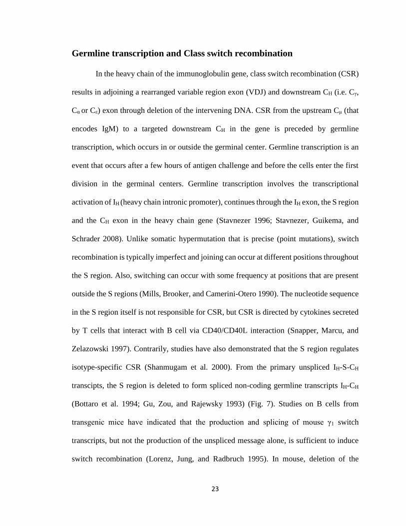
Germline transcription and Class switch recombination
In the heavy chain of the immunoglobulin gene, class switch recombination (CSR)
results in adjoining a rearranged variable region exon (VDJ) and downstream CH (i.e. Cγ,
Cα or Cε) exon through deletion of the intervening DNA. CSR from the upstream Cμ (that
encodes IgM) to a targeted downstream CH in the gene is preceded by germline
transcription, which occurs in or outside the germinal center. Germline transcription is an
event that occurs after a few hours of antigen challenge and before the cells enter the first
division in the germinal centers. Germline transcription involves the transcriptional
activation of IH (heavy chain intronic promoter), continues through the IH exon, the S region
and the CH exon in the heavy chain gene (Stavnezer 1996; Stavnezer, Guikema, and
Schrader 2008). Unlike somatic hypermutation that is precise (point mutations), switch
recombination is typically imperfect and joining can occur at different positions throughout
the S region. Also, switching can occur with some frequency at positions that are present
outside the S regions (Mills, Brooker, and Camerini-Otero 1990). The nucleotide sequence
in the S region itself is not responsible for CSR, but CSR is directed by cytokines secreted
by T cells that interact with B cell via CD40/CD40L interaction (Snapper, Marcu, and
Zelazowski 1997). Contrarily, studies have also demonstrated that the S region regulates
isotype-specific CSR (Shanmugam et al. 2000). From the primary unspliced IH-S-CH
transcipts, the S region is deleted to form spliced non-coding germline transcripts IH-CH
(Bottaro et al. 1994; Gu, Zou, and Rajewsky 1993) (Fig. 7). Studies on B cells from
transgenic mice have indicated that the production and splicing of mouse γ1 switch
transcripts, but not the production of the unspliced message alone, is sufficient to induce
switch recombination (Lorenz, Jung, and Radbruch 1995). In mouse, deletion of the
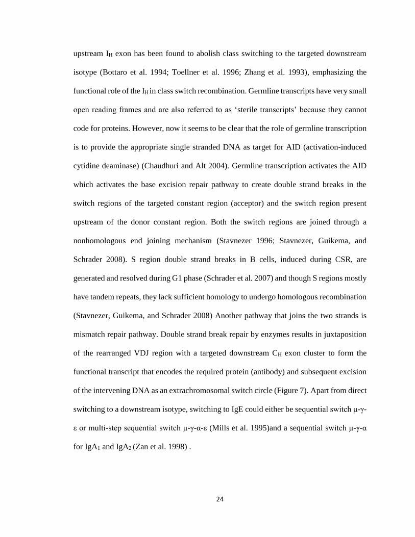
24
upstream IH exon has been found to abolish class switching to the targeted downstream
isotype (Bottaro et al. 1994; Toellner et al. 1996; Zhang et al. 1993), emphasizing the
functional role of the IH in class switch recombination. Germline transcripts have very small
open reading frames and are also referred to as ‘sterile transcripts’ because they cannot
code for proteins. However, now it seems to be clear that the role of germline transcription
is to provide the appropriate single stranded DNA as target for AID (activation-induced
cytidine deaminase) (Chaudhuri and Alt 2004). Germline transcription activates the AID
which activates the base excision repair pathway to create double strand breaks in the
switch regions of the targeted constant region (acceptor) and the switch region present
upstream of the donor constant region. Both the switch regions are joined through a
nonhomologous end joining mechanism (Stavnezer 1996; Stavnezer, Guikema, and
Schrader 2008). S region double strand breaks in B cells, induced during CSR, are
generated and resolved during G1 phase (Schrader et al. 2007) and though S regions mostly
have tandem repeats, they lack sufficient homology to undergo homologous recombination
(Stavnezer, Guikema, and Schrader 2008) Another pathway that joins the two strands is
mismatch repair pathway. Double strand break repair by enzymes results in juxtaposition
of the rearranged VDJ region with a targeted downstream CH exon cluster to form the
functional transcript that encodes the required protein (antibody) and subsequent excision
of the intervening DNA as an extrachromosomal switch circle (Figure 7). Apart from direct
switching to a downstream isotype, switching to IgE could either be sequential switch μ-γ-
ε or multi-step sequential switch μ-γ-α-ε (Mills et al. 1995)and a sequential switch μ-γ-α
for IgA1 and IgA2 (Zan et al. 1998) .
25
Figure 7: Class switch DNA recombination (CSR) event from IgM to IgE involving
the production of germline Iε-Cε transcript, functional VDJ-Cε transcript and Sε-Sμ
DNA switch circle.
26
Hypothesis and Objectives
The mouse 3’IghRR is a sensitive and early target of TCDD. In mouse B cells,
TCDD inhibits IgM secretion (Sulentic, Holsapple, and Kaminski 1998; Sulentic et al.
2004; Marcus, Holsapple, and Kaminski 1998) and in a cell line model, inhibition of Ig
secretion and 3’IghRR are mediated by the AhR (Wourms and Sulentic 2015). In
humans, however, the effect of TCDD on 3’IGHRR and antibody expression is poorly
understood. The current study focuses on the expression and secretion of IgM from
human mature naïve B cells, and their ability to class switch to different Ig isotypes in
response to TCDD. Our working hypothesis is that TCDD inhibits immunoglobulin class
switching through an AhR-mediated mechanism in human B cells. To test this
hypothesis, two main objectives are defined: 1) to determine the role of TCDD in
germline and functional transcription. Since germline transcription is the first and key
step in class switching to different downstream antibody isotypes, and functional
transcripts encode the required antibody, studying the effect of TCDD on these
transcripts would determine if CSR is a sensitive target of TCDD and 2) to determine the
role of the AhR in class switching. The AhR is activated by its ligand, TCDD, and
inhibiting this activation by using an AhR antagonist would help understand the role of
the AhR in human Ig expression.
27
II MATERIALS AND METHODS
Chemicals and Reagents
TCDD was purchased from AccuStandard, Inc (New Haven, CT). It is supplied as
308 μM solution in DMSO. DMSO was purchased from Sigma Aldrich (St. Louis, MO)
and used to dilute TCDD and the AhR Antagonist CH-223191 (Calbiochem, Carlsbad,
CA). Human Mega CD40L (soluble and recombinant) was purchased from Enzo Life
Sciences (San Diego, CA) and reconstituted in 100μl sterile water. When needed, further
dilutions were made in RPMI complete medium containing 10% Bovine Calf Serum
(described later in this section), aliquoted and stored at -20°C. Human IL-4 with carrier
was purchased from Cell Signaling Technology, Inc (Beverly, MA), reconstituted in sterile
1X PBS/10% BCS, aliquoted and stored at -20°C.
Cell Line Model and Culture Conditions
The cell line used in our experiments was Novus CL-01. It is a human monoclonal
Burkitt’s lymphoma B-cell line obtained from a patient that expresses IgM+ and IgD+ on
the cell surface, and can differentiate in response to appropriate stimuli. This cell line is
undergoes class switch from IgM to all seven downstream isotypes (IgA1, IgA2, IgG1, IgG2,
IgG3, IgG4, IgE) via CSR (Cerutti et al. 1998). CL-01 cells express single nucleotide
polymorphisms in the transactivation domain of the AhR.
28
Cells were cultured at 37°C in a 5% CO2 incubator and grown in complete media
consisting of RPMI 1640 Medium 1X Hyclone (Thermo Scientific Laboratories, Logan,
UT) supplemented with 10% bovine calf serum hyclone (Thermo Scientific Laboratories,
Logan, UT), 1M HEPES, 100mM Sodium Pyruvate, 100X MEM Non- Essential Amino
Acids, 100X Pencillin/ Streptomycin, 1M NaOH, 50μM 2-Mercaptoethanol. Cell viability
was monitored by the addition of trypan blue dye to 1ml of the cell suspension using the
Vi-cell Cell Counter (Beckman Coulter, Inc, Pasadena, CA). Cells were also checked
periodically under the microscope for any abnormal growth.
Sandwich Enzyme-Linked Immunosorbent Assay (Sandwich ELISA)
After the desired time of incubation (96 hours for IgG and IgM), cells were
centrifuged at 500xg for 5 minutes. Supernatants were collected, stored in separate 1.5ml
Eppendorf tubes labeled appropriately and used to determine Ig expression levels by using
Sandwich ELISA. The coating and detection antibody are specific for the antigen. The
detection antibody is HRP-conjugated (horseradish peroxidase), an enzyme that cleaves a
substrate producing color change. Colorimetric detection was performed using a
Spectramax Plus 384 UV/VIS Microplate Spectrophotometer at 450nM (Molecular
Devices, Sunnyvale, CA). The SOFTmax PRO software (Molecular Devices) was used to
calculate the sample concentrations using a standard curve generated from the absorption
of the IgG and IgM standard concentrations.
RNA Isolation
After the desired time of incubation (96 hours for IgG and IgM), cells were
centrifuged at 500xg for 5 minutes. Supernatants were collected and further analyzed by
ELISA as described above. The cell pellet was resuspended in Tri Reagent (Sigma Aldrich,
29
St. Louis, MO) and was stored at -80°C until further use. RNA was isolated following the
protocol of Tri Reagent manufacturer.
cDNA Synthesis, Reverse Transcription and Real Time PCR
Quantification of RNA samples was done using NanoDrop ND-1000
spectrophotometer (NanoDrop Products, Wilmington, DE). One microgram of total RNA
was converted into cDNA using BioRad iScript Reverse Transcription Supermix for Real
Time-qPCR (Berkeley, CA) following the instructions of manufacturer. Briefly, samples
were incubated initially for 5 minutes at 25°C, and reverse transcription allowed for 30
minutes at 42°C and finished by incubating samples at 85°C for 5 minutes. Total cDNA
was quantified using NanoDrop ND-1000 spectrophotometer, diluted to 100ng/replicate in
nuclease-free water and used for PCR. Cγ1 and Cα1 germline transcripts and γ1 IGH and α1
IGH functional transcripts were analyzed as follows: 10Xbuffer, dNTPs, forward and
reverse primers, Taq polymerase (New England Biolabs, Ipswich, MA) and template were
added and volume was made up to 25 µl with nuclease-free water. To determine μ IGH
functional transcripts, Cε germline transcripts, α1-2 and γ1-4 germline/functional transcripts
and CYP1A1 expression we used real time PCR (BioRad CFX 96, Berkeley, CA). The
setting for real time PCR was as follows: 2X Luminaris Color HiGreen qPCR Master Mix
(Thermo Scientific, Logan, UT), forward and reverse primers and template were added and
volume was made up to 20µl with nuclease-free water. β-actin was used as loading control.
The PCR primers used for amplifying the primers and their PCR cycling conditions are
indicated in Table 1 and Table 2, respectively. PCR products were run on 1.5% agarose
gels (GeneMate LE Agarose, Kaysville, UT) using a FB200 gel box from Fisher Scientific
(Waltham, MA) and comparing with the O’ Gene Ruler 100bp plus DNA ladder (Thermo

30
Scientific, Logan, UT). Gels were stained with Ethidium Bromide 1% solution (Fisher
BioReagents, Pittsburg, PA). Real Time PCR data were analyzed using the 2(-ΔΔCT) method
by comparing with the appropriate vehicle control.
31
Transcripts Primers
Control genes
CYP1A1 FP- 5’ CAGCTCCAAAGAGGTCCAAG 3’
RP-5’ CATGCAGAAGATGGTCAAGG 3’
β-actin FP- 5’ ATCACCATTGGCAATGAGCGGTTC 3’
RP- 5’ GCGGATGTCCACGTCACACTTCA 3’
Germline (IH-CH) transcripts
Cα1 FP- 5’ CAGCAGCCCTCTTGGCAGGCAGCCAG 3’
RP- 3’ GGGTGGCGGTTAGCGGGGTCTTGG 3’
Cγ1 FP- 5’ GGGCTTCCAAGCCAACAGGGCAGGACA 3’
RP- 5’ GTTTTGTCACAAGATTTGGGCTC 3’
Cε FP- 5’ GACGGGCCACACCATCC 3’
RP- 5’ CGGAGGTGGCATTGGAGG 3’
Functional (VH-DJH-CH) transcripts
α1 IGH FP- 5’ GACACGGCTGTGTATTACTGTGCG 3’
RP- 5’ GGGTGGCGGTTAGCGGGGTCTTGG 3’
γ1 IGH FP- 5’ GACACGGCTGTGTATTACTGTGCG 3’
RP- 5’ GTTTTGTCACAAGATTTGGGCTC 3’
μ IGH FP- 5’ GACACGGCTGTGTATTACTGTGCG 3’
RP- 5’ CCGAATTCAGACGAGGGGGAAAAGGGTT 3’
(Continued)
32
Germline (IH-CH) and Functional (VH-DJH-CH) transcripts
α1 FP- 5’ CGCCATGACAACAGACACAT 3’
RP- 5’ GTATGCTGGTCACAGCGAAG 3’
α2 FP- 5’ ACATTGATGTGGGTGGGTTT 3’
RP- 5’ CCACCACCTACGCTGTAACC 3’
γ1 FP- 5’ GATGTCGCTGGGATAGAAGC 3’
RP- 5’ TGTTGGAGACCTTGCACTTG 3’
γ2 FP- 5’ GAGATGTCGCTGGGGTAGAA 3’
RP- 5’ GGCAAGGAGTACAAGTGCAA 3’
γ3 FP- 5’ TCACGTTGCAGGTGTAGGTC 3’
RP- 5’ CTACTTCCCAGAACCGGTGA 3’
γ4 FP- 5’ GGTTCTTGGTCATCTCCTCCT 3’
RP- 5’ GAACGGCAAGGAGTACAAGTG 3’
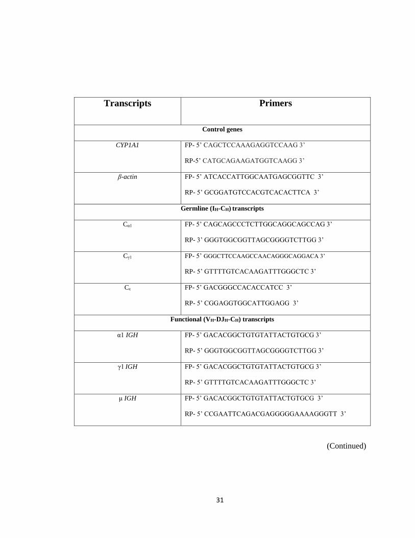
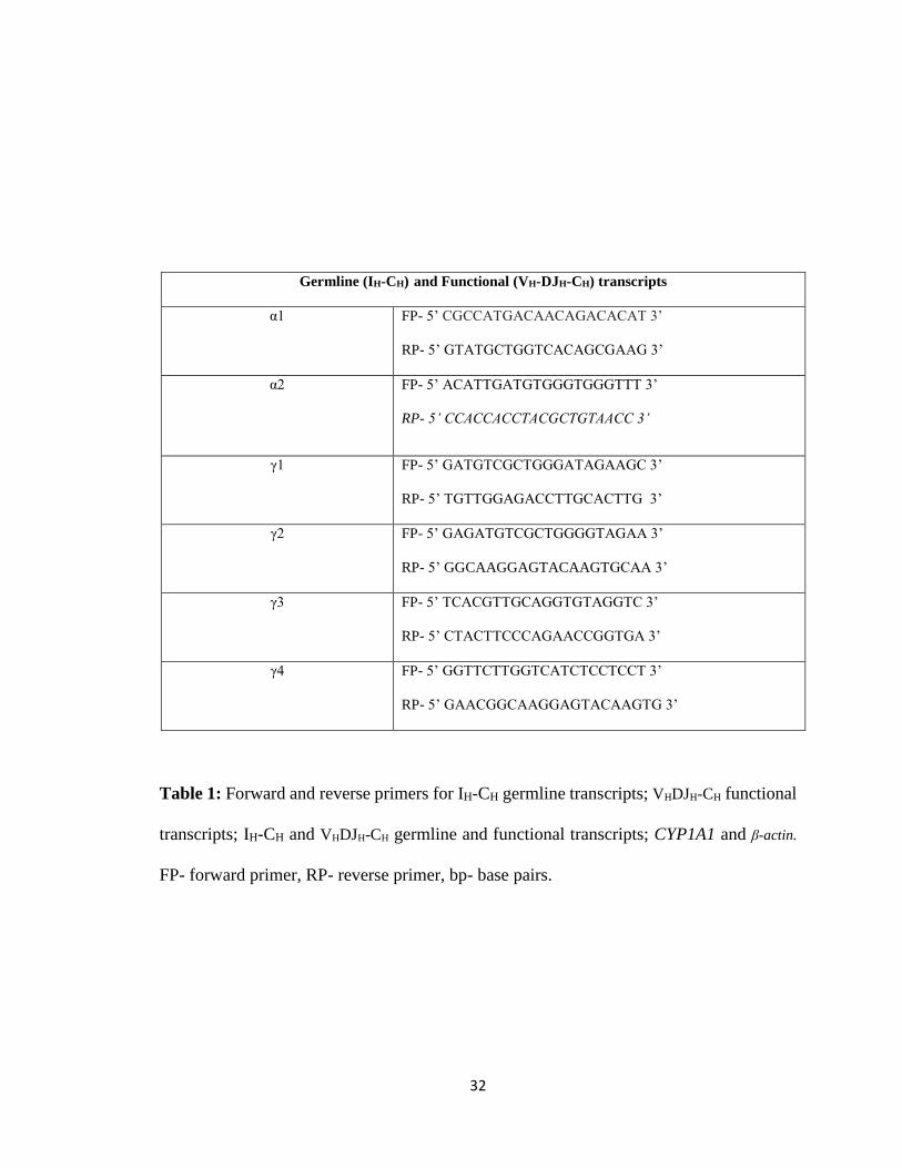
Table 1: Forward and reverse primers for IH-CH germline transcripts; VHDJH-CH functional
transcripts; IH-CH and VHDJH-CH germline and functional transcripts; CYP1A1 and β-actin.
FP- forward primer, RP- reverse primer, bp- base pairs.
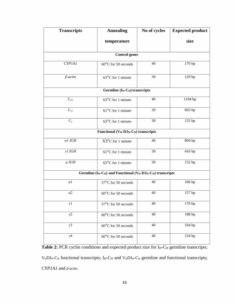
33
Transcripts Annealing
temperature
No of cycles Expected product
size
Control genes
CYP1A1 60°C for 50 seconds 40 170 bp
β-actin 63°C for 1 minute 30 129 bp
Germline (IH-CH) transcripts
Cα1 63°C for 1 minute 40 1194 bp
Cγ1 61°C for 1 minute 30 603 bp
Cε 63°C for 1 minute 30 125 bp
Functional (VH-DJH-CH) transcripts
α1 IGH 63°C for 1 minute 40 804 bp
γ1 IGH 61°C for 1 minute 30 416 bp
μ IGH 63°C for 1 minute 30 152 bp
Germline (IH-CH) and Functional (VH-DJH-CH) transcripts
α1 57°C for 50 seconds 40 166 bp
α2 60°C for 50 seconds 40 157 bp
γ1 57°C for 50 seconds 40 170 bp
γ2 60°C for 50 seconds 40 188 bp
γ3 60°C for 50 seconds 40 164 bp
γ4 60°C for 50 seconds 40 154 bp
Table 2: PCR cyclin conditions and expected product size for IH-CH germline transcripts;
VHDJH-CH functional transcripts; IH-CH and VHDJH-CH germline and functional transcripts;
CYP1A1 and β-actin.
34
Statistical analysis of data
For ELISA and Real Time PCR results, a one-way ANOVA followed by Dunnett’s
Multiple Comparison test was used to analyze the treatments groups (mean ± SE; n=3 for
ELISA and n=3 separate RNA isolations for Routine PCR and Real Time PCR) using
GraphPad Prism software for significant differences between treatment groups and
appropriate vehicle control were denoted by *, **, ***; significant differences between
stimulation and NA were denoted by #, ##, ### which represent significance at p<0.05,
p<0.01 and p<0.001, respectively.
Effects of co-treatment of TCDD and the AhR antagonist on Ig secretion and
transcripts are represented with one-way ANOVA, gray circles represent 10 and 30 nM
TCDD alone, open squares represent 10 and 30 μM of the AhR antagonist alone, green
triangles represent 10 or 30 nM TCDD with 10μM of the AhR antagonist and blue triangles
represent 10 or 30 nM TCDD with 30 μM of the AhR antagonist. Black line represents the
vehicle control at 1. Figures with TCDD and the AhR antagonist are separated from the
original figure with TCDD, the AhR antagonist, TCDD and the AhR antagonist to clearly
indicate the effects of TCDD and the AhR antagonist with respect to corresponding vehicle
control. Only significant differences between the AhR antagonist and TCDD are
represented on the two-way ANOVA graphs.
35
III RESULTS
Differential effects of TCDD and the AhR antagonist on immunoglobulin
secretion
TCDD is known to directly target mouse B cells and inhibit IgM secretion
(Sulentic, Holsapple, and Kaminski 1998). In another study with primary human B cells
from donors, nine of twelve human donor primary B cells showed reduced IgM expression
when treated with TCDD, while two donors showed no response and one demonstrated
enhanced IgM expression (Lu et al. 2010). Expecting an inhibition with TCDD in our
present study, we stimulated our cells with CD40L and IL-4. Surprisingly, stimulation and
TCDD had variable effects on IgM secretion (Fig. 8), as TCDD either increased, decreased
or had no effect on IgM secretion. Conversely, TCDD treatment (10 and 30 nM) inhibited
the stimulation (Fig. 8). Additionally, low levels of IgG were also observed in unstimulated
cells, suggesting that CL-01 cells have mixed subpopulations of cells that have undergone
spontaneous CSR and express either of the IgG isotypes.
The AhR antagonist alone either increased, decreased or had no effect on IgM
secretion or in co-treatment with TCDD, resulting in variable effects on IgM (Fig. 8). In
contrast, the AhR antagonist alone increased IgG secretion by two-fold (10 and 30 μM),
and the AhR antagonist and TCDD antagonized each other’s effects upon co-treatment
(Fig. 9 and 10). Gray arrow from 30 nM TCDD (grey circle) to both green and blue
triangles indicates that 10 and 30 μM of the AhR antagonist significantly reverses the
36
inhibitory effect of 30 nM TCDD on IgG secretion. Green arrow from 10 μM of the AhR
antagonist (open square) to green triangles indicates that 10 and 30 nM TCDD significantly
reverse the increase in IgG secretion by 10 μM of the AhR antagonist (Fig. 10). These
results demonstrate a difference in sensivity of IgM vs. IgG to TCDD and the AhR
antagonist.
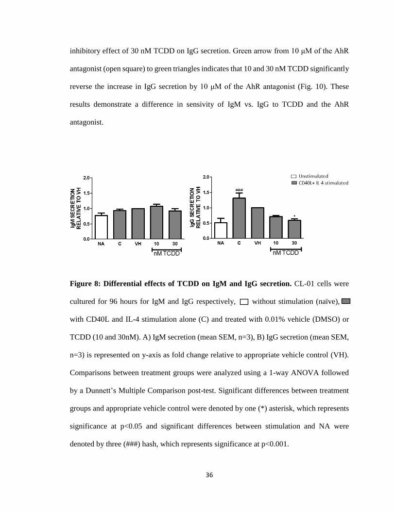
Figure 8: Differential effects of TCDD on IgM and IgG secretion. CL-01 cells were
cultured for 96 hours for IgM and IgG respectively, without stimulation (naïve), or
with CD40L and IL-4 stimulation alone (C) and treated with 0.01% vehicle (DMSO) or
TCDD (10 and 30nM). A) IgM secretion (mean SEM, n=3), B) IgG secretion (mean SEM,
n=3) is represented on y-axis as fold change relative to appropriate vehicle control (VH).
Comparisons between treatment groups were analyzed using a 1-way ANOVA followed
by a Dunnett’s Multiple Comparison post-test. Significant differences between treatment
groups and appropriate vehicle control were denoted by one (*) asterisk, which represents
significance at p<0.05 and significant differences between stimulation and NA were
denoted by three (###) hash, which represents significance at p<0.001.
37
Figure 9: Differential effects of the AhR antagonist on IgM and IgG secretion. CL-01
cells were cultured for 96 hours for IgM and IgG respectively, without stimulation
(naïve), or with CD40L and IL-4 stimulation alone (C) and treated with 0.01% vehicle
(DMSO) or the AhR antagonist (10 and 30μM). A) IgM secretion (mean SEM, n=3), B)
IgG secretion (mean SEM, n=3) is represented on y-axis as fold change relative to
appropriate vehicle control (VH). Comparisons between treatment groups were analyzed
using a 1-way ANOVA followed by a Dunnett’s Multiple Comparison post-test.
Significant differences between treatment groups and appropriate vehicle control were
denoted by three (***) asterisks, which represents significance at p<0.001and significant
differences between stimulation and NA were denoted by two (##) hash, which represents
significance at p<0.01.
38
39
Figure 10: The AhR antagonist and TCDD antagonize each other’s effects on IgG
secretion and have no effect on IgM secretion. CL-01 cells were cultured 96 hours for
IgM and IgG respectively, without stimulation (naïve) or with CD40L and IL-4 stimulation
alone (C) and treated with 0.01% vehicle (DMSO) or TCDD (10 and 30 nM) or the AhR
antagonist (10 and 30 μM). A) IgM secretion (mean SEM, n=3, B) IgG secretion (mean
SEM, n=3) is represented on y-axis as fold change relative to appropriate vehicle control
(VH). Comparisons between treatment groups were analyzed using a 1-way ANOVA
followed by a Dunnett’s Multiple Comparison test. Gray circles represent 10 and 30 nM
TCDD alone, open squares represent 10 and 30 μM of the AhR antagonist alone, green
triangles represent 10 or 30 nM TCDD with 10μM of the AhR antagonist and blue triangles
represent 10 or 30 nM TCDD with 30 μM of the AhR antagonist. The significant
differences between treatment groups and appropriate vehicle control and differences
between stimulation and NA are same as in figures 8 and 9.
40
Differential effects of TCDD and the AhR antagonist on μ IGH functional
transcripts and γ1-4 germline/functional transcripts
TCDD alone and the AhR antagonist alone either increased, decreased or had no
effect on μ IGH functional transcripts, or co-treatment of TCDD and the AhR antagonist
also had variable effects on μ IGH functional transcripts (Fig. 11, 12 and 13). Cγ1 germline
(Fig. 14) and γ1 IGH functional transcripts (Fig. 14) were analyzed qualitatively by gel
electrophoresis of PCR products. To quantitate the germline and functional transcript
expression, real time PCR primers were designed spanning the constant regions for γ1-4.
Stimulation with CD40L plus IL-4 significantly increased the levels of γ1-4
germline/functional transcripts as expected. Addition of TCDD treatment (10 and 30 nM)
inhibited γ1-4 germline/functional transcripts (Fig. 15). The AhR antagonist (10 and 30 μM)
alone increased γ1-4 germline/functional transcripts (Fig. 16). AhR antagonist and TCDD
antagonized each other’s effect on γ transcripts. Green arrow from 30 nM TCDD (gray
circle) to 10 μM of AhR antagonist (open square) indicates that 30 nM TCDD reversed the
increase in γ2-4 transcripts by 10 μM of AhR antagonist (Fig. 17).
41
Figure 11: TCDD has variable effect on μ IGH functional transcripts. CL01 cells were
cultured for 96 hours without stimulation (naïve), or with CD40L and IL-4
stimulation alone (C) and treated with 0.01% vehicle (DMSO) or TCDD (10 and 30 nM).
μ IGH transcript expression (mean SEM, n=3) was measured by real time PCR and
represented on y-axis as fold change relative to appropriate vehicle control (VH).
Comparisons between treatment groups were analyzed using a 1-way ANOVA followed
by a Dunnett’s Multiple Comparison test.
42
Figure 12: The AhR antagonist has variable effect on μ IGH functional transcripts.
CL01 cells were cultured for 96 hours without stimulation (naïve), or with CD40L
and IL-4 stimulation alone (C) and treated with 0.01% vehicle (DMSO) or AhR antagonist
(10 and 30μM). μ IGH transcript expression (mean SEM, n=3) was measured by real time
PCR and represented on y-axis as fold change relative to appropriate vehicle control (VH).
Comparisons between treatment groups were analyzed using a 1-way ANOVA followed
by a Dunnett’s Multiple Comparison test.
43
Figure 13: Co-treatment of the AhR antagonist with TCDD has variable effect on μ
IGH functional transcripts. CL01 cells were cultured for 96 hours without stimulation
(naïve), with CD40L and IL-4 stimulation alone (C) and treated with 0.01% vehicle
(DMSO) or TCDD (10 and 30 nM) or AhR antagonist (10 and 30 μM). μ IGH functional
transcript expression (mean SEM, n=3) was measured by real time PCR and represented
on y-axis as fold change relative to appropriate vehicle control (VH). Comparisons between
treatment groups were analyzed using a 1-way ANOVA followed by a Dunnett’s Multiple
Comparison test. Gray circles represent 10 and 30 nM TCDD alone, open squares represent
10 and 30 μM of the AhR antagonist alone, green triangles represent 10 or 30 nM TCDD
with 10μM of the AhR antagonist and blue triangles represent 10 or 30 nM TCDD with 30
μM of the AhR antagonist. The significant differences between treatment groups and
appropriate vehicle control and differences between stimulation and NA are same as in
figures 11 and 12.
44
Figure 14: Spontaneous class switch from Cμ to Cγ1. CL01 cells were cultured for 96
hours without stimulation (naïve), or with CD40L and IL-4 stimulation and treated with
0.01% vehicle (DMSO) or TCDD (10 and 30nM) or AhR antagonist (10 and 30μM). ‘C’
represents CD40L and IL-4 stimulation, ‘T’ represents nM concentration of TCDD, ‘A’
represents μM concentration of AhR antagonist and ‘T+A’ represents nM concentration of
TCDD and μM concentration of AhR antagonist respectively. Cγ1 germline transcripts,
γ1IGH functional transcripts and β-actin could be observed at 603, 416 and 129 base pairs
(bp), respectively.
45
Figure 15: TCDD inhibits γ1-4 germline/functional transcripts. CL01 cells were
cultured for 96 hours without stimulation (naïve), or with CD40L and IL-4
stimulation alone (C) and treated with 0.01% vehicle (DMSO) or TCDD (10 and 30 nM).
γ1-4 germline/functional transcripts were measured by real time PCR (mean SEM, n=3) and
represented on y-axis as fold change relative to appropriate vehicle control (VH).
Comparisons between treatment groups were analyzed using a 1-way ANOVA followed
by a Dunnett’s Multiple Comparison post-test. Significant differences between treatment
groups and appropriate vehicle control were denoted by one (*), two (**) and three (***)
asterisks, which represents significance at p<0.05, p<0.01 and p<0.001, respectively and
significant differences between stimulation and NA were denoted by two (##) and three
(###) hash, which represents significance at p<0.01 and p<0.001, respectively.
46
Figure 16: The AhR antagonist alone increases γ1-4 germline/functional transcripts.
CL01 cells were cultured for 96 hours without stimulation (naïve), or with CD40L
and IL-4 stimulation and treated with 0.01% vehicle (DMSO) or AhR antagonist (10 and
30μM). γ1-4 germline/functional transcripts were measured by real time PCR (mean SEM,
n=3) and represented on y-axis as fold change relative to appropriate vehicle control (VH).
Comparisons between treatment groups were analyzed using a 1-way ANOVA followed
by a Dunnett’s Multiple Comparison post-test. Significant differences between treatment
groups and appropriate vehicle control were denoted by one (*), two (**) and three (***)
asterisks, which represents significance at p<0.05, p<0.01 and p<0.001, respectively and
significant differences between stimulation and NA were denoted by one (#) and three
(###) hash, which represents significance at p<0.05 and p<0.001, respectively.
47
Figure 17: The AhR antagonist and TCDD antagonize each other’s effects on γ1-4
germline/functional transcripts. CL01 cells were cultured for 96 hours without
stimulation (naïve), or with CD40L and IL-4 stimulation alone (C) and treated with 0.01%
vehicle (DMSO) or TCDD (10 and 30 nM) or AhR antagonist (10 and 30 μM). γ1-4
germline/functional transcripts were measured by real time PCR (mean SEM, n=3) and
represented on y-axis as fold change relative to appropriate vehicle control. Comparisons
between treatment groups were analyzed using a 1-way ANOVA followed by a Dunnett’s
Multiple Comparison test. Gray circles represent 10 and 30 nM TCDD alone, open squares
represent 10 and 30 μM of the AhR antagonist alone, green triangles represent 10 or 30 nM
TCDD with 10μM of the AhR antagonist and blue triangles represent 10 or 30 nM TCDD
with 30 μM of the AhR antagonist. The significant differences between treatment groups
and appropriate vehicle control and differences between stimulation and NA are same as
in figures 15 and 16.
48
Differential effects of TCDD and the AhR antagonist on α1-2
germline/functional transcripts
α1 IGH functional transcripts (Fig. 18) were analyzed by PCR and Cα1 germline
transcripts could not be observed (Data not shown) by PCR analysis. To quantitate the
germline and functional transcript expression, real time PCR primers were designed
spanning the constant regions for α1-2. Surprisingly, α1 as well as α2 germline/functional
transcripts were readily expressed in unstimulated CL-01 cells.
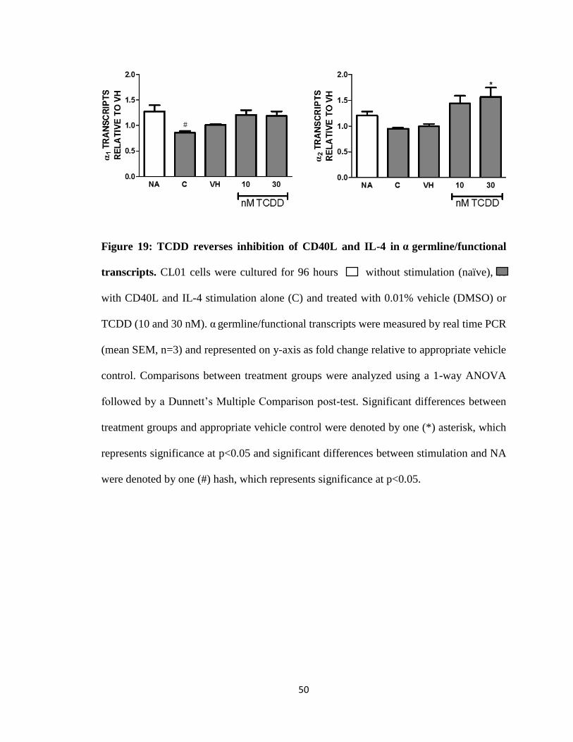
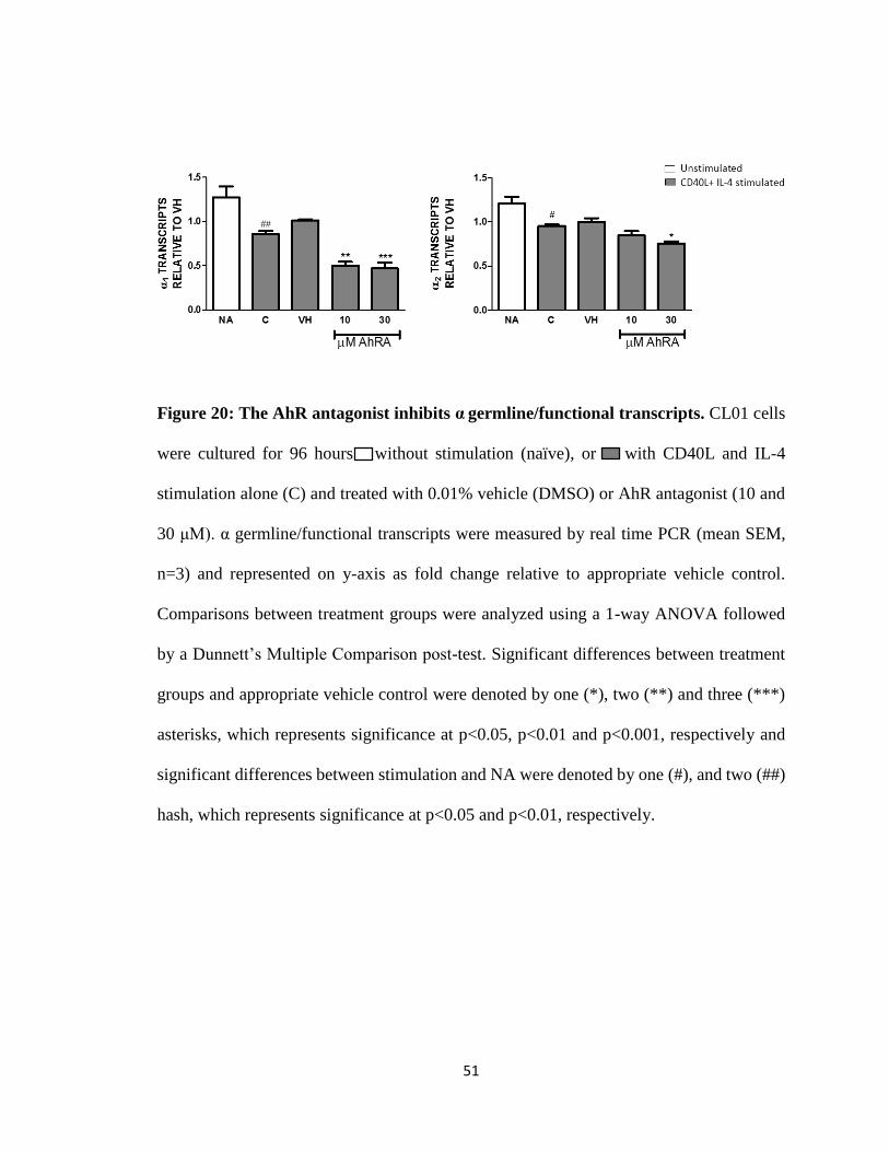
Unexpectedly, α1 and α2 germline/functional transcripts are inhibited with CD40L
and IL-4 stimulation, which is significantly reversed by the addition of TCDD (10 and 30
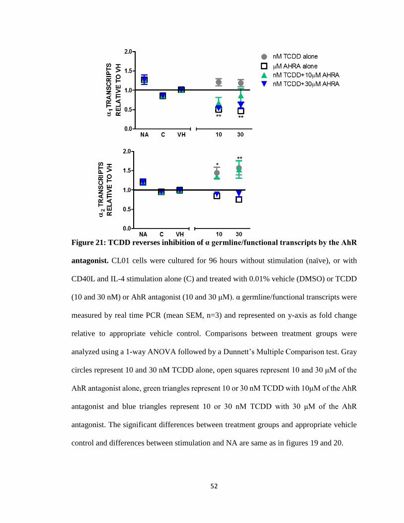
nM) (Fig. 19). Surprisingly, the AhR antagonist alone inhibited α germline/functional
transcripts (Fig. 20). Co-treatment of TCDD with the AhR antagonist reversed the AhR
antagonist-induced inhibition of α1 and α2 germline/functional transcript expression (Fig.
21).
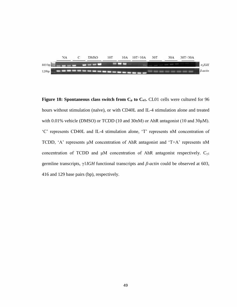
49
Figure 18: Spontaneous class switch from Cμ to Cα1. CL01 cells were cultured for 96
hours without stimulation (naïve), or with CD40L and IL-4 stimulation alone and treated
with 0.01% vehicle (DMSO) or TCDD (10 and 30nM) or AhR antagonist (10 and 30μM).
‘C’ represents CD40L and IL-4 stimulation alone, ‘T’ represents nM concentration of
TCDD, ‘A’ represents μM concentration of AhR antagonist and ‘T+A’ represents nM
concentration of TCDD and μM concentration of AhR antagonist respectively. Cγ1
germline transcripts, γ1IGH functional transcripts and β-actin could be observed at 603,
416 and 129 base pairs (bp), respectively.
50
Figure 19: TCDD reverses inhibition of CD40L and IL-4 in α germline/functional
transcripts. CL01 cells were cultured for 96 hours without stimulation (naïve), or
with CD40L and IL-4 stimulation alone (C) and treated with 0.01% vehicle (DMSO) or
TCDD (10 and 30 nM). α germline/functional transcripts were measured by real time PCR
(mean SEM, n=3) and represented on y-axis as fold change relative to appropriate vehicle
control. Comparisons between treatment groups were analyzed using a 1-way ANOVA
followed by a Dunnett’s Multiple Comparison post-test. Significant differences between
treatment groups and appropriate vehicle control were denoted by one (*) asterisk, which
represents significance at p<0.05 and significant differences between stimulation and NA
were denoted by one (#) hash, which represents significance at p<0.05.
51
Figure 20: The AhR antagonist inhibits α germline/functional transcripts. CL01 cells
were cultured for 96 hours without stimulation (naïve), or with CD40L and IL-4
stimulation alone (C) and treated with 0.01% vehicle (DMSO) or AhR antagonist (10 and
30 μM). α germline/functional transcripts were measured by real time PCR (mean SEM,
n=3) and represented on y-axis as fold change relative to appropriate vehicle control.
Comparisons between treatment groups were analyzed using a 1-way ANOVA followed
by a Dunnett’s Multiple Comparison post-test. Significant differences between treatment
groups and appropriate vehicle control were denoted by one (*), two (**) and three (***)
asterisks, which represents significance at p<0.05, p<0.01 and p<0.001, respectively and
significant differences between stimulation and NA were denoted by one (#), and two (##)
hash, which represents significance at p<0.05 and p<0.01, respectively.
52
Figure 21: TCDD reverses inhibition of α germline/functional transcripts by the AhR
antagonist. CL01 cells were cultured for 96 hours without stimulation (naïve), or with
CD40L and IL-4 stimulation alone (C) and treated with 0.01% vehicle (DMSO) or TCDD
(10 and 30 nM) or AhR antagonist (10 and 30 μM). α germline/functional transcripts were
measured by real time PCR (mean SEM, n=3) and represented on y-axis as fold change
relative to appropriate vehicle control. Comparisons between treatment groups were
analyzed using a 1-way ANOVA followed by a Dunnett’s Multiple Comparison test. Gray
circles represent 10 and 30 nM TCDD alone, open squares represent 10 and 30 μM of the
AhR antagonist alone, green triangles represent 10 or 30 nM TCDD with 10μM of the AhR
antagonist and blue triangles represent 10 or 30 nM TCDD with 30 μM of the AhR
antagonist. The significant differences between treatment groups and appropriate vehicle
control and differences between stimulation and NA are same as in figures 19 and 20.
53
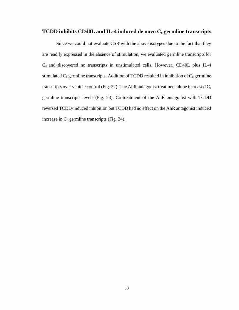
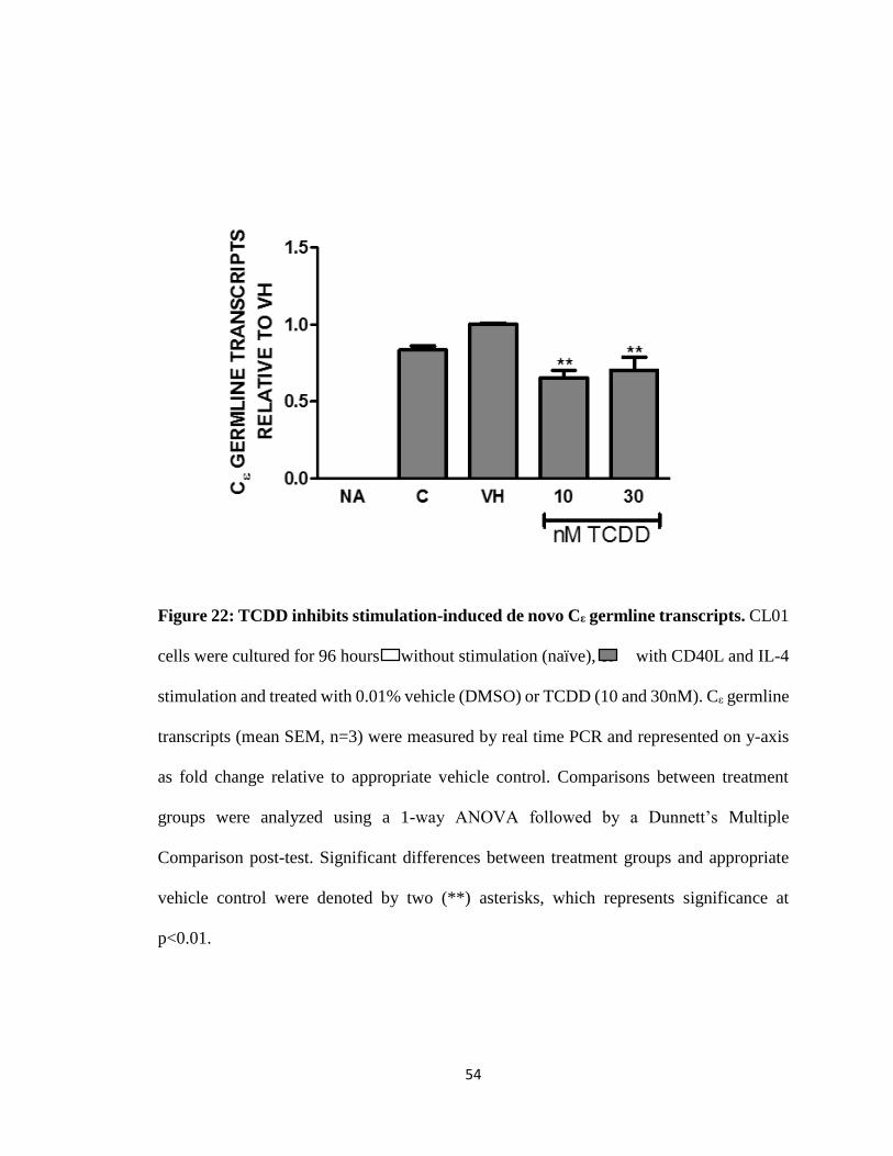
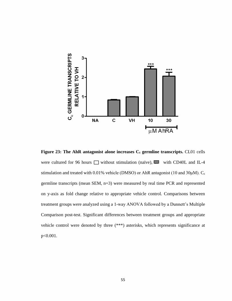
TCDD inhibits CD40L and IL-4 induced de novo Cε germline transcripts
Since we could not evaluate CSR with the above isotypes due to the fact that they
are readily expressed in the absence of stimulation, we evaluated germline transcripts for
Cε and discovered no transcripts in unstimulated cells. However, CD40L plus IL-4
stimulated Cε germline transcripts. Addition of TCDD resulted in inhibition of Cε germline
transcripts over vehicle control (Fig. 22). The AhR antagonist treatment alone increased Cε
germline transcripts levels (Fig. 23). Co-treatment of the AhR antagonist with TCDD
reversed TCDD-induced inhibition but TCDD had no effect on the AhR antagonist induced
increase in Cε germline transcripts (Fig. 24).
54
Figure 22: TCDD inhibits stimulation-induced de novo Cε germline transcripts. CL01
cells were cultured for 96 hours without stimulation (naïve), or with CD40L and IL-4
stimulation and treated with 0.01% vehicle (DMSO) or TCDD (10 and 30nM). Cε germline
transcripts (mean SEM, n=3) were measured by real time PCR and represented on y-axis
as fold change relative to appropriate vehicle control. Comparisons between treatment
groups were analyzed using a 1-way ANOVA followed by a Dunnett’s Multiple
Comparison post-test. Significant differences between treatment groups and appropriate
vehicle control were denoted by two (**) asterisks, which represents significance at
p<0.01.
55
Figure 23: The AhR antagonist alone increases Cε germline transcripts. CL01 cells
were cultured for 96 hours without stimulation (naïve), or with CD40L and IL-4
stimulation and treated with 0.01% vehicle (DMSO) or AhR antagonist (10 and 30μM). Cε
germline transcripts (mean SEM, n=3) were measured by real time PCR and represented
on y-axis as fold change relative to appropriate vehicle control. Comparisons between
treatment groups were analyzed using a 1-way ANOVA followed by a Dunnett’s Multiple
Comparison post-test. Significant differences between treatment groups and appropriate
vehicle control were denoted by three (***) asterisks, which represents significance at
p<0.001.
56
Figure 24: The AhR antagonist reverses inhibition of Cε germline transcripts by
TCDD. CL01 cells were cultured for 96 hours without stimulation (naïve), or with CD40L
and IL-4 stimulation alone (C) and treated with 0.01% vehicle (DMSO) or TCDD (10 and
30 nM) or AhR antagonist (10 and 30 μM). Cε germline transcripts (mean SEM, n=3) were
measured by real time PCR and represented on y-axis as fold change relative to appropriate
vehicle control. Comparisons between treatment groups were analyzed using a 1-way
ANOVA followed by a Dunnett’s Multiple Comparison test. Gray circles represent 10 and
30 nM TCDD alone, open squares represent 10 and 30 μM of the AhR antagonist alone,
green triangles represent 10 or 30 nM TCDD with 10μM of the AhR antagonist and blue
triangles represent 10 or 30 nM TCDD with 30 μM of the AhR antagonist. The significant
differences between treatment groups and appropriate vehicle control and differences
between stimulation and NA are same as in figures 22 and 23.
57
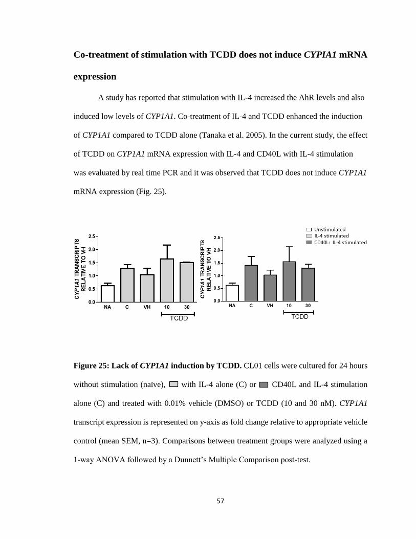
Co-treatment of stimulation with TCDD does not induce CYPIA1 mRNA
expression
A study has reported that stimulation with IL-4 increased the AhR levels and also
induced low levels of CYP1A1. Co-treatment of IL-4 and TCDD enhanced the induction
of CYP1A1 compared to TCDD alone (Tanaka et al. 2005). In the current study, the effect
of TCDD on CYP1A1 mRNA expression with IL-4 and CD40L with IL-4 stimulation
was evaluated by real time PCR and it was observed that TCDD does not induce CYP1A1
mRNA expression (Fig. 25).
Figure 25: Lack of CYP1A1 induction by TCDD. CL01 cells were cultured for 24 hours
without stimulation (naïve), with IL-4 alone (C) or CD40L and IL-4 stimulation
alone (C) and treated with 0.01% vehicle (DMSO) or TCDD (10 and 30 nM). CYP1A1
transcript expression is represented on y-axis as fold change relative to appropriate vehicle
control (mean SEM, n=3). Comparisons between treatment groups were analyzed using a
1-way ANOVA followed by a Dunnett’s Multiple Comparison post-test.
58
IV DISCUSSION
Previous report in mouse mature B cells has shown the inhibitory effect of
TCDD on IgM secretion when stimulated with LPS and when treated with varying
concentrations of TCDD (0.03-30nM) (Sulentic, Holsapple, and Kaminski 1998). In
another study on primary human B cells, the effect of TCDD on CD40L plus IL-4
stimulated cells was inhibitory. Additionally, nine of twelve human donor primary B cells
showed reduced IgM expression when treated with TCDD, while two donors showed no
response and one demonstrated enhanced IgM expression (Lu et al. 2010). In the current
study, CD40L plus IL-4, TCDD and the AhR antagonist had variable effects on IgM
secretion. Similarly, R848 (TLR 7/8)-induced IgM secretion was also variable to TCDD
(Data not shown). Conversely, TCDD inhibited IgG expression and in a concentration
dependent manner (0.1-100nM) (Data not shown). Surprisingly, the AhR antagonist alone
increased IgG expression by two-fold. Both TCDD and the AhR antagonist antagonized
each other’s effects on IgG secretion and also, TCDD reversed the increase observed with
the addition of the AhR antagonist. These results were replicated at the transcript level
supporting an effect on gene regulation.
Presence of γ1-4 germline/functional transcripts in unstimulated cells suggest a
spontaneous class switch to IgG1-4. TCDD treatment inhibited the stimulation induced γ1-4
germline/functional transcripts, and addition of the AhR antagonist alone significantly
increased all the γ1-4 germline/functional transcripts. Both TCDD and the AhR antagonist
59
antagonized each other’s effects on IgG secretion and also, TCDD reversed the increase
observed with the addition of the AhR antagonist. Higher concentration (30 nM) of TCDD
appears to reverse the increase observed by 10 μM concentration of the AhR antagonist in
γ transcripts. This correlates to the IgG ELISA data and confirms that the effect of TCDD
on IgG secretion occurs at transcript level. Another possibility is that TCDD could decrease
the stability of the transcript rather than inhibit transcription, resulting in a decrease in
overall Ig expression. Additionally, NF-κB is known to inhibit CSR to Igγ1 in mouse B
cells (Bhattacharya, Lee, and Sha 2002), and TCDD has been shown to induce the AhR
binding to NF-κB within the mouse 3’IghRR (Richard Salisbury, unpublished data),
therefore TCDD could inhibit IgG by altering transcription factor binding within the
3’IGHRR. Also, differential effects of TCDD and the AhR antagonist on μ IGH functional
transcripts and γ1-4 germline/functional transcripts as well as Ig expression imply a different
role of the AhR in mediating TCDD-induced effects with different antibody isotypes.
In an ongoing study in our lab with shRNA-mediated AhR knockdown cells vs wild
type cells with the AhR, IgM had variable effect to both TCDD and AhR antagonist in wild
type and AhR knockdown cells. TCDD inhibited IgG secretion in wild type cells but AhR
knockdown nearly eliminated IgG secretion in AhR knockdown cells. In contrast, when
treated with AhR antagonist, both the wild type and knockdown cells expressed an increase
in IgG expression (Bassam Kashgari, unpublished data). Different effects of the AhR
antagonist (increase γ expression and IgG secretion) and AhR knockdown cells (decreased
γ expression and IgG secretion) imply that the AhR antagonist could target signaling
pathways other than the AhR. The AhR antagonist (CH223191) inhibits the AhR activation
and thereby prevents its translocation into the nucleus. Since the AhR antagonist inhibits
60
translocation of the AhR, but potentially not the association or dissociation of the AhR with
other cytosolic proteins, it is possible that the AhR antagonist influences these interactions.
For example interaction of the AhR antagonist with endogenous signaling proteins like c-
Src, a protein associated with the AhR activates a chain of events that lead increased Ig
expression (Enan and Matsumura 1996). Alternatively, the AhR antagonist may induce
off-target effects; however the concentration-dependent effect of TCDD and the AhR
antagonist on γ expression suggests a competition for AhR binding.
Analysis of α1 IGH functional transcripts; α1 and α2 germline and functional
transcripts demonstrated the presence of α1 IGH functional transcripts as well as α1 and α2
germline and functional transcripts in unstimulated cells. It is also likely that among α1 and
α2 germline/functional transcripts, α1 and α2 functional transcripts are majorly being
expressed compared to germline transcripts, which supports the theory that the CL-01 cells
have mixed subpopulations of cells, albeit very small populations, that have undergone
spontaneous CSR and express either of the IgA isotypes. Another unexpected observation
was the inhibition of both α1 and α2 germline and functional transcript expression with
stimulation, in contrast to a previous report demonstrating increased expression with
stimulation (Cerutti et al. 1998). Interestingly, addition of TCDD increased α
germline/functional transcripts. Also, the AhR antagonist alone inhibited both α
germline/functional transcripts but α1 transcripts were inhibited by almost 50%, and α2
transcripts seem less sensitive to the AhR antagonist. Differences in the effects of TCDD
on α1 vs α2 germline/functional transcripts may reflect differences in the α1 3’IGHRR vs α2
3’IGHRR. In CL-01 cells, the α2 3’IGHRR has 4 repeats of the IS in the hs1,2 enhancer
and the α1 3’IGHRR has 4 repeats of the IS in the hs1,2 enhancer. This could suggest an
61
increase in sensitivity of the α2 3’IGHRR to TCDD. With the presence of α1 and α2 heavy
chain genes adjacent to the 3’IGHRR, there is a possibility that the polymorphic hs1,2
enhancer is affecting the gene transcription. In fact, higher concentrations of the AhR
antagonist (30 μM) appear to reverse the increase in α transcripts observed with TCDD (10
and 30 nM), suggesting a competitive binding to a common target (the AhR). We have
previously confirmed that TCDD-mediated inhibition of IgA secretion in a mouse cell line
is reversed by using an AhR antagonist or by AhR knockdown through shRNA (Wourms
and Sulentic 2015). In another study, TCDD inhibited mucosal IgA secretion and this was
not observed in AhR deficient mice, suggesting a role of the AhR in mediating TCDD-
induced inhibition of IgA (Kinoshita et al. 2006).
Since we cannot evaluate CSR with the above isotypes due to the fact that they are
readily expressed in the absence of stimulation, we evaluated germline transcripts for Cε
and discovered no transcripts in unstimulated cells. However, CD40L plus IL-4 stimulated
Cε germline transcripts. Addition of TCDD resulted in inhibition of Cε germline transcripts
over vehicle control. The AhR antagonist alone increased Cε germline transcripts levels.
The AhR antagonist reversed the TCDD-induced inhibition, but the reverse is not observed,
suggesting a different mechanism is involved in mediating the effects of TCDD and the
AhR antagonist. In contrast, a study on tonsillar human B cells from atopic and non-atopic
patients demonstrated that TCDD elevated the levels of IgE in atopic patients, but did not
alter the levels of IgE in non-atopic patients (Kimata 2003). In another ongoing study in
our lab, transient transfection of the Iε luciferase reporter plasmid in CL01 cells revealed
basal activity of the Iε promoter that was increased with CD40L and IL-4 stimulation.
Addition of TCDD resulted in inhibition of the Iε promoter and pre-treatment with the AhR
62
antagonist reversed the TCDD-induced inhibition (Zahra Alfaheeda, unpublished data).
Also, evidence suggests that class switching to IgE from IgM could be an indirect switch
via μ-γ-ε (Mills et al. 1995). This could mean that TCDD-induced inhibition of γ and ε
could be related. But, the decreased levels of IgG in AhR knockdown cells in response to
TCDD contradicts this possibility. The presence of an IL-4 response element (Schaffer et
al. 2003) and DRE core motif in the Iε promoter region might mediate activation of the
promoter by IL-4 and inhibition by TCDD, respectively, but the mechanism behind TCDD-
induced inhibition on Cε germline transcripts is unknown.
The AhR is known to play a pivotal role in the xenobiotic metabolism through
upregulation of many metabolizing enzymes, such as CYP1A1 and the induction of
CYP1A1 mRNA is used as a biomarker for the activation of the AhR signaling pathway
(Landers and Bunce 1991; Nebert and Karp 2008; Okey, Riddick, and Harper 1994;
Roman, Pollenz, and Peterson 1998). The human AhR is known to have a relatively low
affinity for TCDD compared to animals and requires 10-fold higher concentrations of
TCDD to induce an effect (Connor and Aylward 2006; Okey, Riddick, and Harper 1994;
Harper, Golas, and Okey 1988; Zeiger et al. 2001). But, human AhR has high affinity for
other ligands such as indirubin [(2Z)-2,3-biindole- 2,3-(1H,1H)-dione] and quercetin [2-
(3,4dihydroxyphenyl)-3,5,7-trihydroxy-4H-chromen-4-one] compared to mouse AHR
(Flaveny et al. 2009) . This could explain why animals are more susceptible to TCDD
compared to humans. Both human and mouse AhR are known to be polymorphic and SNPs
in the transactivation domain of the human AhR have shown to result in the loss of CYP1A1
induction by TCDD (Harper et al. 2002). A study has reported that stimulation with IL-4
increased the AhR levels and also induced low levels of CYP1A1. Co-treatment of IL-4
63
and TCDD enhanced the induction of CYP1A1 compared to TCDD alone (Tanaka et al.
2005). In the current study, we evaluated for the effect of stimulation (IL-4 and CD40L
with IL-4) and TCDD on CYP1A1 induction in CL-01 cells. In fact, there was no
upregulation of CYP1A1 mRNA expression in response to stimulation and TCDD,
suggesting that the AhR in CL-01 cells might not be functional. Upon sequence analysis
of the AhR in CL-01 cells, we found that the AhR is heterozygous with a non-functional
transactivation domain in one of its alleles due to 3 SNPs (P517S, R554K, and V570I) (Fig.
26 and 27) present in Exon-10. Exon-10 encodes the transactivation domain that directly
regulates expression of other genes (Harper et al. 2002). Among the three SNPs mentioned
before, R554K is the most common and well-studied (Harper et al. 2002). The
heterozygous SNPs in the AhR of the CL-01 cells are present at nucleotides positions 2367,
2321 and 2274, respectively. The heterozygous SNPs (P517S and V570I) are primarily
associated with the African-American population (Rowlands et al. 2010) and the CL-01
cells used in the current study were isolated from a Burkitt’s lymphoma patient from
African-American population. 3 SNPs P517S, R554K, and V570I demonstrated to impair
the ability of CYP1A1 induction by TCDD (Wong, Okey, and Harper 2001). In contrast, a
study on human breast cancer cells with lower levels of AhR-554/570 expression has
demonstrated that the expression of these two SNPs did not affect the transcriptional
regulation of CYP1A1 (Celius and Matthews 2010). Additionally, most polymorphisms
occurring in Exon-10 don’t impede the ligand binding ability of the AhR (Harper et al.
2002). Evaluation of CYP1A1 mRNA expression in CL-01 cells in response to stimulation
and TCDD demonstrated the lack of CYP1A1 mRNA induction, suggesting that SNPs in
the transactivation domain result in the loss of CYP1A1 induction by TCDD but not affect
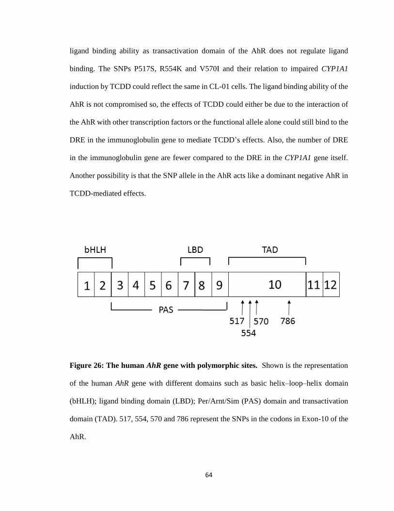
64
ligand binding ability as transactivation domain of the AhR does not regulate ligand
binding. The SNPs P517S, R554K and V570I and their relation to impaired CYP1A1
induction by TCDD could reflect the same in CL-01 cells. The ligand binding ability of the
AhR is not compromised so, the effects of TCDD could either be due to the interaction of
the AhR with other transcription factors or the functional allele alone could still bind to the
DRE in the immunoglobulin gene to mediate TCDD’s effects. Also, the number of DRE
in the immunoglobulin gene are fewer compared to the DRE in the CYP1A1 gene itself.
Another possibility is that the SNP allele in the AhR acts like a dominant negative AhR in
TCDD-mediated effects.
Figure 26: The human AhR gene with polymorphic sites. Shown is the representation
of the human AhR gene with different domains such as basic helix–loop–helix domain
(bHLH); ligand binding domain (LBD); Per/Arnt/Sim (PAS) domain and transactivation
domain (TAD). 517, 554, 570 and 786 represent the SNPs in the codons in Exon-10 of the
AhR.
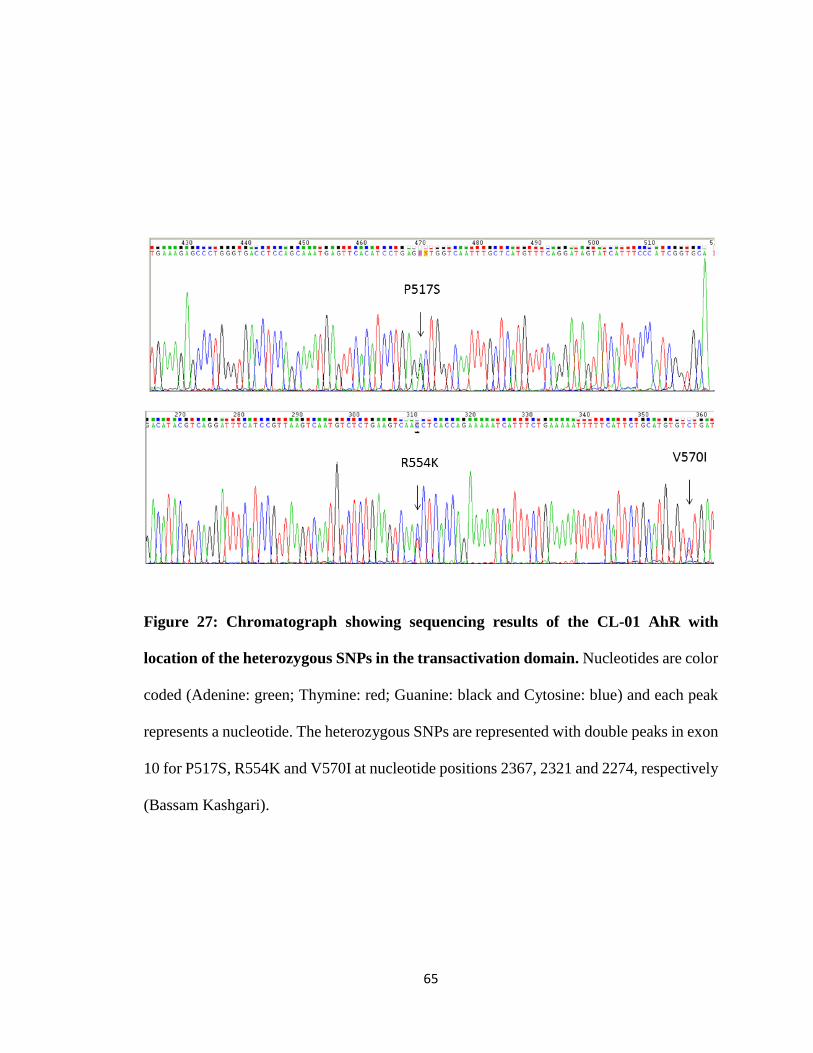
65
Figure 27: Chromatograph showing sequencing results of the CL-01 AhR with
location of the heterozygous SNPs in the transactivation domain. Nucleotides are color
coded (Adenine: green; Thymine: red; Guanine: black and Cytosine: blue) and each peak
represents a nucleotide. The heterozygous SNPs are represented with double peaks in exon
10 for P517S, R554K and V570I at nucleotide positions 2367, 2321 and 2274, respectively
(Bassam Kashgari).
66
CL-01 is a human Burkitt lymphoma cell line with c-myc translocated with one of
the heavy chain genes. c-myc is generally translocated to the 3' enhancer region present
downstream of the Cα gene and in all human Burkitt lymphomas the translocated c-myc is
linked to the transcriptional regulatory regions of the IGH gene (Judde and Max 1992;
Muller et al. 1997). However, it is unclear if the μ enhancer or one of the 3’IGHRRs is
regulating c-myc expression. Animal studies strongly support the ability of 3’IghRR to
dysregulate c-myc. A translocated 3’IGHRR will inactivate one IGH allele might affect the
CSR and Ig expression. Though it is established that mouse 3’IghRR is involved in Ig
expression (Sulentic, Holsapple, and Kaminski 1998), it is poorly understood if the human
3’IGHRR is also involved in Ig expression and CSR. Hence, translocation at any of the
3’IGHRR in human B cells might affect the CSR and Ig expression as well as potentially
leading to an oncogenic event. Ongoing efforts in our lab to delete the entire 3’IGHRR
(both α1 3’IGHRR and the α2 3’IGHRR) using the genome-editing system CRISPR could
provide a permanent deletion with limited off-target effects.
We used real time PCR primers spanning the constant regions of the downstream
antibody isotypes, which does not differentiate between germline and functional
transcripts. Another limitation is that the results that we observed from studying the
germline/functional transcripts cannot differentiate the effects of the AhR, TCDD and the
AhR antagonist on post switched cells vs cells that are ready to undergo CSR. As the CL-
01 cells readily express all the downstream isotypes except for ε, it is difficult to interpret
if the AhR, TCDD or the AhR antagonist really affect CSR. For further studies, studying
the AhR and ARNT binding to the DRE present in the invariant sequence of the hs1,2
enhancer in the 3’IGHRR through chromatin immunoprecipitation (ChIP) analysis could
67
help understand the mechanism of TCDD-induced effects in our cells and protein-protein
interactions within the transcription factor binding sites in the 3’IGHRR. As the AhR in
the CL-01 cells is heterozygous with one allele having a non-functional transactivation
domain that regulates the gene expression but not ligand binding, there is also a possibility
of other potential protein-protein interactions and cross-talk with other signaling pathways.
Ongoing efforts in our lab are focused on the AhR knockout with genome-editing system,
CRISPR, and this could result in the AhR with a fully functional allele in our cells. A future
study involving a fully functional AhR could primarily help understand TCDD-induced
effects in human Ig expression. Another possible future study could focus on studying the
different AhR ligands and observe their effects on CSR as human AhR has lower affinity
for TCDD, but has higher affinity for other ligands such as indirubin [(2Z)-2,3-biindole-
2,3-(1H,1H)-dione] and quercetin [2-(3,4dihydroxyphenyl)-3,5,7-trihydroxy-4H-
chromen-4-one] compared to mouse AHR (Flaveny et al. 2009). This may give us a better
understanding of the effect of the AhR on Ig expression and its relation to the 3’IGHRR.
Previous data in our lab has shown that the α1B repeat of the invariant sequence in the hs1,2
enhancer is sensitive to TCDD in CL-01 cells (Abdullah Freiwan, unpublished data) and
also the hs1,2 enhancer is known to be involved in many autoimmune disorders (Aupetit
et al. 2000; Cianci et al. 2008; Frezza et al. 2004; Giambra et al. 2009; Tolusso et al. 2009).
In conclusion, our results suggest that TCDD has a variable effect on IgM secretion, but
significantly inhibits IgG secretion, an effect reversed by addition of the AhR antagonist.
Surprisingly, the AhR antagonist alone markedly increased IgG secretion above
stimulation. At transcript level, TCDD has variable effects on μ IGH functional transcripts
but significantly inhibits γ1-4 germline/functional transcripts and Cε germline transcripts.
68
Additionally, CD40L and IL-4 stimulation induced de novo synthesis of Cε germline
transcripts, a precursor to CSR. However, α1-2 germline/functional transcripts increased in
response to TCDD. Notably, TCDD and stimulation had no effect on CYP1A1 expression.
Additionally, in CL-01 cells, we recently discovered SNPs in Exon-10 of the AhR, which
encodes the transactivation domain that regulates expression of other genes but does not
affect ligand binding. The AhR is heterozygous with one non-functional transactivation
domain. Results also indicate that a small proportion of the cells have undergone
spontaneous class switch to all of the γ and α isotypes rather than being induced to CSR.
As the human 3’IGHRR is involved in many autoimmune disorders and also in the
regulation of Ig expression, the current study could elucidate the role of the AhR in Ig gene
expression but also the relevance of AhR ligands and the 3’IGHRR.
69
V BIBLIOGRAPHY
Abel, J., and T. Haarmann-Stemmann. 2010. 'An introduction to the molecular basics of
aryl hydrocarbon receptor biology', Biol Chem, 391: 1235-48.
Amakura, Y., T. Tsutsumi, M. Nakamura, H. Kitagawa, J. Fujino, K. Sasaki, T. Yoshida,
and M. Toyoda. 2002. 'Preliminary screening of the inhibitory effect of food
extracts on activation of the aryl hydrocarbon receptor induced by 2,3,7,8-
tetrachlorodibenzo-p-dioxin', Biol Pharm Bull, 25: 272-4.
Aupetit, C., M. Drouet, E. Pinaud, Y. Denizot, J. C. Aldigier, F. Bridoux, and M. Cogne.
2000. 'Alleles of the alpha1 immunoglobulin gene 3' enhancer control evolution of
IgA nephropathy toward renal failure', Kidney Int, 58: 966-71.
Bhattacharya, D., D. U. Lee, and W. C. Sha. 2002. 'Regulation of Ig class switch
recombination by NF-kappaB: retroviral expression of RelB in activated B cells
inhibits switching to IgG1, but not to IgE', Int Immunol, 14: 983-91.
Borghesi, L., and C. Milcarek. 2006. 'From B cell to plasma cell: regulation of V(D)J
recombination and antibody secretion', Immunol Res, 36: 27-32.
Bottaro, A., R. Lansford, L. Xu, J. Zhang, P. Rothman, and F. W. Alt. 1994. 'S region
transcription per se promotes basal IgE class switch recombination but additional
factors regulate the efficiency of the process', Embo j, 13: 665-74.
Brian, A. A. 1988. 'Stimulation of B-cell proliferation by membrane-associated molecules
from activated T cells', Proc Natl Acad Sci U S A, 85: 564-8.
70
Carlstedt-Duke, J. M. 1979. 'Tissue distribution of the receptor for 2,3,7,8-
tetrachlorodibenzo-p-dioxin in the rat', Cancer Res, 39: 3172-6.
Celius, T., and J. Matthews. 2010. 'Functional analysis of six human aryl hydrocarbon
receptor variants in human breast cancer and mouse hepatoma cell lines',
Toxicology, 277: 59-65.
Cerutti, A., H. Zan, A. Schaffer, L. Bergsagel, N. Harindranath, E. E. Max, and P. Casali.
1998. 'CD40 ligand and appropriate cytokines induce switching to IgG, IgA, and
IgE and coordinated germinal center and plasmacytoid phenotypic differentiation
in a human monoclonal IgM+IgD+ B cell line', J Immunol, 160: 2145-57.
Chaudhuri, J., and F. W. Alt. 2004. 'Class-switch recombination: interplay of transcription,
DNA deamination and DNA repair', Nat Rev Immunol, 4: 541-52.
Chauveau, C., E. Pinaud, and M. Cogne. 1998. 'Synergies between regulatory elements of
the immunoglobulin heavy chain locus and its palindromic 3' locus control region',
Eur J Immunol, 28: 3048-56.
Chen, C., and B. K. Birshtein. 1997. 'Virtually identical enhancers containing a segment of
homology to murine 3'IgH-E(hs1,2) lie downstream of human Ig C alpha 1 and C
alpha 2 genes', J Immunol, 159: 1310-8.
Chen, K., W. Xu, M. Wilson, B. He, N. W. Miller, E. Bengten, E. S. Edholm, P. A. Santini,
P. Rath, A. Chiu, M. Cattalini, J. Litzman, B. Bussel J, B. Huang, A. Meini, K.
Riesbeck, C. Cunningham-Rundles, A. Plebani, and A. Cerutti. 2009.
'Immunoglobulin D enhances immune surveillance by activating antimicrobial,
proinflammatory and B cell-stimulating programs in basophils', Nat Immunol, 10:
889-98.
71
Cianci, R., V. Giambra, C. Mattioli, M. Esposito, G. Cammarota, G. Scibilia, G. Magazzu,
A. Orlando, G. Sandri, L. Bianchi, G. B. Gasbarrini, F. Pandolfi, and D. Frezza.
2008. 'Increased frequency of Ig heavy-chain HS1,2-A enhancer *2 allele in
dermatitis herpetiformis, plaque psoriasis, and psoriatic arthritis', J Invest
Dermatol, 128: 1920-4.
Connor, K. T., and L. L. Aylward. 2006. 'Human response to dioxin: aryl hydrocarbon
receptor (AhR) molecular structure, function, and dose-response data for enzyme
induction indicate an impaired human AhR', J Toxicol Environ Health B Crit Rev,
9: 147-71.
Denison, M. S., and S. Heath-Pagliuso. 1998. 'The Ah receptor: a regulator of the
biochemical and toxicological actions of structurally diverse chemicals', Bull
Environ Contam Toxicol, 61: 557-68.
Denizot, Y., E. Pinaud, C. Aupetit, C. Le Morvan, E. Magnoux, J. C. Aldigier, and M.
Cogne. 2001. 'Polymorphism of the human alpha1 immunoglobulin gene 3'
enhancer hs1,2 and its relation to gene expression', Immunology, 103: 35-40.
Enan, E., and F. Matsumura. 1996. 'Identification of c-Src as the integral component of the
cytosolic Ah receptor complex, transducing the signal of 2,3,7,8-
tetrachlorodibenzo-p-dioxin (TCDD) through the protein phosphorylation
pathway', Biochem Pharmacol, 52: 1599-612.
Endler, A., L. Chen, and F. Shibasaki. 2014. 'Coactivator recruitment of AhR/ARNT1', Int
J Mol Sci, 15: 11100-10.
Fernando, T. M., S. D. Ochs, J. Liu, R. C. Chambers-Turner, and C. E. Sulentic. 2012.
'2,3,7,8-tetrachlorodibenzo-p-dioxin induces transcriptional activity of the human
72
polymorphic hs1,2 enhancer of the 3'Igh regulatory region', J Immunol, 188: 3294-
306.
Flaveny, C. A., I. A. Murray, C. R. Chiaro, and G. H. Perdew. 2009. 'Ligand selectivity
and gene regulation by the human aryl hydrocarbon receptor in transgenic mice',
Mol Pharmacol, 75: 1412-20.
Flaveny, C., R. K. Reen, A. Kusnadi, and G. H. Perdew. 2008. 'The mouse and human Ah
receptor differ in recognition of LXXLL motifs', Arch Biochem Biophys, 471: 215-
23.
Frezza, D., V. Giambra, R. Cianci, A. Fruscalzo, M. Giufre, G. Cammarota, C. Martinez-
Labarga, O. Rickards, G. Scibilia, C. Sferlazzas, F. Bartolozzi, S. Starnino, G.
Magazzu, G. B. Gasbarrini, and F. Pandolfi. 2004. 'Increased frequency of the
immunoglobulin enhancer HS1,2 allele 2 in coeliac disease', Scand J
Gastroenterol, 39: 1083-7.
Geusau, A., E. Tschachler, M. Meixner, S. Sandermann, O. Papke, C. Wolf, E. Valic, G.
Stingl, and M. McLachlan. 1999. 'Olestra increases faecal excretion of 2,3,7,8-
tetrachlorodibenzo-p-dioxin', Lancet, 354: 1266-7.
Giambra, V., R. Cianci, S. Lolli, C. Mattioli, G. Tampella, M. Cattalini, S. S. Kilic, F.
Pandolfi, A. Plebani, and D. Frezza. 2009. 'Allele *1 of HS1.2 enhancer associates
with selective IgA deficiency and IgM concentration', J Immunol, 183: 8280-5.
Grewal, I. S., and R. A. Flavell. 1998. 'CD40 and CD154 in cell-mediated immunity', Annu
Rev Immunol, 16: 111-35.
73
Gu, H., Y. R. Zou, and K. Rajewsky. 1993. 'Independent control of immunoglobulin switch
recombination at individual switch regions evidenced through Cre-loxP-mediated
gene targeting', Cell, 73: 1155-64.
Hansen, D. A., P. Esakky, A. Drury, L. Lamb, and K. H. Moley. 2014. 'The aryl
hydrocarbon receptor is important for proper seminiferous tubule architecture and
sperm development in mice', Biol Reprod, 90: 8.
Harper, P. A., C. L. Golas, and A. B. Okey. 1988. 'Characterization of the Ah receptor and
aryl hydrocarbon hydroxylase induction by 2,3,7,8-tetrachlorodibenzo-p-dioxin
and benz(a)anthracene in the human A431 squamous cell carcinoma line', Cancer
Res, 48: 2388-95.
Harper, P. A., Jm Wong, M. S. Lam, and A. B. Okey. 2002. 'Polymorphisms in the human
AH receptor', Chem Biol Interact, 141: 161-87.
Heckman, C. A., T. Cao, L. Somsouk, H. Duan, J. W. Mehew, C. Y. Zhang, and L. M.
Boxer. 2003. 'Critical elements of the immunoglobulin heavy chain gene enhancers
for deregulated expression of bcl-2', Cancer Res, 63: 6666-73.
Henseler, R. A., E. J. Romer, and C. E. Sulentic. 2009. 'Diverse chemicals including aryl
hydrocarbon receptor ligands modulate transcriptional activity of the
3'immunoglobulin heavy chain regulatory region', Toxicology, 261: 9-18.
Hu, W., C. Sorrentino, M. S. Denison, K. Kolaja, and M. R. Fielden. 2007. 'Induction of
cyp1a1 is a nonspecific biomarker of aryl hydrocarbon receptor activation: results
of large scale screening of pharmaceuticals and toxicants in vivo and in vitro', Mol
Pharmacol, 71: 1475-86.
74
Judde, J. G., and E. E. Max. 1992. 'Characterization of the human immunoglobulin kappa
gene 3' enhancer: functional importance of three motifs that demonstrate B-cell-
specific in vivo footprints', Mol Cell Biol, 12: 5206-16.
Kerkvliet, N. I. 1995. 'Immunological effects of chlorinated dibenzo-p-dioxins', Environ
Health Perspect, 103 Suppl 9: 47-53.
Kerkvliet, N. I . 2009. 'AHR-mediated immunomodulation: the role of altered gene
transcription', Biochem Pharmacol, 77: 746-60.
Kerkvliet, N. I., D. M. Shepherd, and L. Baecher-Steppan. 2002. 'T lymphocytes are direct,
aryl hydrocarbon receptor (AhR)-dependent targets of 2,3,7,8-tetrachlorodibenzo-
p-dioxin (TCDD): AhR expression in both CD4+ and CD8+ T cells is necessary
for full suppression of a cytotoxic T lymphocyte response by TCDD', Toxicol Appl
Pharmacol, 185: 146-52.
Kim, S. H., E. C. Henry, D. K. Kim, Y. H. Kim, K. J. Shin, M. S. Han, T. G. Lee, J. K.
Kang, T. A. Gasiewicz, S. H. Ryu, and P. G. Suh. 2006. 'Novel compound 2-
methyl-2H-pyrazole-3-carboxylic acid (2-methyl-4-o-tolylazo-phenyl)-amide
(CH-223191) prevents 2,3,7,8-TCDD-induced toxicity by antagonizing the aryl
hydrocarbon receptor', Mol Pharmacol, 69: 1871-8.
Kimata, H. 2003. '2,3,7,8-tetrachlorodibenzo-p-dioxin selectively enhances spontaneous
IgE production in B cells from atopic patients', Int J Hyg Environ Health, 206: 601-
4.
Kinoshita, H., J. Abe, K. Akadegawa, H. Yurino, T. Uchida, S. Ikeda, K. Matsushima, and
S. Ishikawa. 2006. 'Breakdown of mucosal immunity in gut by 2,3,7,8-
tetraclorodibenzo-p-dioxin (TCDD)', Environ Health Prev Med, 11: 256-63.
75
Landers, J. P., and N. J. Bunce. 1991. 'The Ah receptor and the mechanism of dioxin
toxicity', Biochem J, 276 ( Pt 2): 273-87.
Litman, G. W., J. P. Cannon, and L. J. Dishaw. 2005. 'Reconstructing immune phylogeny:
new perspectives', Nat Rev Immunol, 5: 866-79.
Lorenz, M., S. Jung, and A. Radbruch. 1995. 'Switch transcripts in immunoglobulin class
switching', Science, 267: 1825-8.
Lu, H., R. B. Crawford, B. L. Kaplan, and N. E. Kaminski. 2011. '2,3,7,8-
Tetrachlorodibenzo-p-dioxin-mediated disruption of the CD40 ligand-induced
activation of primary human B cells', Toxicol Appl Pharmacol, 255: 251-60.
Lu, H., R. B. Crawford, J. E. Suarez-Martinez, B. L. Kaplan, and N. E. Kaminski. 2010.
'Induction of the aryl hydrocarbon receptor-responsive genes and modulation of the
immunoglobulin M response by 2,3,7,8-tetrachlorodibenzo-p-dioxin in primary
human B cells', Toxicol Sci, 118: 86-97.
Madisen, L., and M. Groudine. 1994. 'Identification of a locus control region in the
immunoglobulin heavy-chain locus that deregulates c-myc expression in
plasmacytoma and Burkitt's lymphoma cells', Genes Dev, 8: 2212-26.
Mandal, P. K. 2005. 'Dioxin: a review of its environmental effects and its aryl hydrocarbon
receptor biology', J Comp Physiol B, 175: 221-30.
Marcus, R. S., M. P. Holsapple, and N. E. Kaminski. 1998. 'Lipopolysaccharide activation
of murine splenocytes and splenic B cells increased the expression of aryl
hydrocarbon receptor and aryl hydrocarbon receptor nuclear translocator', J
Pharmacol Exp Ther, 287: 1113-8.
76
Marinkovic, N., D. Pasalic, G. Ferencak, B. Grskovic, and A. Stavljenic Rukavina. 2010.
'Dioxins and human toxicity', Arh Hig Rada Toksikol, 61: 445-53.
Market, E., and F. N. Papavasiliou. 2003. 'V(D)J recombination and the evolution of the
adaptive immune system', PLoS Biol, 1: E16.
Matthias, P., and D. Baltimore. 1993. 'The immunoglobulin heavy chain locus contains
another B-cell-specific 3' enhancer close to the alpha constant region', Mol Cell
Biol, 13: 1547-53.
McGregor, D. B., C. Partensky, J. Wilbourn, and J. M. Rice. 1998. 'An IARC evaluation
of polychlorinated dibenzo-p-dioxins and polychlorinated dibenzofurans as risk
factors in human carcinogenesis', Environ Health Perspect, 106 Suppl 2: 755-60.
Meyer, B. K., and G. H. Perdew. 1999. 'Characterization of the AhR-hsp90-XAP2 core
complex and the role of the immunophilin-related protein XAP2 in AhR
stabilization', Biochemistry, 38: 8907-17.
Mills, F. C., J. S. Brooker, and R. D. Camerini-Otero. 1990. 'Sequences of human
immunoglobulin switch regions: implications for recombination and transcription',
Nucleic Acids Res, 18: 7305-16.
Mills, F. C., N. Harindranath, M. Mitchell, and E. E. Max. 1997. 'Enhancer complexes
located downstream of both human immunoglobulin Calpha genes', J Exp Med,
186: 845-58.
Mills, F. C., M. P. Mitchell, N. Harindranath, and E. E. Max. 1995. 'Human Ig S gamma
regions and their participation in sequential switching to IgE', J Immunol, 155:
3021-36.
77
Mimura, J., and Y. Fujii-Kuriyama. 2003. 'Functional role of AhR in the expression of
toxic effects by TCDD', Biochim Biophys Acta, 1619: 263-8.
Mombaerts, P., J. Iacomini, R. S. Johnson, K. Herrup, S. Tonegawa, and V. E.
Papaioannou. 1992. 'RAG-1-deficient mice have no mature B and T lymphocytes',
Cell, 68: 869-77.
Muller, D., C. Bouchard, B. Rudolph, P. Steiner, I. Stuckmann, R. Saffrich, W. Ansorge,
W. Huttner, and M. Eilers. 1997. 'Cdk2-dependent phosphorylation of p27
facilitates its Myc-induced release from cyclin E/cdk2 complexes', Oncogene, 15:
2561-76.
Nebert, D. W., and C. L. Karp. 2008. 'Endogenous functions of the aryl hydrocarbon
receptor (AHR): intersection of cytochrome P450 1 (CYP1)-metabolized
eicosanoids and AHR biology', J Biol Chem, 283: 36061-5.
Noakes, R. 2015. 'The aryl hydrocarbon receptor: a review of its role in the physiology and
pathology of the integument and its relationship to the tryptophan metabolism', Int
J Tryptophan Res, 8: 7-18.
Oettinger, M. A., D. G. Schatz, C. Gorka, and D. Baltimore. 1990. 'RAG-1 and RAG-2,
adjacent genes that synergistically activate V(D)J recombination', Science, 248:
1517-23.
Okey, A. B., D. S. Riddick, and P. A. Harper. 1994. 'The Ah receptor: mediator of the
toxicity of 2,3,7,8-tetrachlorodibenzo-p-dioxin (TCDD) and related compounds',
Toxicol Lett, 70: 1-22.
Pancer, Z., and M. D. Cooper. 2006. 'The evolution of adaptive immunity', Annu Rev
Immunol, 24: 497-518.
78
Perlot, T., F. W. Alt, C. H. Bassing, H. Suh, and E. Pinaud. 2005. 'Elucidation of IgH
intronic enhancer functions via germ-line deletion', Proc Natl Acad Sci U S A, 102:
14362-7.
Pinaud, E., M. Marquet, R. Fiancette, S. Peron, C. Vincent-Fabert, Y. Denizot, and M.
Cogne. 2011. 'The IgH locus 3' regulatory region: pulling the strings from behind',
Adv Immunol, 110: 27-70.
Pollenz, R. S. 2002. 'The mechanism of AH receptor protein down-regulation (degradation)
and its impact on AH receptor-mediated gene regulation', Chem Biol Interact, 141:
41-61.
Quintana, F. J. 2013. 'The aryl hydrocarbon receptor: a molecular pathway for the
environmental control of the immune response', Immunology, 138: 183-9.
Quintana, F. J., A. S. Basso, A. H. Iglesias, T. Korn, M. F. Farez, E. Bettelli, M. Caccamo,
M. Oukka, and H. L. Weiner. 2008. 'Control of T(reg) and T(H)17 cell
differentiation by the aryl hydrocarbon receptor', Nature, 453: 65-71.
Roman, B. L., R. S. Pollenz, and R. E. Peterson. 1998. 'Responsiveness of the adult male
rat reproductive tract to 2,3,7,8-tetrachlorodibenzo-p-dioxin exposure: Ah receptor
and ARNT expression, CYP1A1 induction, and Ah receptor down-regulation',
Toxicol Appl Pharmacol, 150: 228-39.
Rowlands, C. J., D. F. Staskal, B. Gollapudi, and R. Budinsky. 2010. 'The human AHR:
identification of single nucleotide polymorphisms from six ethnic populations',
Pharmacogenet Genomics, 20: 283-90.
Saji, F., Y. Samejima, S. Kamiura, and M. Koyama. 1999. 'Dynamics of immunoglobulins
at the feto-maternal interface', Rev Reprod, 4: 81-9.
79
Saleque, S., M. Singh, and B. K. Birshtein. 1999. 'Ig heavy chain expression and class
switching in vitro from an allele lacking the 3' enhancers DNase I-hypersensitive
hs3A and hs1,2', J Immunol, 162: 2791-803.
Schaffer, A., E. C. Kim, X. Wu, H. Zan, L. Testoni, S. Salamon, A. Cerutti, and P. Casali.
2003. 'Selective inhibition of class switching to IgG and IgE by recruitment of the
HoxC4 and Oct-1 homeodomain proteins and Ku70/Ku86 to newly identified
ATTT cis-elements', J Biol Chem, 278: 23141-50.
Schecter, A., P. Cramer, K. Boggess, J. Stanley, O. Papke, J. Olson, A. Silver, and M.
Schmitz. 2001. 'Intake of dioxins and related compounds from food in the U.S.
population', J Toxicol Environ Health A, 63: 1-18.
Schecter, A., H. T. Quynh, O. Papke, K. C. Tung, and J. D. Constable. 2006. 'Agent
Orange, dioxins, and other chemicals of concern in Vietnam: update 2006', J Occup
Environ Med, 48: 408-13.
Schnekenburger, M., L. Peng, and A. Puga. 2007. 'HDAC1 bound to the Cyp1a1 promoter
blocks histone acetylation associated with Ah receptor-mediated trans-activation',
Biochim Biophys Acta, 1769: 569-78.
Schrader, C. E., J. E. Guikema, E. K. Linehan, E. Selsing, and J. Stavnezer. 2007.
'Activation-induced cytidine deaminase-dependent DNA breaks in class switch
recombination occur during G1 phase of the cell cycle and depend upon mismatch
repair', J Immunol, 179: 6064-71.
Sepulveda, M. A., A. V. Emelyanov, and B. K. Birshtein. 2004. 'NF-kappa B and Oct-2
synergize to activate the human 3' Igh hs4 enhancer in B cells', J Immunol, 172:
1054-64.
80
Sepulveda, M. A., F. E. Garrett, A. Price-Whelan, and B. K. Birshtein. 2005. 'Comparative
analysis of human and mouse 3' Igh regulatory regions identifies distinctive
structural features', Mol Immunol, 42: 605-15.
Shanmugam, A., M. J. Shi, L. Yauch, J. Stavnezer, and A. L. Kenter. 2000. 'Evidence for
class-specific factors in immunoglobulin isotype switching', J Exp Med, 191: 1365-
80.
Snapper, C. M., K. B. Marcu, and P. Zelazowski. 1997. 'The immunoglobulin class switch:
beyond "accessibility"', Immunity, 6: 217-23.
Sorg, O., M. Zennegg, P. Schmid, R. Fedosyuk, R. Valikhnovskyi, O. Gaide, V.
Kniazevych, and J. H. Saurat. 2009. '2,3,7,8-tetrachlorodibenzo-p-dioxin (TCDD)
poisoning in Victor Yushchenko: identification and measurement of TCDD
metabolites', Lancet, 374: 1179-85.
Stavnezer, J. 1996. 'Immunoglobulin class switching', Curr Opin Immunol, 8: 199-205.
Stavnezer, J., J. E. Guikema, and C. E. Schrader. 2008. 'Mechanism and regulation of class
switch recombination', Annu Rev Immunol, 26: 261-92.
Stavnezer, J., and J. Kang. 2008. 'The Surprising Discovery That TGF Specifically Induces
the IgA Class Switch', The Journal of Immunology, 182: 5-7.
Sulentic, C. E., M. P. Holsapple, and N. E. Kaminski. 1998. 'Aryl hydrocarbon receptor-
dependent suppression by 2,3,7, 8-tetrachlorodibenzo-p-dioxin of IgM secretion in
activated B cells', Mol Pharmacol, 53: 623-9.
Sulentic, C. E., M. P. Holsapple, and N. E. Kaminski . 2000. 'Putative link between
transcriptional regulation of IgM expression by 2,3,7,8-tetrachlorodibenzo-p-
81
dioxin and the aryl hydrocarbon receptor/dioxin-responsive enhancer signaling
pathway', J Pharmacol Exp Ther, 295: 705-16.
Sulentic, C. E., and N. E. Kaminski. 2011. 'The long winding road toward understanding
the molecular mechanisms for B-cell suppression by 2,3,7,8-tetrachlorodibenzo-p-
dioxin', Toxicol Sci, 120 Suppl 1: S171-91.
Sulentic, C. E., J. S. Kang, Y. J. Na, and N. E. Kaminski. 2004. 'Interactions at a dioxin
responsive element (DRE) and an overlapping kappaB site within the hs4 domain
of the 3'alpha immunoglobulin heavy chain enhancer', Toxicology, 200: 235-46.
Tanaka, G., S. Kanaji, A. Hirano, K. Arima, A. Shinagawa, C. Goda, S. Yasunaga, K.
Ikizawa, Y. Yanagihara, M. Kubo, Y. Kuriyama-Fujii, Y. Sugita, A. Inokuchi, and
K. Izuhara. 2005. 'Induction and activation of the aryl hydrocarbon receptor by IL-
4 in B cells', Int Immunol, 17: 797-805.
Toellner, K. M., A. Gulbranson-Judge, D. R. Taylor, D. M. Sze, and I. C. MacLennan.
1996. 'Immunoglobulin switch transcript production in vivo related to the site and
time of antigen-specific B cell activation', J Exp Med, 183: 2303-12.
Tolusso, B., D. Frezza, C. Mattioli, A. L. Fedele, S. Bosello, F. Faustini, G. Peluso, V.
Giambra, D. Pietrapertosa, A. Morelli, E. Gremese, M. De Santis, and G. F.
Ferraccioli. 2009. 'Allele *2 of the HS1,2A enhancer of the Ig regulatory region
associates with rheumatoid arthritis', Ann Rheum Dis, 68: 416-9.
van Gent, D. C., J. F. McBlane, D. A. Ramsden, M. J. Sadofsky, J. E. Hesse, and M. Gellert.
1995. 'Initiation of V(D)J recombination in a cell-free system', Cell, 81: 925-34.
Vogel, C. F., E. M. Khan, P. S. Leung, M. E. Gershwin, W. L. Chang, D. Wu, T. Haarmann-
Stemmann, A. Hoffmann, and M. S. Denison. 2014. 'Cross-talk between aryl
82
hydrocarbon receptor and the inflammatory response: a role for nuclear factor-
kappaB', J Biol Chem, 289: 1866-75.
Vos, J. G., C. De Heer, and H. Van Loveren. 1997. 'Immunotoxic effects of TCDD and
toxic equivalency factors', Teratog Carcinog Mutagen, 17: 275-84.
Wong, J. M., A. B. Okey, and P. A. Harper. 2001. 'Human aryl hydrocarbon receptor
polymorphisms that result in loss of CYP1A1 induction', Biochem Biophys Res
Commun, 288: 990-6.
Wormke, M., M. Stoner, B. Saville, K. Walker, M. Abdelrahim, R. Burghardt, and S. Safe.
2003. 'The aryl hydrocarbon receptor mediates degradation of estrogen receptor
alpha through activation of proteasomes', Mol Cell Biol, 23: 1843-55.
Wourms, M. J., and C. E. Sulentic. 2015. 'The aryl hydrocarbon receptor regulates an
essential transcriptional element in the immunoglobulin heavy chain gene', Cell
Immunol, 295: 60-6.
Zan, H., A. Cerutti, P. Dramitinos, A. Schaffer, and P. Casali. 1998. 'CD40 engagement
triggers switching to IgA1 and IgA2 in human B cells through induction of
endogenous TGF-beta: evidence for TGF-beta but not IL-10-dependent direct S
mu-->S alpha and sequential S mu-->S gamma, S gamma-->S alpha DNA
recombination', J Immunol, 161: 5217-25.
Zeiger, M., R. Haag, J. Hockel, D. Schrenk, and H. J. Schmitz. 2001. 'Inducing effects of
dioxin-like polychlorinated biphenyls on CYP1A in the human hepatoblastoma cell
line HepG2, the rat hepatoma cell line H4IIE, and rat primary hepatocytes:
comparison of relative potencies', Toxicol Sci, 63: 65-73.
83
Zhang, J., A. Bottaro, S. Li, V. Stewart, and F. W. Alt. 1993. 'A selective defect in IgG2b
switching as a result of targeted mutation of the I gamma 2b promoter and exon',
Embo j, 12: 3529-37.
Zhang, S., C. Qin, and S. H. Safe. 2003. 'Flavonoids as aryl hydrocarbon receptor
agonists/antagonists: effects of structure and cell context', Environ Health Perspect,
111: 1877-82.
Zhao, B., D. E. Degroot, A. Hayashi, G. He, and M. S. Denison. 2010. 'CH223191 is a
ligand-selective antagonist of the Ah (Dioxin) receptor', Toxicol Sci, 117: 393-403.