differential roles for the interferon-inducible ifi16 and aim2 … · cell cycle, cell death, and...
TRANSCRIPT

Cell Cycle, Cell Death, and Senescence
Differential Roles for the Interferon-Inducible IFI16 andAIM2 Innate Immune Sensors for Cytosolic DNAin Cellular Senescence of Human Fibroblasts
Xin Duan1, Larissa Ponomareva1,2, Sudhakar Veeranki1, Ravichandran Panchanathan1,Eric Dickerson1,2, and Divaker Choubey1,2
AbstractThe IFN-inducible IFI16 and AIM2 proteins act as innate immune sensors for cytosolic double-stranded DNA
(dsDNA). On sensing dsDNA, the IFI16 protein induces the expression of IFN-b whereas the AIM2 proteinforms an inflammasome, which promotes the secretion of IL-1b. Given that the knockdown of IFI16 expressionin human diploid fibroblasts (HDF) delays the onset of cellular senescence, we investigated the potential roles forthe IFI16 and AIM2 proteins in cellular senescence. We found that increased IFI16 protein levels in old(vs. young) HDFs were associated with the induction of IFN-b. In contrast, increased levels of the AIM2 proteinin the senescent (vs. old) HDFs were associated with increased production of IL-1b. The knockdown of type IIFN-a receptor subunit, which reduced the basal levels of the IFI16 but not of the AIM2, protein delayed theonset of cellular senescence. Accordingly, increased constitutive levels of IFI16 and AIM2 proteins in ataxiatelangiectasia mutated (ATM) HDFs were associated with the activation of the IFN signaling and increased levelsof IL-1b. The IFN-b treatment of the young HDFs, which induced the expression of IFI16 and AIM2 proteins,activated a DNA damage response and also increased basal levels of IL-1b. Interestingly, the knockdown of AIM2expression in HDFs increased the basal levels of IFI16 protein and activated the IFN signaling. In contrast, theknockdown of the IFI16 expression in HDFs decreased the basal and dsDNA-induced activation of the IFNsignaling. Collectively, our observations show differential roles for the IFI16 and AIM2 proteins in cellularsenescence and associated secretory phenotype. Mol Cancer Res; 9(5); 589–602. �2011 AACR.
Introduction
Cellular senescence refers to permanent cell-cycle arrest incells (1, 2). The cell-cycle arrest is associated with theactivation of a DNA damage response and the expressionof IFN-inducible genes. The onset of cellular senescencelimits the proliferation of damaged cells, which have poten-tial to develop cancers. However, the senescent cells secretproinflammatory cytokines and chemokines (2, 3). Thisphenotype of the senescent cells is termed senescence-associated secretory phenotype (SASP; ref. 3). The SASPin human diploid fibroblasts (HDF) is associated withhigher levels of inflammatory cytokines, including theinterleukin (IL)-1b, IL-6, and IL-8 (2, 3). Secretion ofthe inflammatory cytokines and chemokines by the senes-cent cells promotes the aging-associated inflammatory
diseases (2). Currently, it is not known whether the innateimmune sensors for cytosolic DNA play any role in cellularsenescence–associated cell growth arrest and/or SASP.With increasing passage, the HDFs in culture exhibit an
increase in apoptotic markers and cell death (4). Further-more, aging cells in culture also release "exosomes" (micro-vesicles), which contain proteins, small RNA [<100 basepair (bp)], and double-stranded DNA [dsDNA; >1,000 bp;ref. 5). These observations are consistent with the idea thatfusion of exosomes with neighboring cells could deliver thecontents (including the DNA) into the cytoplasm. Becausecytosolic dsDNA in most cell types is sensed by the innateimmune sensors as a danger signal (6, 7), the activation of aninnate immune response results in the production of IFN-band proinflammatory cytokines, such as IL-1b.Type I IFNs (IFN-a/b) are multifunctional cytokines
with the growth-inhibitory activities (8, 9). All cells producelow constitutive levels of the type I IFNs in culture (10) andactivation of IFN-regulated factors (IRF) by the innateimmune response can induce the IFN-b expression (11).Binding of type I IFNs to cell surface receptor activates thereceptor-associated Janus tyrosine kinases, Jak1 and Tyk2,which lead to the activating tyrosine phosphorylation oflatent transcription factors that are termed STATs. Theactivated STATs, on translocation into the nucleus, bind to
Authors' Affiliations: 1Department of Environmental Health, University ofCincinnati; and 2Cincinnati VA Medical Center, Cincinnati, Ohio
Corresponding Author: Divaker Choubey, Department of EnvironmentalHealth, University of Cincinnati, 3223 Eden Avenue, P.O. Box 670056,Cincinnati, OH 45267. Phone: 513-558-1014; Fax: 513-558-0925. E-mail:[email protected]
doi: 10.1158/1541-7786.MCR-10-0565
�2011 American Association for Cancer Research.
MolecularCancer
Research
www.aacrjournals.org 589
Research. on January 24, 2020. © 2011 American Association for Cancermcr.aacrjournals.org Downloaded from
Published OnlineFirst April 6, 2011; DOI: 10.1158/1541-7786.MCR-10-0565

the IFN stimulated response element (ISRE) and induce thetranscription of a set of IFN-stimulated genes (ISG; refs. 8,9).On approaching cellular senescence (becoming old) in
culture, HDFs express increased levels of IFN-b (12) andISGs (13). HDFs from ataxia telangiectasia mutated (ATM)patients, who age prematurely (14), express increased levelsof IFN-inducible proteins (15) and the knockdown of theATM expression induces the expression of a set of IFN-inducible genes (16). Accordingly, the loss of type I IFNsignaling, which results in the lack of the expression of ISGs,is associated with immortalization of HDFs (17–20).Furthermore, prolonged IFN-b treatment of young HDFsinduces a senescence-like phenotype (21). Although theseobservations are consistent with the activation of the innateimmune responses in old HDFs, the molecular mechanismsthat induce the expression of the IFNB gene remain largelyunknown. Moreover, it remains unclear whether the activa-tion of IFN signaling, which results in the expression ofcertain ISGs, is required for senescence-associated cell-cyclearrest and the SASP. The SASP in HDFs and epithelial cellsis associated with increased production of proinflammatorycytokines, such as IL-6, IL-8, and IL-1 (3).One family of the ISGs is the Ifi200-gene family, which
encodes for structurally related proteins (p200-familyproteins; refs. 22–24). The p200-family proteins shareat least one partially conserved repeat of 200-amino acidresidue (or the HIN-200 domain), which contains 2consecutive oligonucleotide/oligosaccharide-binding folds(OB-fold; ref. 24). The repeat is often found in proteinsthat bind either single- or double-stranded DNA (24).Most p200-family proteins (except the murine p202protein) also contain a homotypic protein–protein inter-action pyrin domain (PYD) to recruit adaptor proteinASC, which recruits and activates the caspase-1 (24). Thep200-family proteins include the IFI16 and AIM2 pro-teins. Interestingly, recent studies revealed that both IFI16(6) and AIM2 (25–27) proteins could sense cytosolicdsDNA and initiate an innate immune response. TheAIM2 protein, on sensing dsDNA, forms an inflamma-some (28), which through the activation of caspase-1,increases the secretion of proinflammatory cytokines,including IL-1b and IL-18, and induces cell death bypyroptosis (caspase-1–dependent death) in macrophages(25–28). Interestingly, the generation of Aim2-deficientmice revealed that the Aim2 protein is not needed forIFN-b production (29–31). Moreover, Aim2 proteinseems to negatively regulate the IFN-b expression andthe activation of IFN-inducible genes (32). In contrast tothe AIM2 protein, on sensing dsDNA, the IFI16 proteinrecruits stimulator of IFN genes (STING) protein tostimulate the expression of IFN-b through activation ofIRF3 (6).The expression of IFI16 gene is upregulated by IFNs (a,
b, or g) and the activated p53 (22, 23, 33). The IFI16 geneencodes for 3 isoforms (A, B, and C) of the protein (�80kDa) due to alternative splicing (22, 23). Increased levels ofIFI16 protein in old HDFs (lung fibroblast WI-38 and
IMR-90) are associated with the onset of cellular senes-cence, and the knockdown of the IFI16 expression in HDFsdelays the onset of cellular senescence (13). Interestingly,the extent of subcellular localization of the IFI16 protein inthe cytoplasm seems to depend on the cell type (23).Increased levels of IFI16 protein in cells negatively regulatecell proliferation in part by potentiating the p53/p21CIP1
and Rb/E2F-mediated inhibition of cell-cycle progression(13, 23, 34) and inhibition of the hTERT activity (35).Moreover, increased levels of IFI16 protein in primaryhuman umbilical vein endothelial cells upregulate theexpression of inflammatory cytokines through the activationof the transcriptional activity of NF-kB (36, 37). The IFI16protein activates the NF-kB activity through downregula-tion of the IkBa expression, a negative regulator of the NF-kB (36).Many cell types are reported to express the AIM2 gene
(38). However, it remains unclear which signaling pathwaysregulate the expression of the AIM2 gene. Notably, theAIM2 gene contains a microsatellite instability site thatresults in inactivation of the gene in approximately 47% ofcolorectal tumors with high microsatellite instability (39).Interestingly, a search of DNA sequence databases revealedthat different isoforms of the human AIM2 protein may beexpressed in cells and, depending on the isoform of theAIM2 protein that is expressed, the subcellular localizationof the protein may vary (24). For example, in HL-60 cells,the endogenous AIM2 protein is primarily detected in thenucleus (40). In contrast, the approach involving over-expression of the protein in transfected cells indicated acytoplasmic localization of the AIM2 protein (25–27).Given that reduced levels of IFI16 protein are associated
with immortalization of HDFs (23) and both IFI16 andAIM2 proteins seem to initiate different innate immuneresponses on sensing cytosolic dsDNA (6, 24), we investi-gated the role of these 2 proteins in cellular senescence ofHDFs.
Materials and Methods
Cell culture and treatmentsHuman lung fibroblasts WI-38 (at population doubling
level or PDL 23) and IMR-90 (at PDL 26) were purchasedfrom the American Type Culture Collection). Human skinfibroblasts (AG03057 and AG02496A) from ATM indivi-duals were obtained from the National Institute on AgingCell Culture Repository (Coriell Institute for MedicalResearch, Camden, NJ). Human benign prostate hyperpla-sia (BPH)-1 cell line (41) was generously provided by SimonW. Hayward (Vanderbilt University Medical Center, Nash-ville, TN). All HDFs were maintained in culture (CO2, 5%;oxygen, 20%) as suggested by the supplier. Cultures ofHDFs were split (1:4) on approaching confluence. Conse-quently, each cell passage was equivalent to approximately 2PDLs. When 50% to 60% cells in culture at the PDL 50appeared morphologically large and flat, we consideredthese cells old (13). Moreover, when the most (>95%)HDFs in culture lost the ability to divide and also stained
Duan et al.
Mol Cancer Res; 9(5) May 2011 Molecular Cancer Research590
Research. on January 24, 2020. © 2011 American Association for Cancermcr.aacrjournals.org Downloaded from
Published OnlineFirst April 6, 2011; DOI: 10.1158/1541-7786.MCR-10-0565

positive for the senescence-associated acidic b-galactosidase,we considered them senescent (vs. old).When indicated, subconfluent cultures of HDFs were
treated with the human IFN-a (universal IFN) or IFN-b(1,000 units/mL; from PBL Biomedical Laboratories)for the indicated times. In addition, when indicated,HDFs were treated with the indicated units of bleomycin(Sigma–Aldrich). The BPH-1 cell line was treated withhuman IFN-g (10 ng/mL; PBL Biomedical Laboratories)for the indicated times.
Knockdown of the expression of genesTo knockdown the expression of the indicated genes in
young WI-38 HDFs, subconfluent cultures of cells (in a6-well plate) were infected with lentivirus (purchased fromSanta Cruz Biotechnology) encoding short hairpin RNA(shRNA) to the IFI16 (sc-35633-V), AIM2 (sc-88166-V),IFNa/bRa (sc-35637-V), or ATM (sc-29761-V) gene. As acontrol, cells were infected with the lentivirus encoding acontrol shRNA (sc-108080). Twenty-four hours after infec-tions of HDFs, cells were selected in puromycin (1 mg/mL)for 3 to 5 days. Puromycin-resistant HDFs were pooled andcell cultures were maintained without the puromycin in themedium for several days before conducting any experi-ments. Similarly, to knockdown the AIM2 expression inBPH-1 cell line, cells were infected either with the controlvirus or with the virus expressing shRNA AIM2. Twenty-four hours after infections, cell were selected in puromycin(for a week) and resistant cells were pooled.
Plasmids and expression vectorsThe pISRE-luc plasmid, which contains 5 copies of
ISRE-responsive element, was purchased from ClontechLaboratories. The IFI16-luc plasmid, which contains thepromoter region (1.677 kb) of the IFI16 gene, has beendescribed (33). The pCMV-IFI16 plasmid allowing theexpression of the IFI16B protein has been described (34).The pCMV-AIM2 plasmid allowing the expression of Flag-tagged full-length human AIM2 protein was purchasedfrom Origene.
Transfections, nucleofections, and reporter assaysSubconfluent cultures of HDFs were transfected with the
indicated reporter plasmid (1.8 mg) along with the pRL-TKreporter plasmid (0.2 mg; as an internal control), usingFuGene6 transfection reagent (Roche) as suggested by thesupplier. Forty-eight hours after transfections of cells, thefirefly luciferase and Renilla luciferase activities were assayedusing dual-luciferase reporter assay kit (Promega). Therelative firefly luciferase activity is expressed as the ratioof the firefly luciferase/Renilla luciferase. Student's t test forpaired samples was used to determine statistical significanceof the reporter activity data. The differences were consideredstatistically significant at the P � 0.05.The young WI-38 HDFs were nucleofected with highly
purified (endotoxin-free) pEGFP plasmid that is suppliedwith the Nucleofector-II Device (Amaxa Biosystems). Weused the kit R and program V-001 for nucleofections as
suggested by the supplier. After nucleofections, cells wereincubated for the indicated times to isolate total RNA or toprepare total cell extracts for further analyses.
Regular and quantitative real-time PCRTotal RNA was isolated using the TRIzol reagent
(Invitrogen). cDNA was synthesized using primers thatwere supplied with the SuperScript First-strand SynthesisSystem from Invitrogen. Quantitative real-time TaqManPCR technology (Applied Biosystems) was used to com-pare the expression of the indicated genes. The PCR cyclingprogram consisted of denaturing at 95�C for 10 minutesand 40 cycles at 95�C for 15 seconds, and annealingand elongation at 60�C for 1 minute. The TaqMan assaysfor the IFI16 (assay ID Hs00194216_m1), humanAIM2 (Hs00175457_m1), human IFN-b (IFNB;Hs01077958_s1), and the endogenous control b-actin(assay ID Hs99999903_ml), were purchased from theApplied Biosystems and used as suggested by the supplier.For regular PCR, the human IFNB1 primers (forward: 50-
gaatgggaggcttgaatactgcct-30; reverse: 50-tagcaaagatgttctggag-catctc-30) were used. The conditions for regular PCR havebeen described (33).
ImmunoblottingCell lysates were prepared in the radioimmunoprecipita-
tion assay buffer (RIPA buffer; 50 mmol/L Tris-HCl, pH7.5, 150 mmol/L NaCl, 1% Nonidet P-40, 0.5% deox-ycholate, 0.1% sodium dodecyl sulfate). The RIPA bufferwas supplemented with protease inhibitor cocktail tablet(Mini Complete from Roche Diagnostics) and phosphatesinhibitor cocktails 1 and 2 (Sigma). Equal amounts ofprotein were resolved by SDS-PAGE and transferred topolyvinylidene difluoride membranes. Membranes wereprobed overnight at 4�C with the primary antibodiesspecific to the indicated proteins. Antibodies for IFI16(sc-8023), IFNa/bRa (sc-7391), IFI35 (sc-100769),IFI44L (sc-101981), ASC (sc-22514-R), p53 (sc-126),and thioredoxin (sc-20146) were purchased from SantaCruz Biotechnology. Antibodies for STING (ab82960)were purchased from Abcam. Antibodies for p-IRF3(#4947), IRF3 (#4962), PY-STAT1 (#9167), STAT1(#9172), actin (#4970), cleaved IL-1b (#2022), p-p53(Ser-15; #9284), ATM (#2873), p-H2AX (#9718), p-ATM (Ser-1981; #4526), p-p53 (Ser-392; #9281), IkBa(#9242), histone H3 (#9715), p-TBK1 (#5483), and TBK1(#3013) were purchased from Cell Signaling Technology.Antibodies for caspase-1 (AH20082) were purchased fromInvitrogen. Polyclonal antibodies to the human AIM2protein were raised against the C-terminal peptide(GVHSTIKVIKAKKKT) in rabbits, and the specificityof the antiserum was established using immunoblotting.Protein–antibody complexes were detected with horseradishperoxidase–conjugated secondary antibodies by using anECL chemiluminescence system (Amersham Biosciences).The procedure to fractionate HDFs into the nuclear and
cytoplasmic fractions has been described (42). The detec-tion of IkBa protein primarily in the cytoplasmic fraction
IFN-Inducible IFI16 and AIM2 in Cellular Senescence
www.aacrjournals.org Mol Cancer Res; 9(5) May 2011 591
Research. on January 24, 2020. © 2011 American Association for Cancermcr.aacrjournals.org Downloaded from
Published OnlineFirst April 6, 2011; DOI: 10.1158/1541-7786.MCR-10-0565

and the histone H3 in the nuclear fraction served as thequality control for the cell fractionations.
Immunoprecipitation and Western assaysCultures of human embryonic kidney cells (HEK-293) in
60-mm plates were transfected with pCMV-IFI16 plasmid(2 mg) alone or along with pCMV-AIM2 plasmid (2 mg),using the FuGene 6 transfection reagent as suggested by thesupplier. Twenty-four to 36 hours after transfections, totallysates were prepared in immunoprecipitation buffer (20mmol/L HEPES, pH 7.0, 250 mmol/L NaCl, and 1%Nonidet P-40) and lysates containing equal amounts ofproteins (�500 mg) were incubated with an isotype anti-body (2 mg/tube) or an antibody to the Flag-tag supplied inthe Flag Immunoprecipitation Kit (Sigma–Aldrich).Immune complexes were collected by incubating withprotein A/G-sepahrose beads (Thermo Fisher). The beadswere washed 5 times with the washing buffer (20 mmol/LHEPES, pH 7.0, 150 mmol/L NaCl, and 1% Nonidet P-40), boiled with the 1� protein sample buffer, and analyzedby immunoblotting.
Senescence-associated b-galactosidase assaysThese assays were carried out essentially as described by us
(13). In brief, HDFs in culture were fixed using the in situb-galactosidase staining kit (catalogue no. 200384; Strata-gene). The fixed cells were incubated 4 to 6 hours at 37�Cwith the staining solution. HDFs, which stained blue, were
considered positive for the activity of acidic senescence-associated b-galactosidase (SA-b-gal) and were counted andphotographed.
Results
Differential expressions and roles for the IFI16 andAIM2 proteins during cellular senescence of HDFsA recent study noted that on sensing cytosolic dsDNA,
the IFN-inducible IFI16 protein recruits the STINGprotein to stimulate the expression of IFN-b throughthe activation of IRF3 (6). Moreover, studies involvingthe Aim2-deficient mice indicated that the Aim2 proteinnegatively regulates the expression of IFN-b (32). There-fore, to explore the role of IFI16 and AIM2 proteins incellular senescence, we decided to compare steady-statelevels of IFI16 and AIM2 proteins (and their predicteddownstream effector proteins such as STING, IRF3, andSTAT1) in young, old, and senescent populations ofHDFs. We chose human WI-38 lung fibroblasts forour studies because we had identified a potential rolefor the IFI16 protein in cellular senescence-associated cellgrowth arrest in these cells (13). As shown in Figure 1, theyoung WI-38 HDFs (PDL �30) expressed low, but de-tectable, levels of both IFI16 and AIM2 proteins. Con-sistent with our previous observations (13), old WI-38HDFs (PDL �50) expressed much higher levels of theIFI16 protein than young HDFs (Fig. 1A). Interestingly,
A BY
IFNαα/βRα
IFN
-β m
RN
A levels
IFI16
STING
p-IRF3
IRF3
PY-STAT1
STAT1
AIM2Procaspase-1
Pro-IL-1βIL-1βActin
Y
IFNB
O S
5
4
3
2
1
0Young
**
*
Old Senescent
1
Actin
2 31
O2
S3
C
1.5
1
0.5
Y O
ISRE-luc
*
0
Rel L
uc a
cti
vit
y
Figure 1. Differential expression and roles for the IFI16 and AIM2 proteins during cellular senescence of HDFs. A, total cell extracts prepared fromyoung (Y), old (O), or senescent (S)WI-38 HDFswere analyzed by immunoblotting using antibodies specific to the indicated proteins. B, total RNA isolated fromyoung (Y), old (O), or senescent (S) WI-38 HDFs were analyzed by semiquantitative PCR (left) or quantitative real-time PCR (right) for the indicated genes.The ratio of the test gene to actin mRNA was calculated in units (1 unit being the ratio of the test gene to actin mRNA). The relative steady-state levelsof IFNB mRNA in young HDFs are indicated as 1. Results are mean values of triplicate experiments, and error bars represent SD (*,P < 0.05; **, P < 0.01).C, subconfluent cultures of young (Y) or old (O) WI-38 HDFs were infected with Cignal Lenti ISRE Reporter (Luc) virus as suggested by the supplier. Theinfected cells were harvested after 40 to 44 hours to assays for the firefly and Renilla luciferase activities as described in Materials and Methods. Normalizedrelative firefly luciferase activity is shown.
Duan et al.
Mol Cancer Res; 9(5) May 2011 Molecular Cancer Research592
Research. on January 24, 2020. © 2011 American Association for Cancermcr.aacrjournals.org Downloaded from
Published OnlineFirst April 6, 2011; DOI: 10.1158/1541-7786.MCR-10-0565

the increase in the IFI16 protein levels was associated withan increase in the levels of the IFNa/bRa subunit,STING protein, and the activating phosphorylation ofthe IRF3 and STAT1 proteins. Furthermore, the senes-cent HDFs (PDL �56; > 95% cells staining for SA-b-gal;ref. 13) had lower levels of IFI16 protein than old oryoung HDFs. We also tested whether the AIM2 proteinlevels also increase when cells become old. In contrast toour expectations, the AIM2 protein levels were higher inthe senescent HDFs than in the old HDFs and the levelswere inversely correlated with the levels of p-STAT1,STAT1, and IFI16 proteins. Notably, the increase inthe AIM2 protein levels was associated with the cleavageof the pro-IL-1b to IL-1b (Fig. 1A).The aforementioned observations that increased levels
of IFI16 protein in old WI-38 HDFs are associated withincreased levels of STAT1 and its activated form (the p-STAT1) prompted us to compare steady-state levels ofIFN-b mRNA. As shown in Figure 1B, steady-state levelsof IFN-b mRNA were higher in old and senescent WI-38HDFs than in the young HDFs. Furthermore, quantita-tive real-time PCR indicated that the levels of IFN-bmRNA were higher in old HDFs than in senescent cells(Fig. 1B). This observation that increased levels of IFN-bmRNA in senescent cells did not correlate with theactivation of STAT1 protein is consistent with theincreased levels of type I IFN receptor (the IFNa/bRasubunit) in the old HDFs as compared with senescentHDFs, thus, probably accounting for the increased levelsof STAT1 protein and its activation in old (vs. senescent)HDFs. We also compared the activity of an IFN-respon-sive reporter (the ISRE-luc) between the young and oldHDFs. As shown in Figure 1C, the activity of the reporterwas approximately 40% higher in old HDFs than inyoung HDFs. Consistent with this observation, levelsof several mRNAs corresponding to the known IFN-inducible genes (including the CXCL10, IFI27, OAS1,and Mx1) were higher (>5-fold) in old HDFs than inyoung HDFs (data not shown). Collectively, these obser-vations show differential expression and roles for the IFI16and AIM2 proteins during the onset of the cellularsenescence and associated production of proinflammatorycytokines such as IL-1b (Table 1).
The IFN signaling in HDFs is required to maintain theconstitutive expression of the IFI16 protein and theonset of cellular senescenceStudies indicate that the loss of type I IFN signaling,
which results in the lack of the expression of ISGs, isassociated with immortalization of HDFs (19, 20). More-over, the knockdown of IFI16 expression in WI-38 HDFsdelays the onset of cellular senescence (13). Therefore, ouraforementioned observations that increased levels of IFI16protein in old WI-38 HDFs are associated with the induc-tion of the expression of IFN-b and IFN-inducible genesprompted us to investigate whether type I IFN signalingregulates the expression of the IFI16 gene and cellularsenescence in these cells. As shown in Figure 2A, theknockdown of IFNa/bRa subunit expression in WI-38HDFs significantly reduced basal and IFN-a–inducedlevels of the IFI16 protein and IFN-inducible IFI35 pro-tein. However, levels of the AIM2 and IFI44L proteins didnot decrease. Similarly, the knockdown reduced basal andIFN-b–induced levels of IFI16 protein (Fig. 2B). Notably,the knockdown also reduced basal levels of adaptor proteinASC and cleaved IL-1b but not of procaspase-1. In addi-tion, the basal levels of p53 also decreased in the knockeddown HDFs (Fig. 2C). Consistent with these observations,the knockdown of IFNa/bRa subunit expression decreasedthe number of SA-b-gal–positive cells (Fig. 2D), which isindicative of the onset of cellular senescence in HDFs (13).Collectively, these observations showed that type I IFNsignaling is required to maintain the basal and the IFN-induced levels of certain IFN-inducible proteins, includingthe IFI16 protein, but not the AIM2 protein, and the onsetof cellular senescence in WI-38 HDFs. In addition, theseobservations revealed differential regulation of the IFI16and AIM2 expression by IFN signaling (Table 1).
Activation of DNA damage response in HDFsdifferentially regulates the expression of the IFI16 andAIM2 proteinsCellular senescence in HDFs is associated with activation
of DNA damage response (21). Moreover, the activation ofp53 in response to DNA damage activates the transcriptionof the IFI16 gene (33). Because prolonged IFN treatment ofHDFs activates the DNA damage response and p53 (21),
Table 1. Differential regulation of the IFI16 and AIM2 proteins level during cellular senescence of HDFs
HDFs IFI16 levels AIM2 levels Cytokine/signaling
Old Increase No change IFN-b, IS, and DDISSenescent Decrease Increase IL-1b, IISIFN receptor knockdown Decrease No change ISDNA damage response Increase No change DDISATM Increase Increase IS, IISAIM2 knockdown Increase Decrease IS, DDISIFI16 knockdown Decrease Increase IS
Abbreviations: IS, IFN signaling; DDIS, DNA damage–induced signaling; IIS, IL-b–induced signaling.
IFN-Inducible IFI16 and AIM2 in Cellular Senescence
www.aacrjournals.org Mol Cancer Res; 9(5) May 2011 593
Research. on January 24, 2020. © 2011 American Association for Cancermcr.aacrjournals.org Downloaded from
Published OnlineFirst April 6, 2011; DOI: 10.1158/1541-7786.MCR-10-0565

we also compared levels of IFI16 protein in young WI-38and IMR-90 HDFs after bleomycin treatment, a mimetic ofdsDNA break. As shown in Figure 3A, the treatmentincreased levels of IFI16 protein and the increase wasassociated with increased levels of p53 (which indicatesthe activation of DNA damage response). However, bleo-mycin treatment of young WI-38 HDFs did not increaselevels of the AIM2 protein (Fig. 3B). Consistent with theseobservations, treatment of young WI-38 HDFs with bleo-mycin increased steady-state levels of IFI16 mRNA(Fig. 3C). However, levels of the AIM2 mRNA decreased,thus indicating a negative regulation of the AIM2 expressionby the activation of DNA damage response. Together, theseobservations support the idea that the activation of DNAdamage response in young HDFs differentially regulates theexpression of IFI16 and AIM2 proteins (Table 1).
Constitutively increased levels of the IFI16 and AIM2proteins in ATM HDFs are associated with theactivation of the IFN signaling and the production ofIL-1bHDFs from ATM patients express increased levels of
IFN-inducible proteins (15) and the knockdown of theATM expression in HeLa cells induces the expression ofIFN-inducible genes (16). Therefore, our aforementioned
observations that increased levels of IFI16 protein in oldHDFs are associated with the activation of IFN signaling(Fig. 1) encouraged us to compare the steady-state levels ofIFI16 mRNA and protein between WI-38 and ATM(AG03057) HDFs. As shown in Figure 4A, basal levelsof IFI16 mRNAwere higher in the young ATMHDFs thanin WI-38 HDFs. Consistent with this observation, theactivities of the IFI16-luc and ISRE-luc reporters wereapproximately 3-fold higher in the young ATM HDFsthan in WI-38 HDFs (Fig. 4B). Accordingly, the basallevels of IFI16 protein were higher in ATM HDFs than inWI-38 HDFs (Fig. 4C, compare lane 3 with lane 1).Importantly, the increased basal levels of the IFI16 proteinin ATM HDFs were associated with elevated basal levels ofSTAT1 and the activated p-STAT1 (Fig. 4C). We alsocompared the basal and IFN-induced levels of the AIM2protein between the young ATMHDFs and WI-38 HDFs.As shown in Figure 4D, the basal levels of AIM2 proteinwere higher in ATM HDFs than WI-38 HDFs (comparelane 3 with lane 1). Importantly, the increased basal levels ofthe AIM2 protein in ATM HDFs were associated withelevated basal levels of procaspase-1, cleaved caspase-1, pro-IL-1b, and IL-1b. To further investigate the role of ATM inthe regulation of the expression of IFI16 and AIM2 genes,we knockdown the expression of ATM in young WI-38
A B
C D
IFNα/βRα
IFN-β
IFI16
AIM2
ASC
Procaspase-1
Caspase-1 (p20)
Actin
IFN-α
IFI16
shlFNα/βRα
IFI35
IFI44L
Actin
Actin
p53
PY-STAT1
STAT1
AIM2
Control shlFNα/βRα– + – +1 2
– +1 2
3 4
Control shlFNα/βRα
Control shlFNα/βRα
– + – +
1 2 3 4
Figure 2. The knockdown of theIFNa/bRa subunit expression inHDFs decreases IFI16 expressionand delays the onset of asenescence phenotype.A, subconfluent cultures of youngWI-38 HDFs infected with controllentivirus (control) or virusencoding for shIFNa/bRa RNA(shIFNa/bRa) were either leftuntreated or treated with IFN-a for18 hours. After the treatment, totalcell extracts were prepared andanalyzed by immunoblotting usingantibodies specific to theindicated proteins. B, control orshIFNa/bRa HDFs described inA were either left untreated ortreated with IFN-b for 18 hours.Total cell extracts were analyzedby immunoblotting. C, total cellextracts from control or shIFNa/bRa HDFs were analyzed byimmunoblotting for the indicatedproteins. D, phase-contrastphotographs indicatingmorphologic changes (top) anddifferences in the number ofSA-b-gal–positive cells (bottom)between control HDFs and HDFsafter the knockdown of the IFNa/bRa subunit expression.
Duan et al.
Mol Cancer Res; 9(5) May 2011 Molecular Cancer Research594
Research. on January 24, 2020. © 2011 American Association for Cancermcr.aacrjournals.org Downloaded from
Published OnlineFirst April 6, 2011; DOI: 10.1158/1541-7786.MCR-10-0565

HDFs. As shown in Figure 4E, the knockdown increasedbasal and IFN-b–induced levels of IFI16 protein. Notably,the induced, but not the basal, levels of IFI16 protein in theATM knocked down HDFs were associated with anincrease in the steady-state levels of STAT1 and p-STAT1.These observations revealed that the ATM negatively reg-ulates the expression of the IFI16 gene in WI-38 HDFs.Moreover, these observations indicated that the knockdownof ATM (or mutation in the ATM gene) potentiates the IFNsignaling and the expression of the IFN-inducible proteins,including the IFI16 protein. Interestingly, no measurabledifferences were noted between the basal or IFN-b–inducedlevels of the AIM2 protein between control and WI-38HDFs with the reduced ATM expression. Together, theseobservations indicated that the constitutively increasedlevels of the IFI16 and AIM2 proteins in ATM HDFsare associated with the activation of IFN signaling and theproduction of IL-1b.
Prolonged IFN-b treatment of young HDFs activatesDNA damage response and increases the production ofIL-1bProlonged IFN-b treatment of IMR-90 HDFs increases
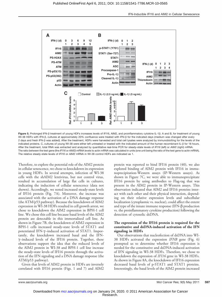
the reactive oxygen species (ROS), activates DNA damageresponse (as determined by phosphorylation of H2AX,ATM, and p53), and induces a senescence-like phenotype(21). Therefore, we tested whether prolonged constitutiveactivation of IFN signaling in young WI-38 HDFsinduces a DNA damage response. As shown in Figure5A, prolonged IFN-b treatment of HDFs, whichincreased steady-state levels of both IFI16 and AIM2proteins, activated a DNA damage response (the ATM/p53 pathway) as determined by phosphorylation ofH2AX, ATM, and p53 proteins.The presence of inflammasome component proteins,
such as AIM2, ASC, procaspase-1, and pro-IL-1b, has been
predicted in nonmyeloid cells (43). However, their expres-sion has not been shown in HDFs. Therefore, we exploredwhether inflammasome proteins are expressed in the youngWI-38 HDFs and whether the IFN-b treatment affectstheir expression and/or activation. As shown in Figure 5B,the IFN-b treatment of young WI-38 HDFs increasedsteady-state levels of AIM2, adaptor protein ASC, procas-pase-1, and pro-IL-1b (Fig. 5B, left). Interestingly, thetreatment also increased levels of the cleaved IL-1b, aproinflammatory cytokine, which is indicative of the activa-tion of an inflammasome. Moreover, the treatment alsoincreased levels of thioredoxin (Fig. 5B, right), indicative ofthe generation of ROS by the IFN-b treatment. Together,these observations support the idea that a prolonged IFN-btreatment of WI-38 HDFs results in ROS production,activation of DNA damage response, and an inflammasomeactivity (as determined by increases in the IL-1b levelswithin the cells).Given that levels of the AIM2 protein in the senescent
WI-38 HDFs were inversely correlated with the levels ofIFI16 protein and production of IL-1b (Fig. 1), the afore-mentioned observation that a prolonged IFN-b treatmentof young WI-38 HDFs increased the levels of IL-1bprompted us to test whether IL-1b could differentiallyregulate the expression of IFI16 and AIM2 genes in HDFs,which could account for differential expression of these 2genes between the old and senescent HDFs (Fig. 1). Asshown in Figure 5C, treatment of young HDFs with IL-1bonly moderately increased steady-state levels of the IFI16mRNA. In contrast, the treatment increased levels of theAIM2 mRNA robustly (>6-fold). Together, these observa-tions show that a prolonged activation of IFN signaling inHDFs can activate a DNA damage response (the ATM/p53pathway) and the production of proinflammatory cytokineIL-1b, which, in turn, can differentially regulate theexpression of IFI16 and AIM2 genes.
A B C
IFI16
1
BleomycinBleomycin
Bleomycin
Rela
tive m
RN
A levels
IFI16 AIM2
- 1x 2x
5Control
4
3
2
1
0
1 2 3- -
2 3 4 5 6
Actin
WI-38 IMR-90
p53
IFI16
Actin
AIM2
Figure 3. DNA damage response also contributes to the constitutive levels of the IFI16 protein. A, subconfluent cultures of young WI-38 or IMR-90HDFs were either left untreated (lanes 1 and 4) or treated with increasing units of bleomycin (lanes 2, 3, 5, and 6) for 24 hours. After the treatment, total cellextracts were analyzed by immunoblotting for the indicated proteins. B, subconfluent cultures of young WI-38 were either left untreated (lane 1) ortreated with increasing units of bleomycin (lanes 2 and 3) for 24 hours. After the treatment, total cell extracts were analyzed by immunoblotting for theindicated proteins. C, subconfluent cultures of young WI-38 were either left untreated or treated with bleomycin for 24 hours. After the treatment, totalRNA was analyzed by quantitative real-time PCR for IFI16 and AIM2 mRNA levels. The ratio between the test gene (the IFI16 or AIM2) mRNA levelsto actin mRNAwas calculated in units (1 unit being the ratio of the test gene to actin mRNA). The relative steady-state levels of IFI16mRNA in control HDFsare indicated as 1.
IFN-Inducible IFI16 and AIM2 in Cellular Senescence
www.aacrjournals.org Mol Cancer Res; 9(5) May 2011 595
Research. on January 24, 2020. © 2011 American Association for Cancermcr.aacrjournals.org Downloaded from
Published OnlineFirst April 6, 2011; DOI: 10.1158/1541-7786.MCR-10-0565

IFI16 protein is primarily detected in the cytoplasm ofold or IFN-b–treated HDFs and dsDNA induces theIFN-b expressionStudies indicate that the extent of the cytoplasmic and
nuclear localization of the endogenous IFI16 protein variesamong various cell types (23). Therefore, we investigatedthe subcellular localization of the IFI16 protein in youngversus old and IFN-b–treated WI-38 HDFs. As shown inFigure 6A, the bulk of the IFI16 proteins (all 3 isoforms)were detected primarily in the cytoplasm of the youngHDFs and only a fraction was detectable in the nucleus.Similarly, increased levels of IFI16 proteins in old HDFswere detected primarily in the cytoplasm and only a fractionin the nucleus. Furthermore, IFN-b–induced levels ofIFI16 proteins in young WI-38 HDFs were detectedprimarily in the cytoplasm (Fig. 6B). As expected (24),the bulk of the AIM2 protein was detected primarily in thecytoplasm after IFN-b treatment ofWI-38 HDFs (Fig. 6B).However, a fraction was detectable in the nucleus.IFI16 protein is a cytosolic sensor of dsDNA in macro-
phages, and on sensing of dsDNA, the IFI16 protein induces
the expression of IFN-b through the activation of TBK1 andIRF3 (6). Therefore, our aforementioned observations thatthe IFI16 protein is primarily localized in the cytoplasm ofWI-38 HDFs (Fig. 6A and B) encouraged us to test whethernucleofection of dsDNA into HDFs induces the expressionof IFN-b. As shown in Figure 6C, nucleofection of dsDNAplasmid into young WI-38 HDFs increased steady-statelevels of IFN-b mRNA. Importantly, the increase wasassociated with the activation of TBK1 and IRF3(Fig. 6D). In addition, the increase in the IFN-b expressionwas associated with an increase in IFI16, but not AIM2,protein levels. Together, these observations support the ideathat increased cytoplasmic levels of the IFI16 protein in oldHDFs on sensing cytosolic dsDNA induce the expression ofIFN-b through the activation of TBK1 and IRF3.
The knockdown of the AIM2 expression in cellsactivates the DNA damage response, IFN signaling, andincreases IFI16 protein levelsAim2 deficiency in the murine immune cells increases
the expression of IFN-b and IFN-inducible genes (32).
A B C
D E
Rel IF
I16
mR
NA
levels
No
rm. R
el.
Lu
c A
ct.
ActinWI-38 ATM WI-38
ISRE-lucIFN-β
IFN-β:
IFI16-luc
PY-STAT1STAT1
PS-p53 (S15)
IFI16
ATM
WI-38 ATM
– +
1 2
– +
3 4
WI-38 ATM– +
1 2
– +
3 4
Vector
Bleomycin
shATM
– +
1 2
– –
3 4
+
5
–– – + – – +
6
Control15
10
5
0
4
3
2
1
0p53
Actin
PY-STAT1
STAT1
PS-p53
IFI16
AIM2
Actin
Procaspase-1
Cleaved caspase-1
Pro-IL-1βCleaved-IL-1β
IFI16
AIM2
ATM
p53
IFN-β
IFN-β
Figure 4. Constitutively increased levels of the IFI16 protein in ATM HDFs are associated with the activation of IFN signaling. A, subconfluent culturesof young WI-38 or ATM HDFs were either left untreated or treated with IFN-b for 18 hours. After the treatment, total RNA was extracted and analyzed byquantitative real-time PCR for steady-state levels of IFI16 mRNA. The ratio between the test gene IFI16 mRNA levels to actin mRNA was calculated inunits (1 unit being the ratio of the test gene to actin mRNA). The relative steady-state levels of IFI16 mRNA in WI-38 control HDFs are indicated as 1. B,subconfluent culture of young WI-38 or ATM HDFs was transfected with either ISRE-luc or IFI16-luc reporter plasmid (1.8 mg) along with a secondpRL-TK reporter (0.2 mg). The transfected cells were harvested after 40 to 44 hours to conduct assays for the firefly andRenilla luciferase activities as describedin Materials and Methods. Normalized relative firefly luciferase activity in WI-38 cells is shown as 1. C, subconfluent cultures of young WI-38 or ATM HDFswere either left untreated (lanes 1 and 3) or treated with IFN-b (1,000 units/mL) for 18 hours. After the treatment, total cell extracts were analyzed byimmunoblotting for the indicated proteins. D, subconfluent cultures of young WI-38 or ATM HDFs were either left untreated (lanes 1 and 3) or treated withIFN-b (1,000 units/mL) for 18 hours. After the treatment, total cell extracts were analyzed by immunoblotting for the indicated proteins. E, subconfluentcultures of youngWI-38 HDFs that were infected with control virus (Vector) or shATM virus (shATM) were left untreated (lanes 1 and 4), treated with IFN-b (lanes2 and 5), or treated with bleomycin (lanes 3 and 6) for 18 hours. After the treatment, total cell extracts were analyzed by immunoblotting for the indicatedproteins.
Duan et al.
Mol Cancer Res; 9(5) May 2011 Molecular Cancer Research596
Research. on January 24, 2020. © 2011 American Association for Cancermcr.aacrjournals.org Downloaded from
Published OnlineFirst April 6, 2011; DOI: 10.1158/1541-7786.MCR-10-0565
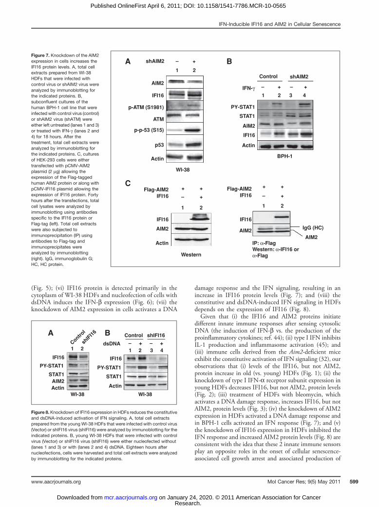
Therefore, to explore the potential role of the AIM2 proteinin cellular senescence, we chose to knockdown its expressionin young HDFs. In several attempts, infection of WI-38cells with the shAIM2 lentivirus, but not control virus,resulted in accumulation of large flat cells in cultures,indicating the induction of cellular senescence (data notshown). Accordingly, we noted increased steady-state levelsof IFI16 protein (Fig. 7A). Moreover, the increase wasassociated with the activation of a DNA damage response(the ATM/p53 pathway). Because the knockdown of AIM2expression inWI-38 HDFs resulted in cell growth arrest, wechose to knockdown the AIM2 expression in BPH-1 cellline. We chose this cell line because basal levels of the AIM2protein are detectable in this immortalized cell line. Asshown in Figure 7B, the knockdown of AIM2 expression inBPH-1 cells increased steady-state levels of STAT1 andpotentiated IFN-g–induced activation of STAT1. Impor-tantly, the knockdown increased basal and the IFN-g–induced levels of the IFI16 protein. Together, theseobservations support the idea that the reduced levels ofthe AIM2 protein in WI-38 and BPH-1 cell line increasethe steady-state levels of IFI16 protein through the activa-tion of the IFN signaling and a DNA damage response (theATM/p53 pathway).Given that levels of AIM2 protein in HDFs are inversely
correlated with IFI16 protein (Figs. 1 and 7) and AIM2
protein was reported to bind IFI16 protein (40), we alsoexplored binding of AIM2 protein with IFI16 in immu-noprecipitation-Western assays (IP-Western assays). Asshown in Figure 7C, we were able to immunoprecipitateIFI16 protein by using antibodies to Flag-tag that waspresent in the AIM2 protein in IP-Western assays. Thisobservation indicated that AIM2 and IFI16 proteins inter-act with each other and their physical interaction, depend-ing on their relative expression levels and subcellularlocalization (cytoplasmic vs. nuclear), could affect the extentand type of the innate immune response (IFN-b productionvs. the proinflammatory cytokine production) following thedetection of cytosolic dsDNA.
The expression of the IFI16 protein is required for theconstitutive and dsDNA-induced activation of the IFNsignaling in HDFsOur observations that nucleofection of dsDNA into WI-
38 HDFs activated the expression IFNB gene (Fig. 6)prompted us to determine whether IFI16 expression isneeded for the constitutive and dsDNA-induced activationof IFN signaling in WI-38 HDFs. Therefore, we chose toknockdown the expression of IFI16 gene in WI-38 HDFs.As shown in Figure 8A, the knockdown of IFI16 expressiondecreased basal levels of p-STAT1 and STAT1 proteins.Interestingly, the basal levels of the AIM2 protein increased
A
C
B
1 2 30 1 2
0 1 2 3
1 2 3 4
1 2 3 4 50 3 6 9 12
ActinActin
10864
2
1
01 2 3R
el
IFI1
6 m
RN
A lev
els
Rel
AIM
2 m
RN
A lev
els
Control
5
4
3
2
1
01 2 3
IL-1β (2.5 ng/mL) IL-1β (5 ng/mL)
PS-H2AX
PS-ATM
PS-p53(15)
PS-p53(392)
ATM
p53
IFI16 p-STAT1 (Y701)
STAT1 (Ser-727)
STAT1
AIM2
ASC
Procaspase-1
Pro-IL-1βCleaved IL-1β
AIM2
IFI16
AIM2Thioredoxin
Actin
IFN-β (d) IFN-β (d)
IFN-β (d)
Figure 5. Prolonged IFN-b treatment of young HDFs increases levels of IFI16, AIM2, and proinflammatory cytokine IL-1b. A and B, for treatment of youngWI-38 HDFs with IFN-b, cultures at approximately 20% confluence were treated with IFN-b for the indicated days (medium was changed after every2 days and fresh IFN-b was added). After the treatment, HDFs were harvested and total cell lysates were analyzed by immunoblotting for the levels of theindicated proteins. C, cultures of young WI-38 were either left untreated or treated with the indicated amount of the human recombinant IL-b for 18 hours.After the treatment, total RNA was extracted and analyzed by quantitative real-time PCR for steady-state levels of IFI16 (left) or AIM2 (right) mRNA.The ratio between the test gene (the IFI16 or AIM2) mRNA levels to actin mRNAwas calculated in units (one unit being the ratio of the test gene to actin mRNA).The relative steady-state levels of IFI16 or AIM2 mRNA in WI-38 control HDFs are indicated as 1.
IFN-Inducible IFI16 and AIM2 in Cellular Senescence
www.aacrjournals.org Mol Cancer Res; 9(5) May 2011 597
Research. on January 24, 2020. © 2011 American Association for Cancermcr.aacrjournals.org Downloaded from
Published OnlineFirst April 6, 2011; DOI: 10.1158/1541-7786.MCR-10-0565

moderately. Moreover, the knockdown also reduced basaland dsDNA-induced levels of p-STAT1 and STAT1 pro-teins after nucleofection of cells. Together, these observa-tions showed that the expression of IFI16 protein is neededfor the basal and dsDNA-induced activation of the IFNsignaling in HDFs.
Discussion
The loss of IFN signaling in human cells that results inthe lack of the expression of IFN-inducible genes isassociated with the development of certain cancers (17–20). Consistent with the aforementioned observations, theloss of IFI16 expression in cells is associated with thedevelopment of cancers including the breast and prostate(23). Moreover, increased levels of IFI16 protein inhuman normal prostate epithelial cells (34) and HDFs(13) are associated with cellular senescence-associated cellgrowth arrest. Accordingly, reduced expression of IFI16protein in Li–Fraumeni HDFs is associated with sponta-
neous immortalization (19) and the knockdown of IFI16expression in WI-38 HDFs delays the onset of cellularsenescence (13). Given that the recent studies identifiedIFN-inducible IFI16 and AIM2 proteins as sensors forcytosolic dsDNA, we investigated their roles in cellularsenescence of HDFs. Our observations revealed that (i)increased levels of IFI16, but not AIM2, protein in old (vs.young or senescent) HDFs are associated with increasedexpression of the IFN-b and activation of IFN signaling(Fig. 1); (ii) increased levels of the AIM2 protein insenescent (vs. old or young) HDFs are associated withincreased levels of IL-1b (Fig. 1); (iii) the knockdown oftype I IFN-a receptor subunit expression in HDFsdecreases IFI16, but not AIM2, protein levels and delaysthe onset of a senescence phenotype (Fig. 2); (iv) increasedbasal levels of IFI16 and AIM2 proteins in ATM HDFsare associated with the activation of IFN signaling andincreased levels of IL-1b (Fig. 4); (v) a prolonged IFN-btreatment of young HDFs increases steady-state levels ofIFI16, AIM2, and the proinflammatory cytokine IL-1b
A B
C D
1 2 3
1 2 3
- + +
+ - +
4
C N C N
1 2 3 4
C
Young Old
Control IFN-β
N C N
Control
1 μg
2 μg
IFI16
AIM2
IFI16
PS-TBK1
TBK1
PS-IRF3
IRF3
AIM2
Actin
IFI16
IKBα
Histone H3
IKBα
Histone H3
WI-38
WI-38 (Y)
DNA
80
60
40
IFN
B m
RN
A level
20
1086420
1 2 3
PULSE
WI-38
Figure 6. IFI16 protein is primarily detected in the cytoplasm of old or IFN-b–treated HDFs, and nucleofection of young HDFs with dsDNA inducesthe IFN-b expression. A, cultures of young (PDL �30) proliferating or old (PDL �50) WI-38 HDFs were harvested, and cells were subjected to the nuclearand cytoplasmic fractionation. The fractions containing equal amounts of proteins were analyzed by immunoblotting using antibodies specific to the indicatedproteins. B, cultures of young proliferatingWI-38 HDFs were either left untreated or treated with IFN-b for 24 hours. After the treatment, cells were subjected tothe nuclear and cytoplasmic fractionation. The fractions containing approximately equal amounts of proteins were analyzed by immunoblotting. C,young proliferating (PDL�30) WI-38 HDFs were nucleofected without DNA (control) or with the indicated amounts of plasmid DNA (GFP plasmid) as describedin Materials and Methods. After 18 hours of nucleofection, total RNA was extracted and analyzed by quantitative real-time PCR for steady-state levelsof IFNB mRNA. The ratio between the IFNB mRNA levels to actin mRNA was calculated in units (1 unit being the ratio of the IFNB mRNA to actin mRNA).The relative steady-state levels of IFNBmRNA in control HDFs are indicated as 1. D, young proliferating WI-38 HDFs were either left without nucleofection ornucleofected without DNA or nucleofected with 2 mg of plasmid DNA. After 18 hours of nucleofection, total cell lysates were analyzed by immunoblottingfor the indicated proteins.
Duan et al.
Mol Cancer Res; 9(5) May 2011 Molecular Cancer Research598
Research. on January 24, 2020. © 2011 American Association for Cancermcr.aacrjournals.org Downloaded from
Published OnlineFirst April 6, 2011; DOI: 10.1158/1541-7786.MCR-10-0565
(Fig. 5); (vi) IFI16 protein is detected primarily in thecytoplasm of WI-38 HDFs and nucleofection of cells withdsDNA induces the IFN-b expression (Fig. 6); (vii) theknockdown of AIM2 expression in cells activates a DNA
damage response and the IFN signaling, resulting in anincrease in IFI16 protein levels (Fig. 7); and (viii) theconstitutive and dsDNA-induced IFN signaling in HDFsdepends on the expression of IFI16 (Fig. 8).Given that (i) the IFI16 and AIM2 proteins initiate
different innate immune responses after sensing cytosolicDNA (the induction of IFN-b vs. the production of theproinflammatory cytokines; ref. 44); (ii) type I IFN inhibitsIL-1 production and inflammasome activation (45); and(iii) immune cells derived from the Aim2-deficient miceexhibit the constitutive activation of IFN signaling (32), ourobservations that (i) levels of the IFI16, but not AIM2,protein increase in old (vs. young) HDFs (Fig. 1); (ii) theknockdown of type I IFN-a receptor subunit expression inyoung HDFs decreases IFI16, but not AIM2, protein levels(Fig. 2); (iii) treatment of HDFs with bleomycin, whichactivates a DNA damage response, increases IFI16, but notAIM2, protein levels (Fig. 3); (iv) the knockdown of AIM2expression in HDFs activated a DNA damage response andin BPH-1 cells activated an IFN response (Fig. 7); and (v)the knockdown of IFI16 expression in HDFs inhibited theIFN response and increased AIM2 protein levels (Fig. 8) areconsistent with the idea that these 2 innate immune sensorsplay an opposite roles in the onset of cellular senescence-associated cell growth arrest and associated production of
Figure 7. Knockdown of the AIM2expression in cells increases theIFI16 protein levels. A, total cellextracts prepared from WI-38HDFs that were infected withcontrol virus or shAIM2 virus wereanalyzed by immunoblotting forthe indicated proteins. B,subconfluent cultures of thehuman BPH-1 cell line that wereinfected with control virus (control)or shAIM2 virus (shATM) wereeither left untreated (lanes 1 and 3)or treated with IFN-g (lanes 2 and4) for 18 hours. After thetreatment, total cell extracts wereanalyzed by immunoblotting forthe indicated proteins. C, culturesof HEK-293 cells were eithertransfected with pCMV-AIM2plasmid (2 mg) allowing theexpression of the Flag-taggedhuman AIM2 protein or along withpCMV-IFI16 plasmid allowing theexpression of IFI16 protein. Fortyhours after the transfections, totalcell lysates were analyzed byimmunoblotting using antibodiesspecific to the IFI16 protein orFlag-tag (left). Total cell extractswere also subjected toimmunoprecipitation (IP) usingantibodies to Flag-tag andimmunoprecipitates wereanalyzed by immunoblotting(right). IgG, immunoglobulin G;HC, HC protein.
IFI16
p-ATM (S1981)
p-p-53 (S15)
p53
AIM2
shAIM2 –
1 2
+
ATM
Actin
WI-38
+ +
+–
21
1
IFN-γ
A B
C
IFI16
IFI16
Flag-AIM2IFI16
– –+ +
Control shAIM2
2 3 4
PY-STAT1
STAT1
AIM2
AIM2
Actin
Actin
Western
+ +
+–
21
IFI16
Flag-AIM2IFI16
AIM2IgG (HC)
AIM2IP: α-FlagWestern: α-IFI16 orα-Flag
BPH-1
IFI161Control
shIFI16
2 1dsDNA
A B– –+ +
Control shIFI16
2 3 4
PY-STAT1STAT1AIM2Actin
IFI16PY-STAT1
STAT1
ActinWI-38WI-38
Figure 8. Knockdown of IFI16 expression in HDFs reduces the constitutiveand dsDNA-induced activation of IFN signaling. A, total cell extractsprepared from the young WI-38 HDFs that were infected with control virus(Vector) or shIFI16 virus (shIFI16) were analyzed by immunoblotting for theindicated proteins. B, young WI-38 HDFs that were infected with controlvirus (Vector) or shIFI16 virus (shIFI16) were either nucleofected without(lanes 1 and 3) or with (lanes 2 and 4) dsDNA. Eighteen hours afternucleofections, cells were harvested and total cell extracts were analyzedby immunoblotting for the indicated proteins.
IFN-Inducible IFI16 and AIM2 in Cellular Senescence
www.aacrjournals.org Mol Cancer Res; 9(5) May 2011 599
Research. on January 24, 2020. © 2011 American Association for Cancermcr.aacrjournals.org Downloaded from
Published OnlineFirst April 6, 2011; DOI: 10.1158/1541-7786.MCR-10-0565

IL-1b (Fig. 9). Therefore, further work will be needed toelucidate their roles.ATM HDFs, which express higher levels of IFN-indu-
cible proteins, exhibit premature senescence (15). There-fore, our observations that ATM HDFs expressconstitutively higher levels of IFI16 protein and exhibitthe activation of IFN signaling (Fig. 4) support the notionthat increased levels of IFI16 protein in ATM HDFscontribute to premature cellular senescence through theactivation of the IFN signaling.Overexpression of IFI16 protein in prostate cancer cell
lines induces a senescence-like phenotype, which is accom-panied by the stimulation of p53-mediated transcription, anincrease in the p21CIP1 protein levels (a transcriptionaltarget of p53), and an inhibition of the E2F-mediatedtranscription of target genes (34). Furthermore, increasedexpression of IFI16 protein in HDFs decreases the hTERTexpression by inhibiting c-Myc–mediated transcription(35). In contrast, the knockdown of IFI16 expression inHDFs increases c-Myc protein levels and decreases thep21CIP1 protein levels (35). Similarly, overexpression ofthe AIM2 protein in breast cancer cell lines inhibits cellproliferation in vitro (46). Together, these observationssupport the idea that increased levels of both IFI16 andAIM2 proteins contribute to inhibition of cell-cycle pro-gression whereas their reduced levels in cells provide thegrowth advantage.Subcellular localization of IFI16 and other p200-family
proteins (including the AIM2 protein) seem to depend onthe cell type (23). In WI-38 HDFs, the bulk of the IFI16protein was detected in the cytoplasm (Fig. 6). However,it was also detectable in the nuclear fraction. The nuclearlocalization of IFI16 protein is consistent with its pro-posed transcriptional regulatory functions (22). Giventhat the IFI16 and AIM2 proteins can form a hetero-
dimers (Fig. 7), further work is required to determinewhether the binding of IFI16 protein with AIM2 proteincould regulate the subcellular localization of the AIM2protein.Most p200-family proteins share 2 protein domains: the
PYD and HIN-200 domain (24). The PYD of the AIM2protein heterodimerizes with an adaptor protein ASC inresponse to cytoplasmic dsDNA and forms ASC speckles(25–27). However, the PYD of IFI16 protein does notform ASC speckles. This is consistent with the limited(only 29%) amino acid residue identities between thePYD of the AIM2 and IFI16 proteins. The HIN-200domain consists of 2 OB-folds, which recognize nucleicacids (24). Consistent with this observation, the AIM2protein requires this domain to sense cytosolic dsDNAand to assemble an inflammasome (24). Similarly, onbinding to dsDNA through the HIN-200 domain, theIFI16 protein recruits the STING protein to stimulate theexpression of IFN-b (6). Thus, supporting the idea thatcytosolic IFI16 protein, on sensing dsDNA that is takenup by the HDFs in cultures of old HDFs (5), stimulatesthe expression of IFN-b. In turn, the constitutivelyincreased levels of the IFN-b in cultures of old HDFsstimulate the expression of IFI16 protein further, as well asthe expression of other IFN-inducible proteins, activate aDNA damage response, and potentiate senescence-asso-ciated cell growth arrest (Fig. 9).Most p200-family proteins have the ability to homo- and
heterodimerize (23, 24). Consistent with this observation,the human AIM2 protein was shown to heterodimerize withthe IFI16 protein in vitro (40). Similarly, we noted thatAIM2 protein can bind to IFI16 protein in IP-Westernassays (Fig. 7). These observations support the idea that theability of AIM2 protein to heterodimerize with the IFI16protein (and possibly other p200 proteins) may limit the
Sterile DNA(Necroticor ApoptoticSource)
CytoplasmDNA
AIM2AIM2
ASC
CASP-1
Nucleus(IFNAIFNB)
IRF3
TBK1
STING
IFI16
CLEAVAGE
Pro-IL-1β
IFN-β
IFN-β
IL-1β
NF-κBIFI16
DNA damage responseand IFN signaling
Senescence-associatedcell growth arrest
Secretory phenotype
IL-1β
IL-6, IL-8, and IL-8
Figure 9. Proposed roles of theIFI16 and AIM2 proteins in cellularsenescence-associated cellgrowth arrest and secretoryphenotype in HDFs. CASP-1,caspase-1.
Duan et al.
Mol Cancer Res; 9(5) May 2011 Molecular Cancer Research600
Research. on January 24, 2020. © 2011 American Association for Cancermcr.aacrjournals.org Downloaded from
Published OnlineFirst April 6, 2011; DOI: 10.1158/1541-7786.MCR-10-0565

ability of cells to produce IFN-b on sensing cytosolicdsDNA. Accordingly, we noted that the knockdown ofAIM2 expression in BPH-1 human prostate cell line, whichexpresses detectable levels of both AIM2 and IFI16 pro-teins, resulted in the activation of IFN signaling andincreases in IFI16 protein levels (Fig. 7).Given that the IFN-b is produced during the innate
immune responses that are induced after infections (47),that HDFs from individuals with ATM express increasedlevels of IFN-inducible proteins (15), that the immunesystem of patients with autoimmune diseases, which areassociated with increased levels of type I IFNs (IFN-a and-b), shows signs of an accelerated aging (48), that IFN-bpromotes atherosclerosis by stimulating macrophagerecruitment to lesions (49), it is important to understandthe regulation and role of IFN-b and the IFN-inducibleproteins in cellular aging and aging-associated inflammatorydiseases. Our observations that IFN-inducible IFI16 and
AIM2 proteins differentially regulate the IFN signaling inHDFs will serve the basis to understand the role ofIFN-inducible proteins in diseases that are associated withincreased production of IFN-b and proinflammatory cyto-kines.
Disclosure of Potential Conflicts of Interest
No potential conflicts of interest were disclosed.
Grant Support
This work was supported by a grant (AG 025036) from the NIH and a MeritAward from the Veterans Affairs to D. Choubey.
The costs of publication of this article were defrayed in part by the payment of pagecharges. This article must therefore be hereby marked advertisement in accordancewith 18 U.S.C. Section 1734 solely to indicate this fact.
Received December 14, 2010; revised March 16, 2011; acceptedMarch 30, 2011;published OnlineFirst April 6, 2011.
References1. Campisi J. Cellular senescence as a tumor-suppressor mechanism.
Trends Cell Biol 2001;11:S27–31.2. Campisi J. Cancer and ageing: rival demons?Nat Rev Cancer
2003;3:339–49.3. Young AR, Narita M. SASP reflects senescence. EMBO Rep
2009;10:228–30.4. Mammone T, Gan D, Foyouzi-Youssefi R. Apoptotic cell death
increases with senescence in normal human dermal fibroblast cul-tures. Cell Biol Int 2006;30:903–9.
5. LehmannBD, PaineMS, Brooks AM,McCubrey JA, Renegar RH, et al.Senescence-associated exosome release from human prostate can-cer cells. Cancer Res 2008;68:7864–71.
6. Unterholzner L, Keating SE, Baran M, Horan KA, Jensen SB, et al.IFI16 is an innate immune sensor for intracellular DNA. Nat Immunol2010;11:997–1004.
7. Takaoka A, Wang Z, Choi MK, Yanai H, Negishi H, et al. DAI (DLM-1/ZBP1) is a cytosolic DNA sensor and an activator of innate immuneresponse. Nature 2007;448:501–5.
8. Borden EC, Sen GC, Uze G, Silverman RH, Ransohoff RM, et al.Interferons at age 50: past, current and future impact on biomedicine.Nat Rev Drug Discov 2007;6:975–90.
9. Stark GR. How cells respond to interferons revisited: from early historyto current complexity. Cytokine Growth Factor Rev 2007;18:419–23.
10. Taniguchi T, Takaoka A. A weak signal for strong responses: inter-feron-alpha/beta revisited. Nat Rev Mol Cell Biol 2001;2:378–86.
11. Honda K, Yanai H, Takaoka A, Taniguchi T. Regulation of the type-IIFN induction: a current view. Int Immunol 2005;17:1367–78.
12. Tahara H, Kamada K, Sato E, Tsuyama N, Kim JK, et al. Increase inexpression levels of interferon-inducible genes in senescent humandiploid fibroblasts and in SV40-transformed human fibroblasts withextended lifespan. Oncogene 1995;11:1125–32.
13. Xin H, Pereira-Smith OM, Choubey D. Role of IFI 16 in cellularsenescence of human fibroblasts. Oncogene 2004;23:6209–17.
14. Barzilai A, Rotman G, Shiloh Y. ATM deficiency and oxidative stress: anew dimension of defective response to DNA damage. DNA Repair2002;1:3–25.
15. Siddoo-Atwal C, Haas AL, Rosin MP. Elevation of interferon beta-inducible proteins in ataxia telangiectasia cells. Cancer Res1996;56:443–7.
16. Chen S,Wang G, Makrigiorgos GM, Price BD. Stable siRNA-mediatedsilencing of ATM alters the transcriptional profile of HeLa cells.Biochem Biophys Res Commun 2004;317:1037–44.
17. Shou J, Soriano R, Hayward SW, Cunha GR, Williams PM, et al.Expression profiling of a human cell line model of prostatic cancer
reveals a direct involvement of interferon signaling in prostate tumorprogression. Proc Natl Acad Sci U S A 2002;99:2830–5.
18. Untergasser G, Koch HB, Menssen A, Hermeking H. Characterizationof epithelial senescence by serial analysis of gene expression: identi-fication of genes potentially involved in prostate cancer. Cancer Res2002;62:6255–62.
19. Kulaeva OI, Draghici S, Tang L, Kraniak JM, Land SJ, et al. Epigeneticsilencing of multiple interferon pathway genes after cellular immorta-lization. Oncogene 2003;22:4118–27.
20. Fridman AL, Tainsky MA. Critical pathways in cellular senescence andimmortalization revealed by gene expression profiling. Oncogene2008;27:5975–87.
21. Moiseeva O, Mallette FA, Mukhopadhyay UK, Moores A, Ferbeyre G.DNA damage signaling and p53-dependent senescence after pro-longed b-interferon stimulation. Mol Biol Cell 2006;17:1583–92.
22. Johnstone RW, Trapani JA. Transcription and growth regulatoryfunctions of the HIN-200 family of proteins. Mol Cell Biol1999;19:5833–8.
23. Choubey D, Deka R, Ho SM. Interferon-inducible IFI16 protein inhuman cancers and autoimmune diseases. Front Biosci2008;13:598–608.
24. Choubey D, Duan X, Dickerson E, Ponomareva L, Panchanathan R,et al. Interferon-inducible p200-family proteins as novel sensors ofcytoplasmic DNA: role in inflammation and autoimmunity. J InterferonCytkine Res 2010;30:371–80.
25. Hornung V, Ablasser A, Charrel-Dennis M, Bauernfeind F, Horvath G,et al. AIM2 recognizes cytosolic dsDNA and forms a caspase-1-activating inflammasome with ASC. Nature 2009;458:514–8.
26. Fernandes-Alnemri T, Yu JW, Datta P, Wu J, Alnemri ES. AIM2activates the inflammasome and cell death in response to cytoplasmicDNA. Nature 2009;458:509–13.
27. B€urckst€ummer T, Baumann C, Bl€uml S, Dixit E, D€urnberger G, et al.An orthogonal proteomic-genomic screen identifies AIM2 as acytoplasmic DNA sensor for the inflammasome. Nat Immunol2009;10:266–72.
28. Schroder K, Muruve DA, Tschopp J. Innate immunity: cytoplasmicDNA sensing by the AIM2 inflammasome. Curr Biol 2009;19:R262–5.
29. Rathinam VA, Jiang Z, Waggoner SN, Sharma S, Cole LE, et al. TheAIM2 inflammasome is essential for host defense against cytosolicbacteria and DNA viruses. Nat Immunol 2010;11:395–402.
30. Fernandes-Alnemri T, Yu JW, Juliana C, Solorzano L, Kang S, et al.The AIM2 inflammasome is critical for innate immunity to Francisellatularensis. Nat Immunol 2010;11:385–93.
IFN-Inducible IFI16 and AIM2 in Cellular Senescence
www.aacrjournals.org Mol Cancer Res; 9(5) May 2011 601
Research. on January 24, 2020. © 2011 American Association for Cancermcr.aacrjournals.org Downloaded from
Published OnlineFirst April 6, 2011; DOI: 10.1158/1541-7786.MCR-10-0565

31. Jones JW, Kayagaki N, Broz P, Henry T, Newton K, et al. Absent inmelanoma 2 is required for innate immune recognition of Francisellatularensis. Proc Natl Acad Sci U S A 2010;107:9771–6.
32. Panchanathan R, Duan X, Shen H, Rathinam VA, Erickson LD, et al.Aim2 deficiency stimulates the expression of IFN-inducible Ifi202,a lupus susceptibility murine gene within the Nba2 autoimmunesusceptibility locus. J Immunol 2010;185:7385–93.
33. Song LL, Alimirah F, Panchanathan R, Xin H, Choubey D. Expressionof an IFN-inducible cellular senescence gene, IFI16, is up-regulated byp53. Mol Cancer Res 2008;6:1732–41.
34. Xin H, Curry J, Johnstone RW, Nickoloff BJ, et al. Role of IFI16, amember of the interferon-inducible p200-protein family, in prostateepithelial cellular senescence. Oncogene 2003;22:4831–40.
35. Song LL, Ponomareva L, Shen H, Duan X, Alimirah F, et al. Interferon-inducible IFI16, a negative regulator of cell growth, down-regulatesexpression of human telomerase reverse transcriptase (hTERT) gene.PLoS One 2010;5:e8569.
36. Caposio P, Gugliesi F, Zannetti C, Sponza S, Mondini M, et al. A novelrole of the interferon-inducible protein IFI16 as inducer of proinflamma-tory molecules in endothelial cells. J Biol Chem 2007;282:33515–29.
37. Baggetta R, De Andrea M, Gariano GR, Mondini M, Ritt�a M, et al. Theinterferon-inducible gene IFI16 secretome of endothelial cells drivesthe early steps of the inflammatory response. Eur J Immunol2010;40:2182–9.
38. DeYoung KL, RayME, Su YA, Anzick SL, Johnstone RW, et al. Cloninga novel member of the human interferon-inducible gene family asso-ciated with control of tumorigenicity in a model of human melanoma.Oncogene 1997;15:453–7.
39. Woerner SM, Kloor M, Schwitalle Y, Youmans H, Doeberitz MK, et al.The putative tumor suppressor AIM2 is frequently affected by differentgenetic alterations in microsatellite unstable colon cancers. GenesChromosomes Cancer 2007;46:1080–9.
40. Cresswell KS, Clarke CJ, Jackson JT, Darcy PK, Trapani JA, et al.Biochemical and growth regulatory activities of the HIN-200 familymember and putative tumor suppressor protein, AIM2. BiochemBiophys Res Commun 2005;326:417–24.
41. Hayward SW, Dahiya R, Cunha GR, Bartek J, Deshpande N, et al.Establishment and characterization of an immortalized but non-trans-formed human prostate epithelial cell line: BPH-1. In Vitro Cell Dev BiolAnim 1995;31:14–24.
42. Choubey D, Lengyel P. Interferon action: cytoplasmic and nuclearlocalization of the interferon-inducible 52-kD protein that is encodedby the Ifi 200 gene from the gene 200 cluster. J Interferon Res1993;13:43–52.
43. Yazdi AS, Drexler SK, Tschopp J. The role of the inflammasome innonmyeloid cells. J Clin Immunol 2010;30:623–7.
44. Goubau D, Rehwinkel J, Reis e Sousa C. PYHIN proteins: center stagein DNA sensing. Nat Immunol 2010;11:984–6.
45. Guarda G, Braun M, Staehli F, Tardivel A, Mattmann C, et al. Type Iinterferon inhibits interleukin-1 production and inflammasome activa-tion. Immunity 2011;34:213–23.
46. Chen IF, Ou-Yang F, Hung JY, Liu JC,Wang H, et al. AIM2 suppresseshuman breast cancer cell proliferation in vitro and mammary tumorgrowth in a mouse model. Mol Cancer Ther 2006;5:1–7.
47. Theofilopoulos AN, Baccala R, Beutler B, Kono DH. Type I interferons(alpha/beta) in immunity and autoimmunity. Annu Rev Immunol2005;23:307–36.
48. Thewissen M, Somers V, Venken K, Linsen L, van Paassen P, et al.Analyses of immunosenescent markers in patients with autoimmunedisease. Clin Immunol 2007;123:209–18.
49. Goossens P, GijbelsMJ, Zernecke A, EijgelaarW, VergouweMN, et al.Myeloid type I interferon signaling promotes atherosclerosis by sti-mulating macrophage recruitment to lesions. Cell Metab 2010;12:142–53.
Duan et al.
Mol Cancer Res; 9(5) May 2011 Molecular Cancer Research602
Research. on January 24, 2020. © 2011 American Association for Cancermcr.aacrjournals.org Downloaded from
Published OnlineFirst April 6, 2011; DOI: 10.1158/1541-7786.MCR-10-0565
2011;9:589-602. Published OnlineFirst April 6, 2011.Mol Cancer Res Xin Duan, Larissa Ponomareva, Sudhakar Veeranki, et al. Senescence of Human FibroblastsInnate Immune Sensors for Cytosolic DNA in Cellular Differential Roles for the Interferon-Inducible IFI16 and AIM2
Updated version
10.1158/1541-7786.MCR-10-0565doi:
Access the most recent version of this article at:
Cited articles
http://mcr.aacrjournals.org/content/9/5/589.full#ref-list-1
This article cites 49 articles, 11 of which you can access for free at:
Citing articles
http://mcr.aacrjournals.org/content/9/5/589.full#related-urls
This article has been cited by 5 HighWire-hosted articles. Access the articles at:
E-mail alerts related to this article or journal.Sign up to receive free email-alerts
SubscriptionsReprints and
To order reprints of this article or to subscribe to the journal, contact the AACR Publications
Permissions
Rightslink site. (CCC)Click on "Request Permissions" which will take you to the Copyright Clearance Center's
.http://mcr.aacrjournals.org/content/9/5/589To request permission to re-use all or part of this article, use this link
Research. on January 24, 2020. © 2011 American Association for Cancermcr.aacrjournals.org Downloaded from
Published OnlineFirst April 6, 2011; DOI: 10.1158/1541-7786.MCR-10-0565