difficult airway society guidelines for the management of...
TRANSCRIPT
Guidelines
Difficult Airway Society Guidelines for the management oftracheal extubationMembership of the Difficult Airway Society Extubation Guidelines Group: M. Popat (Chairman),1
V. Mitchell,2 R. Dravid,3 A. Patel,4 C. Swampillai5 and A. Higgs6
1 Consultant Anaesthetist, Nuffield Department of Anaesthetics, Oxford Radcliffe Hospital NHS Trust, Oxford, UK2 Consultant Anaesthetist, University College London Hospital, London, UK3 Consultant Anaesthetist, Kettering General Hospital, Kettering, UK4 Consultant Anaesthetist, The Royal National Throat Nose and Ear Hospital, London, UK5 Anaesthetic Specialist Registrar, Lister Hospital, Stevenage, UK6 Consultant in Anaesthesia & Intensive Care Medicine, Warrington and Halton Hospitals Warrington, UK
SummaryTracheal extubation is a high-risk phase of anaesthesia. The majority of problems that occur during extubation andemergence are of a minor nature, but a small and significant number may result in injury or death. The need for astrategy incorporating extubation is mentioned in several international airway management guidelines, but the subject isnot discussed in detail, and the emphasis has been on extubation of the patient with a difficult airway. The DifficultAirway Society has developed guidelines for the safe management of tracheal extubation in adult peri-operative practice.The guidelines discuss the problems arising during extubation and recovery and promote a strategic, stepwise approachto extubation. They emphasise the importance of planning and preparation, and include practical techniques for use inclinical practice and recommendations for post-extubation care.................................................................................................................................................................
Correspondence to: Dr M. PopatEmail: [email protected]*This article is accompanied by an Editorial. See page 213 of this issue
Accepted: 5 January 2012
What other guideline statements are available on this topic?The need for a strategy incorporating extubation is mentioned in several international airway management guidelines: theCanadian Airway Focus Group’s 1998 recommendations for the management of the unanticipated difficult airway; the 2003American Society of Anesthesiologists (ASA) difficult airway guidelines; the Societa Italiana Anaesthesia AnalgesiaRianimazione Terapia Intensiva (SIAARTI) recommendations for airway control and difficult airway management 2005. TheDifficult Airway Society (DAS) difficult intubation guidelines of 2004 mention the need for a pre-formulated extubation plan,but no details are given.
Why was this guideline developed?Complications are common at extubation and during recovery and may result in significant morbidity and mortality. Althoughextubation is addressed in some airway management guidelines, it has not received the same attention as intubation.
How does this statement differ from existing guidelines?These guidelines recommend that an extubation strategy should be developed before the start of anaesthesia. A stepwiseapproach is used to aid risk stratification, the practical management of routine and at-risk situations, and to highlight theimportance of continued postextubation care. Flowcharts have been produced to summarise this philosophy. The guidelinesare applicable to adult peri-operative practice; they do not address paediatric or critical care patients.
Why does this statement differ from existing guidelines?These guidelines explore the pathophysiology of problems arising during extubation and emergence. They address theimportance of planning extubation to avoid difficulties. They provide a structured framework around which extubation can bemanaged and taught and offer practical strategies for use in clinical practice.
Anaesthesia 2012, 67, 318–340 doi:10.1111/j.1365-2044.2012.07075.x
318 Anaesthesia ª 2012 The Association of Anaesthetists of Great Britain and Ireland
Tracheal extubation is a critical step during emergencefrom general anaesthesia. It is not simply a reversal ofthe process of intubation because conditions are oftenless favourable than at the start of anaesthesia. Atextubation, there is a transition from a controlled to anuncontrolled situation. Anatomical and physiologicalchanges, compounded by time pressures and otherconstraints, contribute to a situation that can be morechallenging for the anaesthetist than tracheal intubation.
Although the majority of problems followingextubation are of a minor nature, a small but significantnumber have serious consequences, including hypoxicbrain injury and death [1–6].
Data from the UK suggest that respiratory compli-cations are common at extubation and during recovery[7, 8]. In the fourth National Audit Project (NAP4) ofthe Royal College of Anaesthetists and the DAS, majorairway complications occurred during emergence or inrecovery in approximately one third of the reportedcases relating to anaesthesia [9].
Closed-claims data from the US have demonstratedmorbidity and mortality associated with extubation [10].Following the publication of the ASA guidelines formanagement of the difficult airway, there was astatistically significant reduction in airway claims arisingfrom injury at induction of anaesthesia. However, claimsarising from injury intra-operatively, at extubation andduring recovery did not change. Death or brain injurywas more common in claims associated with extubationand recovery than those occurring at the time ofinduction of anaesthesia. Problems at extubation weremore common in patients who were obese and in thosewith obstructive sleep apnoea.
Despite evidence of high complication risks, trachealextubation and emergence from anaesthesia havegenerated less interest than induction and intubation.Many international guidelines for the management ofdifficult intubation are available, but few discussextubation in any detail [11–16]. In the UK, the RoyalCollege of Anaesthetists’ training syllabus contains verylittle information on extubation [17]. The DAS difficultintubation guidelines have been widely adopted by UKanaesthetists since their publication in 2004, andalthough extubation is mentioned, it is not addressedin any detail [18]. For these reasons, DAS decided to
produce guidelines for the management of trachealextubation in adult peri-operative practice.
By way of a disclaimer, it is not intended that theseguidelines should constitute a minimum standard ofpractice, nor are they to be regarded as a substitute forgood clinical judgement.
MethodsThe need for extubation guidelines was established at theDAS Annual General Meeting in 2007, and a workinggroup was convened.
Identification of evidenceA preliminary search of international guidelines andpublished literature was performed. Anaesthetic airwaymanagement guidelines published by nationally recog-nised scientific societies were used to determine thestandards required to formulate recommendations [11–15, 18, 19].
A structured literature search of available scientificpublications from 1970 to 2008 was carried out usingdatabases (Medline, Embase, PubMed, National Guide-lines Clearinghouse), search engines (Google Scholarand Scirus) and officially recognised websites (DAS(http://www.das.uk.com), Society of Airway Manage-ment (http://samhq.com), ASA (http://www.asahq.org),European Society of Anaesthesiologists (http://www.euroanesthesia.org)). English language and Englishabstract publications were searched using keywordsand filters. The most commonly used words and phrasesincluded ‘tracheal extubation’, ‘extubation’, ‘complica-tions’, ‘difficult airway’, ‘difficult extubation’, ‘generalanaesthesia’, ‘laryngospasm’, ‘post-obstructive pulmon-ary oedema’, ‘cardiovascular’, ‘airway exchange catheter’,‘AEC’, ‘remifentanil’, ‘laryngeal mask’, ‘supraglotticairway’, ‘subglottic’, ‘tracheostomy’, ‘steroids’, ‘recovery’.The search was repeated every six months until June2011. Initially, 6215 abstracts were retrieved, of which327 were considered relevant. These were examined fordata overlap and original research. These findings wereextended by cross-referencing the data and hand-searching. As we did not find any large randomisedcontrolled trials in extubation practice, expert opinion inthe form of editorials, book chapters and comments wastaken into consideration.
Popat et al. | Management of tracheal extubation Anaesthesia 2012, 67, 318–340
Anaesthesia ª 2012 The Association of Anaesthetists of Great Britain and Ireland 319
Classification of evidenceAll scientific publications were reviewed according tothe Oxford Centre for Evidence Based Medicine criteria[20]. The literature was grouped according to both levelsof evidence and core topic (primary physiologicalresearch, complications and management, airway tech-niques). The aim of this process was to obtain studieswith high levels of evidence to support any recommen-dations made.
The lack of any large, prospective, randomisedcontrolled trials or meta-analyses in extubation practiceand the difficulty in making recommendations based onhigh grades of evidence was discussed at DAS annualmeetings in 2008 and 2009. With DAS Committeeapproval and general agreement of members, it wasdecided to produce guidelines that would be simple,pragmatic and useful in day-to-day practice. A draftversion of the guidelines was circulated to interestedmembers of DAS and acknowledged internationalexperts for comment. Before submission for publication,the algorithms were displayed on the DAS website andall members were invited to comment.
Problems at extubation: why isextubation hazardous?The purpose of tracheal intubation is to provide airwaypatency, ensure airway protection, aid ventilation of thelungs and improve surgical access. In most patients,removal of the tracheal tube – the process of extubation –is uneventful. However, in a minority of cases, anatomicaland ⁄ or physiological compromise can result in morbidityand mortality. These problems arise more frequently inpatients who fall into the ‘at-risk’ group (see below). Theproblems related to extubation are not only technical andmay be compounded by human factors [2–4, 21].
Problems related to airway reflexesThe return of airway reflexes depends on many factors,and may be delayed for some hours after removal of thetracheal tube. In practice, exaggerated, reduced (ob-tunded) and dysfunctional reflexes may all causeproblems [22].
Exaggerated laryngeal reflexesBreath holding, coughing and bucking (a forceful andprotracted cough that mimics a Valsalva manoeuvre) are
physiological responses to airway stimulation and areassociated with increases in arterial blood pressure,venous pressure and heart rate.
Laryngospasm is a protective exaggeration of thenormal glottic closure reflex, and is produced bystimulation of the superior laryngeal nerve [23–27].Laryngospasm is often triggered by the presence of blood,secretions or surgical debris, particularly in a light plane ofanaesthesia. Nasal, buccal, pharyngeal or laryngealirritation, upper abdominal stimulation or manipulationand smell have all been implicated in the aetiology oflaryngospasm. Clinical experience suggests that intrave-nous anaesthesia using a propofol-based technique isassociated with a lower incidence of complications relatedto exaggerated airway reflexes, and there is some evidenceto support this [28–31]. Typically, laryngospasm causessigns of upper airway obstruction (including stridor) thatcan precede complete airway obstruction and requires animmediate response (Appendix 1A). If not relievedpromptly, laryngospasm may result in post-obstructivepulmonary oedema (also known as negative pressurepulmonary oedema) and hypoxic cardiac arrest (Appen-dix 2B) [32–36]. The equivalent response in the lowerairway is bronchospasm.
Reduced airway reflexesUpper airway reflexes maintain tone and upper airwaypatency; laryngeal reflexes protect the lower airway.
Many factors can contribute to a reduction inpharyngeal tone, causing collapse and airway obstruction[37, 38]. This is a particular problem in obese patients andin those with obstructive sleep apnoea (OSA), who aremore sensitive to the effects of opioids and residualanaesthesia [39, 40]. Late airway obstruction followingopioid administration is a recognised problem in OSApatients [41]. Residual neuromuscular blockade has beenshown to increase the incidence of postoperativerespiratory complications. Train-of-four ratios of 0.7–0.9 are associated with impaired pharyngeal function,airway obstruction, increased risk of aspiration andattenuation of the hypoxic ventilatory response [42–44].
Reduced laryngotracheal reflexes increase the risk ofaspiration and airway soiling. Partial or complete airwayobstruction with forceful inspiratory effort generates asignificant negative intrathoracic pressure, which opensthe oesophagus increasing the risk of regurgitation [45].
Anaesthesia 2012, 67, 318–340 Popat et al. | Management of tracheal extubation
320 Anaesthesia ª 2012 The Association of Anaesthetists of Great Britain and Ireland
Forceful positive pressure ventilation via a facemaskor supraglottic airway device, for example duringdifficult bag ⁄ mask ventilation, may overcome loweroesophageal sphincter tone and distend the stomach.
The presence of blood in the airway is significantif airway reflexes are obtunded, because the aspirationof blood clots can cause complete airway obstruction[46].
Protective laryngeal reflexes are impaired aftertracheal extubation, and may be compromised followingairway management with a supraglottic airway device[47–49].
Dysfunctional laryngeal reflexesParadoxical vocal cord motion describes a rare conditionin which vocal cord adduction occurs on inspiration,and can cause stridor following extubation. It is morecommon in young females and in those with emotionalstress. The condition is often misdiagnosed and treatedas laryngospasm or bronchospasm. The diagnosis canonly be made by direct observation of the vocal cords,and responds to treatment with anxiolytic, sedative oropioid agents [50–53].
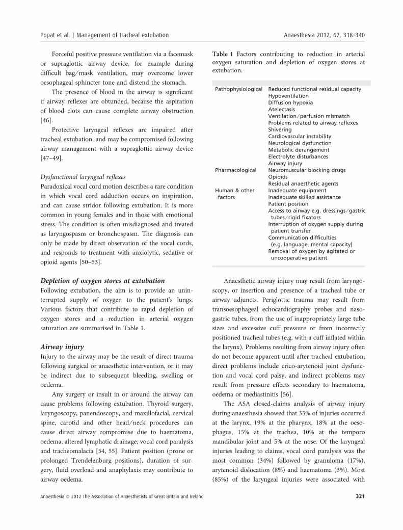
Depletion of oxygen stores at extubationFollowing extubation, the aim is to provide an unin-terrupted supply of oxygen to the patient’s lungs.Various factors that contribute to rapid depletion ofoxygen stores and a reduction in arterial oxygensaturation are summarised in Table 1.
Airway injuryInjury to the airway may be the result of direct traumafollowing surgical or anaesthetic intervention, or it maybe indirect due to subsequent bleeding, swelling oroedema.
Any surgery or insult in or around the airway cancause problems following extubation. Thyroid surgery,laryngoscopy, panendoscopy, and maxillofacial, cervicalspine, carotid and other head ⁄ neck procedures cancause direct airway compromise due to haematoma,oedema, altered lymphatic drainage, vocal cord paralysisand tracheomalacia [54, 55]. Patient position (prone orprolonged Trendelenburg positions), duration of sur-gery, fluid overload and anaphylaxis may contribute toairway oedema.
Anaesthetic airway injury may result from laryngo-scopy, or insertion and presence of a tracheal tube orairway adjuncts. Periglottic trauma may result fromtransoesophageal echocardiography probes and naso-gastric tubes, from the use of inappropriately large tubesizes and excessive cuff pressure or from incorrectlypositioned tracheal tubes (e.g. with a cuff inflated withinthe larynx). Problems resulting from airway injury oftendo not become apparent until after tracheal extubation;direct problems include crico-arytenoid joint dysfunc-tion and vocal cord palsy, and indirect problems mayresult from pressure effects secondary to haematoma,oedema or mediastinitis [56].
The ASA closed-claims analysis of airway injuryduring anaesthesia showed that 33% of injuries occurredat the larynx, 19% at the pharynx, 18% at the oeso-phagus, 15% at the trachea, 10% at the temporomandibular joint and 5% at the nose. Of the laryngealinjuries leading to claims, vocal cord paralysis was themost common (34%) followed by granuloma (17%),arytenoid dislocation (8%) and haematoma (3%). Most(85%) of the laryngeal injuries were associated with
Table 1 Factors contributing to reduction in arterialoxygen saturation and depletion of oxygen stores atextubation.
Pathophysiological Reduced functional residual capacityHypoventilationDiffusion hypoxiaAtelectasisVentilation ⁄ perfusion mismatchProblems related to airway reflexesShiveringCardiovascular instabilityNeurological dysfunctionMetabolic derangementElectrolyte disturbancesAirway injury
Pharmacological Neuromuscular blocking drugsOpioidsResidual anaesthetic agents
Human & otherfactors
Inadequate equipmentInadequate skilled assistancePatient positionAccess to airway e.g. dressings ⁄ gastrictubes ⁄ rigid fixators
Interruption of oxygen supply duringpatient transfer
Communication difficulties(e.g. language, mental capacity)
Removal of oxygen by agitated oruncooperative patient
Popat et al. | Management of tracheal extubation Anaesthesia 2012, 67, 318–340
Anaesthesia ª 2012 The Association of Anaesthetists of Great Britain and Ireland 321
short-term tracheal intubation and 80% followed routine(not difficult) tracheal intubation [57]. In adults, theglottis is the narrowest part of the airway, and theposterior glottis supports the tracheal tube. Movementof oversized or poorly positioned tracheal tubes, oroverinflated cuffs, on the posterior glottis and arytenoidscartilages can lead to oedema and compromised airflow[58]. Supraglottic swelling and oedema can causeposterior displacement of the epiglottis and (typically)inspiratory obstruction. Glottic, subglottic and trachealoedema can cause life-threatening airway compromise.
Physiological compromise in other systemsThe process of extubation causes exaggerated reflexes inother physiological systems resulting in hypertension,tachycardia (with associated myocardial ischaemia),raised venous pressures and increase in intra-ocularand intracerebral pressures [59–63].
Human factorsThe environment at extubation is not as favourable as atintubation. Equipment, monitoring and assistance maybe inadequate. Patient factors contributing to extubationproblems may be compounded by distraction, timepressure, operator fatigue, lack of equipment or skilledassistance and poor communication [64–66].
Managing extubationThere is a lack of compelling evidence to support a ‘onesize fits all’ extubation strategy for every patient. Thereis, however, a general agreement that good preparationis key to successful airway management and that anextubation strategy should be in place for every patient[10, 18, 67, 68].
General principlesExtubation is an elective process, and it is important toplan and execute it well. The goal is to ensureuninterrupted oxygen delivery to the patient’s lungs,avoid airway stimulation, and have a back-up plan, thatwould permit ventilation and re-intubation with mini-mum difficulty and delay should extubation fail. Sincethe introduction of the DAS unanticipated difficultintubation guidelines, the concept of a stepwiseapproach has been widely accepted. This approach has
been used to aid decision making and safe managementof extubation.
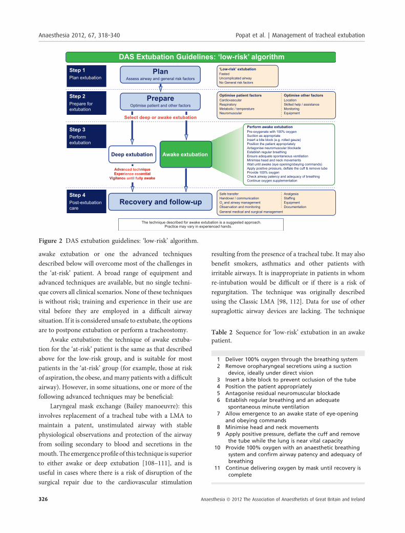
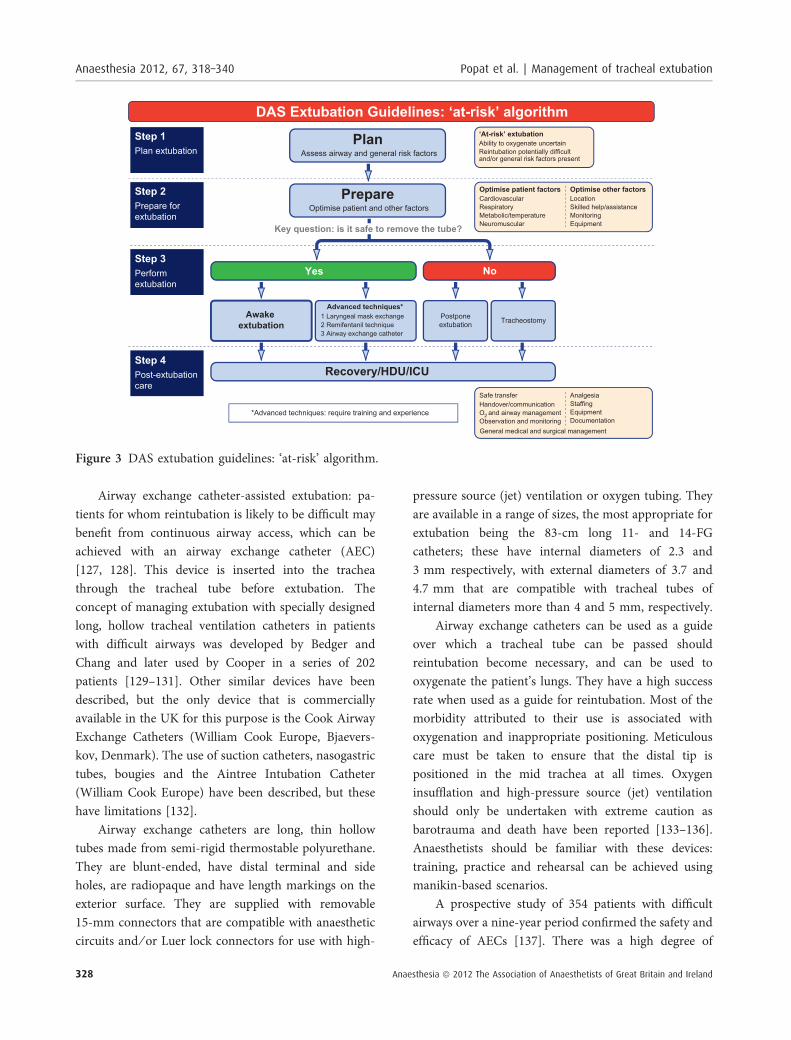
The DAS extubation guidelines (Figs. 1–3)The guidelines describe the following four steps:
Step 1: plan extubation.Step 2: prepare for extubation.Step 3: perform extubation.Step 4: post-extubation care: recovery and follow-up.
Step 1: plan extubationAn outline extubation plan should be in place beforeinduction of anaesthesia and reviewed throughout andimmediately before performing extubation. Planninginvolves an assessment of the airway and general riskfactors. The following questions may aid in the decisionmaking process [69], answers to which will helpdetermine whether extubation is ‘low-risk’ or ‘at-risk’[67]:
1 Are there airway risk factors?• was the airway normal ⁄ uncomplicated at induction?• has the airway changed?
2 Are there general risk factors?
Low-risk extubation. This is a routine or uncomplicatedextubation. The airway was normal ⁄ uncomplicated atinduction and remains unchanged at the end of surgery,and no general risk factors are present.
‘At-risk’ extubation. This is an extubation ‘at risk’ ofpotential complications. Airway risk factors are present:
1 Pre-existing airway difficulties. Airway access wasdifficult at induction (anticipated or unanticipated)and may have worsened intra-operatively. This groupincludes patients with obesity and OSA, and those atrisk of aspiration of gastric contents;
2 Peri-operative airway deterioration. The airway wasnormal at induction, but may have become difficult tomanage, for example, due to distorted anatomy,haemorrhage, haematoma or oedema resulting fromsurgery, trauma or non-surgical factors;
3 Restricted airway access. Airway access was straight-forward at induction, but is limited at the end ofsurgery, for example, where the airway is shared, orhead ⁄ neck movements restricted (halo fixation,mandibular wiring, surgical implants, cervical spinefixation).
Anaesthesia 2012, 67, 318–340 Popat et al. | Management of tracheal extubation
322 Anaesthesia ª 2012 The Association of Anaesthetists of Great Britain and Ireland
General risk factors may also be present; these maycomplicate or even preclude extubation, and includeimpaired respiratory function, cardiovascular instability,neurological ⁄ neuromuscular impairment, hypo ⁄hyperthermia, and abnormalities of clotting, acid-basebalance or electrolyte levels.
Smooth emergence from anaesthesia is desirable forthe success of some surgical procedures. For example,coughing and straining can cause raised venous pressureresulting in haematoma formation, airway compressionand suture disruption. Raised intra-ocular and intra-cranial pressures can compromise surgical outcomes.Cardiovascular changes may put the patient with severeischaemic heart disease at risk [62, 70].
Step 2: prepare for extubationPreparation is aimed at the final optimisation ofairway, general and logistical factors to ensure the bestpossible conditions for success extubation. Togetherwith planning (step 1), preparation (step 2) enables therisk stratification of extubation into ‘low-risk’ and ‘at-risk’ categories, and should always precede extubation(step 3).
Final evaluation and optimisation of airway fac-tors. The airway should be reassessed at the end ofsurgery and before extubation. This review should beused to finalise the extubation plan and to determine themost appropriate rescue plan for re-intubation shouldextubation fail.
Assessment should follow a logical sequence:
1 Airway. It is essential to consider whether bag-maskventilation would be achievable. Oedema, bleeding,blood clots, trauma, foreign bodies and airwaydistortion can be assessed by direct or indirectlaryngoscopy. It is important to remember both thatthe presence of a tracheal tube may give a falselyoptimistic view of the larynx at direct laryngoscopy,and that oedema may progress very rapidly;
2 Larynx. A cuff-leak test may be used to assesssubglottic calibre. Clinically, the presence of a largeaudible leak when the tracheal tube cuff is deflated isreassuring: the absence of a leak around an appro-priately sized tube generally precludes safe extubation.If the clinical conditions suggest airway oedema,caution should be exercised even if there is a cuff leak.Spirometry allows quantitative assessment of a cuffleak and is sensitive, but lacks specificity [71–76].
3 Lower airway. It is important to consider factors inthe lower airway that may contraindicate extubation,such as lower airway trauma, oedema, infection andsecretions. Chest radiography may be necessary toexclude bronchial intubation, pneumothorax, surgicalemphysema or other pulmonary pathology, if intuba-tion was difficult or oxygenation suboptimal duringsurgery.
Gastric distension splints the diaphragm andrestricts breathing. Gastric decompression with anoro ⁄ nasogastric tube is advisable if high-pressurefacemask ⁄ supraglottic airway ventilation has beennecessary.
If the airway rescue plan involves subglottic accessthen ability to access the neck should be confirmed.
Final evaluation and optimisation of general factors.Neuromuscular block should be fully reversed tomaximise the likelihood of adequate ventilation, andrestore protective airway reflexes and the ability toclear upper airway secretions. The use of a peripheralnerve stimulator to ensure a train-of-four ratio of 0.9or above is recommended and has been shown toreduce the incidence of postoperative airway complica-tions. An accelerometer is more accurate than visualassessment for train-of-four response [42, 77].Sugammadex provides more reliable antagonism ofrocuronium- (and to a lesser extent vecuronium-)induced neuromuscular blockade than neostigmine.Cardiovascular instability should be corrected andadequate fluid balance assured. The patient’s bodytemperature, acid-base balance, electrolyte and coagu-lation status should be optimised. Adequate analgesiashould be provided.
Final evaluation and optimisation of logisticalfactors. Extubation is an elective process, which shouldbe carried out in a controlled manner with the samestandards of monitoring, equipment and assistance thatare available at induction. Tracheal extubation can takeas long to perform safely as tracheal intubation, and thisshould be considered when organising list schedules,or sending for the next patient. Communication isessential, and the anaesthetist, surgeon and theatre teamall play an important role. Additional resources may berequired for the ‘at risk’ patient.
Popat et al. | Management of tracheal extubation Anaesthesia 2012, 67, 318–340
Anaesthesia ª 2012 The Association of Anaesthetists of Great Britain and Ireland 323
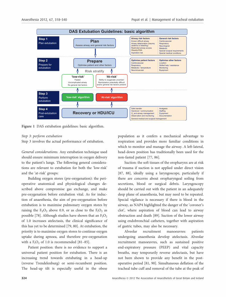
Step 3: perform extubationStep 3 involves the actual performance of extubation.
General considerations. Any extubation technique usedshould ensure minimum interruption in oxygen deliveryto the patient’s lungs. The following general considera-tions are relevant to extubation for both the ‘low-risk’and the ‘at-risk’ groups:
Building oxygen stores (pre-oxygenation): the peri-operative anatomical and physiological changes de-scribed above compromise gas exchange, and makepre-oxygenation before extubation vital. As for induc-tion of anaesthesia, the aim of pre-oxygenation beforeextubation is to maximise pulmonary oxygen stores byraising the FEO2 above 0.9, or as close to the FIO2 aspossible [78]. Although studies have shown that an FIO2
of 1.0 increases atelectasis, the clinical significance ofthis has yet to be determined [79, 80]. At extubation, thepriority is to maximise oxygen stores to continue oxygenuptake during apnoea, and therefore pre-oxygenationwith a FIO2 of 1.0 is recommended [81–85].
Patient position: there is no evidence to support auniversal patient position for extubation. There is anincreasing trend towards extubating in a head-up(reverse Trendelenburg) or semi-recumbent position.The head-up tilt is especially useful in the obese
population as it confers a mechanical advantage torespiration and provides more familiar conditions inwhich to monitor and manage the airway. A left-lateral,head-down position has traditionally been used for thenon-fasted patient [77, 86].
Suction: the soft tissues of the oropharynx are at riskof trauma if suction is not applied under direct vision[87, 88], ideally using a laryngoscope, particularly ifthere are concerns about oropharyngeal soiling fromsecretions, blood or surgical debris. Laryngoscopyshould be carried out with the patient in an adequatelydeep plane of anaesthesia, but may need to be repeated.Special vigilance is necessary if there is blood in theairway, as NAP4 highlighted the danger of the ‘coroner’sclot’, where aspiration of blood can lead to airwayobstruction and death [89]. Suction of the lower airwayusing endobronchial catheters, together with aspirationof gastric tubes, may also be necessary.
Alveolar recruitment manoeuvres: patientsundergoing anaesthesia develop atelectasis. Alveolarrecruitment manoeuvres, such as sustained positiveend-expiratory pressure (PEEP) and vital capacitybreaths, may temporarily reverse atelectasis, but havenot been shown to provide any benefit in the post-operative period [81, 90]. Simultaneous deflation of thetracheal tube cuff and removal of the tube at the peak of
Figure 1 DAS extubation guidelines: basic algorithm.
Anaesthesia 2012, 67, 318–340 Popat et al. | Management of tracheal extubation
324 Anaesthesia ª 2012 The Association of Anaesthetists of Great Britain and Ireland
a sustained inflation generates a passive exhalation, andmay be sensibly employed to expel secretions andpossibly reduce the incidence of laryngospasm andbreathholding.
Bite block: a bite block prevents occlusion of thetracheal tube should the patient bite down duringemergence from anaesthesia [91–93]. Forced inspira-tory efforts against an obstructed airway can rapidlylead to pulmonary oedema (see Appendix 2B) [94].Should biting occur, deflating the cuff of the tube orlaryngeal mask airway (LMA) may prevent post-obstructive pulmonary oedema, as significant negativepressure cannot be generated if air can flow around thedevice. Various devices have been used as bite blocks,including the Guedel airway. When rolled gauze isused, it is important that it is tied or taped to thetracheal tube to prevent displacement or accidentalairway obstruction.
Avoidance of the sequelae of airway stimulation:traditionally, extubation has been performed when thepatient is either fully ‘awake’ or deeply anaesthetised.
Awake extubation is generally safer as the return ofairway tone, reflexes and respiratory drive allows thepatient to maintain their own airway.
Deep extubation reduces the incidence of coughing,bucking and the haemodynamic effects of tracheal tubemovement, but these advantages are offset by anincreased incidence of upper airway obstruction [95–97]. This is an advanced technique, which should bereserved for patients in whom airway managementwould be easy and who are not at increased risk ofaspiration.
It is possible to reduce the risk of airway obstructionby exchanging the tracheal tube for a LMA beforeemergence (Bailey maneouvre; see below) [98].
Opioids such as alfentanil, fentanyl and morphinehave been used to suppress any cough reflex. Currently,the ultrashort-acting opioid remifentanil, administeredby infusion, is the drug of choice for this technique, butrequires careful administration (see below). The benefitsof cough suppression must be weighed against theincreased risks of sedation and respiratory depression.Lidocaine has been used to reduce coughing; it may beadministered topically at intubation, into the cuff of thetracheal tube or intravenously before extubation, withsome benefit [77].
Other pharmacological agents have been used toattenuate the cardiovascular and respiratory changesassociated with extubation, including opioids, calciumchannel antagonists, magnesium, lidocaine, clonidine,ketamine and beta blockers [28, 99–103]. Doxapram hasbeen used to prevent and ⁄ or treat laryngospasm,although it is associated with cardiovascular stimulationand robust evidence to support its use for this indicationis lacking [104]. The use of steroids to reduceinflammatory airway oedema is described below [105–107].
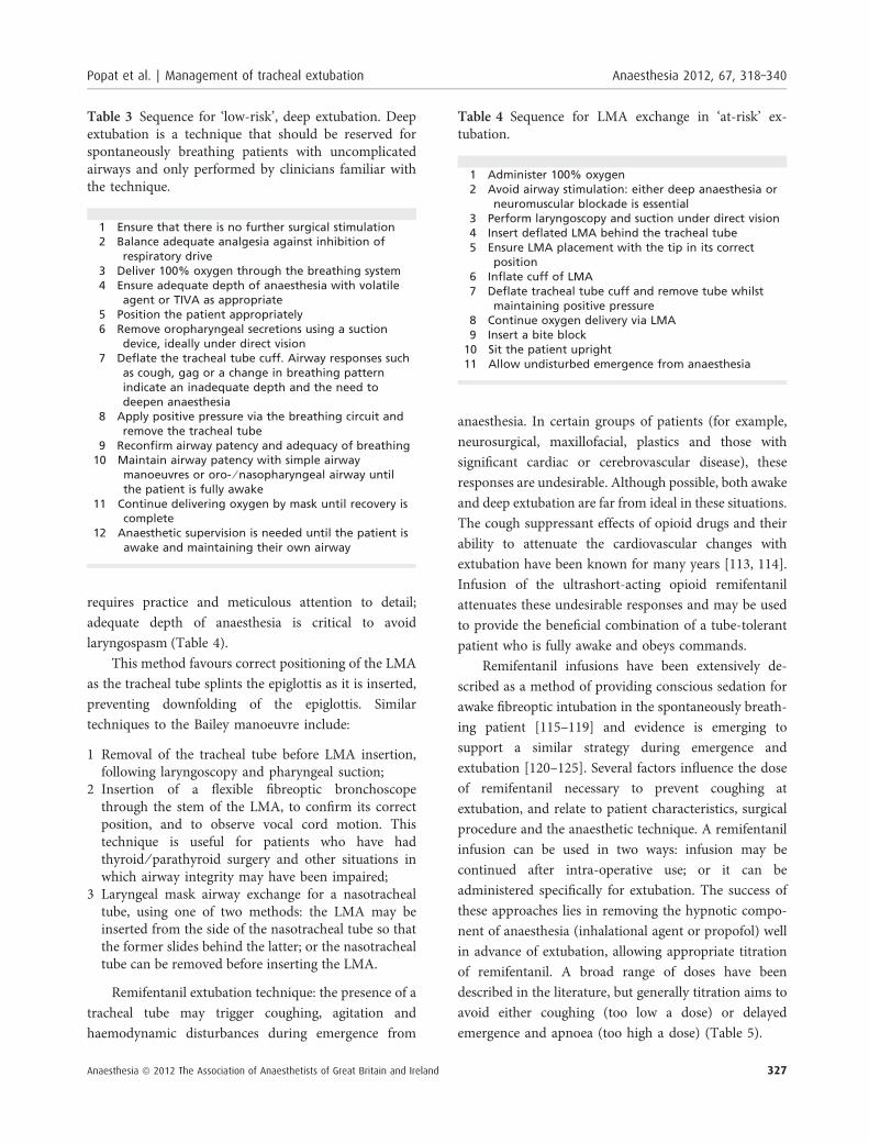
The performance of ‘low-risk’ (routine) extubation.Whilst no extubation is without risk, routine extubationis characterised by the expectation that reintubationcould be managed without difficulty, if required.
Stepwise approaches to awake and deep ‘low-risk’extubations are given in Tables 2 and 3, respectively.
The performance of ‘at-risk’ extubation. An ‘at-risk’extubation is one in which the risk stratification (steps 1and 2 above) has identified general and ⁄ or airway riskfactors that suggest that a patient may not be able tomaintain his ⁄ her own airway after removal of thetracheal tube. ‘At-risk’ extubation is characterised by theconcern that airway management may not be straight-forward should reintubation be required.
An example of an ‘at-risk’ extubation might involvethe patient having emergency surgery to repair a leakingaortic aneurysm for whom general factors such as a fullstomach, unstable cardiovascular physiology, acid-basederangement or temperature control can make extuba-tion more challenging.
An example of ‘at-risk’ extubation due to airwayfactors might involve the patient undergoing head andneck surgery after awake fibreoptic intubation beforeinduction of general anaesthesia, because of previoushead and neck radiotherapy.
Step 1 would stratify both these patients into the ‘at-risk’ extubation group. Step 2 would enable stabilisationof general factors and optimisation of logistical factorse.g. communication with the intensive care unit,assembling equipment, getting help.
The key decision to be made is whether it is safer toextubate, or preferable for the patient’s trachea to remainintubated. If it is considered safe to extubate, then an
Popat et al. | Management of tracheal extubation Anaesthesia 2012, 67, 318–340
Anaesthesia ª 2012 The Association of Anaesthetists of Great Britain and Ireland 325
awake extubation or one the advanced techniquesdescribed below will overcome most of the challenges inthe ‘at-risk’ patient. A broad range of equipment andadvanced techniques are available, but no single techni-que covers all clinical scenarios. None of these techniquesis without risk; training and experience in their use arevital before they are employed in a difficult airwaysituation. If it is considered unsafe to extubate, the optionsare to postpone extubation or perform a tracheostomy.
Awake extubation: the technique of awake extuba-tion for the ‘at-risk’ patient is the same as that describedabove for the low-risk group, and is suitable for mostpatients in the ‘at-risk’ group (for example, those at riskof aspiration, the obese, and many patients with a difficultairway). However, in some situations, one or more of thefollowing advanced techniques may be beneficial:
Laryngeal mask exchange (Bailey manoeuvre): thisinvolves replacement of a tracheal tube with a LMA tomaintain a patent, unstimulated airway with stablephysiological observations and protection of the airwayfrom soiling secondary to blood and secretions in themouth. The emergence profile of this technique is superiorto either awake or deep extubation [108–111], and isuseful in cases where there is a risk of disruption of thesurgical repair due to the cardiovascular stimulation
resulting from the presence of a tracheal tube. It may alsobenefit smokers, asthmatics and other patients withirritable airways. It is inappropriate in patients in whomre-intubation would be difficult or if there is a risk ofregurgitation. The technique was originally describedusing the Classic LMA [98, 112]. Data for use of othersupraglottic airway devices are lacking. The technique
Table 2 Sequence for ‘low-risk’ extubation in an awakepatient.
1 Deliver 100% oxygen through the breathing system2 Remove oropharyngeal secretions using a suction
device, ideally under direct vision3 Insert a bite block to prevent occlusion of the tube4 Position the patient appropriately5 Antagonise residual neuromuscular blockade6 Establish regular breathing and an adequate
spontaneous minute ventilation7 Allow emergence to an awake state of eye-opening
and obeying commands8 Minimise head and neck movements9 Apply positive pressure, deflate the cuff and remove
the tube while the lung is near vital capacity10 Provide 100% oxygen with an anaesthetic breathing
system and confirm airway patency and adequacy ofbreathing
11 Continue delivering oxygen by mask until recovery iscomplete
Figure 2 DAS extubation guidelines: ‘low-risk’ algorithm.
Anaesthesia 2012, 67, 318–340 Popat et al. | Management of tracheal extubation
326 Anaesthesia ª 2012 The Association of Anaesthetists of Great Britain and Ireland
requires practice and meticulous attention to detail;adequate depth of anaesthesia is critical to avoidlaryngospasm (Table 4).
This method favours correct positioning of the LMAas the tracheal tube splints the epiglottis as it is inserted,preventing downfolding of the epiglottis. Similartechniques to the Bailey manoeuvre include:
1 Removal of the tracheal tube before LMA insertion,following laryngoscopy and pharyngeal suction;
2 Insertion of a flexible fibreoptic bronchoscopethrough the stem of the LMA, to confirm its correctposition, and to observe vocal cord motion. Thistechnique is useful for patients who have hadthyroid ⁄ parathyroid surgery and other situations inwhich airway integrity may have been impaired;
3 Laryngeal mask airway exchange for a nasotrachealtube, using one of two methods: the LMA may beinserted from the side of the nasotracheal tube so thatthe former slides behind the latter; or the nasotrachealtube can be removed before inserting the LMA.
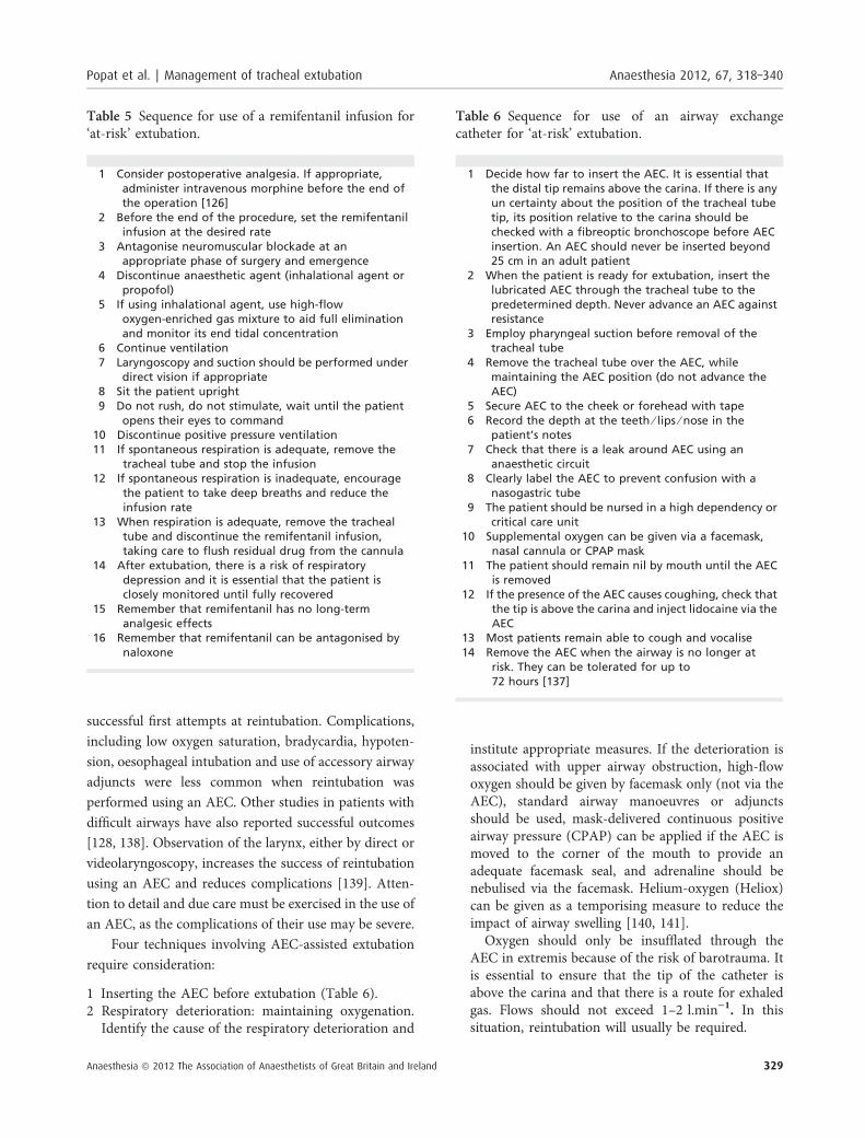
Remifentanil extubation technique: the presence of atracheal tube may trigger coughing, agitation andhaemodynamic disturbances during emergence from
anaesthesia. In certain groups of patients (for example,neurosurgical, maxillofacial, plastics and those withsignificant cardiac or cerebrovascular disease), theseresponses are undesirable. Although possible, both awakeand deep extubation are far from ideal in these situations.The cough suppressant effects of opioid drugs and theirability to attenuate the cardiovascular changes withextubation have been known for many years [113, 114].Infusion of the ultrashort-acting opioid remifentanilattenuates these undesirable responses and may be usedto provide the beneficial combination of a tube-tolerantpatient who is fully awake and obeys commands.
Remifentanil infusions have been extensively de-scribed as a method of providing conscious sedation forawake fibreoptic intubation in the spontaneously breath-ing patient [115–119] and evidence is emerging tosupport a similar strategy during emergence andextubation [120–125]. Several factors influence the doseof remifentanil necessary to prevent coughing atextubation, and relate to patient characteristics, surgicalprocedure and the anaesthetic technique. A remifentanilinfusion can be used in two ways: infusion may becontinued after intra-operative use; or it can beadministered specifically for extubation. The success ofthese approaches lies in removing the hypnotic compo-nent of anaesthesia (inhalational agent or propofol) wellin advance of extubation, allowing appropriate titrationof remifentanil. A broad range of doses have beendescribed in the literature, but generally titration aims toavoid either coughing (too low a dose) or delayedemergence and apnoea (too high a dose) (Table 5).
Table 4 Sequence for LMA exchange in ‘at-risk’ ex-tubation.
1 Administer 100% oxygen2 Avoid airway stimulation: either deep anaesthesia or
neuromuscular blockade is essential3 Perform laryngoscopy and suction under direct vision4 Insert deflated LMA behind the tracheal tube5 Ensure LMA placement with the tip in its correct
position6 Inflate cuff of LMA7 Deflate tracheal tube cuff and remove tube whilst
maintaining positive pressure8 Continue oxygen delivery via LMA9 Insert a bite block
10 Sit the patient upright11 Allow undisturbed emergence from anaesthesia
Table 3 Sequence for ‘low-risk’, deep extubation. Deepextubation is a technique that should be reserved forspontaneously breathing patients with uncomplicatedairways and only performed by clinicians familiar withthe technique.
1 Ensure that there is no further surgical stimulation2 Balance adequate analgesia against inhibition of
respiratory drive3 Deliver 100% oxygen through the breathing system4 Ensure adequate depth of anaesthesia with volatile
agent or TIVA as appropriate5 Position the patient appropriately6 Remove oropharyngeal secretions using a suction
device, ideally under direct vision7 Deflate the tracheal tube cuff. Airway responses such
as cough, gag or a change in breathing patternindicate an inadequate depth and the need todeepen anaesthesia
8 Apply positive pressure via the breathing circuit andremove the tracheal tube
9 Reconfirm airway patency and adequacy of breathing10 Maintain airway patency with simple airway
manoeuvres or oro- ⁄ nasopharyngeal airway untilthe patient is fully awake
11 Continue delivering oxygen by mask until recovery iscomplete
12 Anaesthetic supervision is needed until the patient isawake and maintaining their own airway
Popat et al. | Management of tracheal extubation Anaesthesia 2012, 67, 318–340
Anaesthesia ª 2012 The Association of Anaesthetists of Great Britain and Ireland 327
Airway exchange catheter-assisted extubation: pa-tients for whom reintubation is likely to be difficult maybenefit from continuous airway access, which can beachieved with an airway exchange catheter (AEC)[127, 128]. This device is inserted into the tracheathrough the tracheal tube before extubation. Theconcept of managing extubation with specially designedlong, hollow tracheal ventilation catheters in patientswith difficult airways was developed by Bedger andChang and later used by Cooper in a series of 202patients [129–131]. Other similar devices have beendescribed, but the only device that is commerciallyavailable in the UK for this purpose is the Cook AirwayExchange Catheters (William Cook Europe, Bjaevers-kov, Denmark). The use of suction catheters, nasogastrictubes, bougies and the Aintree Intubation Catheter(William Cook Europe) have been described, but thesehave limitations [132].
Airway exchange catheters are long, thin hollowtubes made from semi-rigid thermostable polyurethane.They are blunt-ended, have distal terminal and sideholes, are radiopaque and have length markings on theexterior surface. They are supplied with removable15-mm connectors that are compatible with anaestheticcircuits and ⁄ or Luer lock connectors for use with high-
pressure source (jet) ventilation or oxygen tubing. Theyare available in a range of sizes, the most appropriate forextubation being the 83-cm long 11- and 14-FGcatheters; these have internal diameters of 2.3 and3 mm respectively, with external diameters of 3.7 and4.7 mm that are compatible with tracheal tubes ofinternal diameters more than 4 and 5 mm, respectively.
Airway exchange catheters can be used as a guideover which a tracheal tube can be passed shouldreintubation become necessary, and can be used tooxygenate the patient’s lungs. They have a high successrate when used as a guide for reintubation. Most of themorbidity attributed to their use is associated withoxygenation and inappropriate positioning. Meticulouscare must be taken to ensure that the distal tip ispositioned in the mid trachea at all times. Oxygeninsufflation and high-pressure source (jet) ventilationshould only be undertaken with extreme caution asbarotrauma and death have been reported [133–136].Anaesthetists should be familiar with these devices:training, practice and rehearsal can be achieved usingmanikin-based scenarios.
A prospective study of 354 patients with difficultairways over a nine-year period confirmed the safety andefficacy of AECs [137]. There was a high degree of
Figure 3 DAS extubation guidelines: ‘at-risk’ algorithm.
Anaesthesia 2012, 67, 318–340 Popat et al. | Management of tracheal extubation
328 Anaesthesia ª 2012 The Association of Anaesthetists of Great Britain and Ireland
successful first attempts at reintubation. Complications,including low oxygen saturation, bradycardia, hypoten-sion, oesophageal intubation and use of accessory airwayadjuncts were less common when reintubation wasperformed using an AEC. Other studies in patients withdifficult airways have also reported successful outcomes[128, 138]. Observation of the larynx, either by direct orvideolaryngoscopy, increases the success of reintubationusing an AEC and reduces complications [139]. Atten-tion to detail and due care must be exercised in the use ofan AEC, as the complications of their use may be severe.
Four techniques involving AEC-assisted extubationrequire consideration:
1 Inserting the AEC before extubation (Table 6).2 Respiratory deterioration: maintaining oxygenation.
Identify the cause of the respiratory deterioration and
institute appropriate measures. If the deterioration isassociated with upper airway obstruction, high-flowoxygen should be given by facemask only (not via theAEC), standard airway manoeuvres or adjunctsshould be used, mask-delivered continuous positiveairway pressure (CPAP) can be applied if the AEC ismoved to the corner of the mouth to provide anadequate facemask seal, and adrenaline should benebulised via the facemask. Helium-oxygen (Heliox)can be given as a temporising measure to reduce theimpact of airway swelling [140, 141].
Oxygen should only be insufflated through theAEC in extremis because of the risk of barotrauma. Itis essential to ensure that the tip of the catheter isabove the carina and that there is a route for exhaledgas. Flows should not exceed 1–2 l.min)1. In thissituation, reintubation will usually be required.
Table 6 Sequence for use of an airway exchangecatheter for ‘at-risk’ extubation.
1 Decide how far to insert the AEC. It is essential thatthe distal tip remains above the carina. If there is anyun certainty about the position of the tracheal tubetip, its position relative to the carina should bechecked with a fibreoptic bronchoscope before AECinsertion. An AEC should never be inserted beyond25 cm in an adult patient
2 When the patient is ready for extubation, insert thelubricated AEC through the tracheal tube to thepredetermined depth. Never advance an AEC againstresistance
3 Employ pharyngeal suction before removal of thetracheal tube
4 Remove the tracheal tube over the AEC, whilemaintaining the AEC position (do not advance theAEC)
5 Secure AEC to the cheek or forehead with tape6 Record the depth at the teeth ⁄ lips ⁄ nose in the
patient’s notes7 Check that there is a leak around AEC using an
anaesthetic circuit8 Clearly label the AEC to prevent confusion with a
nasogastric tube9 The patient should be nursed in a high dependency or
critical care unit10 Supplemental oxygen can be given via a facemask,
nasal cannula or CPAP mask11 The patient should remain nil by mouth until the AEC
is removed12 If the presence of the AEC causes coughing, check that
the tip is above the carina and inject lidocaine via theAEC
13 Most patients remain able to cough and vocalise14 Remove the AEC when the airway is no longer at
risk. They can be tolerated for up to72 hours [137]
Table 5 Sequence for use of a remifentanil infusion for‘at-risk’ extubation.
1 Consider postoperative analgesia. If appropriate,administer intravenous morphine before the end ofthe operation [126]
2 Before the end of the procedure, set the remifentanilinfusion at the desired rate
3 Antagonise neuromuscular blockade at anappropriate phase of surgery and emergence
4 Discontinue anaesthetic agent (inhalational agent orpropofol)
5 If using inhalational agent, use high-flowoxygen-enriched gas mixture to aid full eliminationand monitor its end tidal concentration
6 Continue ventilation7 Laryngoscopy and suction should be performed under
direct vision if appropriate8 Sit the patient upright9 Do not rush, do not stimulate, wait until the patient
opens their eyes to command10 Discontinue positive pressure ventilation11 If spontaneous respiration is adequate, remove the
tracheal tube and stop the infusion12 If spontaneous respiration is inadequate, encourage
the patient to take deep breaths and reduce theinfusion rate
13 When respiration is adequate, remove the trachealtube and discontinue the remifentanil infusion,taking care to flush residual drug from the cannula
14 After extubation, there is a risk of respiratorydepression and it is essential that the patient isclosely monitored until fully recovered
15 Remember that remifentanil has no long-termanalgesic effects
16 Remember that remifentanil can be antagonised bynaloxone
Popat et al. | Management of tracheal extubation Anaesthesia 2012, 67, 318–340
Anaesthesia ª 2012 The Association of Anaesthetists of Great Britain and Ireland 329
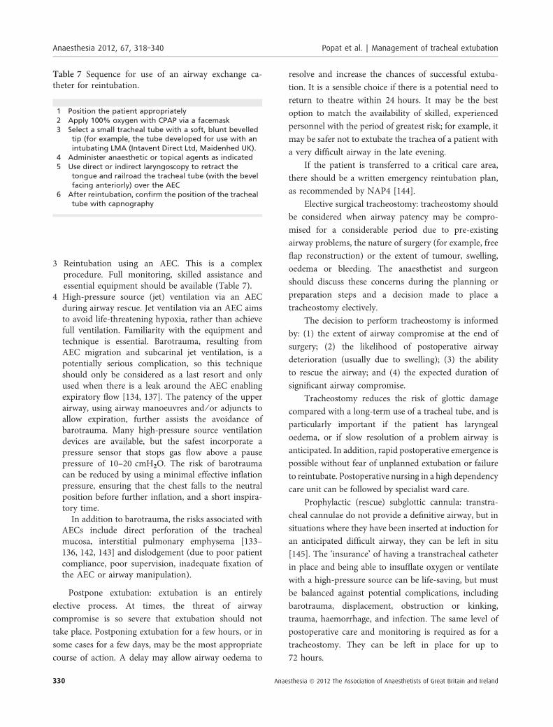
3 Reintubation using an AEC. This is a complexprocedure. Full monitoring, skilled assistance andessential equipment should be available (Table 7).
4 High-pressure source (jet) ventilation via an AECduring airway rescue. Jet ventilation via an AEC aimsto avoid life-threatening hypoxia, rather than achievefull ventilation. Familiarity with the equipment andtechnique is essential. Barotrauma, resulting fromAEC migration and subcarinal jet ventilation, is apotentially serious complication, so this techniqueshould only be considered as a last resort and onlyused when there is a leak around the AEC enablingexpiratory flow [134, 137]. The patency of the upperairway, using airway manoeuvres and ⁄ or adjuncts toallow expiration, further assists the avoidance ofbarotrauma. Many high-pressure source ventilationdevices are available, but the safest incorporate apressure sensor that stops gas flow above a pausepressure of 10–20 cmH2O. The risk of barotraumacan be reduced by using a minimal effective inflationpressure, ensuring that the chest falls to the neutralposition before further inflation, and a short inspira-tory time.
In addition to barotrauma, the risks associated withAECs include direct perforation of the trachealmucosa, interstitial pulmonary emphysema [133–136, 142, 143] and dislodgement (due to poor patientcompliance, poor supervision, inadequate fixation ofthe AEC or airway manipulation).
Postpone extubation: extubation is an entirelyelective process. At times, the threat of airwaycompromise is so severe that extubation should nottake place. Postponing extubation for a few hours, or insome cases for a few days, may be the most appropriatecourse of action. A delay may allow airway oedema to
resolve and increase the chances of successful extuba-tion. It is a sensible choice if there is a potential need toreturn to theatre within 24 hours. It may be the bestoption to match the availability of skilled, experiencedpersonnel with the period of greatest risk; for example, itmay be safer not to extubate the trachea of a patient witha very difficult airway in the late evening.
If the patient is transferred to a critical care area,there should be a written emergency reintubation plan,as recommended by NAP4 [144].
Elective surgical tracheostomy: tracheostomy shouldbe considered when airway patency may be compro-mised for a considerable period due to pre-existingairway problems, the nature of surgery (for example, freeflap reconstruction) or the extent of tumour, swelling,oedema or bleeding. The anaesthetist and surgeonshould discuss these concerns during the planning orpreparation steps and a decision made to place atracheostomy electively.
The decision to perform tracheostomy is informedby: (1) the extent of airway compromise at the end ofsurgery; (2) the likelihood of postoperative airwaydeterioration (usually due to swelling); (3) the abilityto rescue the airway; and (4) the expected duration ofsignificant airway compromise.
Tracheostomy reduces the risk of glottic damagecompared with a long-term use of a tracheal tube, and isparticularly important if the patient has laryngealoedema, or if slow resolution of a problem airway isanticipated. In addition, rapid postoperative emergence ispossible without fear of unplanned extubation or failureto reintubate. Postoperative nursing in a high dependencycare unit can be followed by specialist ward care.
Prophylactic (rescue) subglottic cannula: transtra-cheal cannulae do not provide a definitive airway, but insituations where they have been inserted at induction foran anticipated difficult airway, they can be left in situ[145]. The ‘insurance’ of having a transtracheal catheterin place and being able to insufflate oxygen or ventilatewith a high-pressure source can be life-saving, but mustbe balanced against potential complications, includingbarotrauma, displacement, obstruction or kinking,trauma, haemorrhage, and infection. The same level ofpostoperative care and monitoring is required as for atracheostomy. They can be left in place for up to72 hours.
Table 7 Sequence for use of an airway exchange ca-theter for reintubation.
1 Position the patient appropriately2 Apply 100% oxygen with CPAP via a facemask3 Select a small tracheal tube with a soft, blunt bevelled
tip (for example, the tube developed for use with anintubating LMA (Intavent Direct Ltd, Maidenhed UK).
4 Administer anaesthetic or topical agents as indicated5 Use direct or indirect laryngoscopy to retract the
tongue and railroad the tracheal tube (with the bevelfacing anteriorly) over the AEC
6 After reintubation, confirm the position of the trachealtube with capnography
Anaesthesia 2012, 67, 318–340 Popat et al. | Management of tracheal extubation
330 Anaesthesia ª 2012 The Association of Anaesthetists of Great Britain and Ireland
Step 4: post-extubation care: recovery and follow-upLife-threatening complications following extubation arenot restricted to the immediate postoperative period.Anaesthetists have a continuing duty of care to thepatient [146].
Oxygen should be administered during transfer torecovery, and portable monitoring should be consideredif the recovery area is distant from the operating theatreor if the patient’s condition is unstable.
Staffing and communication. Trained staff shouldnurse the patient until airway reflexes have returnedand the patient is physiologically stable. There shouldbe one recovery nurse for each patient, with neverfewer than two personnel in recovery. An appropriatelyskilled anaesthetist must be immediately available [147,148].
Good communication is essential. Surgical andanaesthetic concerns for recovery and the postoperativeperiod should be discussed at the end of the case. A clearverbal handover and written instructions should beavailable for both recovery and the ward or highdependency unit. In high-risk cases, the on-call teamshould be briefed about the patient and a written airwaymanagement plan should be in place. A calm atmo-sphere and reassurance are particularly helpful for thepatient with airway compromise as anxiety increases thework of breathing.
Observations and warning signs. Observations shouldinclude level of consciousness, respiratory rate, heartrate, blood pressure, peripheral oxygen saturation,temperature and pain score. Capnography (using aspecially designed facemask) has the potential to aidearly detection of airway obstruction [149, 150]. Closeobservation of the patient is necessary during recovery.A pulse oximeter is not designed to be a monitor ofventilation. Oximeters can give incorrect readings in avariety of circumstances and should never be reliedupon as the sole monitor [151–154].
Warning signs include early problems with theairway (stridor, obstructed pattern of breathing, agita-tion) and resulting from surgery (drain losses, free flapperfusion, airway bleeding, haematoma formation andairway swelling), and late problems after return to theward, relating to mediastinitis and airway injury.
Mediastinitis can occur after airway perforation, forexample, after difficult intubation, and is characterisedby pain (severe sore throat, deep cervical pain, chestpain, dysphagia, painful swallowing), fever and crepitus[57]. Patients should be informed about the symptomsof mediastinitis, and advised to seek medical adviceshould they occur.
The ASA closed-claim analysis suggests that airwaytrauma most commonly involves the larynx (afterroutine intubation), the pharynx and the oesophagus(after difficult intubation) [56]. Pharyngeal and oeso-phageal injury are difficult to diagnose, with pneu-mothorax, pneumo mediastinum or surgical emphysemapresent in only 50% of cases.
A patient who is agitated or complains of difficultybreathing should never be ignored, even if objectivesigns are absent.
Equipment and monitors. A difficult airway trolleyshould be immediately available, as should relevantitems such as clip removers and wire cutters. Standardmonitoring should be continued in recovery. Capno-graphy should be available.
Location and safe transfer. All extubations should besupervised by an anaesthetist. ‘At-risk’ extubationshould occur in the operating theatre. Patients in whomthere is concern about the airway should either stay inrecovery or go to a critical care environment. Duringtransfer to recovery or critical care areas, the patientshould be supervised by an anaesthetist.
Transfer of ‘at-risk’ patients from intensive care tothe operating theatre for extubation may be appropriateto ensure availability of necessary equipment andexpertise.
Respiratory care for patients with airway compromise.Patients with airway compromise should be nursedupright, and administered high-flow humidified oxygen.End-tidal carbon dioxide monitoring is desirable. Thepatient should be kept starved, as laryngeal competencemay be impaired despite full consciousness [48]. Factorsthat would impede venous drainage should be avoided.Deep breaths and coughing to clear secretions should beencouraged. In patients with OSA, a nasopharyngealairway may overcome upper airway obstruction. If the
Popat et al. | Management of tracheal extubation Anaesthesia 2012, 67, 318–340
Anaesthesia ª 2012 The Association of Anaesthetists of Great Britain and Ireland 331
patient uses a CPAP device at home, it should beavailable for use in recovery and on the ward.
Steroids reduce inflammatory airway oedema result-ing from direct airway injury (surgical ⁄ anaesthe-tic ⁄ thermal ⁄ chemical) [105–107, 155], but have noeffect on mechanical oedema secondary to venousobstruction (e.g. neck haematoma). The evidencesuggests that all steroids are equally effective, providedthey are given in adequate doses (equivalent to 100 mghydrocortisone every 6 hours). Steroids should bestarted as soon as possible in patients who are at highrisk of inflammatory airway oedema and continued forat least 12 hours. Single-dose steroids given immediatelybefore extubation are ineffective [105–107, 155, 156].
If upper respiratory obstruction ⁄ stridor develops,nebulised adrenaline (1 mg) may reduce airway oedema.Heliox may be helpful, but limits the FIO2 [140, 141,157–160].
Analgesia. Good analgesia optimises postoperativerespiratory function. Sedative analgesia should beavoided or titrated cautiously. Effective anti-emesis isimportant.
Documentation and recommendations for futuremanagement. Clinical details and instructions forrecovery and postoperative care should be recordedon the anaesthetic chart. Difficulties should bedocumented in the ‘Alerts’ section of the medical notesand in the local difficult intubation database. Details ofairway management and future recommendationsshould be recorded. A letter should be sent to thepatient’s general practitioner and a copy given to thepatient (DAS Airway Alert form), who should also begiven a full explanation when they are able to retain thisinformation [161, 162]. The patient should also bewarned about the delayed symptoms of airway traumaand advised to seek medical help should they develop.Patients with difficult airways should be advised toregister with an accessible, dependable database such asMedicAlert.
ConclusionGuidelines are useful in infrequent, life-threateningsituations, and have been shown to improve outcomes[16, 163–170]. Several national guidelines for manage-
ment of the airway have been published, but none hasaddressed extubation in detail [11–15, 18, 19].
Extubation differs from intubation, in that it shouldalways be an elective process with adequate timeavailable to the anaesthetist for methodical management.Extubation practice is highly variable, and is not oftenformally addressed in training. Technical and non-technical factors can contribute to adverse events atextubation [36, 135, 171, 172], but outcomes areimproved by planning, organisation and communication[65, 66, 173].
The DAS extubation guidelines promote the conceptof an extubation strategy, involving a stepwise approachto planning, preparation and risk stratification, aimed atclear identification and management of patients ‘at risk’during extubation.
The evidence base for extubation practice is limited,so inevitably some of the recommendations in theseguidelines are based on expert opinion. Awake extuba-tion is the preferred technique for most patients.However, deep extubation, laryngeal mask exchange,remifentanil infusion and the use of airway exchangecatheters may be beneficial in certain clinical situations.Delaying extubation or performing an elective tracheost-omy should be considered when it is unsafe to extubate.
Representing the first attempt specifically to addressextubation in a national guideline, we commend thisdocument to the anaesthetic community, and hope thatit will be used to inform clinical practice with the samedegree of success as the DAS difficult intubationguidelines.
AcknowledgementsThe authors thank the expert review panel for theirsubstantial contribution to the guidelines: David Ball; RaviBagrath; Radhika Bhishma; David Bogod; Ian Calder;Simon Clarke; Tim Cook; Ali Diba; Sylva Dolenska; PeterGroom; John Henderson; Atul Kapila; Cyprian Mendon-ca; Barry Mcguire; Alistair McNarry; Thomas Mort; MaryMushambi; Ellen O’Sullivan; Adrian Pearce; Subrahma-nyam Radhakrishna; Jairaj Rangasami; Mridula Rai; MarkStacey; Tim Strang; Matthew Turner; and Nick Woodall.Special thanks to Professor Richard Cooper and Dr RalphVaughan, pioneers in extubation whose work inspired us.We also thank those DAS members who gave theirfeedback whilst the draft algorithms were displayed on the
Anaesthesia 2012, 67, 318–340 Popat et al. | Management of tracheal extubation
332 Anaesthesia ª 2012 The Association of Anaesthetists of Great Britain and Ireland
DAS website. Finally, we are indebted to Michelle Whitefor her help with the algorithms.
Competing interestsDr Patel has received an honorarium from the LaryngealMask Company and has received free samples of single-use LMAs that have been used for evaluation andresearch.
Dr Popat has received free samples of airwayequipment for evaluation and research and support forworkshops from Intavent Direct UK, Intersurgical UK,KARL STORZ Endoscopy (UK) Ltd and SmithsMedical.
Dr Mitchell has received free samples of single-useLMAs for evaluation and research support for airwayworkshops from Intavent Direct UK, Smiths MedicalUK, & KARL STORZ Endoscopy (UK) Ltd and KeymedLtd (UK).
Dr Dravid has received free samples of airwayequipment and support for airway workshops fromIntavent, Intersurgicals, Freelance surgicals, Fannin, UK,Liteoptics, Keymed Ltd (UK) & KARL STORZ Endo-scopy (UK) Ltd. The Guidelines have been presented inpart at the following meetings: AAGBI Core Topics,Manchester, October 2010; AAGBI Core Topics,Oxford, June 2011; DAS Annual Meeting, November2011; British Association of Indian Anaesthetists Annualmeeting, London 2011; Irish College of Anaesthetists’CME Day, November 2011; and various regionaltraining days.
No other competing interests declared.
References1. Cook TM, Scott S, Mihai R. Litigation related to airway and
respiratory complications of anaesthesia: an analysis of claimsagainst the NHS in England 1995–2007. Anaesthesia 2010; 65:556–63.
2. Peskett MJ. Clinical indicators and other complications in therecovery room or postanaesthetic care unit. Anaesthesia 1999;54: 1143–9.
3. Rose DK, Cohen MM, Wigglesworth DF, DeBoer DP. Criticalrespiratory events in the postanesthesia care unit. Patient,surgical, and anesthetic factors. Anesthesiology 1994; 81: 410–8.
4. Mhyre JM, Riesner MN, Polley LS, Naughton NN. A series ofanesthesia-related maternal deaths in Michigan, 1985-2003.Anesthesiology 2007; 106: 1096–104.
5. Auroy Y, Benhamou D, Péquignot F, Bovet M, Jougla E, LienhartA. Mortality related to anaesthesia in France: analysis of deathsrelated to airway complications. Anaesthesia 2009; 64: 366–70.
6. Lewis G. The Confidential Enquiry into Maternal andChild Health (CEMACH). Saving Mothers’ Lives:Reviewing Maternal Deaths to make MotherhoodSafer—2003–2005. The Seventh Report on ConfidentialEnquiries into Maternal Deaths in the United Kingdom.London: CEMACH, 2007.
7. Asai T, Koga K, Vaughan RS. Respiratory complicationsassociated with tracheal intubation and extubation. BritishJournal of Anaesthesia 1998; 80: 767–75.
8. Abdy S. An audit of airway problems in the recovery room.Anaesthesia 1999; 54: 372–5.
9. Cook TM, Woodall N, Frerk C. Royal College of Anaesthetists.4th National Audit Project: Major Complications of AirwayManagement in the UK. Royal College of Anaesthetists,London, 2011: 62–70.
10. Peterson GN, Domino KB, Caplan RA, Posner KL, Lee LA, CheneyFW. Management of the difficult airway: a closed claimsanalysis. Anesthesiology 2005; 103: 33–9.
11. American Society of Anesthesiologists Task Force on Manage-ment of the Difficult Airway. Practice guidelines for manage-ment of the difficult airway: an updated report by the AmericanSociety of Anesthesiologists Task Force on Management of theDifficult Airway. Anesthesiology 2003; 98: 1269–77.
12. SIAARTI Task Force on Difficult Airway Management. L’intuba-zione difficile e la difficoltà di controllo delle vie aeree nell’adulto(SIAARTI). Minerva Anestesiologica 1998; 64: 361–71.
13. Braun U, Goldmann K, Hempel V, Krie C. Airway management.Guidelines of the German Society of Anesthesiology andIntensive Care. Anasthesiologie, Intensivmedizin, Notfallmedi-zin, Schmerztherapie 2004; 45: 302–6.
14. Boisson-Bertrand D, Bourgain JL, Camboulives J, et al. Difficultintubation. French Society of Anesthesia and Intensive Care. Acollective expertise. Annales Francaises D’Anesthesie et deRéanimation 1996; 15: 207–14.
15. Crosby ET, Cooper RM, Douglas MJ, et al. The unanticipateddifficult airway with recommendations for management.Canadian Journal of Anesthesia 1998; 45: 757–76.
16. Heidegger T, Gerig HJ, Henderson JJ. Strategies and algorithmsfor management of the difficult airway. Best Practice &Research Clinical Anaesthesiology 2005; 19: 661–74.
17. Royal College of Anaesthetists. The CCT in anaesthetics trainingprogramme august 2010 http://www.rcoa.ac.uk/index.asp?-PageID=1479 (accessed 07 ⁄ 12 ⁄ 2011).
18. Henderson JJ, Popat MT, Latto IP, Pearce AC. Difficult AirwaySociety guidelines for management of the unanticipateddifficult intubation. Anaesthesia 2004; 59: 675–94.
19. American Society of Anesthesiologists Task Force onManagement of the Difficult Airway. Practice guidelines formanagement of the difficult airway. Anesthesiology 1993; 78:597–602.
20. Oxford centre for evidence-based medicine – Levels ofevidence. http://www.cebm.net/index.aspx?o = 1025 (ac-cessed 07 ⁄ 12 ⁄ 2011).
21. Cooper GM, McClure JH. Anaesthesia chapter from savingmothers’ lives; reviewing maternal deaths to make pregnancysafer. British Journal of Anaesthesia 2008; 100: 17–22.
22. Langton JA. Airway reflexes. In: Calder I, Pearce A, eds. CoreTopics in Airway Management. Cambridge: Cambridge Uni-versity Press, Cambridge, 2011: 28–35.
23. Miller KA, Harkin CP, Bailey PL. Postoperative tracheal extuba-tion. Anesthesia and Analgesia 1995; 80: 149–72.
24. Ikari T, Sasaki CT. Glottic closure reflex: control mechanisms.Annals of Otology, Rhinology, and Laryngology 1980; 89: 220–4.
Popat et al. | Management of tracheal extubation Anaesthesia 2012, 67, 318–340
Anaesthesia ª 2012 The Association of Anaesthetists of Great Britain and Ireland 333
25. Fink BR. The curse of Adam: effort closure of the larynx.Anesthesiology 1973; 39: 325–7.
26. Landsman IS. Mechanisms and treatment of laryngospasm.International Anesthesiology Clinics 1997; 35: 67–73.
27. Rex MA. A review of the structural and functional basis oflaryngospasm and a discussion of the nerve pathways involvedin the reflex and its clinical significance in man and animals.British Journal of Anaesthesia 1970; 42: 891–9.
28. Pak HJ, Lee WH, Ji SM, Choi YH. Effect of a small dose of propofolor ketamine to prevent coughing and laryngospasm in childrenawakening from general anesthesia. Korean Journal ofAnesthesiology 2011; 60: 25–9.
29. Hans P, Marechal H, Bonhomme V. Effect of propofol andsevoflurane on coughing in smokers and non-smokers awa-kening from general anaesthesia at the end of a cervical spinesurgery. British Journal of Anaesthesia 2008; 101: 731–7.
30. Barker P, Langton JA, Wilson IG, Smith G. Movements of thevocal cords on induction of anaesthesia with thiopentone orpropofol. British Journal of Anaesthesia 1992; 69: 23–5.
31. McKeating K, Bali IM, Dundee JW. The effects of thiopentoneand propofol on upper airway integrity. Anaesthesia 1988; 43:638–40.
32. Olsson GL, Hallen B. Laryngospasm during anaesthesia. Acomputer-aided incidence study in 136,929 patients. ActaAnaesthesiologica Scandinavica 1984; 28: 567–75.
33. Patton WC, Baker CL. Prevalence of negative-pressure pulmon-ary edema at an orthopaedic hospital. Journal of the SouthernOrthopaedic Association 2000; 9: 248–53.
34. Lang SA, Duncan PG, Shephard DA, Ha HC. Pulmonary oedemaassociated with airway obstruction. Canadian Journal ofAnesthesia 1990; 37: 210–8.
35. Scarbrough FE, Wittenberg JM, Smith BR, Adcock DK. Pulmonaryedema following postoperative laryngospasm: case reportsand review of the literature. Anesthesia Progress 1997; 44:110–6.
36. Cook TM, Woodall N, Frerk C, Fourth National Audit Project.Major complications of airway management in the UK: resultsof the 4th National Audit Project of the Royal College ofAnaesthetists and the Difficult Airway Society. Part 1:Anaesthesia. British Journal of Anaesthesia 2011; 106: 617–31.
37. Nishino T. Swallowing as a protective reflex for the upperrespiratory tract. Anesthesiology 1993; 79: 588–601.
38. Mathew JP, Rosenbaum SH, O’Connor T, Barash PG. Emergencytracheal intubation in the postanesthesia care unit: physicianerror or patient disease? Anesthesia and Analgesia 1990; 71:691–7.
39. Gupta RM, Parvizi J, Hanssen AD, Gay PC. Postoperativecomplications in patients with obstructive sleep apneasyndrome undergoing hip or knee replacement: a case-controlstudy. Mayo Clinic Proceedings 2001; 79: 897–905.
40. Adesanya AO, Lee W, Greilich NB, Joshi GP. Perioperativemanagement of obstructive sleep apnea. Chest 2010; 138:1489–98.
41. Gross JB, Bachenberg KL, Benumof JL, et al. Practice guidelinesfor the perioperative management of patients with obstructivesleep apnea: a report by the American Society of Anesthesiol-ogists Task Force on perioperative management of patientswith obstructive sleep apnea. Anesthesiology 2006; 104:1081–93.
42. Murphy GS, Brull SJ. Residual neuromuscular block: lessonsunlearned. Part I: definitions, incidence, and adverse physio-logic effects of residual neuromuscular block. Anesthesia andAnalgesia 2010; 111: 120–8.
43. Plaud B, Debaene B, Donati F, Marty J. Residual paralysis afteremergence from anesthesia. Anesthesiology 2010; 112: 1013–22.
44. Kluger MT, Bullock MF. Recovery room incidents: a review of419 reports from the Anaesthetic Incident Monitoring Study(AIMS). Anaesthesia 2002; 57: 1060–6.
45. Matadial CM, Slonin JH. Surgery in the morbidly obese. In: AtleeJL, eds. Complications in Anesthesia. USA: Saunders, 2006:810–13.
46. Cook T, Frerk C. Aspiration of gastric contents and blood. In:Cook T, Woodall N, Frerk C, eds. Royal College of Anaesthetists.4th National Audit Project: Major Complications of AirwayManagement in the UK. London: Royal College of Anaesthe-tists, London, 2011: 155–64.
47. Tanaka A, Isono S, Ishikawa T, Nishino T. Laryngeal reflex beforeand after placement of airway interventions: endotracheal tubeand laryngeal mask airway. Anesthesiology 2005; 102: 20–5.
48. Burgess GE, Cooper JR, Marino RJ, Peuler MJ, Warriner RA.Laryngeal competence after tracheal extubation. Anesthesiol-ogy 1979; 51: 73–7.
49. Caranza R, Nandwani N, Tring JP, Thompson JP, Smith G. Upperairway reflex sensitivity following general anaesthesia for day-case surgery. Anaesthesia 2000; 55: 367–70.
50. Larsen B, Caruso LJ, Villariet DB. Paradoxical vocal cord motion:an often misdiagnosed cause of postoperative stridor. Journalof Clinical Anesthesia 2004; 16: 230–4.
51. Arndt GA, Voth BR. Paradoxical vocal cord motion in therecovery room: a masquerader of pulmonary dysfunction.Canadian Journal of Anesthesia 1996; 43: 1249–51.
52. Kinghorn K, Dhamee S. Paradoxical vocal cord motion: apostoperative dilemma–a case report. Middle East Journal ofAnesthesiology 2006; 18: 1203–7.
53. Neustein SM, Taitt-Wynter LM, Rosenblatt MA. Treating stridorwith opioids: a challenging case of paradoxical vocal cordmovement. Journal of Clinical Anesthesia 2010; 22: 130–1.
54. Shaha AR, Jaffe BM. Practical management of post-thyroidect-omy hematoma. Journal of Surgical Oncology 1994; 57: 235–8.
55. Rosenbaum MA, Haridas M, McHenry CR. Life-threatening neckhematoma complicating thyroid and parathyroid surgery.American Journal of Surgery 2008; 195: 339–43.
56. Hagberg C, Georgi R, Krier C. Complications of managing theairway. Best Practice & Research Clinical Anaesthesiology2005; 19: 641–59.
57. Domino KB, Posner KL, Caplan RA, Cheney FW. Airway injuryduring anesthesia: a closed claims analysis. Anesthesiology1999; 91: 1703–11.
58. Sandhu GS. Laryngeal and Esophageal Trauma. In: Flint PW,Haughey BH, Lund VJ, et al., eds. Cummings Otolaryngology -Head and Neck Surgery. New York: Mosby Elsevier, 2010: 933–42.
59. Barham NJ, Boomers OW, Sherry KM, Locke TJ. Myocardialischaemia during tracheal extubation in patients after cardiacsurgery: an observational study. British Journal of Anaesthesia1998; 80: 832–3.
60. Rose DK, Cohen MM, DeBoer DP. Cardiovascular events in thepostanesthesia care unit: contribution of risk factors. Anesthe-siology 1996; 84: 772–81.
61. Wellwood M, Ayler ND, Teasdale MD, et al. Extubation andmyocardial ischemia. Anesthesiology 1984; 61: A132.
62. Elia S, Liu P, Chrusciel C, Hilgenberg A, Skourtis C, Lappas D.Effects of tracheal extubation on coronary blood flow,myocardial metabolism and systemic haemodynamic re-sponses. Canadian Journal of Anesthesia 1989; 36: 2–8.
Anaesthesia 2012, 67, 318–340 Popat et al. | Management of tracheal extubation
334 Anaesthesia ª 2012 The Association of Anaesthetists of Great Britain and Ireland
63. Leech P, Barker J, Fitch W. Proceedings: changes in intracranialpressure and systemic arterial pressure during the terminationof anaesthesia. British Journal of Anaesthesia 1974; 46: 315–6.
64. Glavin RJ. Excellence in anesthesiology: the role of nontechnicalskills. Anesthesiology 2009; 110: 201–3.
65. Fletcher GC, McGeorge P, Flin RH, Glavin RJ, Maran NJ. The roleof non-technical skills in anaesthesia: a review of currentliterature. British Journal of Anaesthesia 2002; 88: 418–29.
66. Flin R, Crichton M. Safety at the Sharp End. A Guide to Non-Technical Skills. Farnham, Surrey: Ashgate Publishing Ltd,2008.
67. Cooper RM. Extubation and Changing Endotracheal Tubes. In:Hagberg CA, eds. Benumof’s Airway Management: Principlesand Practice. Philadelphia: Mosby, 2007: 1164–80.
68. Calder I, Pearce A. Basic Principles of Airway Management. In:Calder I, Pearce A, eds. Core Topics in Airway Management.Cambridge: Cambridge University Press, 2011: 43–52.
69. Dravid R, Lee G. Extubation and Re-Intubation Strategy. In:Popat M, ed. Difficult Airway Management. Oxford: OxfordUniversity Press, 2009: 131–44.
70. Kulkarni A, Price G, Saxena M, Skowronski G. Difficultextubation: calming the sympathetic storm. Anaesthesia andIntensive Care 2004; 32: 413–6.
71. De Bast Y, De Backer D, Moraine JJ, Lemaire M, Vandenborght C,Vincent JL. The cuff leak test to predict failure of trachealextubation for laryngeal edema. Intensive Care Medicine 2002;28: 1267–72.
72. Epstein SK. Preventing postextubation respiratory failure.Critical Care Medicine 2006; 34: 1547–8.
73. Jaber S, Chanques G, Matecki S, et al. Post-extubation stridor inintensive care unit patients. Risk factors evaluation andimportance of the cuff-leak test. Intensive Care Medicine2003; 29: 69–74.
74. Shin SH, Heath K, Reed S, Collins J, Weireter LJ, Britt LD. The cuffleak test is not predictive of successful extubation. AmericanSurgeon 2008; 74: 1182–5.
75. De Backer D. The cuff-leak test: what are we measuring? CriticalCare (London, England) 2005; 9: 31–3.
76. Fisher MM, Raper RF. The ‘cuff-leak’ test for extubation.Anaesthesia 1992; 47: 10–2.
77. Jubb P, Ford P. Extubation after anaesthesia: a systematicreview. http://felipeairway.sites.medinfo.ufl.edu/files/2011/04/2009-Jubb.pdf (accessed 07 ⁄ 12 ⁄ 2011).
78. Bell MD. Routine pre-oxygenation – A new ‘minimum standard’of care? Anaesthesia 2004; 59: 943–5.
79. Duggan M, Kavanagh BP. Pulmonary atelectasis: a pathogenicperioperative entity. Anesthesiology 2005; 102: 838–54.
80. Hedenstierna G, Edmark L. Mechanisms of atelectasis in theperioperative period. Best Practice & Research Clinical Anaes-thesiology 2010; 24: 157–69.
81. Lumb A. Just a little oxygen to breathe as you go off tosleep…is it always a good idea? British Journal of Anaesthesia2007; 99: 769–71.
82. Benoît Z, Wicky S, Fischer JF, et al. The effect of increased FIO(2)before tracheal extubation on postoperative atelectasis.Anesthesia and Analgesia 2002; 95: 1777–81.
83. Lindahl SGE, Mure M. Dosing oxygen: a tricky matter or a pieceof cake? Anesthesia and Analgesia 2002; 95: 147–3.
84. Edmark L, Kostova-Aherdan K, Enlund M, Hedenstierna G.Optimal oxygen concentration during induction of generalanesthesia. Anesthesiology 2003; 98: 28–33.
85. Hardman JG, Wills JS, Aitkenhead AR. Factors determining theonset and course of hypoxemia during apnea: an investigation
using physiological modeling. Anesthesia and Analgesia 2000;90: 619–24.
86. Vaughan RS. Extubation – Yesterday and today. Anaesthesia2003; 58: 949–50.
87. Bogetz MS, Tupper BJ, Vigil AC. Too much of a good thing:uvular trauma caused by overzealous suctioning. Anesthesiaand Analgesia 1991; 72: 125–6.
88. Evans DP, Lo BM. Uvular necrosis after orotracheal intubation.The American Journal of Emergency Medicine 2009; 27: 631.
89. Quinn A, Woodall N. The End of Anaesthesia and Recovery. In:Cook T, Woodall N, Frerk C, eds. 4th National Audit Project ofthe Royal College of Anaesthetists and the Difficult AirwaySociety. Major complications of airway management in the UK.London: Royal College of Anaesthetists, 2011: 62–70.
90. Clarke JP, Schuitemaker MN, Sleigh JW. The effect of intrao-perative ventilation strategies on perioperative atelectasis.Anaesthesia and Intensive Care 1998; 26: 262–6.
91. Negus B. Gauze bite block. Anaesthesia and Intensive Care1997; 25: 589.
92. Kojima Y, Ina H, Yokota S. Double gauze bite block fororotracheally intubated patients. Journal of Anesthesia 2005;19: 271–2.
93. Liu EH, Yih PS. Negative pressure pulmonary oedema caused bybiting and endotracheal tube occlusion – A case for orophar-yngeal airways. Singapore Medical Journal 1999; 40: 174–5.
94. Dicpinigaitis PV, Mehta DC. Postobstructive pulmonary edemainduced by endotracheal tube occlusion. Intensive CareMedicine 1995; 21: 1048–50.
95. Patel RI, Hannallah RS, Norden J, Casey WF, Verghese ST.Emergence airway complications in children: a comparison oftracheal extubation in awake and deeply anesthetized patients.Anesthesia and Analgesia 1991; 73: 266–70.
96. Kempen P. Extubation in adult patients: who, what, when,where, how, and why? Journal of Clinical Anesthesia 1999; 11:441–4.
97. Parr SM, Robinson BJ, Glover PW, Galletly DC. Level ofconsciousness on arrival in the recovery room and thedevelopment of early respiratory morbidity. Anaesthesia andIntensive Care 1991; 19: 369–72.
98. Nair I, Bailey PM. Use of the laryngeal mask for airwaymaintenance following tracheal extubation. Anaesthesia 1995;50: 174–5.
99. Zalunardo MP, Zollinger A, Spahn DR, Seifert B, Pasch T.Preoperative clonidine attenuates stress response duringemergence from anesthesia. Journal of Clinical Anesthesia2000; 12: 343–9.
100. Fuhrman TM, Ewell CL, Pippin WD, Weaver JM. Comparison ofthe efficacy of esmolol and alfentanil to attenuate thehemodynamic responses to emergence and extubation. Journalof Clinical Anesthesia 1992; 4: 444–7.
101. Kovac AL, Masiongale A. Comparison of nicardipine versusesmolol in attenuating the hemodynamic responses toanesthesia emergence and extubation. Journal of Cardiothor-acic and Vascular Anesthesia 2007; 21: 45–50.
102. Bidwai AV, Bidwai VA, Rogers CR, Stanley TH. Blood-pressureand pulse-rate responses to endotracheal extubation with andwithout prior injection of lidocaine. Anesthesiology 1979; 51:171–3.
103. Arar C, Colak A, Alagol A, et al. The use of esmolol andmagnesium to prevent haemodynamic responses to extubationafter coronary artery grafting. European Journal of Anaesthe-siology 2007; 24: 826–31.
104. Owen H. Postextubation laryngospasm abolished by doxapram.Anaesthesia 1982; 37: 1112–4.
Popat et al. | Management of tracheal extubation Anaesthesia 2012, 67, 318–340
Anaesthesia ª 2012 The Association of Anaesthetists of Great Britain and Ireland 335
105. Fan T, Wang G, Mao B, et al. Prophylactic administration ofparenteral steroids for preventing airway complications afterextubation in adults: meta-analysis of randomised placebocontrolled trials. British Medical Journal 2008; 337: a1841.
106. Epstein SK. Corticosteroids to prevent postextubation upperairway obstruction: the evidence mounts. Critical Care (London,England) 2007; 11: 156.
107. Jaber S, Jung B, Chanques G, Bonnet F, Marret E. Effects ofsteroids on reintubation and post-extubation stridor in adults:meta-analysis of randomized controlled trials. Critical Care(London, England) 2009; 13: R49.
108. Koga K, Asai T, Vaughan RS, Latto IP. Respiratory complicationsassociated with tracheal extubation. Timing of trachealextubation and use of the laryngeal mask during emergencefrom anaesthesia. Anaesthesia 1998; 53: 540–4.
109. Stix MS, Borromeo CJ, Sciortino GJ, Teague PD. Learning toexchange an endotracheal tube for a laryngeal mask prior toemergence. Canadian Journal of Anesthesia 2001; 48: 795–9.
110. Fujii Y, Toyooka H, Tanaka H. Cardiovascular responses to trachealextubation or LMA removal in normotensive and hypertensivepatients. Canadian Journal of Anesthesia 1997; 44: 1082–6.
111. Costa e Silva L, Brimacombe JR. Tracheal tube ⁄ laryngeal maskexchange for emergence. Anesthesiology 1996; 85: 218.
112. George SL, Blogg CE. Role of the LMA in tracheal extubation?British Journal of Anaesthesia 1994; 72: 610.
113. Mendel P, Fredman B, White PF. Alfentanil suppresses coughingand agitation during emergence from isoflurane anesthesia.Journal of Clinical Anesthesia 1995; 7: 114–8.
114. Nishina K, Mikawa K, Maekawa N, Obara H. Fentanylattenuates cardiovascular responses to tracheal extubation.Acta Anaesthesiologica Scandinavica 1995; 39: 85–9.
115. Mingo OH, Ashpole KJ, Irving CJ, Rucklidge MWM. Remifentanilsedation for awake fibreoptic intubation with limited applica-tion of local anaesthetic in patients for elective head and necksurgery. Anaesthesia 2008; 63: 1065–9.
116. Goodman CTD, Kessell G. Remifentanil for fibre-optic intuba-tion: use in difficult airways. Anaesthesia 2009; 64: 220.
117. Lallo A, Billard V, Bourgain JL. A comparison of propofol andremifentanil target-controlled infusions to facilitate fiberopticnasotracheal intubation. Anesthesia and Analgesia 2009; 108:852–7.
118. Rai MR, Parry TM, Dombrovskis A, Warner OJ. Remifentaniltarget-controlled infusion vs propofol target-controlled infusionfor conscious sedation for awake fibreoptic intubation: adouble-blinded randomized controlled trial. British Journal ofAnaesthesia 2008; 100: 125–30.
119. Machata AM, Illievich UM, Gustorff B, Gonano C, Fassler K, SpissCK. Remifentanil for tracheal tube tolerance: a case controlstudy. Anaesthesia 2007; 62: 796–801.
120. Aouad MT, Al-Alami AA, Nasr VG, Souki FG, Zbeidy RA, Siddik-Sayyid SM. The effect of low-dose remifentanil on responses tothe endotracheal tube during emergence from general anesthe-sia. Anesthesia and Analgesia 2009; 108: 1157–60.
121. Lee B, Lee JR, Na S. Targeting smooth emergence: the effectsite concentration of remifentanil for preventing cough duringemergence during propofol-remifentanil anaesthesia for thyr-oid surgery. British Journal of Anaesthesia 2009; 102: 775–8.
122. Nho JS, Lee SY, Kang JM, et al. Effects of maintaining aremifentanil infusion on the recovery profiles during emer-gence from anaesthesia and tracheal extubation. British Journalof Anaesthesia 2009; 103: 817–21.
123. Shajar MA, Thompson JP, Hall AP, Leslie NA, Fox AJ. Effect of aremifentanil bolus dose on the cardiovascular response to
emergence from anaesthesia and tracheal extubation. BritishJournal of Anaesthesia 1999; 83: 654–6.
124. Saravanan P, Marnane C, Morris EA. Extubation of the surgicallyresected airway – A role for remifentanil and propofol infusions.Canadian Journal of Anesthesia 2006; 53: 507–11.
125. Lee JH, Koo BN, Jeong JJ, Kim HS, Lee JR. Differential effects oflidocaine and remifentanil on response to the tracheal tubeduring emergence from general anaesthesia. British Journal ofAnaesthesia 2011; 106: 410–5.
126. Muñoz HR, Guerrero ME, Brandes V, Cortínez LI. Effect of timingof morphine administration during remifentanil-based anaes-thesia on early recovery from anaesthesia and postoperativepain. British Journal of Anaesthesia 2002; 88: 814–8.
127. Cooper RM, Cohen DR. The use of an endotracheal ventilationcatheter for jet ventilation during a difficult intubation.Canadian Journal of Anesthesia 1994; 41: 1196–9.
128. Dosemeci L, Yilmaz M, Yegin A, Cengiz M, Ramazanoglu A. Theroutine use of pediatric airway exchange catheter afterextubation of adult patients who have undergone maxillofacialor major neck surgery: a clinical observational study. CriticalCare (London, England) 2004; 8: R385–90.
129. Bedger RC, Chang JL. A jet-stylet endotracheal catheter fordifficult airway management. Anesthesiology 1987; 66: 221–3.
130. Cooper RM. Use of an endotracheal ventilation catheter fordifficult extubations. Anesthesiology 1992; 77: A1110.
131. Cooper RM. The use of an endotracheal ventilation catheter inthe management of difficult extubations. Canadian Journal ofAnesthesia 1996; 43: 90–3.
132. Biro P, Priebe HJ. Staged extubation strategy: is an airwayexchange catheter the answer? Anesthesia and Analgesia2007; 105: 1182–5.
133. Baraka AS. Tension pneumothorax complicating jet ventilationvia a cook airway exchange catheter. Anesthesiology 1999; 91:557–8.
134. Benumof JL. Airway exchange catheters: simple concept,potentially great danger. Editorial views. Anesthesiology1999; 91: 342–4.
135. Determination of Sherriff Linda Margaret Ruxton in fatalaccident inquiry into the death of Gordon Ewing. http://www.scotcourts.gov.uk/opinions/2010FAI15.html (accessed07 ⁄ 12 ⁄ 2011).
136. Duggan LV, Law JA, Murphy MF. Brief review: supplementingoxygen through an airway exchange catheter: efficacy,complications, and recommendations. Canadian Journal ofAnesthesia 2011; 58: 560–8.
137. Mort TC. Continuous airway access for the difficult extubation:the efficacy of the airway exchange catheter. Anesthesia andAnalgesia 2007; 105: 1357–62.
138. Loudermilk EP, Hartmannsgruber M, Stoltzfus DP, Langevin PB. Aprospective study of the safety of tracheal extubation using apediatric airway exchange catheter for patients with a knowndifficult airway. Chest 1997; 111: 1660–5.
139. Mort TC. Tracheal tube exchange: feasibility of continuousglottic viewing with advanced laryngoscopy assistance. An-esthesia and Analgesia 2009; 108: 1228–31.
140. Ho AM, Dion PW, Karmarkar MK, Chung DC, Tay BA. Use ofheliox in critical upper airway obstruction. Physical andphysiologic considerations in choosing the optimal helium:oxy-gen mix. Resuscitation 2002; 52: 297–300.
141. Solomons NB, Livesey JR. Acute upper airway obstructionfollowing Teflon injection of a vocal cord; the value of nebulizedadrenaline and a helium ⁄ oxygen mixture in its management.Journal of Laryngology and Otology 1990; 104: 654–5.
Anaesthesia 2012, 67, 318–340 Popat et al. | Management of tracheal extubation
336 Anaesthesia ª 2012 The Association of Anaesthetists of Great Britain and Ireland
142. Candido KD, Saatee S, Appavu SK, Khorasani A. Revisiting theASA guidelines for management of a difficult airway. Anesthe-siology 2000; 93: 295–8.
143. Haridas RP. Jet ventilation through jet stylets. Anesthesiology2000; 93: 295.
144. Cook TM, Woodall N, Harper J, Benger J, Fourth National AuditProject. Major complications of airway management in the UK:results of the Fourth National Audit Project of the Royal Collegeof Anaesthetists and the Difficult Airway Society. Part 2:intensive care and emergency departments. British Journal ofAnaesthesia 2011; 106: 632–42.
145. Ryan JC, McGuire B. Role of cricothyroid cannulation in headand neck surgery. Journal of Laryngology and Otology 2008;122: 1096–9.
146. Association of Anaesthetists Great Britain and Ireland. Im-mediate post-anaesthetic recovery. http://www.aagbi.org/publications/guidelines/docs/postanaes02.pdf (accessed 07 ⁄12 ⁄ 2011).
147. Association of Anaesthetists Great Britain and Ireland. Recom-mendations for standards of monitoring during anaesthesiaand recovery 4th edition. http://www.aagbi.org/publications/guidelines/docs/standardsofmonitoring07.pdf (accessed 07 ⁄12 ⁄ 2011).
148. Vimlati L, Gilsanz F, Goldik Z. Quality and safety guidelines ofpostanaesthesia care: working party on post anaesthesia care(approved by the European Board and Section of Anaesthesiol-ogy, Union Européenne des Médecins Spécialistes). EuropeanJournal of Anaesthesiology 2009; 26: 715–21.
149. O’Sullivan E, Laffey J, Pandit JJ. A rude awakening after ourfourth ‘NAP’: lessons for airway management. Anaesthesia2011; 66: 331–4.
150. Whitaker DK. Time for capnography – everywhere. Anaesthesia2011; 66: 544–9.
151. Hutton P, Clutton-Brock T. The benefits and pitfalls of pulseoximetry. British Medical Journal 1993; 307: 457–8.
152. Davidson JA, Hosie HE. Limitations of pulse oximetry: respira-tory insufficiency – A failure of detection. British MedicalJournal 1993; 307: 372–3.
153. Fu ES, Downs JB, Schweiger JW, Miguel RV, Smith RA.Supplemental oxygen impairs detection of hypoventilation bypulse oximetry. Chest 2004; 126: 1552–8.
154. Farmery AD. Physiology of Apnoea and Hypoxia. In: Calder I,eds. Core Topics in Airway Management. Cambridge: Cam-bridge University Press, 2011: 9–18.
155. François B, Bellissant E, Gissot V, et al. 12-h pretreatment withmethylprednisolone versus placebo for prevention of post-extubation laryngeal oedema: a randomised double-blind trial.Lancet 2007; 369: 1083–9.
156. Darmon JY, Rauss A, Dreyfuss D, et al. Evaluation of riskfactors for laryngeal edema after tracheal extubation inadults and its prevention by dexamethasone. A placebo-controlled, double-blind, multicenter study. Anesthesiology1992; 77: 245–51.
157. Boorstein JM, Boorstein SM, Humphries GN, Johnston CC. Usinghelium-oxygen mixtures in the emergency management ofacute upper airway obstruction. Annals of Emergency Medicine1989; 18: 688–90.
158. Diehl J-I, Peigne V, Guerot E, Faisy C, Lecourt L, Mercat A. Helium inthe adult critical care setting. Annals of Intensive Care 2011; 1: 24.
159. Jaber S, Carlucci A, Boussarsar M, et al. Helium-oxygen in thepostextubation period decreases inspiratory effort. AmericanJournal of Respiratory and Critical Care Medicine 2001; 164: 633–7.
160. Skrinskas GJ, Hyland RH, Hutcheon MA. Using helium-oxygenmixtures in the management of acute upper airway
obstruction. Canadian Medical Association Journal 1983;128: 555–8.
161. Barron FA, Ball DR, Jefferson P, Norrie J. ‘Airway Alerts’. How UKanaesthetists organise, document and communicate difficultairway management. Anaesthesia 2003; 58: 73–7.
162. Difficult Airway Society. Airway alert form. http://www.das.uk.-com/guidelines/downloads.html (accessed 07 ⁄ 12 ⁄ 2011).
163. Roche N, Durieux P. Clinical practice guidelines: from metho-dological to practical issues. Intensive Care Medicine 1994; 20:593–601.
164. Cook DJ, Greengold NL, Ellrodt AG, Weingarten SR. The relationbetween systematic reviews and practice guidelines. Annals ofInternal Medicine 1997; 127: 210–6.
165. Woolf SH, Grol R, Hutchinson A, Eccles M, Grimshaw J. Clinicalguidelines: potential benefits, limitations, and harms of clinicalguidelines. British Medical Journal 1999; 318: 527–30.
166. Sigurdsson GH, McAteer E. Morbidity and mortality associatedwith anaesthesia. Acta Anaesthesiologica Scandinavica 1996;40: 1057–63.
167. Armitage M, Flanagan D. Improving quality measures in theemergency services. Journal of the Royal Society of Medicine2001; 94: S39.
168. Sanders AB. The development of AHA (American HeartAssociation) guidelines for emergency cardiac care. RespiratoryCare 1995; 40: 338–44.
169. Thomas RD, Waites JH, Hubbard WN, Wicks M. Cardiopulmonaryresuscitation in a district general hospital: increased successover 7 years. Archives of Emergency Medicine 1990; 7: 200–5.
170. McGowan J, Graham CA, Gordon MW. Appointment of aResuscitation Training Officer is associated with improvedsurvival from in-hospital ventricular fibrillation ⁄ ventriculartachycardia cardiac arrest. Resuscitation 1999; 41: 169–73.
171. Helmreich RL. On error management: lessons from aviation.British Medical Journal 2000; 320: 781–5.
172. Westrum R. Human factors experts beginning to focus onorganizational factors in safety. International Civil AviationOrganization Journal 1996; 51: 6–8.
173. O’Connor T, Papanikolaou V, Keogh I. Safe surgery, the humanfactors approach. The Surgeon: Journal of the Royal Colleges ofSurgeons of Edinburgh and Ireland 2010; 8: 93–5.
174. Visvanathan T, Kluger MT, Webb RK, Westhorpe RN. Crisismanagement during anaesthesia: laryngospasm. Quality andSafety in Health Care 2005; 14: e3.
175. Bhananker SM, Ramamoorthy C, Geiduschek JM, et al.Anesthesia-related cardiac arrest in children: update from thePediatric Perioperative Cardiac Arrest Registry. Anesthesia andAnalgesia 2007; 105: 344–50.
176. Tsui BC, Wagner A, Cave D, Elliott C, El-Hakim H, Malherbe S. Theincidence of laryngospasm with a ‘‘no touch’’ extubationtechnique after tonsillectomy and adenoidectomy. Anesthesiaand Analgesia 2004; 98: 327–9.
177. Koç C, Kocaman F, Aygenç E, Ozdem C, Cekiç A. The use ofpreoperative lidocaine to prevent stridor and laryngospasmafter tonsillectomy and adenoidectomy. Otolaryngology-Headand Neck Surgery 1998; 118: 880–2.
178. Aviv JE, Sanders I, Silva D, Kraus WM, Wu BL, Biller HF.Overcoming laryngospasm by electrical stimulation of theposterior cricoarytenoid muscle. Otolaryngology-Head andNeck Surgery 1989; 100: 110–8.
179. Kollef MH, Pluss J. Noncardiogenic pulmonary edema followingupper airway obstruction. 7 cases and a review of theliterature. Medicine (Baltimore) 1991; 70: 91–8.
180. McConkey PP. Postobstructive pulmonary oedema – A case seriesand review. Anaesthesia and Intensive Care 2000; 28: 72–6.
Popat et al. | Management of tracheal extubation Anaesthesia 2012, 67, 318–340
Anaesthesia ª 2012 The Association of Anaesthetists of Great Britain and Ireland 337
181. Krodel DJ, Bittner EA, Abdulnour R, Brown R, Eikermann M. Casescenario: acute postoperative negative pressure pulmonaryedema. Anesthesiology 2010; 113: 200–7.
182. Minogue SC, Ralph J, Lampa MJ. Laryngotracheal topicalizationwith lidocaine before intubation decreases the incidence ofcoughing on emergence from general anesthesia. Anesthesiaand Analgesia 2004; 99: 1253–7.
183. Gonzalez RM, Bjerke RJ, Drobycki T, et al. Prevention ofendotracheal tube-induced coughing during emergence fromgeneral anesthesia. Anesthesia and Analgesia 1994; 79: 792–5.
184. Oberer C, von Ungern-Sternberg BS, Frei FJ, Erb TO. Respiratoryreflex responses of the larynx differ between sevoflurane andpropofol in pediatric patients. Anesthesiology 2005; 103:1142–8.
185. Hohlrieder M, Tiefenthaler W, Klaus H, et al. Effect of totalintravenous anaesthesia and balanced anaesthesia on thefrequency of coughing during emergence from the anaesthesia.British Journal of Anaesthesia 2007; 99: 587–91.
186. Valley RD, Freid EB, Bailey AG, et al. Tracheal extubation ofdeeply anesthetized pediatric patients: a comparison ofdesflurane and sevoflurane. Anesthesia and Analgesia 2003;96: 1320–4.
187. McKay RE, Bostrom A, Balea MC, McKay WR. Airway responsesduring desflurane versus sevoflurane administration via alaryngeal mask airway in smokers. Anesthesia and Analgesia2006; 103: 1147–54.
188. Arain SR, Shankar H, Ebert TJ. Desflurane enhances reactivityduring the use of the laryngeal mask airway. Anesthesiology2005; 103: 495–9.
189. Gulhas N, Durmus M, Demirbilek S, Togal T, Ozturk E, Ersoy MO.The use of magnesium to prevent laryngospasm aftertonsillectomy and adenoidectomy: a preliminary study. Pedia-tric Anesthesia 2003; 13: 43–7.
190. Lee CK, Chien TJ, Hsu JC, et al. The effect of acupuncture on theincidence of postextubation laryngospasm in children. Anaes-thesia 1998; 53: 917–20.
191. Brock-Utne JG. Biting on ET tubes. Anaesthesia and IntensiveCare 1997; 25: 309.
192. Glasser SA, Siler JN. Delayed onset of laryngospasm-inducedpulmonary edema in an adult outpatient. Anesthesiology1985; 62: 370–1.
193. Tami TA, Chu F, Wildes TO, Kaplan M. Pulmonary edema andacute upper airway obstruction. Laryngoscope 1986; 96: 506–9.
194. Oswalt CE, Gates GA, Holmstrom MG. Pulmonary edema as acomplication of acute airway obstruction. Journal of theAmerican Medical Association 1977; 238: 1833–5.
195. Fremont RD, Kallet RH, Matthay MA, Ware LB. Postobstructivepulmonary edema: a case for hydrostatic mechanisms. Chest2007; 131: 1742–6.
196. Hakim TS, van der Zee H, Malik AB. Effects of sympathetic nervestimulation on lung fluid and protein exchange. Journal ofApplied Physiology: Respiratory, Environmental and ExercisePhysiology 1979; 47: 1025–30.
197. Thiagarajan RR, Laussen PC. Negative pressure pulmonaryedema in children–pathogenesis and clinical management.Pediatric Anesthesia 2007; 17: 307–10.
198. Hopkins SR, Schoene RB, Henderson WR, Spragg RG, Martin TR,West JB. Intense exercise impairs the integrity of thepulmonary blood-gas barrier in elite athletes. American Journalof Respiratory and Critical Care Medicine 1997; 155: 1090–4.
199. Koh MS, Hsu AA, Eng P. Negative pressure pulmonary oedemain the medical intensive care unit. Intensive Care Medicine2003; 29: 1601–4.
200. Pelosi P, Jaber S. Noninvasive respiratory support in theperioperative period. Current Opinion in Anesthesiology 2010;23: 233–8.
AppendicesAppendix 1: LaryngospasmLaryngospasm is a complication of airway manipulation,that can adversely affect patient outcome [36, 174]. It isan exaggeration of the normal glottic closure reflex, aresponse to airway stimulation. Although there is someevidence from animal studies that hypoxia and hyper-capnia may have an inhibitory effect on laryngospasm, itis untrue that the vocal cords will open before deathoccurs [175]. A large prospective Scandinavian studyfound an overall incidence of laryngospasm of 8.7 ⁄ 1000patients, but it is more common in children, smokers,patients with pre-existing airways infections, and follow-ing the use of specific anaesthetic agents [32]. Proceduresinvolving airway manipulation, increased secretions, andblood and surgical debris around the glottic area,particularly during light planes of anaesthesia, areassociated with higher risks of laryngospasm [176, 177].
The precise pathophysiological mechanism under-lying laryngospasm remains unclear, but the end resultis a persistent apposition of the vocal cords [25, 27, 178].Classically, laryngospasm presents with a characteristicinspiratory ‘crowing’ sound. If the obstruction worsens,marked suprasternal recession (‘tracheal tug’), use ofaccessory respiratory muscles and paradoxical move-ments of the thorax and abdomen may develop.Complete obstruction presents with silent inspiration.If unrelieved, laryngospasm may lead to post-obstructivepulmonary oedema and can progress to hypoxic cardiacarrest and death [32, 33, 35, 179–181].
The management of laryngospasm can be dividedinto strategies for prevention and for treatment.
Strategies to prevent laryngospasm at extubationThe risk of laryngospasm is greatest if extubation isattempted in a lighter plane of anaesthesia. Suctionshould be performed under direct vision with the patientdeeply anaesthetised, to ensure that the upper airway isclear of any debris; further stimulation should beavoided until the patient is awake [177]. Topicallidocaine sprayed onto the vocal cords at inductionhas been shown to reduce the risk of laryngospasmfollowing short procedures [182, 183]. Airway reactivity
Anaesthesia 2012, 67, 318–340 Popat et al. | Management of tracheal extubation
338 Anaesthesia ª 2012 The Association of Anaesthetists of Great Britain and Ireland
varies with anaesthetic agent, with sevoflurane andpropofol being the least irritant [29, 184–188]. Otheradjuncts that have been used to prevent laryngospasminclude intravenous lidocaine, doxapram, magnesium,and ketamine [104, 181, 189], and acupuncture [190].
Strategies to treat laryngospasm at extubationLaryngospasm is most commonly seen in the post-extubation phase of anaesthesia, either in theatre or inthe recovery area, but can also occur with a supraglotticairway device in situ [36]. Appropriate equipment,monitoring and personnel should be available through-out the theatre suite.
The management of laryngospasm is summarised inTable A1.
Treatment of laryngospasm
1 Call for help2 Apply continuous positive airway pressure with 100%
oxygen using a reservoir bag and facemask whilstensuring the upper airway is patent. Avoidunnecessary upper airway stimulation
3 Larson’s manoeuvre: place the middle finger of eachhand in the ‘laryngospasm notch’ between theposterior border of the mandible and the mastoidprocess whilst also displacing the mandible forward ina jaw thrust. Deep pressure at this point may helprelieve laryngospasm
4 Low-dose propofol e.g. 0.25 mg.kg)1 intravenouslymay help
If laryngospasm persists and ⁄ or oxygen saturation isfalling:5 Propofol (1–2 mg.kg)1 intravenously) Whilst low doses
of propofol may be effective in early laryngospasm,larger doses are needed in severe laryngospasm ortotal cord closure
6 Suxamethonium 1 mg.kg)1 intravenously. Worseninghypoxia in the face of continuing severe laryngospasmwith total cord closure unresponsive to propofolrequires immediate treatment with intravenoussuxamethonium succinylcholine. The rationale for1 mg.kg)1 is to provide cord relaxation, permittingventilation, re-oxygenation and intubation should itbe necessary
7 In the absence of intravenous access suxamethoniumcan be administered via the intramuscular(2–4 mg.kg)1), intralingual (2–4 mg.kg)1) orintra-osseous (1 mg.kg)1) routes
8 Atropine may be required to treat bradycardia9 In extremis, consider a surgical airway
Appendix 2: Post-obstructive pulmonaryoedemaThe negative intrathoracic pressure created by forcefulinspiratory e!orts against an obstructed airway can lead
to post-obstructive (non-cardiogenic) pulmonary oede-ma. The commonest cause is laryngospasm (> 50%), butpost-obstructive pulmonary oedema may also occur if apatient forcibly bites on a tracheal tube or LMA com-pletely occluding the lumen [93, 94, 191]. The conditionpresents with dyspnoea, agitation, cough, pink, frothysputum and low oxygen saturations. Di!use, bilateralalveolar opacities consistent with pulmonary oedema areseen on the chest radiograph [192]. Di!erential diag-nosis includes the other causes of acute pulmonaryoedema and aspiration of gastric contents.
Post-obstructive pulmonary oedema occurs after 0.1%of all general anaesthetics [32, 193, 194]. It is morecommon in young muscular adults (male:female ratio4:1) [195].
Prompt diagnosis and management usually result in clin-ical and radiological resolution within a few hours (unlessthere are secondary complications), although delayed pre-sentation of up to two and a half hours has been described[192,193]. Death is rare and usually attributable to hypoxicbrain injury at the time of the airway obstruction.
The pathophysiology is uncertain and is likely to bemultifactorial, but negative pleural pressure is the mostimportant.
Negative pleural pressures are generated by forcefulinspiratory e!orts, which increase the hydrostatic pres-sure gradient across the pulmonary capillary wall andcause fluid leak into the interstitial space. E!orts to ex-hale against airway obstruction are protective as theyresult in PEEP, which reduces the capillary wall pressuregradient and fluid leak into the interstitium; PEEP alsocounters alveolar collapse and de-recruitment.
Negative intrathoracic pressure results in increased ve-nous return (preload) to the right ventricle and increasein the pulmonary capillary blood volume. Hypoxic pul-monary vasoconstriction facilitates fluid shifts into theinterstitium. Right ventricular afterload also increases ashypoxia, acidosis and negative intrathoracic pressureincrease pulmonary vascular tone. This may result in ashift of the interventricular septum into the left ven-tricular outflow tract, increasing left ventricular diastolicdysfunction and promoting pulmonary oedema.
Popat et al. | Management of tracheal extubation Anaesthesia 2012, 67, 318–340
Anaesthesia ª 2012 The Association of Anaesthetists of Great Britain and Ireland 339
Together with reactive catecholamine release, hy-poxia, hypercarbia and acidosis cause systemic andpulmonary vasoconstriction, increasing left and rightventricular afterload [34, 196]. Increased hydrostaticpressure in the pulmonary capillaries causes disruptionof the alveolar capillary membrane (stress failure), in-creasing permeability, and may contribute to the de-velopment of pulmonary oedema, or cause frankbronchial bleeding [197, 198], although the generallybenign nature and rapid resolution of post-obstructivepulmonary oedema suggest that this is not the pre-dominant mechanism.
Post-obstructive pulmonary oedema may be preventedthrough use of a bite block during emergence [199].Should biting occlude the tracheal tube, deflation of thecu! may allow some inward gas flow and reduce nega-tive intrathoracic pressure.
The management of post-obstructive pulmonary oede-ma is shown in Table 8.
Table 8 Management of post-obstructive pulmonaryoedema.
1 Treat the cause: relieve the airway obstruction2 Administer 100% O2 with full facial CPAP mask.
In addition to relieving upper airway obstruction,CPAP may reduce oedema formation by increasingmean intrathoracic pressure and minimise alveolarcollapse by increasing functional residual capacity,improving gas exchange and reducing the work ofbreathing
3 Nurse the patient sitting upright4 If there is fulminant pulmonary oedema with critical
hypoxaemia, tracheal intubation and mechanicalventilation with PEEP are necessary. Less severehypoxia responds to supplemental oxygen and ⁄ ornon-invasive ventilation, or CPAP [200]
5 Intravenous opioids may help reduce subjectivedyspnoea
6 Chest radiography may exclude other complications ofdifficult airway management and causes of hypoxia(gastric aspiration, pre-existing infection,pneumothorax, barotrauma, pulmonary collapse)
7 Frank haemoptysis may necessitate direct laryngoscopyand ⁄ or flexible bronchoscopy
8 Diuretics are often administered, but their efficacy isunproven [199]
The Association of Anaesthetists of Great Britain & Ireland grants readers the right to reproduce the algorithms includedin this article (Figs 1, 2 and 3) for non-commercial purposes (including in scholarly journals, books and non-commercialwebsites), without the need to request permission. Each reproduction of any algorithm must be accompanied by thefollowing text: ‘Reproduced from Popat M, Mitchell V, Dravid R, Patel A, Swampillai C, Higgs A. Difficult AirwaySociety Guidelines for the management of tracheal extubation. Anaesthesia 2012; 67: 318–340, with permission from theAssociation of Anaesthetists of Great Britain & Ireland/Blackwell Publishing Ltd.
Anaesthesia 2012, 67, 318–340 Popat et al. | Management of tracheal extubation
340 Anaesthesia ª 2012 The Association of Anaesthetists of Great Britain and Ireland