diffusion across cell membranewhichbobareyou.com/uploads/2/9/4/6/2946053/cell_membrane_transp… ·...
TRANSCRIPT

The Cell Membrane and Cellular Transport
Diffusion across cell membrane Cell membrane is the boundary between
inside & outside… separates cell from its environment
INfoodcarbohydratessugars, proteinsamino acidslipidssalts, O2, H2O
OUTwasteammoniasaltsCO2H2O products
cell needs materials in & products or waste out
IN
OUT
Can it be an impenetrable boundary? NO!
3

Phospholipids
Fatty acid
Phosphate
Phosphate head hydrophilic
Fatty acid tails hydrophobic
Arranged as a bilayer“repelled by water”
“attracted to water”
Cell membrane defines cell Cell membrane separates living cell from
aqueous environment thin barrier = 8nm thick
Controls traffic in & out of the cell allows some substances to cross more
easily than others - “selectively permeable” hydrophobic (nonpolar) vs. hydrophilic (polar)
Diffusion through phospholipid bilayer What molecules can get through directly?
fats & other lipids
inside cell
outside cell
lipid
salt
aa H2Osugar
NH3
What molecules can NOT get through directly? polar molecules
H2O ions (charged)
salts, ammonia large molecules
starches, proteins

Arranged as a Phospholipid bilayer
polarhydrophilic
heads
nonpolarhydrophobic
tails
polarhydrophilic
heads
Serves as a cellular barrier / borderH2Osugar
lipids
salt
waste
impermeable to polar molecules
Permeability to polar molecules? Membrane becomes semi-permeable via
protein channels specific channels allow specific material
across cell membrane
inside cell
outside cell
sugaraaH2O
saltNH3
Why are proteins the perfect molecule to build structures in the cell membrane?
Within membrane nonpolar amino acids
hydrophobic anchors protein
into membrane On outer surfaces of
membrane in fluid polar amino acids
hydrophilic extend into
extracellular fluid & into cytosol
Polar areasof protein
Nonpolar areas of protein

Classes of amino acidsWhat do these amino acids have in common?
nonpolar & hydrophobic
Classes of amino acidsWhat do these amino acids have in common?
polar & hydrophilic
I like thepolar onesthe best!
NH2
H+
COOH
Cytoplasm
Retinalchromophore
Nonpolar(hydrophobic)α-helices in thecell membrane H+
Porin monomerβ-pleated sheets
Bacterialoutermembrane
proton pump channel in photosynthetic bacteria
aquaporin = water channel in bacteria
function through conformational change = protein changes shape
Examples
H2O
H2O
H+
H+

Many Functions of Membrane Proteins
Outside
Plasmamembrane
InsideTransporter Cell surface
receptorEnzymeactivity
Cell surface identity marker
Attachment to thecytoskeleton
Cell adhesion
“Antigen”
“Channel”
Membrane Proteins Proteins determine membrane’s specific functions
cell membrane & organelle membranes each have unique collections of proteins
Classes of membrane proteins: peripheral proteins
loosely bound to surface of membrane ex: cell surface identity marker (antigens)
integral proteins penetrate lipid bilayer, usually across whole membrane transmembrane protein ex: transport proteins
channels, permeases (pumps)
Membrane is a collage of proteins & other molecules embedded in the fluid matrix of the lipid bilayer
Extracellular fluid
Cholesterol
Cytoplasm
Glycolipid
Transmembraneproteins
Filaments ofcytoskeleton
Peripheralprotein
Glycoprotein
Phospholipids
1972, S.J. Singer & G. Nicolson proposed Fluid Mosaic Model

Membrane carbohydrates Play a key role in cell-cell recognition
ability of a cell to distinguish one cell from another antigens
important in organ & tissue development
basis for rejection of foreign cells by immune system
Membrane fat composition varies Fat composition affects flexibility
membrane must be fluid & flexible about as fluid as thick salad oil
% unsaturated fatty acids in phospholipids keep membrane less viscous cold-adapted organisms, like winter wheat
increase % in autumn cholesterol in membrane
Movement across the Cell Membrane

19
Diffusion (Dialysis) 2nd Law of Thermodynamics
governs biological systems universe tends towards disorder (entropy)
Diffusion movement from HIGH → LOW concentration
Simple Diffusion Move from HIGH to LOW concentration
“passive transport” movement down the concentration gradient no energy needed
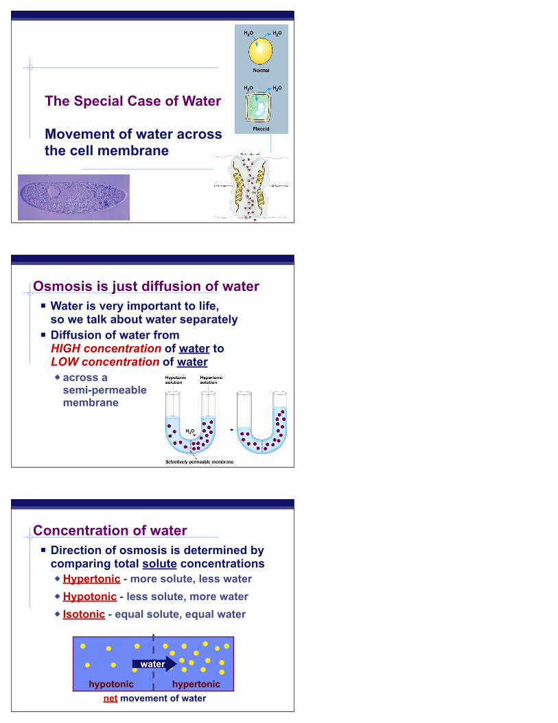
The Special Case of Water
Movement of water across the cell membrane
Osmosis is just diffusion of water Water is very important to life,
so we talk about water separately Diffusion of water from
HIGH concentration of water to LOW concentration of water across a
semi-permeable membrane
Concentration of water Direction of osmosis is determined by
comparing total solute concentrations Hypertonic - more solute, less water Hypotonic - less solute, more water Isotonic - equal solute, equal water
hypotonic hypertonic
water
net movement of water

freshwater balanced saltwater
Managing water balance Cell survival depends on balancing
water uptake & loss
Managing water balance Hypotonic
a cell in fresh water high concentration of water around cell
problem: cell gains water, swells & can burst
example: Paramecium ex: water continually enters
Paramecium cell solution: contractile vacuole
pumps water out of cell ATP
plant cells turgid = full cell wall protects from bursting
freshwater
ATP
1
No problem,here
KABOOM!
Pumping water out Contractile vacuole in Paramecium
ATP

28
Managing water balance Hypertonic
a cell in salt water low concentration of water
around cell problem: cell loses water &
can die example: shellfish solution: take up water or
pump out salt plant cells
plasmolysis = wilt can recover
saltwater
2
I willsurvive!
I’m shrinking,I’m shrinking!
plasmolysis in onion cells
30

Managing water balance Isotonic
animal cell immersed in mild salt solution
no difference in concentration of water between cell & environment problem: none
no net movement of water flows across membrane equally, in
both directions cell in equilibrium volume of cell is stable
example: blood cells in blood plasma slightly salty IV solution in hospital
balanced
3
I couldbe better…
That’sperfect!
Aquaporins Water moves rapidly into & out of cells
evidence that there were water channels protein channels allowing flow of water
across cell membrane
Peter AgreJohn Hopkins
Roderick MacKinnonRockefeller
Cell (compared to beaker) → hypertonic or hypotonicBeaker (compared to cell) → hypertonic or hypotonicWhich way does the water flow? → in or out of cell
.05 M .03 M
Do you understand Osmosis…

Facilitated Diffusion Diffusion through protein channels
channels move specific molecules across cell membrane
no energy needed
“The Bouncer”
open channel = fast transport
facilitated = with help
HIGH
LOW
Active Transport
“The Doorman”
conformational change
Cells may need to move molecules against concentration gradient conformational shape change transports solute
from one side of membrane to other protein “pump” “costs” energy = ATP
ATP
LOW
HIGH
Active Transport
36

symportantiport
Active transport Many models & mechanisms
ATP ATP
Transport summary
simplediffusion
facilitateddiffusion
activetransport
ATP
Getting through cell membrane Passive Transport
Simple diffusion diffusion of nonpolar, hydrophobic molecules
lipids HIGH → LOW concentration gradient
Facilitated transport diffusion of polar, hydrophilic molecules through a protein channel
HIGH → LOW concentration gradient
Active transport diffusion against concentration gradient
LOW → HIGH uses a protein pump requires ATP
ATP

40
How about large molecules? Moving large molecules into & out of cell
through vesicles & vacuoles endocytosis
phagocytosis = “cellular eating” pinocytosis = “cellular drinking”
exocytosis
exocytosis
Endocytosis
phagocytosis
pinocytosis
receptor-mediated endocytosis
fuse with lysosome for digestion
non-specificprocess
triggered bymolecular signal

43
Membranes help join cells to form tissues• Types of intercellular junctions in animals
Tight junctions prevent fluid from moving across a layer of cells
Tight junction
0.5 µm
1 µm
Spacebetweencells Plasma membranes
of adjacent cells
Extracellularmatrix
Gap junction
Tight junctions
0.1 µm
Intermediatefilaments
Desmosome
Gapjunctions
At tight junctions, the membranes ofneighboring cells are very tightly pressedagainst each other, bound together byspecific proteins (purple). Forming continu-ous seals around the cells, tight junctionsprevent leakage of extracellular fluid acrossA layer of epithelial cells.
Desmosomes (also called anchoringjunctions) function like rivets, fastening cellsTogether into strong sheets. IntermediateFilaments made of sturdy keratin proteinsAnchor desmosomes in the cytoplasm.
Gap junctions (also called communicatingjunctions) provide cytoplasmic channels fromone cell to an adjacent cell. Gap junctions consist of special membrane proteins that surround a pore through which ions, sugars,amino acids, and other small molecules maypass. Gap junctions are necessary for commu-nication between cells in many types of tissues,including heart muscle and animal embryos.
TIGHT JUNCTIONS
DESMOSOMES
GAP JUNCTIONS
45

46