diffusion tensor imaging (dti) for the study of disorders of consciousness
TRANSCRIPT
Diffusion Tensor Imaging (DTI) for the study of disorders of consciousness
Stephen Larroque
Coma Science Group, GIGA research
University of Liège
24/03/2017
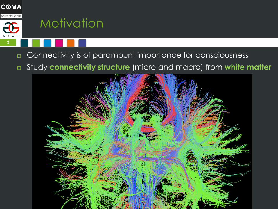
Motivation
Connectivity is of paramount importance for consciousness
Study connectivity structure (micro and macro) from white matter
2
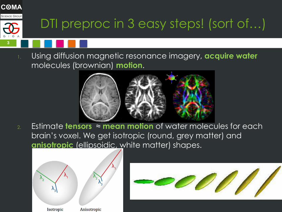
DTI preproc in 3 easy steps! (sort of…)
1. Using diffusion magnetic resonance imagery, acquire water
molecules (brownian) motion.
2. Estimate tensors ≈ mean motion of water molecules for each
brain’s voxel. We get isotropic (round, grey matter) and
anisotropic (ellipsoidic, white matter) shapes.
3
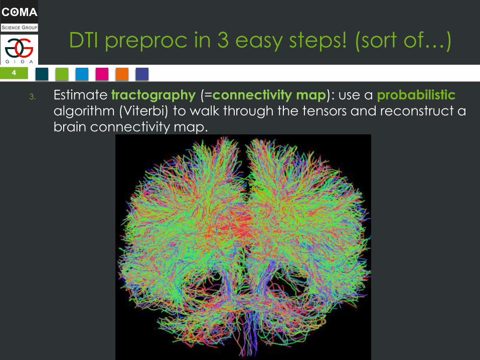
DTI preproc in 3 easy steps! (sort of…)
3. Estimate tractography (=connectivity map): use a probabilistic
algorithm (Viterbi) to walk through the tensors and reconstruct a
brain connectivity map.
4

Result
5
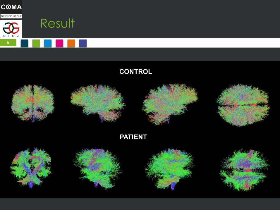
Result
6
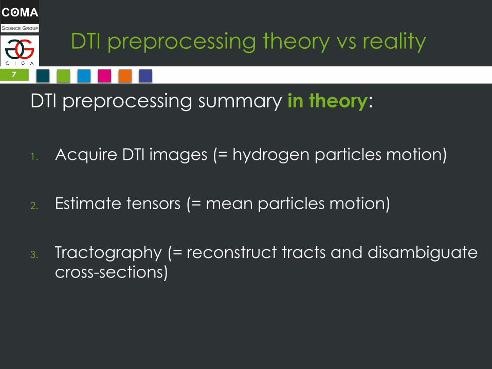
DTI preprocessing theory vs reality
DTI preprocessing summary in theory:
1. Acquire DTI images (= hydrogen particles motion)
2. Estimate tensors (= mean particles motion)
3. Tractography (= reconstruct tracts and disambiguate
cross-sections)
7

DTI preprocessing theory vs reality
DTI preprocessing summary in practice: 1. Acquire DTI images + T1
2. Reorient both
3. Extract gradients (bvecs and bvals)
4. Brain Extraction (BET) mask on DWI and T1
5. Correct eddy currents
6. Estimate tensors & FA metrics
7. Segment T1
8. Coregister DWI on T1
9. Downsample T1
10. Estimate DWI response function
11. Tractography
12. And more steps depending on your objectives…
8
DTI is still in the process of
standardization… but not there yet!

2nd-level analysis (group comparison)
Fixel-based (local metrics) approach:
1. Normalize all subjects on a (tracts) template
2. Compare locally difference of tracts metrics (eg, AFD for density)
Advantage: compare directly the whole structure, but at the expense of
losing info at normalization.
Connectome approach:
1. Parcellation (Freesurfer) to get regions (or use map provided in MRTRIX)
2. Connectivity matrix (tck2connectome)
3. Graph theory measures and comparison
Advantage: respects each subject’s structure and global brain approach,
but lose info at parcellation (your analysis is as good as your
parcellation)
Average/global measures approach:
1. Compute a global measure for each subject (eg, average FA)
2. T-test on the values of one group with the other group
9
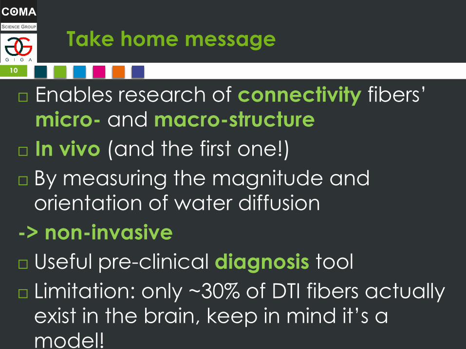
Take home message
Enables research of connectivity fibers’
micro- and macro-structure
In vivo (and the first one!)
By measuring the magnitude and
orientation of water diffusion
-> non-invasive
Useful pre-clinical diagnosis tool
Limitation: only ~30% of DTI fibers actually
exist in the brain, keep in mind it’s a
model!
10
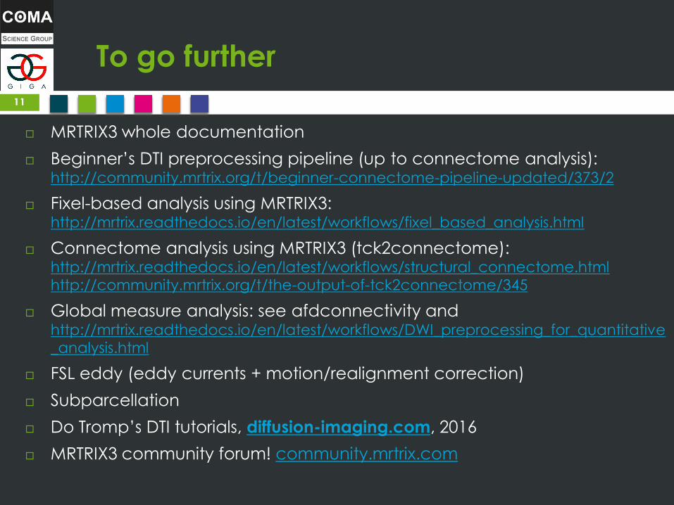
To go further
MRTRIX3 whole documentation
Beginner’s DTI preprocessing pipeline (up to connectome analysis): http://community.mrtrix.org/t/beginner-connectome-pipeline-updated/373/2
Fixel-based analysis using MRTRIX3: http://mrtrix.readthedocs.io/en/latest/workflows/fixel_based_analysis.html
Connectome analysis using MRTRIX3 (tck2connectome): http://mrtrix.readthedocs.io/en/latest/workflows/structural_connectome.html
http://community.mrtrix.org/t/the-output-of-tck2connectome/345
Global measure analysis: see afdconnectivity and http://mrtrix.readthedocs.io/en/latest/workflows/DWI_preprocessing_for_quantitative
_analysis.html
FSL eddy (eddy currents + motion/realignment correction)
Subparcellation
Do Tromp’s DTI tutorials, diffusion-imaging.com, 2016
MRTRIX3 community forum! community.mrtrix.com
11
Thank you for your
attention References:
•Posterior cingulate cortex-related co-activation patterns: a resting state FMRI study in propofol-induced loss of
consciousness, Amico, Enrico, et al, PLoS One 9.6 (2014): e100012.
•Multimodal neuroimaging in patients with disorders of consciousness showing “functional hemispherectomy”, Van
Someren, E. J. W. (2011), Slow Brain Oscillations of Sleep, Resting State and Vigilance: Proceedings of the 26th International
Summer School of Brain Research, Held at the Royal Netherlands Academy of Arts and Sciences, Amsterdam, The
Netherlands, 29 June-2 July, 2010, 193, 323.
• Neural correlates of consciousness in patients who have emerged from a minimally conscious state: a cross-
sectional multimodal imaging study, Carol Di Perri & Mohamed Ali Bahri & Enrico Amico & Aurore Thibaut & Lizette Heine
et al., The Lancet Neurology, 2016
•Do Tromp, http://www.diffusion-imaging.com/, 2016
•Amico et al., Conf Proc IEEE Eng Med Biol Soc. 2015
12

BONUS SLIDES
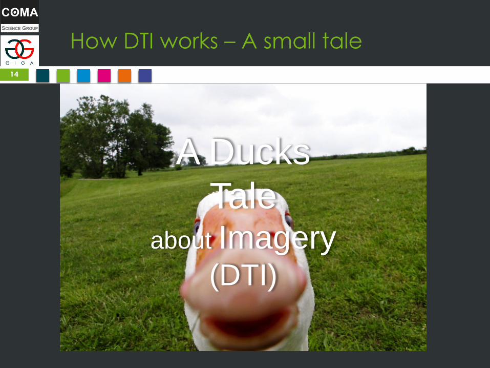
How DTI works – A small tale
14
A Ducks
Tale about Imagery
(DTI)
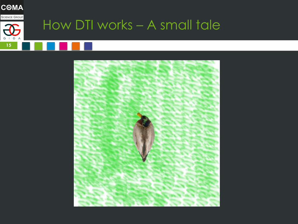
How DTI works – A small tale
15

How DTI works – A small tale
16
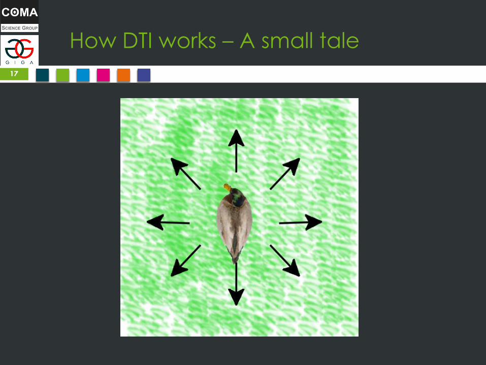
How DTI works – A small tale
17
How DTI works – A small tale
18

How DTI works – A small tale
19

How DTI works – A small tale
20
How DTI works – A small tale
21

How DTI works – A small tale
22
How DTI works – A small tale
23

How DTI works – A small tale
24
How DTI works – A small tale
25
How DTI works – A small tale
26
How DTI works – A small tale
27
How DTI works – A small tale
28
How DTI works – A small tale
29
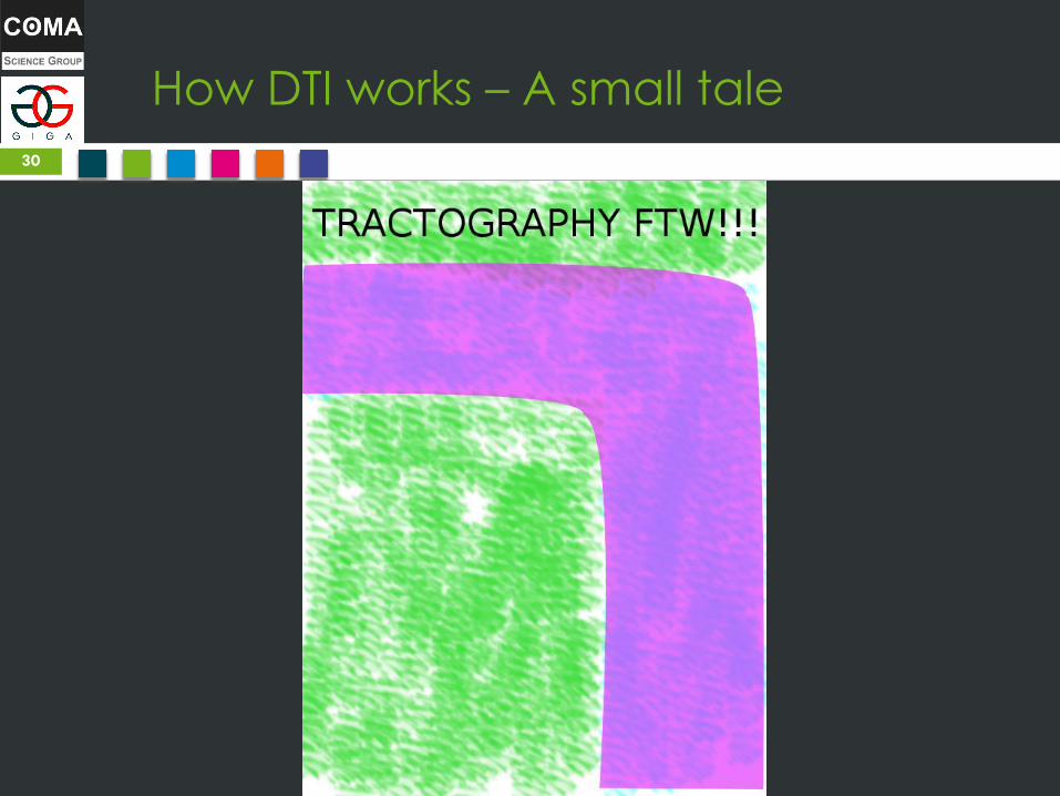
How DTI works – A small tale
30
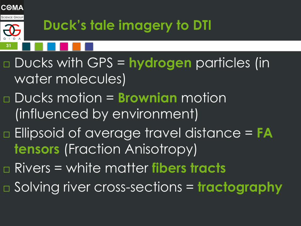
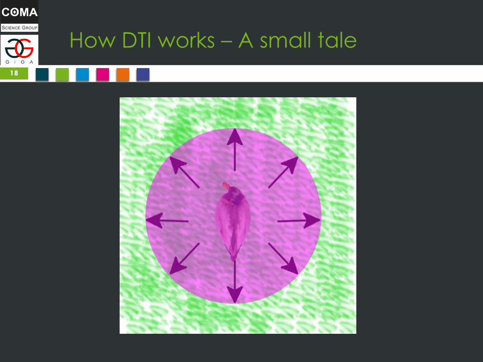
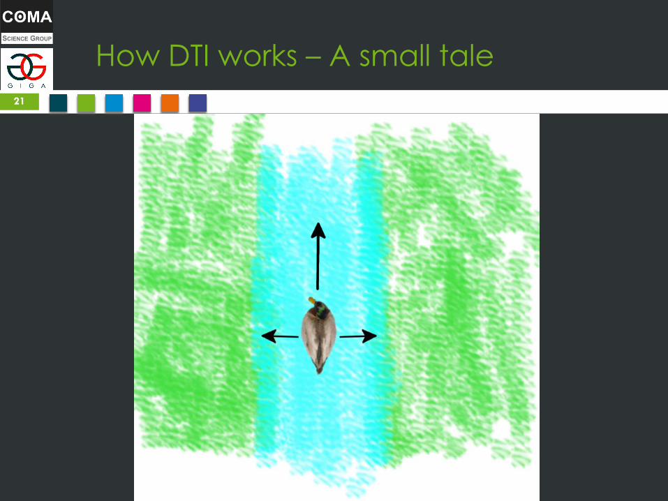
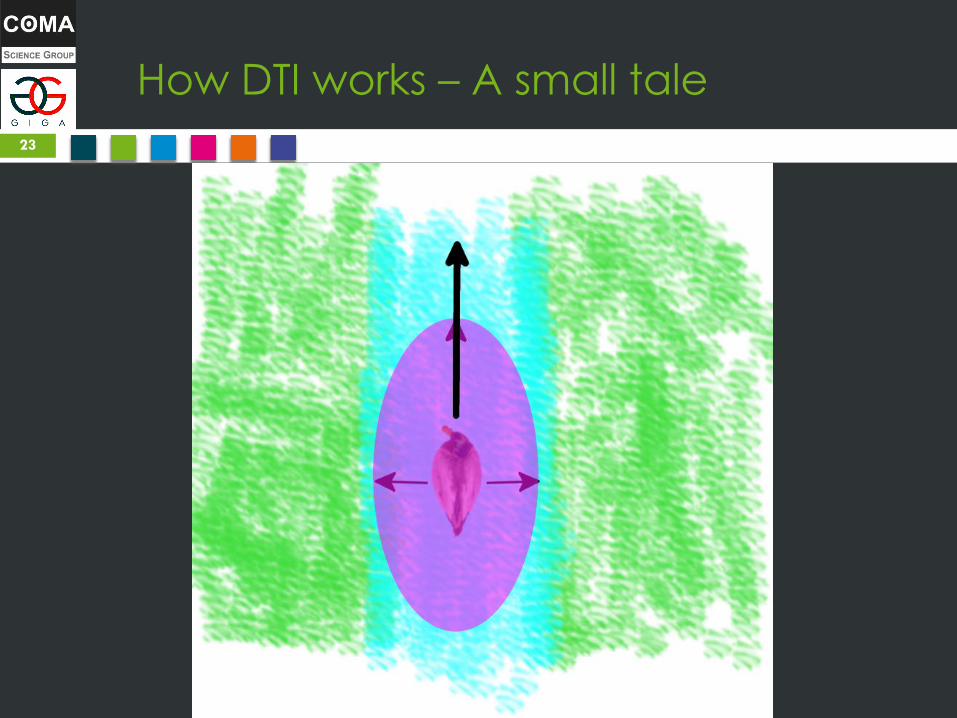
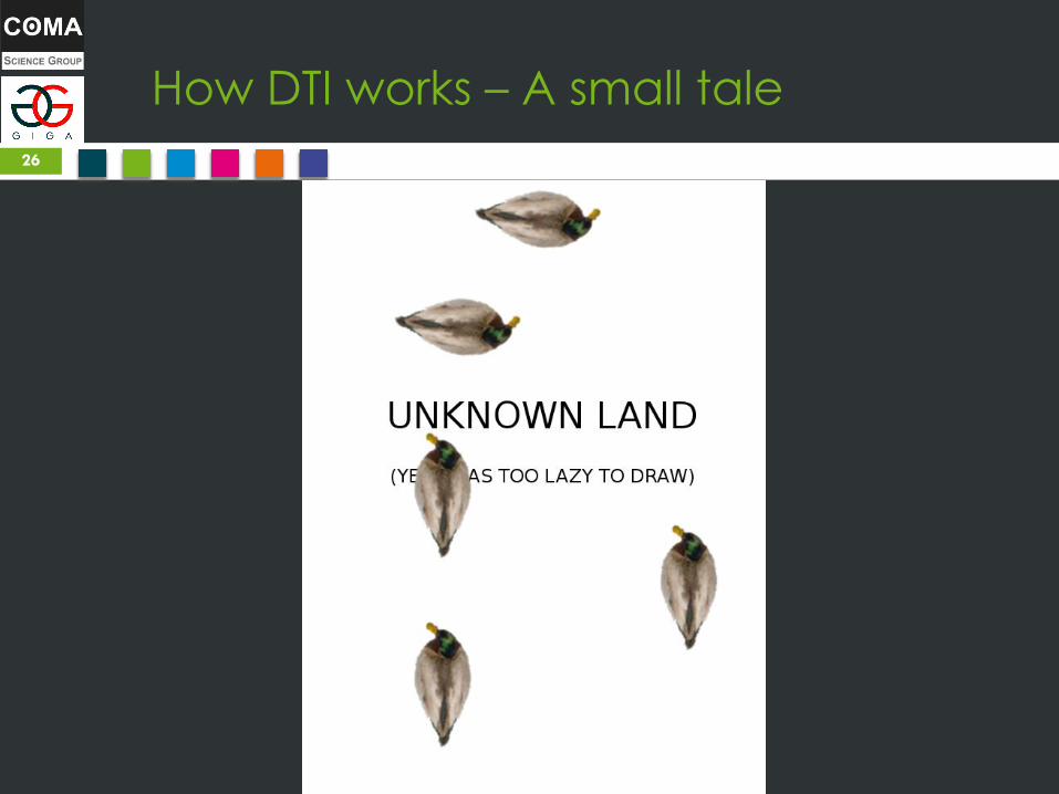
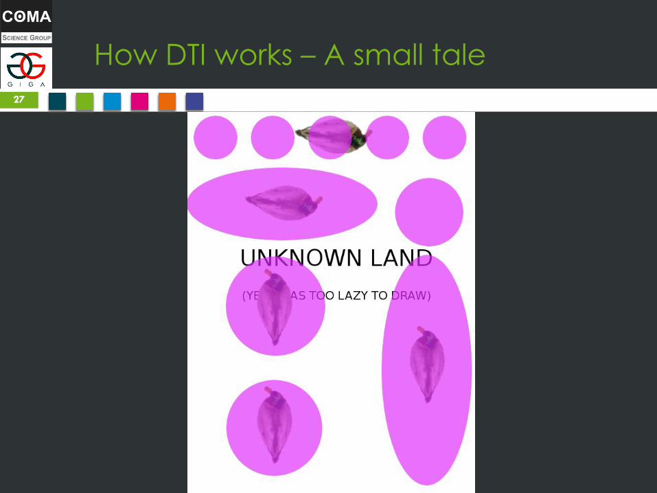
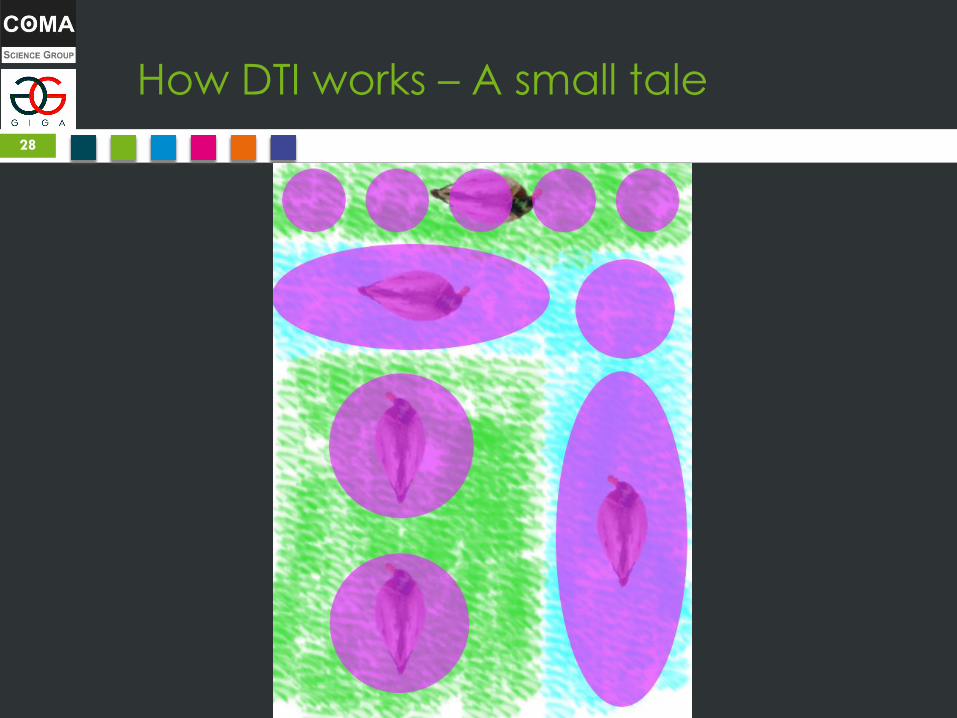
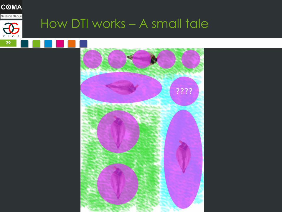
Duck’s tale imagery to DTI
Ducks with GPS = hydrogen particles (in
water molecules)
Ducks motion = Brownian motion
(influenced by environment)
Ellipsoid of average travel distance = FA
tensors (Fraction Anisotropy)
Rivers = white matter fibers tracts
Solving river cross-sections = tractography
31