diffusion tensor imaging of mild tbi - war related … · diffusion tensor imaging of mild ......
TRANSCRIPT

Page
Diffusion Tensor Imaging of Mild Diffusion Tensor Imaging of Mild TBI:TBI:
A Potential Biomarker of A Potential Biomarker of NeurocognitiveNeurocognitiveOutcome?Outcome?
PratikPratik MukherjeeMukherjee, MD , MD PhDPhD

Page
• Conventional Clinical Neuroimaging: current limitations for TBI– CT is still the mainstay of head trauma imaging, but is
grossly insensitive to parenchymal brain injury in mild TBI
– 3T and now 7T MR imaging are growing increasingly sensitive to focal lesions in mild TBI, yet these lesions do not correlate with patient outcome
• Diffusion Tensor Imaging (DTI): a potential biomarker of TBI?– Provides quantitative pathophysiological parameters
(e.g. FA) that correlate with neurocognitive outcome in mild TBI
– In chronic symptomatic mild TBI, provides structure-function correlation that can relate injury in particular
SynopsisSynopsis

Page
• Mild TBI– The great majority of TBI is “mild TBI”, also called
“concussion” or “minor TBI”– Mild TBI is commonly defined as Glasgow Coma
Scale (GCS) 13-15, with loss of consciousness no greater than 30 minutes and post-traumatic amnesia not greater than 24 hrs
– Although most mild TBI victims recover quickly, ~15% suffer persistent post-concussive syndrome(PCS)
– Somatic and emotional complaints in PCS include headache, fatigue, dizziness, insomnia, anxiety, depression, and even seizures
– Impairments in memory and executive attention are the two most common neurocognitive manifestations
Mild TBI is Not Necessarily Mild TBI is Not Necessarily ““MildMild””

Page
• X-ray Computed Tomography (CT)– Detects surgical emergencies (hematoma, mass
effect, etc.)– Detects cortical surface contusions– Depicts the small focal hemorrhages sometimes
associated with axonal shearing injury (“DAI”, “TAI”)
• Magnetic Resonance Imaging (MRI)– Gradient echo T2*-weighted images are more
sensitive than CT for the microhemorrhages of DAI– Fast spin echo T2-weighted and FLAIR images are
more sensitive than CT to contusions and to non-hemorrhagic axonal shearing injuries
Conventional Conventional NeuroimagingNeuroimaging of Mild of Mild TBITBI

Page
Cerebral Contusions: CT Cerebral Contusions: CT vsvs 3T MRI3T MRI
3T T2 3T T2 FLAIRFLAIR
CTCT

Page
3T detects twice as many microhemorrhages as 1.5T on gradient-echo T2*-weighted images
Scheid et al., J Neurotrauma (2007)
CTCT 3T GRE3T GRET2*T2*
MicrohemorrhagesMicrohemorrhages: CT : CT vsvs 3T MRI3T MRI

Page
• 38 mildTBI patients (blunt head trauma) prospectively enrolled
– All with GCS 13-15 in the Emergency Dept.; no prior history of head trauma– All with loss of consciousness; none for more than 30 min.– All with post-traumatic amnesia
» Positive for brain injury• 15/38 (39%) CT scans versus 29/38 (76%) 3T MRI scans
» Positive for hemorrhagic axonal shearing injury• 3/38 (8%) CT scans versus 15/38 (39%) 3T MRI scans
» Positive for non-hemorrhagic axonal shearing injury• 1/38 (3%) CT scans versus 4/38 (11%) 3T MRI scans
» Positive for cerebral contusions• 11/38 (29%) CT scans versus 21/38 (55%) 3T MRI scans
- The difference between CT and 3T MRI in per lesiondetection rates even greater than in per patient detection rates
Mild Traumatic Brain Injury: CT Mild Traumatic Brain Injury: CT vsvs 3T MRI3T MRI
Lee H, Wintermark M, Gean A, Ghajar J, Manley G, Mukherjee P. J Neurotrauma 2008; 25:1049-56

Page
Hemorrhagic Diffuse Axonal InjuryHemorrhagic Diffuse Axonal Injury3T3T 7T7T
veiveinn
veiveinn
PratikPratik MukherjeeMukherjee, MD PhD, MD PhD

Page
Mild Traumatic Brain Injury: Mild Traumatic Brain Injury: Do CT and 3T MR Findings Affect Outcome?Do CT and 3T MR Findings Affect Outcome?
Lee H, Wintermark M, Gean A, Ghajar J, Manley G, Mukherjee P.J Neurotrauma 2008; 25:1049-56

Page
• Acute Mild TBI:– Lack of correlation between 1.0T MR imaging
findings and long-term outcome as determined by neurocognitive tests & functional recoveryHughes et al. Hughes et al. NeuroradiologyNeuroradiology (2004)(2004)
• Chronic TBI (Mild to Severe):– Lack of correlation between microhemorrhages on
3T MR imaging and long-term outcome as determined by the Glasgow Outcome ScaleScheidScheid et al. et al. AJNRAJNR (2003)(2003)
Lack of Utility of Conventional MRI in Lack of Utility of Conventional MRI in Mild TBIMild TBI

Page
• Diffusion Tensor Imaging (Basser et al., 1994)– Is sensitive to microstructural changes within white matter
tracts, which may improve the detection of axonal injuryimprove the detection of axonal injuryArfanakisArfanakis et al. et al. AJNRAJNR (2002) and many other studies(2002) and many other studies
– can localize axonal shearing injury to specific white matter tracts, for structurestructure--function correlationfunction correlationLe et al. Le et al. NeurosurgeryNeurosurgery (2005) and other studies(2005) and other studies
– can provide quantitative quantitative pathophysiologicalpathophysiological informationinformationthat might be useful for determining prognosis and monitoring therapeutic interventions in TBIHuismanHuisman et al. et al. AJNRAJNR (2004) and other studies(2004) and other studies
–– 3T MRI with parallel imaging3T MRI with parallel imaging vastly improves the ability to perform high-resolution, high quality DTI in a clinically feasible scan time
Rationale for DTI in TBIRationale for DTI in TBI

Page
Diffusion Tensor ImagingDiffusion Tensor Imaging
Mukherjee P, et al., AJNR 2008; 29:632-41
Pure water at 37˚C:ADC ~ 3.0 x 10⁻³ mm2/sec
Normal adult brain: (GM & WM)ADC ~ 0.7 x 10⁻³ mm2/sec
in the range b = 0 - 1000 sec/mm2 …
Normal term newborn brain: GM: ADC ~ 1.1 x 10⁻³ mm2/secWM: ADC ~ 1.5 x 10⁻³ mm2/sec
Fractional Anisotropy (FA):0 (spherical) to 1 (linear)

Page
3T MRI3T MRI--DTI of Mild TBIDTI of Mild TBI
•• Conventional 3T MRI sequencesConventional 3T MRI sequences–– FLAIR T2FLAIR T2--weighted:weighted: whole-brain axials @ 3 mm slices (4
min)–– MPGR T2*MPGR T2*--weighted:weighted: whole-brain axials @ 3 mm slices (4
min)–– 3D IR3D IR--FSPGR T1FSPGR T1--weighted:weighted: whole-brain, 1 mm isotropic
(5 min)
• Experimental 3T MRI sequences–– DTI:DTI: 128x128 with FOV 23x23 cm, 72 interleaved slices @
1.8-mmTE=64 ms, TR=14 s, 55 diffusion-encoding directions, b=1000 s/mm2
ASSET parallel imaging with acceleration factor of 2 (13 min)

Page
3 Tesla Diffusion Tensor Imaging (DTI)3 Tesla Diffusion Tensor Imaging (DTI)
centrumsemiovale
superiorlongitudinalfasciculus
cingulum bundle
corpuscallosum,
body
1.8 mm isotropic 1.8 mm isotropic spatial resolutionspatial resolution

Page
1.8 mm isotropic resolution1.8 mm isotropic resolution
optic nerve
pyramidal tract
decussation,superior
cerebellarpeduncle
cingulum
decussation,middle
cerebellarpeduncle
anteriorcommissure
transversepontine fibers
middlecerebellarpeduncle
ILF
SLF
corpuscallosum
3 Tesla Diffusion Tensor Imaging (DTI)3 Tesla Diffusion Tensor Imaging (DTI)

Page
• 34 chronic symptomatic mild TBI patients prospectively enrolled 1-65 months after injury, both in NY & SF
– All with only a single episode of head trauma (predominantly MVAs, assaults, & falls)
– All with no history of chronic medical or neuropsychiatric illness (including drug or EtOHabuse)
– All presented with GCS 13-15 in the Emergency Dept.– All presented with post-traumatic amnesia– All with persistent post-concussive symptoms
• 26 normal volunteers from NY & SF matched for:– age– gender– handedness– years of education
Cornell Cornell -- UCSFUCSF Study: 3T MRIStudy: 3T MRI--DTI of Mild TBIDTI of Mild TBI
Niogi S, Mukherjee P, Ghajar J et al., AJNR 2008; 29:967-73.
Is Extent of Microstructural White Matter Injury Related to Global Cognitive Processing Speed?

Page
3T T2*3T T2*--GRE MRI of Mild TBI: GRE MRI of Mild TBI: No Correlation with Cognitive Processing SpeedNo Correlation with Cognitive Processing Speed
Reaction Time versus # of microbleeds
R = -.08, p=0.701
400
500
600
700
800
900
1000
1100
0 5 10 15 20
# of traumatic microbleeds (conventional MRI)
Mea
n R
T (m
s)
Niogi S, Mukherjee P, Ghajar J et al., AJNR 2008; 29:967-73.

Page
3T DTI of 3T DTI of Mild TBIMild TBI
Niogi S, Mukherjee P, Ghajar J et al., AJNR 2008; 29:967-73.
Page
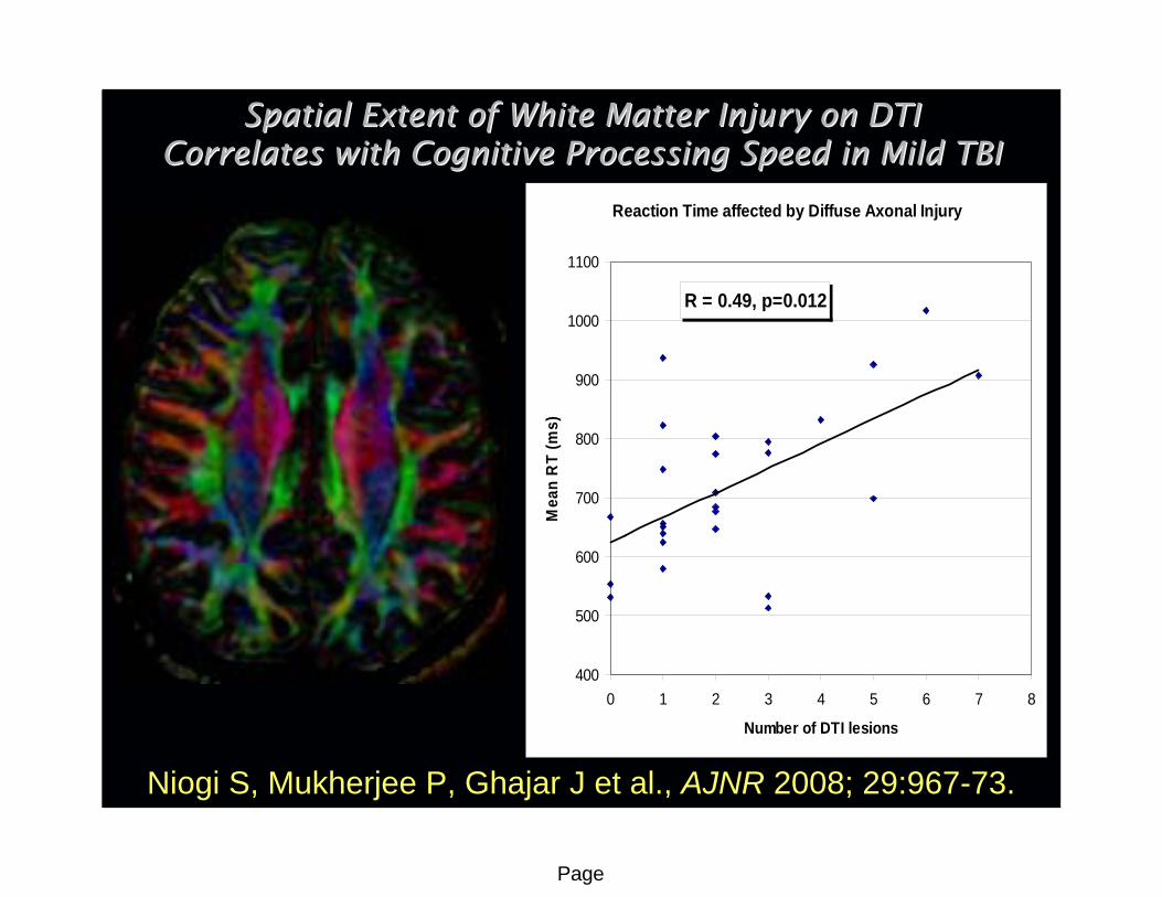
Spatial Extent of White Matter Injury on DTI Spatial Extent of White Matter Injury on DTI Correlates with Cognitive Processing Speed in Mild TBICorrelates with Cognitive Processing Speed in Mild TBI
Reaction Time affected by Diffuse Axonal Injury
R = 0.49, p=0.012
400
500
600
700
800
900
1000
1100
0 1 2 3 4 5 6 7 8
Number of DTI lesions
Mea
n R
T (m
s)
Niogi S, Mukherjee P, Ghajar J et al., AJNR 2008; 29:967-73.

Page
• Microstructural White Matter Injury in Mild TBI– 10 of 11 patients with normal findings on
conventional 3T MR imaging had evidence of reduced FA in one or more WM tracts
– The most frequently injured tracts are large longitudinal fiber bundles anteriorly located in the brain (ACR, UF, genu of CC), farthest from the axis of rotation in rotational TAI
DTI of Mild TBIDTI of Mild TBI
Niogi S, Mukherjee P, Ghajar J et al., AJNR 2008; 29:967-73.

Page
3D Diffusion Tensor Fiber 3D Diffusion Tensor Fiber TractographyTractography
FACT – fiber assignment by continuous tracking in 3D along the primary eigenvector (Mori et al. 1999)
“Streamline Tractography”
dense seeding – multiple seed points within a voxel (Conturoet al. 1999; Mori et al. 1999)
multi-ROI filtering – retain only those tracts passing through start and end ROIs, and other intermediary ROIs (Conturo et al. 1999)
interpolation – step sizes smaller than a voxel (Conturo et al. 1999)

Page
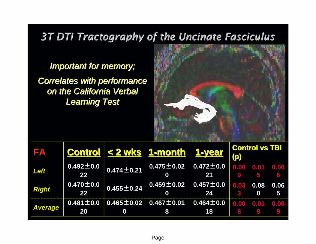
3T DTI 3T DTI TractographyTractography of the of the UncinateUncinateFasciculusFasciculus
Important for memory Important for memory Correlate with performance on the Correlate with performance on the
California Verbal Learning Test (CVLT)California Verbal Learning Test (CVLT)

Page
3T DTI 3T DTI TractographyTractography of the of the CingulumCingulum Bundle and Bundle and Anterior Corona Anterior Corona RadiataRadiata
Important for Important for attentionalattentional control control (focusing on task in the presence of distracters)(focusing on task in the presence of distracters)
Correlate with conflict on the Attention Network Task (ANT)Correlate with conflict on the Attention Network Task (ANT)
““congruentcongruent””
““incongruentincongruent””
Page
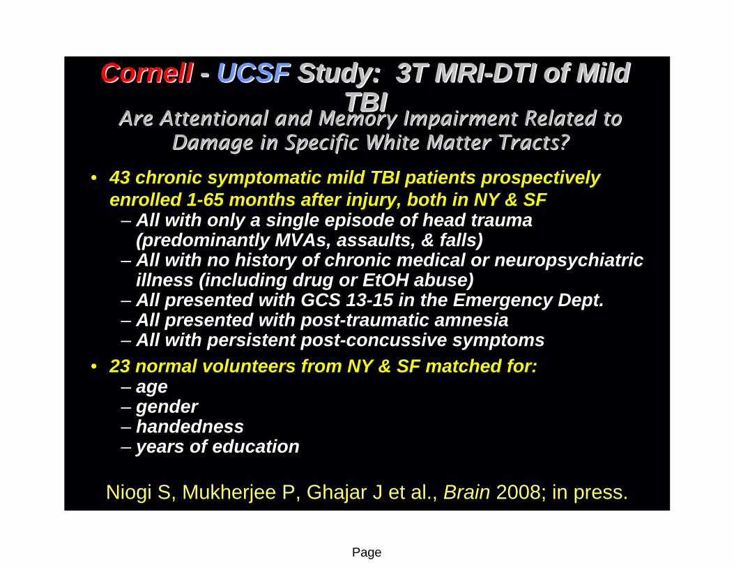
• 43 chronic symptomatic mild TBI patients prospectively enrolled 1-65 months after injury, both in NY & SF
– All with only a single episode of head trauma (predominantly MVAs, assaults, & falls)
– All with no history of chronic medical or neuropsychiatric illness (including drug or EtOH abuse)
– All presented with GCS 13-15 in the Emergency Dept.– All presented with post-traumatic amnesia– All with persistent post-concussive symptoms
• 23 normal volunteers from NY & SF matched for:– age– gender– handedness– years of education
Cornell Cornell -- UCSFUCSF Study: 3T MRIStudy: 3T MRI--DTI of Mild DTI of Mild TBITBI
Niogi S, Mukherjee P, Ghajar J et al., Brain 2008; in press.
Are Are AttentionalAttentional and Memory Impairment Related to and Memory Impairment Related to Damage in Specific White Matter Tracts?Damage in Specific White Matter Tracts?

Page
Bilateral UNC correlates with Memory in Normal Adults
R = 0.52
4
6
8
10
12
14
16
18
0.4 0.45 0.5 0.55 0.6 0.65 0.7
FA of UNC
LDFR
Mem
ory
Sco
re fr
om C
VLT
Left hemisphere ACR correlates with Conflict in Normal Adults
R = -0.42
25
45
65
85
105
125
145
165
185
0.4 0.45 0.5 0.55 0.6
FA of ACR
Con
flict
Sco
re fr
om A
NT
(ms)

Page
Bilateral Uncinate Fasciculus correlates with Memory in mild TBI
R = 0.518
0
2
4
6
8
10
12
14
16
18
0.3 0.4 0.5 0.6 0.7 0.8
FA of UNC
LDFR
Mem
ory
Sco
re fr
om C
VLT
Left hemisphere Anterior Corona Radiata correlates with Conflict in mild TBI
R = -0.47
0
50
100
150
200
250
300
350
0 0.2 0.4 0.6 0.8 1
FA of ACR
Conf
lict S
core
from
ANT
(ms)

Page
• 31 mild TBI patients prospectively enrolled in Emergency Dept.
– All with only a single episode of head trauma (predominantly MVAs, assaults, & falls)
– All with no history of chronic medical or neuropsychiatric illness (including drug or EtOH abuse)
– All presented with GCS 13-15 in the Emergency Dept.– All presented with witnessed loss of consciousness
(LOC)– All presented with post-traumatic amnesia
– Patients scanned serially with 3T MRI and DTI at acute (< 2 wks), 1-month, and 1-year time points after injury
• 19 age-, gender-, & education-matched normal volunteers
UCSFUCSF Prospective Longitudinal Study Prospective Longitudinal Study of Mild TBI with 3T MRIof Mild TBI with 3T MRI--DTIDTI
Page
3T DTI 3T DTI TractographyTractography of the of the UncinateUncinate FasciculusFasciculus
Important for memory; Important for memory;
Correlates with performance Correlates with performance on the California Verbal on the California Verbal
Learning Test Learning Test
FA ControlControl < 2 wks< 2 wks 11--monthmonth 11--yearyear Control Control vsvs TBI TBI (p)(p)
Left0.492±0.0
220.474±0.21 0.475±0.02
00.472±0.0
210.00
90.01
50.00
6
Right0.470±0.0
220.455±0.24 0.459±0.02
00.457±0.0
240.03
30.08
00.06
5
Average0.481±0.0
200.465±0.02
00.467±0.01
80.464±0.0
180.00
80.01
90.00
9

Page
3T DTI 3T DTI TractographyTractography of the of the Inferior Inferior FrontoFronto--Occipital Fasciculus (IFO)Occipital Fasciculus (IFO)
The major tract connecting The major tract connecting the frontal and occipital the frontal and occipital
lobeslobes
FA ControlControl < 2 wks< 2 wks 11--monthmonth 11--yearyear Control Control vsvs TBI TBI (p)(p)
Left0.550±0.0
240.539±0.02
30.543±0.02
40.535±0.0
210.11
10.34
20.04
5
Right0.533±0.0
220.524±0.02
30.521±0.02
30.518±0.0
230.15
80.06
30.03
6
Average0.542±0.0
220.531±0.02
00.532±0.02
10.527±0.0
190.10
60.13
50.02
6

Page
3T DTI 3T DTI TractographyTractography of the of the CingulumCingulum BundleBundle
Important for executive Important for executive attention;attention;
Injury leads to poor conflict Injury leads to poor conflict monitoringmonitoring
FA ControlControl < 2 wks< 2 wks 11--monthmonth 11--yearyear Control Control vsvs TBI TBI (p)(p)
Left0.562±0.0
330.549±0.02
80.553±0.03
60.542±0.0
310.18
60.36
80.05
9
Right0.520±0.0
220.512±0.02
50.515±0.02
90.510±0.0
230.26
30.46
30.14
3
Average0.541±0.0
260.531±0.02
50.534±0.03
10.526±0.0
230.18
00.37
90.06
0
Page
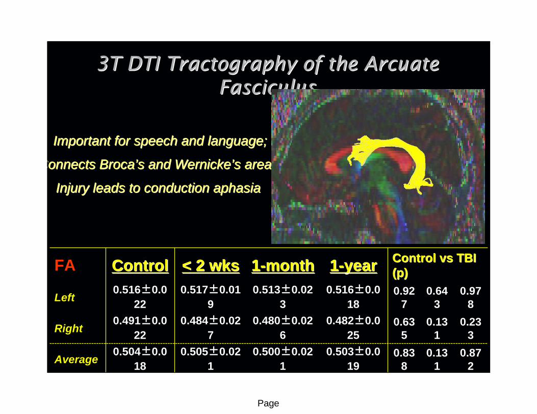
3T DTI 3T DTI TractographyTractography of the of the ArcuateArcuateFasciculusFasciculus
Important for speech and language;Important for speech and language;
Connects Connects BrocaBroca’’ss and and WernickeWernicke’’ss areas;areas;
Injury leads to conduction aphasia Injury leads to conduction aphasia
FA ControlControl < 2 wks< 2 wks 11--monthmonth 11--yearyear Control Control vsvs TBI TBI (p)(p)
Left0.516±0.0
220.517±0.01
90.513±0.02
30.516±0.0
180.92
70.64
30.97
8
Right0.491±0.0
220.484±0.02
70.480±0.02
60.482±0.0
250.63
50.13
10.23
3
Average0.504±0.0
180.505±0.02
10.500±0.02
10.503±0.0
190.83
80.13
10.87
2

Page
3T DTI 3T DTI TractographyTractography of the of the CorticospinalCorticospinal TractTract
The major tract responsible The major tract responsible for motor functionfor motor function
FA ControlControl < 2 wks< 2 wks 11--monthmonth 11--yearyear Control Control vsvs TBI TBI (p)(p)
Left0.587±0.0
280.589±0.02
20.586±0.02
10.581±0.0
240.76
20.94
50.47
9
Right0.573±0.0
230.571±0.02
70.570±0.02
10.561±0.0
220.74
80.72
50.11
9
Average0.580±0.0
240.580±0.02
20.578±0.02
30.571±0.0
221.00
00.82
90.23
9

Page
3T DTI 3T DTI TractographyTractography of the of the GenuGenu of the Corpus of the Corpus CallosumCallosum
The major tract connecting The major tract connecting the left and right frontal the left and right frontal
lobeslobes
ControlControl < 2 wks< 2 wks 11--monthmonth 11--yearyear Control Control vsvs TBI TBI (p)(p)
FA 0.556±0.022
0.542±0.022
0.544±0.025
0.542±0.019
0.038
0.084
0.032

Page
3T DTI 3T DTI TractographyTractography of the of the SpleniumSplenium of the Corpus of the Corpus CallosumCallosum
The major tract connecting The major tract connecting the left and right occipital the left and right occipital
lobeslobes
ControlControl < 2 wks< 2 wks 11--monthmonth 11--yearyear Control Control vsvs TBI TBI (p)(p)
FA 0.669±0.025
0.658±0.027
0.656±0.030
0.653±0.027
0.144
0.094
0.044

Page
• Conventional MR Imaging Techniques– Growing increasingly sensitive to the focal lesions of mild TBI,
especially microbleeds of TAI on T2* GRE or SWI– However, no evidence that focal lesions are relevant to long-term
neurocognitive status or functional recovery in mild TBI• Diffusion Tensor Imaging of Mild TBI
– DTI measures such as FA are correlated with cognitive processing speed, memory, & attention
– Specific (micro)structure-function relationships can be found between particular white matter tracts and their associated neurocognitive domain (UF-memory, ACR-attention)
– Reduced microstructural integrity of specific WM tracts can be detected within 2 wks after mild TBI – prognostic biomarker?
– However, overlap with normal variation may limit utility for clinical diagnosis in individual cases of mild TBI
NeuroimagingNeuroimaging of Mild TBI: of Mild TBI: ConclusionsConclusions

Page
• Diffusion Tensor Imaging (DTI) – structural connectivity– DTI is sensitive in blunt trauma, so might also be sensitive in
blast– Does the distribution of microstructural WM injury differ in
blast?» Blunt TBI: anterior WM tracts (prefrontal connectivity) appear most affected» Blast TBI: are posterior cerebral and posterior fossa tracts most affected?
– What is the relationship between white matter FA and biomechanical susceptibility to blast injury?
» Current limitation of DTI: it is largely unknown what are the biological determinants of DTI parameters such as FA, and what are the pathophysiological changes that cause reduced FA after trauma
– Can DTI be used to help model strain and deformation in the brain due to blast exposure?
DTI DTI –– Application to BlastApplication to Blast--Related TBI?Related TBI?

Page
CornellCornellSumit N. Niogi, PhD
Bruce McCandliss, PhD
UCSF @ SFGHUCSF @ SFGHGeoffrey T. Manley, MD PhD
Alisa Gean, MDHana Lee
Michele Meeker, RN
Supported by the a collaborative grant from the James S. McDonnell Foundation
administered through the Brain Trauma Foundation
(Cornell, UC Berkeley, UCSF)
UCSF RadiologyUCSF RadiologyJoshua Ng
Srivathsa VeeraraghavanDaniel B. Vigneron, PhD
Michael WahlDuan Xu, PhD
Brain Trauma FoundationBrain Trauma FoundationJam Ghajar, MD PhD
AcknowledgementsAcknowledgements