digital breast tomosynthesis: lessons learned from early clinical implementation
TRANSCRIPT
Note: This copy is for your personal non-commercial use only. To order presentation-ready copies for distribution to your colleagues or clients, contact us at www.rsna.org/rsnarights. B
REA
ST IM
AG
ING
E89
Digital Breast Tomosynthesis: Lessons Learned from Early Clinical Implementation1
The limitations of mammography are well known and are partly related to the fact that with conventional imaging, the three-di-mensional volume of the breast is imaged and presented in a two-dimensional format. Because normal breast tissue is similar in x-ray attenuation to some breast cancers, clinically relevant malignancies may be obscured by normal overlapping tissue. In addition, com-plex areas of normal tissue may be perceived as suspicious. The lim-itations of two-dimensional breast imaging lead to low sensitivity in detecting some cancers and high false-positive recall rates. Although mammographic screening has been shown to reduce breast cancer deaths by approximately 30%, controversy exists over when and how often screening mammography should occur. Digital breast tomosynthesis (DBT) is rapidly being implemented in breast imag-ing clinics around the world as early clinical data demonstrate that it may address some of the limitations of conventional mammog-raphy. With DBT, multiple low-dose x-ray images are acquired in an arc and reconstructed to create a three-dimensional image, thus minimizing the impact of overlapping breast tissue and improv-ing lesion conspicuity. Early studies of screening DBT have shown decreased false-positive callback rates and increased rates of cancer detection (particularly for invasive cancers), resulting in increased sensitivity and specificity. In our clinical practice, we have com-pleted more than 2 years of using two-view digital mammography combined with two-view DBT for all screening and select diagnos-tic imaging examinations (over 25,000 patients). Our experience, combined with previously published data, demonstrates that the combined use of DBT and digital mammography is associated with improved outcomes for screening and diagnostic imaging. Online supplemental material is available for this article.©RSNA, 2014 • radiographics.rsna.org
Robyn Gartner Roth, MD Andrew D. A. Maidment, PhD Susan P. Weinstein, MD Susan Orel Roth, MD Emily F. Conant, MD
Abbreviations: DBT = digital breast tomo-synthesis, DCIS = ductal carcinoma in situ, DM = digital mammography, FDA = Food and Drug Administration, IDC = invasive duc-tal carcinoma, MLO = mediolateral oblique, OR = odds ratio, 3D = three-dimensional, 2D = two-dimensional
RadioGraphics 2014; 34:E89–E102
Published online 10.1148/rg.344130087
Content Codes: 1From the Department of Breast Imaging, Department of Radiology, Hospital of the University of Pennsylvania, 3400 Spruce St, 1 Silverstein, Philadelphia, PA 19104. Re-cipient of a Certificate of Merit award for an education exhibit at the 2012 RSNA Annual Meeting. Received July 10, 2013; revision re-quested August 14 and received December 17; accepted February 17, 2014. For this journal-based SA-CME activity, the authors A.D.A.M. and E.F.C. have disclosed financial relation-ships (see p E101); the other authors, editor, and reviewers have no relevant relationships to disclose. Address correspondence to R.G.R. (e-mail: [email protected]).
Funding: The research was supported by the National Institutes of Health [Penn Center for Innovation in Personalized Breast Screening grant number U54CA163313].
See discussion on this article by Feig (pp E103–E105).
After completing this journal-based SA-CME activity, participants will be able to: ■ Describe the principles of DBT and
how information obtained at 3D DBT may replace the need for some 2D di-agnostic imaging in the evaluation of suspicious breast lesions.
■ Discuss how a combination of DM and DBT can be used to decrease call-back rates, increase cancer detection, and assist with problem solving.
■ Identify the limitations of DBT and issues to consider in clinical implemen-tation.
See www.rsna.org/education/search/RG.
SA-CME LEARNING OBJECTIVES
E90 July-August 2014 radiographics.rsna.org
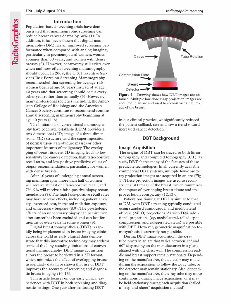
Figure 1. Drawing shows how DBT images are ob-tained. Multiple low-dose x-ray projection images are acquired in an arc and used to reconstruct a 3D im-age of the breast.
in our clinical practice, we significantly reduced the patient callback rate and saw a trend toward increased cancer detection.
DBT Background
Image AcquisitionThe origins of DBT can be traced to both linear tomography and computed tomography (CT); as such, DBT shares many of the features of these predicate technologies. In all currently available commercial DBT systems, multiple low-dose x-ray projection images are acquired in an arc (Fig 1). These projection images are used to recon-struct a 3D image of the breast, which minimizes the impact of overlapping breast tissue and im-proves lesion conspicuity (12,13).
Patient positioning at DBT is similar to that at DM, with DBT screening typically conducted using standard craniocaudal and mediolateral oblique (MLO) projections. As with DM, addi-tional projections (eg, mediolateral, rolled, spot compression, and exaggerated views) are possible with DBT. However, geometric magnification to-mosynthesis is currently not possible.
During DBT image acquisition, the x-ray tube pivots in an arc that varies between 15° and 60° (depending on the manufacturer) in a plane aligned with the chest wall. The compression pad-dle and breast support remain stationary. Depend-ing on the manufacturer, the detector may rotate during the acquisition to follow the x-ray tube, or the detector may remain stationary. Also, depend-ing on the manufacturer, the x-ray tube may move continuously during image acquisition, or it may be held stationary during each acquisition (called a “step-and-shoot” acquisition method).
IntroductionPopulation-based screening trials have dem-onstrated that mammographic screening can reduce breast cancer deaths by 30% (1). In addition, it has been shown that digital mam-mography (DM) has an improved screening per-formance when compared with analog imaging, particularly in premenopausal women, women younger than 50 years, and women with dense breasts (2). However, controversy still exists over when and how often screening mammography should occur. In 2009, the U.S. Preventive Ser-vices Task Force on Screening Mammography recommended that screening for average-risk women begin at age 50 years instead of at age 40 years and that screening should occur every other year rather than annually (3). However, many professional societies, including the Amer-ican College of Radiology and the American Cancer Society, continue to recommend routine annual screening mammography beginning at age 40 years (4–6).
The limitations of conventional mammogra-phy have been well established. DM provides a two-dimensional (2D) image of a three-dimen-sional (3D) structure, and the superimposition of normal tissue can obscure masses or other important features of malignancy. The overlap-ping of breast tissue at 2D imaging leads to low sensitivity for cancer detection, high false-positive recall rates, and low positive predictive values of biopsy recommendations, particularly for women with dense breasts.
After 10 years of undergoing annual screen-ing mammography, more than half of women will receive at least one false-positive recall, and 7%–9% will receive a false-positive biopsy recom-mendation (7). The high false-positive recall rates may have adverse effects, including patient anxi-ety, increased cost, increased radiation exposure, and unnecessary biopsies (8,9). The psychologic effects of an unnecessary biopsy can persist even after cancer has been excluded and can last for months or even years in some women (9).
Digital breast tomosynthesis (DBT) is rap-idly being implemented in breast imaging clinics across the world as early clinical data demon-strate that this innovative technology may address some of the long-standing limitations of conven-tional mammography. DBT image acquisition allows the breast to be viewed in a 3D format, which minimizes the effect of overlapping breast tissue. Early data have shown that use of DBT improves the accuracy of screening and diagnos-tic breast imaging (10–13).
This article focuses on our early clinical ex-periences with DBT in both screening and diag-nostic settings. One year after instituting DBT
RG • Volume 34 Number 4 Roth et al E91
of the object. Thus, small objects such as micro-calcifications and fibrils rapidly blur out of focus as the reader scrolls through the stack of images. Small high-contrast structures, such as surgi-cal clips, can cause readily identifiable artifacts outside the plane of focus. In comparison, large objects, such as large regions of glandularity, will influence many sections distant from the glandu-lar tissue—even influencing the gray level of the skin. Fortunately, these artifactual densities are featureless because no internal structures are pre-served outside the plane of focus, and thus they have no clinical importance.
DBT Image SetThe Selenia Dimensions combined DBT and DM image set consists of three image series: a conventional 2D mammogram, source projec-tion images, and multiple reconstructed images presented as the DBT image stack. The various images are coregistered, which allows the reader to toggle between image sets for problem solving. The 15 source projection images can be viewed sequentially, giving the reader the perception that the breast is being rotated; this has value in the assessment of gross patient motion.
Reconstructed DBT images, spaced in 1-mm increments, can be displayed either in cine mode or individually and scrolled through manually by the reader. The first image of the stack is the re-constructed section from either side of the breast (medial or lateral for the MLO view; cranial or caudal for the craniocaudal view). Therefore, as a lesion is identified by the reader, the 3D local-ization within the breast may be inferred, which helps to triangulate the lesion. This is particularly important when a lesion is seen only at DBT or only on one view. By creating 3D images of the breast, DBT can overcome many of the chal-lenges of DM by minimizing the effects of over-lapping breast tissue and improving lesion detec-tion and localization.
Three-dimensional DBT images are recon-structed from the projection images. There are various methods for reconstruction, including fil-tered back-projection and iterative techniques. In all cases, a 3D image of the breast is created that consists of multiple 2D sections aligned parallel to the breast support; the planes are separated by a fixed increment (typically 1 mm).
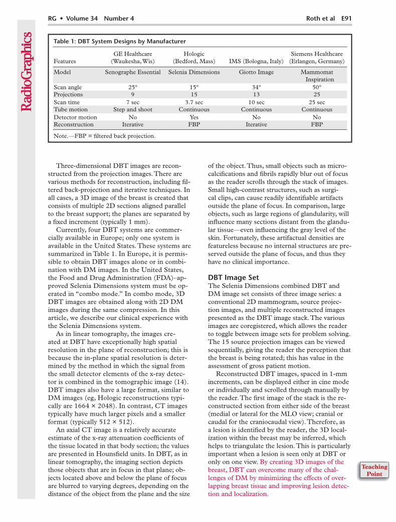
Currently, four DBT systems are commer-cially available in Europe; only one system is available in the United States. These systems are summarized in Table 1. In Europe, it is permis-sible to obtain DBT images alone or in combi-nation with DM images. In the United States, the Food and Drug Administration (FDA)–ap-proved Selenia Dimensions system must be op-erated in “combo mode.” In combo mode, 3D DBT images are obtained along with 2D DM images during the same compression. In this article, we describe our clinical experience with the Selenia Dimensions system.
As in linear tomography, the images cre-ated at DBT have exceptionally high spatial resolution in the plane of reconstruction; this is because the in-plane spatial resolution is deter-mined by the method in which the signal from the small detector elements of the x-ray detec-tor is combined in the tomographic image (14). DBT images also have a large format, similar to DM images (eg, Hologic reconstructions typi-cally are 1664 × 2048). In contrast, CT images typically have much larger pixels and a smaller format (typically 512 × 512).
An axial CT image is a relatively accurate estimate of the x-ray attenuation coefficients of the tissue located in that body section; the values are presented in Hounsfield units. In DBT, as in linear tomography, the imaging section depicts those objects that are in focus in that plane; ob-jects located above and below the plane of focus are blurred to varying degrees, depending on the distance of the object from the plane and the size
Table 1: DBT System Designs by Manufacturer
FeaturesGE Healthcare
(Waukesha, Wis)Hologic
(Bedford, Mass) IMS (Bologna, Italy)Siemens Healthcare
(Erlangen, Germany)
Model Senographe Essential Selenia Dimensions Giotto Image Mammomat Inspiration
Scan angle 25° 15° 34° 50°Projections 9 15 13 25Scan time 7 sec 3.7 sec 10 sec 25 secTube motion Step and shoot Continuous Continuous ContinuousDetector motion No Yes No NoReconstruction Iterative FBP Iterative FBP
Note.—FBP = filtered back projection.
E92 July-August 2014 radiographics.rsna.org
The reader can also display “slab” images, which are maximum intensity projection render-ings of multiple sections. Both the center and thickness of the slab can be varied. Slab views have particular value for viewing calcification clusters; for example, if calcifications are seen on multiple consecutive 1-mm-thick reconstructed sections, the reader can change the thickness of the reconstruction, allowing the entire cluster of calcifications to be viewed in a single slab.
Outcomes with DBTSome early studies have shown improved accuracy in both screening and diagnostic settings with the addition of DBT (10–13). Reductions in false-positive callback rates that range from 6% to 67% have been reported, with stable or slightly increased cancer detection rates (12,13,15,16). Studies have also demonstrated improved sensitivity and specific-ity for breast cancer detection with use of combined DBT and DM versus 2D DM (12,13,17–21).
Interval analysis of 12,631 women in the pro-spective Oslo Tomosynthesis Screening Trial (com-bined DBT and DM vs DM) has shown a 27% increase in the cancer detection rate (P = .001), including a 40% increase in the detection rate for invasive breast cancer across all breast densities (P < .001). The trial has also demonstrated a 15% reduction in the false-positive callback rate (P < .001) (13). Early results from prospective trials in the United States have shown reduced callback rates and increased rates of cancer detection with use of combined DBT and DM (22–24), findings that begin to address the major concerns regarding conventional screening mammography.
Our early clinical experience supports these published data. The following sections highlight lessons learned from early clinical implementation.
Impact of DBT on Our Screening Population
We began screening with DBT (using the Selenia Dimensions system) in September 2011. An in-terim data analysis was performed 1 year after the
introduction of DBT. A total of 11,115 patients were screened by using the bilateral two-view combo mode (DM and DBT images acquired in a single compression). Outcome measures were compared with those from the prior year, when 10,751 patients were screened by using DM alone. Callback rates and cancer detection rates were compared for both years. The readers re-mained stable during the 2-year period.
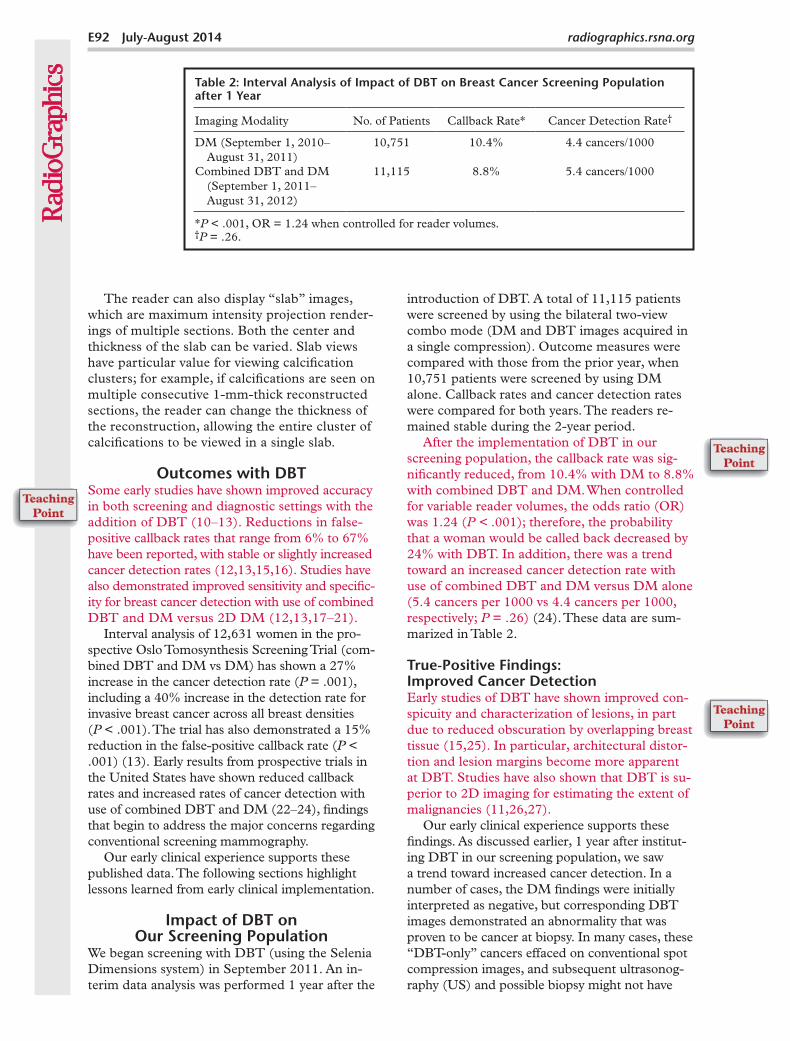
After the implementation of DBT in our screening population, the callback rate was sig-nificantly reduced, from 10.4% with DM to 8.8% with combined DBT and DM. When controlled for variable reader volumes, the odds ratio (OR) was 1.24 (P < .001); therefore, the probability that a woman would be called back decreased by 24% with DBT. In addition, there was a trend toward an increased cancer detection rate with use of combined DBT and DM versus DM alone (5.4 cancers per 1000 vs 4.4 cancers per 1000, respectively; P = .26) (24). These data are sum-marized in Table 2.
True-Positive Findings: Improved Cancer DetectionEarly studies of DBT have shown improved con-spicuity and characterization of lesions, in part due to reduced obscuration by overlapping breast tissue (15,25). In particular, architectural distor-tion and lesion margins become more apparent at DBT. Studies have also shown that DBT is su-perior to 2D imaging for estimating the extent of malignancies (11,26,27).
Our early clinical experience supports these findings. As discussed earlier, 1 year after institut-ing DBT in our screening population, we saw a trend toward increased cancer detection. In a number of cases, the DM findings were initially interpreted as negative, but corresponding DBT images demonstrated an abnormality that was proven to be cancer at biopsy. In many cases, these “DBT-only” cancers effaced on conventional spot compression images, and subsequent ultrasonog-raphy (US) and possible biopsy might not have
Table 2: Interval Analysis of Impact of DBT on Breast Cancer Screening Population after 1 Year
Imaging Modality No. of Patients Callback Rate* Cancer Detection Rate†
DM (September 1, 2010– August 31, 2011)
10,751 10.4% 4.4 cancers/1000
Combined DBT and DM (September 1, 2011– August 31, 2012)
11,115 8.8% 5.4 cancers/1000
*P < .001, OR = 1.24 when controlled for reader volumes.†P = .26.

RG • Volume 34 Number 4 Roth et al E93
Figure 2. Invasive lobular carcinoma in a 68-year-old woman. (a, b) Findings on bilateral MLO (a) and craniocaudal (b) 2D screening mammograms were initially interpreted as negative. Corresponding DBT images (Movies 1, 2) show architectural distortion and an irregular mass in the upper outer right breast, findings not seen at DM. (c) US image of the right breast shows an irregular mass at the 10-o’clock posi-tion, 5 cm from the nipple. (d) Sagittal contrast-enhanced fat-saturated T1-weighted magnetic resonance (MR) subtraction image of the right breast helps confirm the DBT findings. Pathologic analysis demon-strated invasive lobular carcinoma.
been performed if DBT had not been included in the screening study (Fig 2; Movies 1, 2).
Figure 3 depicts a DBT-only cancer that was de-tected in a 72-year-old woman at screening mam-mography. Findings on bilateral craniocaudal DM images were initially interpreted as negative; how-ever, corresponding DBT images showed a spicu-lated mass in the medial right breast (Movie 3). US of the right breast was performed on the basis of the DBT findings and depicted an irregular hypoechoic mass with shadowing, a finding that corresponded
to the abnormality seen at DBT. Pathologic analysis of the core biopsy sample revealed IDC.
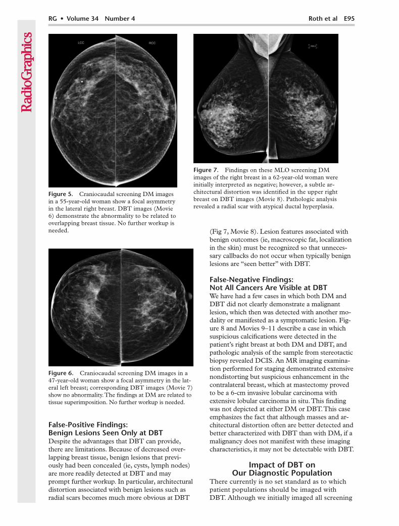
Figure 4 is another example of a DBT-only cancer that was detected at screening mammog-raphy. Bilateral MLO and craniocaudal DM im-ages showed subtle architectural distortion in the lateral subareolar right breast, a finding that was more conspicuous on DBT images. The extensive nature of the abnormal distortion was appreci-ated at DBT (Movies 4, 5). MR images depicted extensive abnormal nonmasslike enhancement in
E94 July-August 2014 radiographics.rsna.org
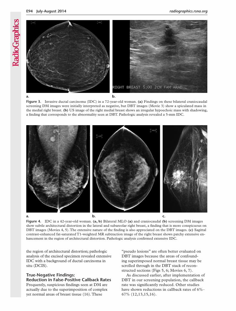
Figure 4. IDC in a 42-year-old woman. (a, b) Bilateral MLO (a) and craniocaudal (b) screening DM images show subtle architectural distortion in the lateral and subareolar right breast, a finding that is more conspicuous on DBT images (Movies 4, 5). The extensive nature of the finding is also appreciated on the DBT images. (c) Sagittal contrast-enhanced fat-saturated T1-weighted MR subtraction image of the right breast shows patchy extensive en-hancement in the region of architectural distortion. Pathologic analysis confirmed extensive IDC.
Figure 3. Invasive ductal carcinoma (IDC) in a 72-year-old woman. (a) Findings on these bilateral craniocaudal screening DM images were initially interpreted as negative, but DBT images (Movie 3) show a spiculated mass in the medial right breast. (b) US image of the right medial breast shows an irregular hypoechoic mass with shadowing, a finding that corresponds to the abnormality seen at DBT. Pathologic analysis revealed a 5-mm IDC.
the region of architectural distortion; pathologic analysis of the excised specimen revealed extensive IDC with a background of ductal carcinoma in situ (DCIS).
True-Negative Findings: Reduction in False-Positive Callback RatesFrequently, suspicious findings seen at DM are actually due to the superimposition of complex yet normal areas of breast tissue (16). These
“pseudo lesions” are often better evaluated on DBT images because the areas of confound-ing superimposed normal breast tissue may be scrolled through in the DBT stack of recon-structed sections (Figs 5, 6; Movies 6, 7).
As discussed earlier, after implementation of DBT in our screening population, the callback rate was significantly reduced. Other studies have shown reductions in callback rates of 6%–67% (12,13,15,16).
RG • Volume 34 Number 4 Roth et al E95
Figure 6. Craniocaudal screening DM images in a 47-year-old woman show a focal asymmetry in the lat-eral left breast; corresponding DBT images (Movie 7) show no abnormality. The findings at DM are related to tissue superimposition. No further workup is needed.
Figure 7. Findings on these MLO screening DM images of the right breast in a 62-year-old woman were initially interpreted as negative; however, a subtle ar-chitectural distortion was identified in the upper right breast on DBT images (Movie 8). Pathologic analysis revealed a radial scar with atypical ductal hyperplasia.
Figure 5. Craniocaudal screening DM images in a 55-year-old woman show a focal asymmetry in the lateral right breast. DBT images (Movie 6) demonstrate the abnormality to be related to overlapping breast tissue. No further workup is needed.
False-Positive Findings: Benign Lesions Seen Only at DBTDespite the advantages that DBT can provide, there are limitations. Because of decreased over-lapping breast tissue, benign lesions that previ-ously had been concealed (ie, cysts, lymph nodes) are more readily detected at DBT and may prompt further workup. In particular, architectural distortion associated with benign lesions such as radial scars becomes much more obvious at DBT
(Fig 7, Movie 8). Lesion features associated with benign outcomes (ie, macroscopic fat, localization in the skin) must be recognized so that unneces-sary callbacks do not occur when typically benign lesions are “seen better” with DBT.
False-Negative Findings: Not All Cancers Are Visible at DBTWe have had a few cases in which both DM and DBT did not clearly demonstrate a malignant lesion, which then was detected with another mo-dality or manifested as a symptomatic lesion. Fig-ure 8 and Movies 9–11 describe a case in which suspicious calcifications were detected in the patient’s right breast at both DM and DBT, and pathologic analysis of the sample from stereotactic biopsy revealed DCIS. An MR imaging examina-tion performed for staging demonstrated extensive nondistorting but suspicious enhancement in the contralateral breast, which at mastectomy proved to be a 6-cm invasive lobular carcinoma with extensive lobular carcinoma in situ. This finding was not depicted at either DM or DBT. This case emphasizes the fact that although masses and ar-chitectural distortion often are better detected and better characterized with DBT than with DM, if a malignancy does not manifest with these imaging characteristics, it may not be detectable with DBT.
Impact of DBT on Our Diagnostic Population
There currently is no set standard as to which patient populations should be imaged with DBT. Although we initially imaged all screening

E96 July-August 2014 radiographics.rsna.org
Figure 8. DCIS and invasive lobular cancer in a 45-year-old woman. (a–c) Suspicious calcifications are seen in the upper outer right breast on craniocaudal (a) and MLO (b) screening DM images. The calcifications are also seen on a zoomed-in DM image (c). DBT images show calcifications in the right breast (Movies 9, 10) and also depict a cyst in the left breast, which otherwise is normal (Movie 11). Pathologic analysis revealed a 2-cm DCIS in the right breast. (d, e) Sagittal contrast-enhanced fat-saturated T1-weighted MR subtraction images of the right (d) and left (e) breasts obtained for staging show enhancement of the known DCIS in the right breast and extensive suspicious enhancement in the left breast, a finding that was originally interpreted as negative on the DM and DBT images. Pathologic analysis showed a 6-cm invasive lobular cancer in the left breast, with extensive lobular carcinoma in situ.
patients with DBT, we have expanded to include all breast cancer survivors, to aid with detection of new or recurrent tumors. We also now use DBT to assist with diagnostic problem solving, in addition to using standard spot compression and magnification views at 2D DM.
Early studies have shown similar or improved performance of DBT for analyzing lesion mar-gins compared with use of standard 2D diag-nostic DM, which suggests that standard 2D diagnostic DM could possibly be replaced with DBT (25,28–31). Additionally, the total radia-
tion dose from combo-mode DBT screening could ultimately prove to be less than the total radiation dose for a patient imaged with only DM who is recalled for multiple additional DM views (29). The ability to problem solve with DBT images may expedite patient workup by alleviating the need for additional diagnostic images, thus minimizing patient anxiety associ-ated with a callback and leading to a potentially decreased overall radiation dose.
These findings indicate that DBT will im-prove the predictive value and diagnostic yield
RG • Volume 34 Number 4 Roth et al E97
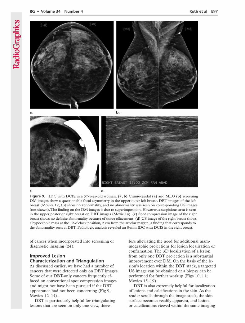
Figure 9. IDC with DCIS in a 57-year-old woman. (a, b) Craniocaudal (a) and MLO (b) screening DM images show a questionable focal asymmetry in the upper outer left breast. DBT images of the left breast (Movies 12, 13) show no abnormality, and no abnormality was seen on corresponding US images (not shown). The finding on the DM images is due to superimposition. However, a suspicious area is seen in the upper posterior right breast on DBT images (Movie 14). (c) Spot compression image of the right breast shows no definite abnormality because of tissue effacement. (d) US image of the right breast shows a hypoechoic mass at the 12-o’clock position, 2 cm from the areolar margin, a finding that corresponds to the abnormality seen at DBT. Pathologic analysis revealed an 8-mm IDC with DCIS in the right breast.
of cancer when incorporated into screening or diagnostic imaging (24).
Improved Lesion Characterization and TriangulationAs discussed earlier, we have had a number of cancers that were detected only on DBT images. Some of our DBT-only cancers frequently ef-faced on conventional spot compression images and might not have been pursued if the DBT appearance had not been concerning (Fig 9, Movies 12–14).
DBT is particularly helpful for triangulating lesions that are seen on only one view, there-
fore alleviating the need for additional mam-mographic projections for lesion localization or confirmation. The 3D localization of a lesion from only one DBT projection is a substantial improvement over DM. On the basis of the le-sion’s location within the DBT stack, a targeted US image can be obtained or a biopsy can be performed for further workup (Figs 10, 11; Movies 15–19).
DBT is also extremely helpful for localization of lesions and calcifications in the skin. As the reader scrolls through the image stack, the skin surface becomes readily apparent, and lesions or calcifications viewed within the same imaging

E98 July-August 2014 radiographics.rsna.org
Figure 10. IDC in a 77-year-old woman. (a, b) Findings on bilateral craniocaudal (a) and MLO (b) screening DM images were initially interpreted as normal. On cranio-caudal DBT images, a subtle area of archi-tectural distortion is seen in the lateral right breast, midway through the stack (Movie 15), and is triangulated to the mid breast on MLO and mediolateral DBT images (Mov-ies 16, 17). (c, d) On spot compression im-ages of the right breast, the lesion is effaced and not readily visible. (e) Targeted US im-age of the right breast obtained on the basis of the triangulated location from the DBT images depicts the lesion. Pathologic analysis revealed IDC.
RG • Volume 34 Number 4 Roth et al E99
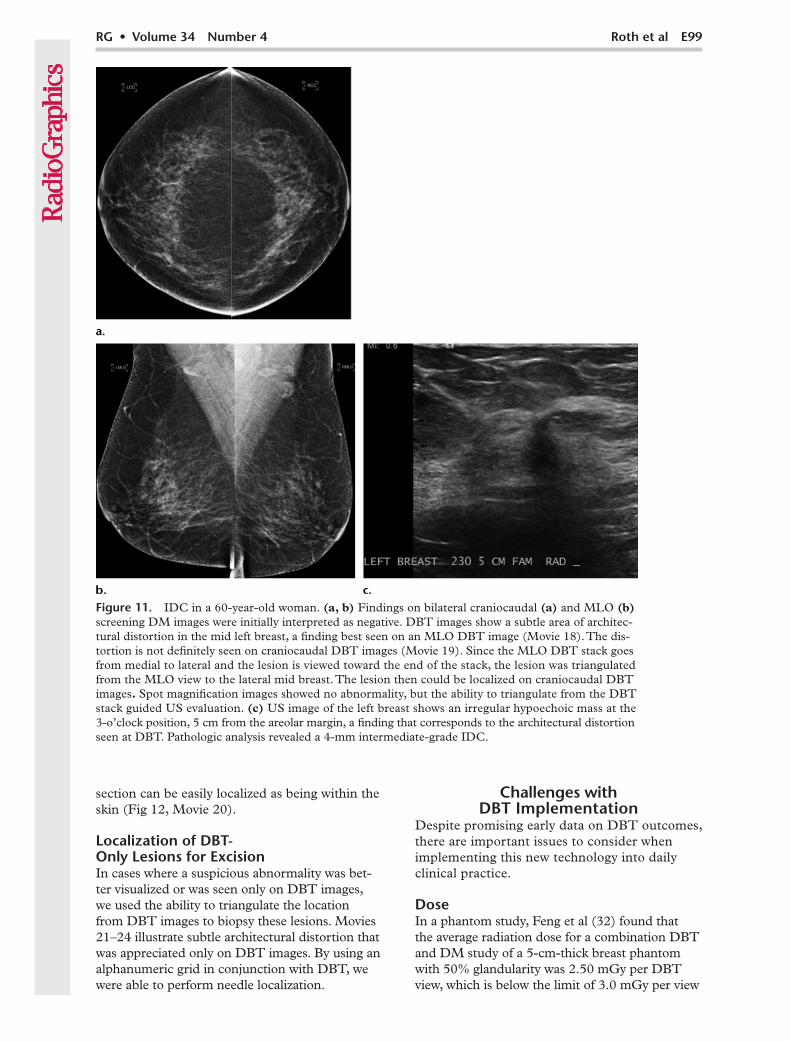
Figure 11. IDC in a 60-year-old woman. (a, b) Findings on bilateral craniocaudal (a) and MLO (b) screening DM images were initially interpreted as negative. DBT images show a subtle area of architec-tural distortion in the mid left breast, a finding best seen on an MLO DBT image (Movie 18). The dis-tortion is not definitely seen on craniocaudal DBT images (Movie 19). Since the MLO DBT stack goes from medial to lateral and the lesion is viewed toward the end of the stack, the lesion was triangulated from the MLO view to the lateral mid breast. The lesion then could be localized on craniocaudal DBT images. Spot magnification images showed no abnormality, but the ability to triangulate from the DBT stack guided US evaluation. (c) US image of the left breast shows an irregular hypoechoic mass at the 3-o’clock position, 5 cm from the areolar margin, a finding that corresponds to the architectural distortion seen at DBT. Pathologic analysis revealed a 4-mm intermediate-grade IDC.
section can be easily localized as being within the skin (Fig 12, Movie 20).
Localization of DBT- Only Lesions for ExcisionIn cases where a suspicious abnormality was bet-ter visualized or was seen only on DBT images, we used the ability to triangulate the location from DBT images to biopsy these lesions. Movies 21–24 illustrate subtle architectural distortion that was appreciated only on DBT images. By using an alphanumeric grid in conjunction with DBT, we were able to perform needle localization.
Challenges with DBT Implementation
Despite promising early data on DBT outcomes, there are important issues to consider when implementing this new technology into daily clinical practice.
DoseIn a phantom study, Feng et al (32) found that the average radiation dose for a combination DBT and DM study of a 5-cm-thick breast phantom with 50% glandularity was 2.50 mGy per DBT view, which is below the limit of 3.0 mGy per view

E100 July-August 2014 radiographics.rsna.org
Figure 12. DBT used to localize calcifications to the skin. Cranio-caudal DM image of the left breast shows clustered calcifications, find-ings that are clearly visible in the skin layer on the DBT images (Movie 20).
set by the Mammography Quality Standard Act. The mean glandular dose was only 8% higher for combined DBT and DM than for a full-field DM acquisition (1.30 mGy and 1.20 mGy, respec-tively). However, at combo-mode imaging, DBT images are obtained with DM images, which leads to an overall increase in dose for a screening exam-ination. Essentially, when the combo mode is used, women are exposed to a radiation dose equivalent to twice that of DM. The actual patient dose varies significantly depending on breast size and compo-sition (13). Recently, the FDA has approved the use of reconstructed synthetic 2D images obtained from the DBT acquisition (33–36). In early stud-ies, the diagnostic accuracy of tomosynthesis with synthetic imaging has been shown to be similar to that of tomosynthesis combined with standard 2D DM. Therefore, in many cases, the additional dose from 2D DM imaging will be unnecessary, thus lowering the overall radiation dose of tomosynthe-sis imaging.
CalcificationsDetection of calcifications may be challeng-ing with DBT. Large calcifications may cause substantial artifacts, which appear on multiple imaging sections as out-of-focus white objects bordered by dark shadows. Conflicting data have been reported regarding detection of microcal-cifications at DBT (37,38). In addition, there currently is no commercially available computer-aided detection (CAD) system for DBT, although active research in this field suggests improved performance of 3D CAD compared with DM CAD (39–43).
Learning Curve and DBT TrainingAs with any new imaging technology, there is a substantial learning curve for interpreting DBT studies. In our practice, we had a slight initial increase in the overall screening callback rate. We believe that this was related to detection of DBT-only cancers early in our screening experience with DBT and the associated need to establish a new operating point to balance the sensitivity and specificity of screening. Some readers began recalling patients with more subtle DBT findings in hopes of finding additional DBT-only cancers. In addition, previously overlooked benign masses and architectural distortion often appeared more conspicuous, which prompted further workup. With increased reader experience, our callback rate stabilized and had decreased significantly within 6 months.
Currently, radiologists, physicists, and technol-ogists are required by the Mammography Quality Standards Act to complete 8 hours of dedicated tomosynthesis training before clinical implemen-
tation (44). Although initial training is invaluable, experience is the only true trainer. In our opin-ion, when DBT is implemented in a clinical prac-tice, it is best to start with high-volume screening so that the large variation in normal and benign findings can be appreciated with DBT before implementing the new technology in more com-plicated settings, such as diagnostic workup or in patients who have had breast conservation ther-apy, where scars and radiation changes may make diagnosis extremely challenging.
Other ConsiderationsCurrently there is no approved current proce-dural terminology (CPT) code or standard ad-ditional reimbursement for DBT imaging, but as additional data from large prospective trials be-come available, it is hoped that a new reimburse-ment code will be created.
With the additional images in the recon-structed DBT stack, interpretation times will undoubtedly increase. Recent studies have esti-mated that the addition of DBT images to DM images in the combo mode will approximately double interpretation times (13,16,45). Although this is a challenge, we were able to handle the increased interpretation time required without

RG • Volume 34 Number 4 Roth et al E101
increasing our staffing or dramatically changing our schedule.
Because both the DM images and DBT pro-jections must be stored, picture archiving and communication system (PACS) storage require-ments will increase. The average combined DBT and DM study produces approximately 1 GB of data. The data can be stored at 4:1 lossless com-pression to decrease the total size of the dataset to 250 GB, although this is more than 10 times greater than the size of a compressed four-view DM set (46).
ConclusionsDBT is a promising new technology that has shown improved accuracy for screening and diagnostic breast imaging. Our early clinical ex-perience supports these findings. One year after implementing DBT for all screening patients, we demonstrated a substantial reduction in our overall callback rate and a trend toward increased cancer detection.
In our diagnostic population, we showed improved conspicuity of lesions with use of DBT, particularly for architectural distortion and masses. The 3D localization that is pos-sible with DBT offers substantial improvements over conventional imaging. The use of DBT in the diagnostic setting can expedite workups by reducing the number of 2D images needed (ie, spot compressions and additional projections for localization).
As with any new technology, several issues must be considered when implementing DBT into daily practice. Ongoing large-scale prospec-tive trials will help guide the evidence-based utili-zation of this new technology.
Disclosures of Conflicts of Interest.—A.D.A.M: Fi-nancial activities not related to the present article: advisory board member for Real-Time Tomography, nonfinancial support from Hologic. E.F.C.: Financial activities related to the present article: advisory board member for Hologic.
References 1. Tabár L, Vitak B, Chen TH, et al. Swedish Two-
County Trial: impact of mammographic screening on breast cancer mortality during 3 decades. Radi-ology 2011;260(3):658–663.
2. Pisano ED, Gatsonis C, Hendrick E, et al. Diag-nostic performance of digital versus film mam-mography for breast-cancer screening. N Engl J Med 2005;353(17):1773–1783.
3. U.S. Preventive Services Task Force. Screening for breast cancer: U.S. Preventive Services Task Force recommendation statement. Ann Intern Med 2009; 151(10):716–726, W236.
4. ACR Practice Guideline for the Performance of Screening and Diagnostic Mammography. American College of Radiology Web site. http://www.acr.org/~/media/ACR/Documents/PGTS/guidelines/Screen-
ing_Mammography.pdf. Updated 2008. Accessed May 20, 2013.
5. American College of Obstetricians-Gynecologists. Practice Bulletin No. 122: Breast cancer screening. Obstet Gynecol 2011;118(2 Pt 1):372–382.
6. American Cancer Society Guidelines for the Early Detection of Cancer. American Cancer Society Web site. http://www.cancer.org/cancer/breastcan-cer/moreinformation/breastcancerearlydetection/breast-cancer-early-detection-acs-recs. Updated May 3, 2013. Accessed May 20, 2013.
7. Hubbard RA, Kerlikowske K, Flowers CI, Yankaskas BC, Zhu W, Miglioretti DL. Cumulative probability of false-positive recall or biopsy recommendation after 10 years of screening mammography: a cohort study. Ann Intern Med 2011;155(8):481–492.
8. Brewer NT, Salz T, Lillie SE. Systematic review: the long-term effects of false-positive mammo-grams. Ann Intern Med 2007;146(7):502–510.
9. Brodersen J, Siersma VD. Long-term psychosocial consequences of false-positive screening mammog-raphy. Ann Fam Med 2013;11(2):106–115 .
10. Skaane P, Gullien R, Bjørndal H, et al. Digital breast tomosynthesis (DBT): initial experience in a clinical setting. Acta Radiol 2012;53(5):524–529.
11. Michell MJ, Iqbal A, Wasan RK, et al. A compari-son of the accuracy of film-screen mammography, full-field digital mammography, and digital breast tomosynthesis. Clin Radiol 2012;67(10):976–981.
12. Rafferty EA, Park JM, Philpotts LE, et al. Assess-ing radiologist performance using combined digital mammography and breast tomosynthesis com-pared with digital mammography alone: results of a multicenter, multireader trial. Radiology 2013; 266(1):104–113.
13. Skaane P, Bandos AI, Gullien R, et al. Comparison of digital mammography alone and digital mam-mography plus tomosynthesis in a population-based screening program. Radiology 2013;267(1):47–56.
14. Acciavatti RJ, Maidment AD. Observation of su-per-resolution in digital breast tomosynthesis. Med Phys 2012;39(12):7518–7539.
15. Poplack SP, Tosteson TD, Kogel CA, Nagy HM. Digital breast tomosynthesis: initial experience in 98 women with abnormal digital screening mam-mography. AJR Am J Roentgenol 2007;189(3): 616–623.
16. Gur D, Abrams GS, Chough DM, et al. Digital breast tomosynthesis: observer performance study. AJR Am J Roentgenol 2009;193(2):586–591.
17. Svahn T, Andersson I, Chakraborty D, et al. The diagnostic accuracy of dual-view digital mam-mography, single-view breast tomosynthesis and a dual-view combination of breast tomosynthesis and digital mammography in a free-response observer performance study. Radiat Prot Dosimetry 2010; 139(1–3):113–117.
18. Gur D, Bandos AI, Rockette HE, et al. Localized detection and classification of abnormalities on FFDM and tomosynthesis examinations rated under an FROC paradigm. AJR Am J Roentgenol 2011;196(3):737–741.
19. Wallis MG, Moa E, Zanca F, Leifland K, Daniels-son M. Two-view and single-view tomosynthesis versus full-field digital mammography: high-resolution x-ray imaging observer study. Radiology 2012;262(3):788–796.
20. Noroozian M, Hadjiiski L, Rahnama-Moghadam S, et al. Digital breast tomosynthesis is comparable

E102 July-August 2014 radiographics.rsna.org
to mammographic spot views for mass character-ization. Radiology 2012;262(1):61–68.
21. Bernardi D, Ciatto S, Pellegrini M, et al. Prospec-tive study of breast tomosynthesis as a triage to assessment in screening. Breast Cancer Res Treat 2012;133(1):267–271.
22. Rose SL, Tidwell AL, Bujnoch LJ, Kushwaha AC, Nordmann AS, Sexton RS Jr. Implementation of breast tomosynthesis in a routine screening prac-tice: an observational study. AJR Am J Roentgenol 2013;200(6):1401–1408.
23. Haas B, Kalra VB, Raghu M, Philpotts LE. Perfor-mance of digital breast tomosynthesis compared to conventional digital mammography for breast cancer screening [abstr]. In: Radiological Society of North America Scientific Assembly and Annual Meeting Program. Oak Brook, Ill: Radiological So-ciety of North America, 2012; 158.
24. Conant EF, Gavenonis SC, Weinstein SP, Schnall MD, Kontos D. Early implementation of digital breast tomosynthesis: comparison of call-back and cancer detection rates in a clinical screening practice [abstr]. In: Radiological Society of North America Scientific Assembly and Annual Meeting Program. Oak Brook, Ill: Radiological Society of North America, 2012; 239.
25. Tagliafico A, Astengo D, Cavagnetto F, et al. One-to-one comparison between digital spot compres-sion view and digital breast tomosynthesis. Eur Radiol 2012;22(3):539–544.
26. Förnvik D, Zackrisson S, Ljungberg O, et al. Breast tomosynthesis: accuracy of tumor measurement compared with digital mammography and ultraso-nography. Acta Radiol 2010;51(3):240–247.
27. Meacock LM, Mombelloni S, Iqbal A, Akbar A, Wang Y, Michell MJ. The accuracy of breast can-cer size measurement: digital breast tomosynthe-sis (DBT) vs. 2D digital mammography (DM). Presented at the 22nd annual meeting of the European Congress of Radiology, Vienna, Austria, March 4–8, 2010.
28. Zuley ML, Bandos AI, Ganott MA, et al. Digital breast tomosynthesis versus supplemental diagnos-tic mammographic views for evaluation of noncal-cified breast lesions. Radiology 2013;266(1):89–95.
29. Brandt KR, Craig DA, Hoskins TL, et al. Can digital breast tomosynthesis replace conventional diagnostic mammography views for screening re-calls without calcifications? A comparison study in a simulated clinical setting. AJR Am J Roentgenol 2013;200(2):291–298.
30. Waldherr C, Cerny P, Altermatt HJ, et al. Value of one-view breast tomosynthesis versus two-view mammography in diagnostic workup of women with clinical signs and symptoms and in women re-called from screening. AJR Am J Roentgenol 2013; 200(1):226–231.
31. Hakim CM, Chough DM, Ganott MA, Sumkin JH, Zuley ML, Gur D. Digital breast tomosyn-thesis in the diagnostic environment: a subjective side-by-side review. AJR Am J Roentgenol 2010; 195(2):W172–W176.
32. Feng SS, Sechopoulos I. Clinical digital breast tomosynthesis system: dosimetric characterization. Radiology 2012;263(1):35–42.
33. Gur D, Zuley ML, Anello MI, et al. Dose re-duction in digital breast tomosynthesis (DBT) screening using synthetically reconstructed pro-jection images: an observer performance study. Acad Radiol 2012;19(2):166–171.
34. Houssami N, Skaane P. Overview of the evidence on digital breast tomosynthesis in breast cancer de-tection. Breast 2013;22(2):101–108.
35. Proceedings of the Radiologic Devices Panel. U.S. Food and Drug Administration Web site. http://www.fda.gov/downloads/AdvisoryCommittees/CommitteesMeetingMaterials/MedicalDevices/MedicalDevicesAdvisoryCommittee/Radiologi-calDevicesPanel/UCM328613.pdf. Published Oc-tober 24, 2012. Accessed September 21, 2013.
36. Skaane P, Gullien R, Eben EB, et al. Implementa-tion of synthesized 2D plus tomosynthesis images in breast cancer screening: comparison of perfor-mance levels with full field digital mammography plus tomosynthesis in a population-based screening program [abstr]. In: Radiological Society of North America Scientific Assembly and Annual Meeting Program. Oak Brook, Ill: Radiological Society of North America, 2013; 62.
37. Kopans D, Gavenonis S, Halpern E, Moore R. Calcifications in the breast and digital breast tomo-synthesis. Breast J 2011;17(6):638–644.
38. Spangler ML, Zuley ML, Sumkin JH, et al. Detec-tion and classification of calcifications on digital breast tomosynthesis and 2D digital mammogra-phy: a comparison. AJR Am J Roentgenol 2011; 196(2):320–324.
39. Chan HP, Wei J, Zhang Y, et al. Computer-aided detection of masses in digital tomosynthesis mam-mography: comparison of three approaches. Med Phys 2008;35(9):4087–4095.
40. Chan HP, Wei J, Sahiner B, et al. Computer-aided detection system for breast masses on digital tomo-synthesis mammograms: preliminary experience. Radiology 2005;237(3):1075–1080.
41. Singh S, Tourassi GD, Baker JA, Samei E, Lo JY. Automated breast mass detection in 3D recon-structed tomosynthesis volumes: a featureless ap-proach. Med Phys 2008;35(8):3626–3636.
42. Reiser I, Nishikawa RM, Giger ML, et al. Com-puterized mass detection for digital breast tomo-synthesis directly from the projection images. Med Phys 2006;33(2):482–491.
43. Reiser I, Nishikawa RM, Edwards AV, et al. Au-tomated detection of microcalcification clusters for digital breast tomosynthesis using projection data only: a preliminary study. Med Phys 2008;35 (4):1486–1493.
44. Breast tomosynthesis: training requirements and site readiness. Hologic Web site. http://www.ho-logic.com/data/Medical%20Professionals/pdf/SS -00099_TrainingRequirementsSheet.pdf. Updated 2011. Accessed February 2, 2014.
45. Bernardi D, Ciatto S, Pellegrini M, et al. Applica-tion of breast tomosynthesis in screening: incre-mental effect on mammography acquisition and reading time. Br J Radiol 2012;85(1020):e1174–e1178. doi:10.1259/bjr/19385909. Published Janu-ary 28, 2014. Accessed February 2, 2014.
46. Baker JA, Lo JY. Breast tomosynthesis: state-of-the-art and review of the literature. Acad Radiol 2011;18(10):1298–1310.
This journal-based SA-CME activity has been approved for AMA PRA Category 1 CreditTM. See www.rsna.org/education/search/RG.

Teaching Points July-August Issue 2014
Digital Breast Tomosynthesis: Lessons Learned from Early Clinical Imple-mentationRobyn Gartner Roth, MD • Andrew D. A. Maidment, PhD • Susan P. Weinstein, MD • Susan Orel Roth, MD • Emily F. Conant, MD
RadioGraphics 2014; 34:E89–E102 • Published online 10.1148/rg.344130087 • Content Codes:
Page E91By creating 3D images of the breast, DBT can overcome many of the challenges of DM by minimizing the effects of overlapping breast tissue and improving lesion detection and localization.
Page E92Some early studies have shown improved accuracy in both screening and diagnostic settings with the ad-dition of DBT. Reductions in false-positive callback rates that range from 6% to 67% have been reported, with stable or slightly increased cancer detection rates. Studies have also demonstrated improved sensitivity and specificity for breast cancer detection with use of combined DBT and DM versus 2D DM.
Page E92After the implementation of DBT in our screening population, the callback rate was significantly re-duced, from 10.4% with DM to 8.8% with combined DBT and DM. When controlled for variable reader volumes, the odds ratio (OR) was 1.24 (P < .001); therefore, the probability that a woman would be called back decreased by 24% with DBT. In addition, there was a trend toward an increased cancer de-tection rate with use of combined DBT and DM versus DM alone (5.4 cancers per 1000 vs 4.4 cancers per 1000, respectively; P = .26)
Page E92Early studies of DBT have shown improved conspicuity and characterization of lesions, in part due to reduced obscuration by overlapping breast tissue. In particular, architectural distortion and lesion mar-gins become more apparent at DBT. Studies have also shown that DBT is superior to 2D imaging for estimating the extent of malignancies.
Page E96The ability to problem solve with DBT images may expedite patient workup by alleviating the need for additional diagnostic images, thus minimizing patient anxiety associated with a callback and leading to a potentially decreased overall radiation dose.