digital radiography - konica minolta
TRANSCRIPT

DIGITAL RADIOGRAPHY
Distributed by:
KONICA MINOLTA MEDICAL & GRAPHIC, INC.No.1, Sakura-machi, Hino-shi, Tokyo, 191-8511, Japan
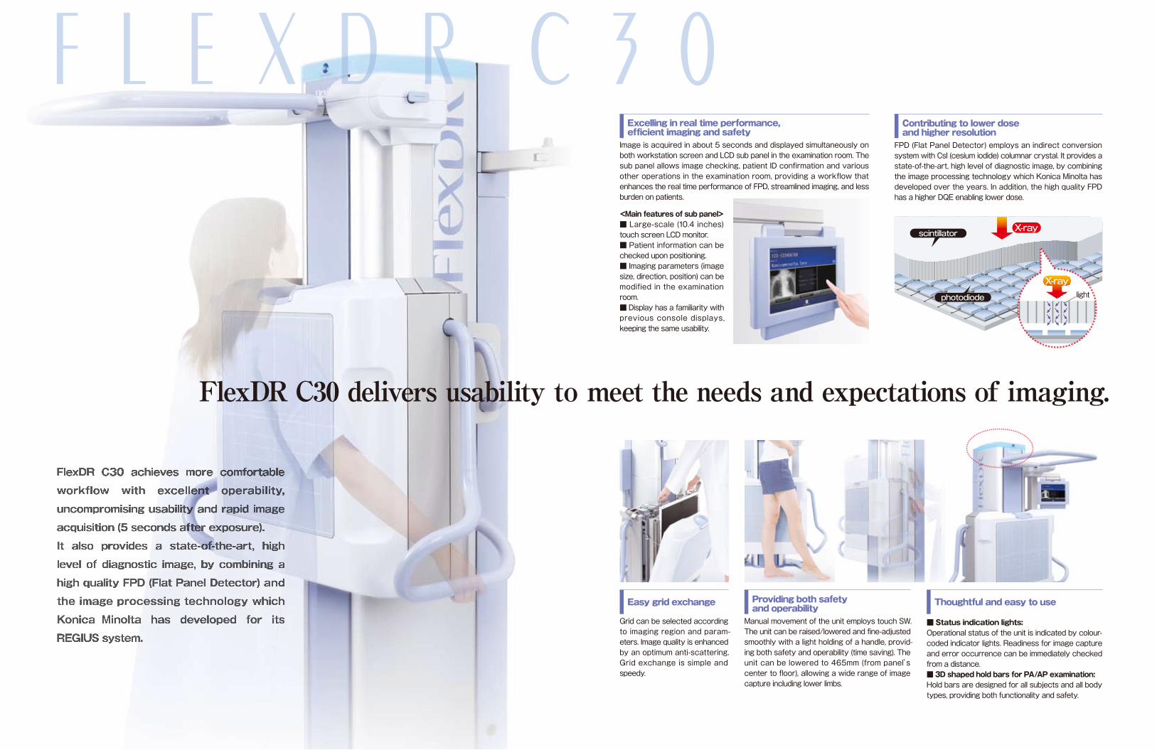
Providing both safety and operabilityManual movement of the unit employs touch SW. The unit can be raised/lowered and fine-adjusted smoothly with a light holding of a handle, provid-ing both safety and operability (time saving). The unit can be lowered to 465mm (from panel’s center to floor), allowing a wide range of image capture including lower limbs.
Easy grid exchange
Grid can be selected according to imaging region and param-eters. Image quality is enhanced by an optimum anti-scattering. Grid exchange is simple and speedy.
F L E X D R C 3 0
FlexDR C30 achieves more comfortable
workflow with excellent operability,
uncompromising usability and rapid image
acquisition (5 seconds after exposure).
It also provides a state-of-the-art, high
level of diagnostic image, by combining a
high quality FPD (Flat Panel Detector) and
the image processing technology which
Konica Minolta has developed for its
REGIUS system.
FlexDR C30 delivers usability to meet the needs and expectations of imaging.
Contributing to lower dose and higher resolutionFPD (Flat Panel Detector) employs an indirect conversion system with CsI (cesium iodide) columnar crystal. It provides a state-of-the-art, high level of diagnostic image, by combining the image processing technology which Konica Minolta has developed over the years. In addition, the high quality FPD has a higher DQE enabling lower dose.
Excelling in real time performance, efficient imaging and safetyImage is acquired in about 5 seconds and displayed simultaneously on both workstation screen and LCD sub panel in the examination room. The sub panel allows image checking, patient ID confirmation and various other operations in the examination room, providing a workflow that enhances the real time performance of FPD, streamlined imaging, and less burden on patients.
<Main features of sub panel>■ Large-scale (10.4 inches) touch screen LCD monitor. ■ Patient information can be checked upon positioning. ■ Imaging parameters (image size, direction, position) can be modified in the examination room. ■ Display has a familiarity with previous console displays, keeping the same usability.
X-ray
photodiode
X-ray
scintillator
light
Thoughtful and easy to use
■ Status indication lights:Operational status of the unit is indicated by colour-coded indicator lights. Readiness for image capture and error occurrence can be immediately checked from a distance. ■ 3D shaped hold bars for PA/AP examination:Hold bars are designed for all subjects and all body types, providing both functionality and safety.

Provision of real-time operation
H Y B R I D - W S C S - 3Diagnostic Imaging Workstation CS-3
is a console for dual use with FlexDR
and REGIUS series. Touch screen
LCD monitor with wide viewing angle
has been introduced to a simple and
intuitive user interface. Reliable
image checking feature follows the
same line of that in FlexDR series.
The system provides total manage-
ment from input of patient/order
information received from HIS/RIS all
the way through to image checking,
as well as more comfortable and
efficient image acquisition environ-
ment.
Equipped with patient information registration function ■Search screen: On this screen, patient information can be searched by ID number. Suitable for operation with patient check-in at each imaging room. ■Order list screen: Registered orders are displayed sequentially in the combined use of patient check-in terminal. Suitable for operation with separate patient check-in.
Various optional functions that enhance usefulness and convenience. ■Arbitrary annotation function: This function allows text editing and layout at any positions on the image. Annotations can be reflected in the output (to host or printer). ■Inter-console image sharing: This function allows image data sharing among CS-3 consoles on a single network.
Hybrid Device Workflow Console
The system centralizes workflow from order information management to image output in general X-ray imaging by the use of “FlexDR and REGIUS system”. Change into Uniformity of handling (including console operations) and consistency of image quality is achieved with simultane-ous CR and DR imaging for a single patient.
Delivering Uncompromising Real Time performance. Centralized control of DR and CR creates
a comfortable work environment.
★Screen design is subject to change without notice for the purpose of performance improvement.
■Hybrid Device Network System
REGIUS190
FlexDRC30
FlexDRC30
CS-3
REGIUS110
Viewer
HIS/RIS
Server
DRYPRO873
FlexDRseries
REGIUSseries
CS-3
Patient selection
FlexDR imaging selection
Image acquisition
Image checking
REGIUS imaging selection
Image acquisition
Image checking
Image adjustment
▲Search screen ▲Order list screen ▲Arbitrary annotation function ▲Inter-console image sharing screen
■Sub panel
■CS-3
Based on the review of image acquisition workflow, Konica Minolta offers an additional touch screen LCD monitor (sub panel) with FlexDR model, providing image display and various other operations in the examination room to achieve a workflow with improved real-time performance. Upper line shows CS-3 display and lower line shows sub panel display.
Image data sharing
CS-3 CS-3
CS-3
CS-3
**
*To be released soon
Simultaneous display
in examination room
Single PC! saving space
No movement between consoles!
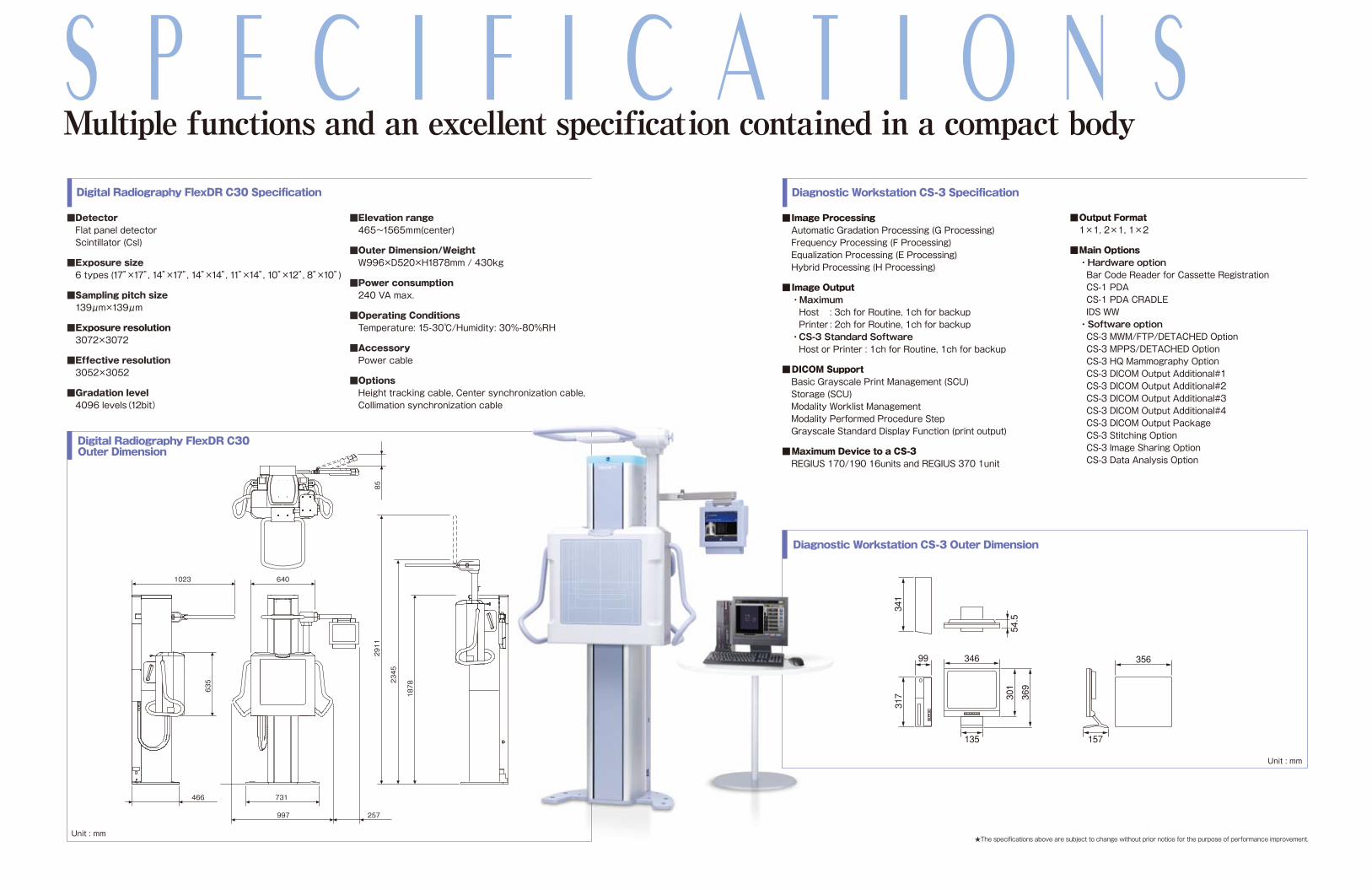
Digital Radiography FlexDR C30 Outer Dimension
Unit : mm
Diagnostic Workstation CS-3 Outer Dimension
Unit : mm
S P E C I F I C A T I O N S
★The specifications above are subject to change without prior notice for the purpose of performance improvement.
■Elevation range 465~1565mm(center)
■Outer Dimension/Weight W996×D520×H1878mm / 430kg
■Power consumption 240 VA max.
■Operating Conditions Temperature: 15-30℃/Humidity: 30%-80%RH
■Accessory Power cable
■Options Height tracking cable, Center synchronization cable, Collimation synchronization cable
Multiple functions and an excellent specification contained in a compact body
1023
635
466
2345
2911
1878
640
731
997 257
85
Digital Radiography FlexDR C30 Specification Diagnostic Workstation CS-3 Specification
■Detector Flat panel detector Scintillator (Csl)
■Exposure size 6 types (17”×17”, 14”×17”, 14”×14”, 11”×14”, 10”×12”, 8”×10”)
■Sampling pitch size 139μm×139μm
■Exposure resolution 3072×3072
■Effective resolution 3052×3052
■Gradation level 4096 levels(12bit)
■Output Format 1×1, 2×1, 1×2
■Main Options ・Hardware option Bar Code Reader for Cassette Registration CS-1 PDA CS-1 PDA CRADLE IDS WW ・Software option CS-3 MWM/FTP/DETACHED Option CS-3 MPPS/DETACHED Option CS-3 HQ Mammography Option CS-3 DICOM Output Additional#1 CS-3 DICOM Output Additional#2 CS-3 DICOM Output Additional#3 CS-3 DICOM Output Additional#4 CS-3 DICOM Output Package CS-3 Stitching Option CS-3 Image Sharing Option CS-3 Data Analysis Option
■Image Processing Automatic Gradation Processing (G Processing) Frequency Processing (F Processing) Equalization Processing (E Processing) Hybrid Processing (H Processing)
■Image Output ・Maximum Host : 3ch for Routine, 1ch for backup Printer : 2ch for Routine, 1ch for backup ・CS-3 Standard Software Host or Printer : 1ch for Routine, 1ch for backup
■DICOM Support Basic Grayscale Print Management (SCU) Storage (SCU) Modality Worklist Management Modality Performed Procedure Step Grayscale Standard Display Function (print output)
■Maximum Device to a CS-3 REGIUS 170/190 16units and REGIUS 370 1unit
346
135
54.5
301
369
356
317
99
341
157