dihydrofolate reductase-thymidylate synthase of leishmania major
TRANSCRIPT

Proc. Nati. Acad. Sci. USAVol. 83, pp. 2584-2588, April 1986Genetics
Primary structure of the gene encoding the bifunctionaldihydrofolate reductase-thymidylate synthase of Leishmania major
(chemotherapy/protein homologies/intervening sequences/gene ampliflcation/protozoan parasites)
STEPHEN M. BEVERLEY, THOMAS E. ELLENBERGER, AND JOHN S. CORDINGLEYDepartment of Pharmacology, Harvard Medical School, Boston, MA 02125
Communicated by George H. Hitchings, December 24, 1985
ABSTRACT We have determined the nucleotide sequenceof the dihydrofolate reductase-thymidylate synthetase (DHFR-TS) gene of the protozoan parasite Leishmania major(dihydrofolate reductase, EC 1.5.1.3 and thymidylate syn-thase, EC 2.1.1.45). The DHFR-TS protein is encoded by asingle 1560-base-pair open reading frame within genomicDNA, in contrast to vertebrate DHFRs or mouse and phage T4TSs, which contain intervening sequences. Comparisons of theDHFR-TS sequence with DHFR and TS sequences of otherorganisms indicate that (i) the order of enzymatic activitieswithin the bifunctional polypeptide chain is DHFR followed byTS, (ii) the Leishmania bifunctional DHFR-TS evolved inde-pendently and not through a phage T4-related intermediate,and (iii) the rate ofevolution ofboth theDHFR and TS domainshas not detectably changed despite the acquisition of newfunctional properties by the bifunctional enzyme. The Leish-mania gene is 86% G+C in the third codon position, in contrastto genes of the parasite Plasmodiumfalciparum, which exhibitan opposite bias toward A+T. The DHFR-TS locus is encodedwithin a region of DNA amplified in methotrexate-resistantlines, as previously proposed.
The enzymes dihydrofolate reductase (DHFR; EC 1.5.1.3)and thymidylate synthase (TS; EC 2.1.1.45) have criticalroles in intermediary metabolism and consequently are im-portant targets for chemotherapy. Drugs used in the treat-ment of human tumors that inhibit DHFR and TS aremethotrexate and metabolites of 5-fluorouracil, respectively(1, 2). Other antifolates such as trimethoprim and pyrimeth-amine have been employed in the treatment of pathogens (3,4). In most organisms DHFR and TS exist as separatemolecular entities, DHFR as a monomer of about 20 kDa andTS as a dimer made up of --35-kDa subunits (5). In contrast,in Leishmania as well as all protists examined to date, theseenzymes are part of a bifunctional DHFR-TS complex,consisting of a homodimer of a 54- to 100-kDa polypeptidechain (6-8). This bifunctional enzyme exhibits distinct bio-chemical properties such as metabolic channeling (9) anddisparate binding of methotrexate and 2'-deoxy-5-fluorouri-dine monophosphate (5-FdUMP) (8, 9). Detailed molecularand biochemical studies may reveal additional features thatwill prove useful in developing agents that may selectivelyinhibit the Leishmania enzyme, analogous to the selectiveinhibition of Plasmodium DHFR by pyrimethamine (4).Mutants of Leishmania have been obtained that overpro-
duce the bifunctional enzyme DHFR-TS, thus facilitating itsisolation and characterization (6, 9). Mutants exhibitingenzyme overproduction also exhibit amplification ofextrachromosomal circular DNAs (10), one of which iscorrelated with enzyme overproduction (the R region). Wehave now determined the nucleotide sequence of the gene
within the R region encoding the bifunctional enzyme DHFR-TS of Leishmania major.
MATERIALS AND METHODSCells, Genomic, and Recombinant DNAs. The LT 252 cell
line (11) and the methotrexate-resistant R1000 derivative cellline (6) were utilized as sources of genomic DNA and RNAas described (10). We have shown by DNA hybridization andserological tests that this line actually is of the speciesLeishmania major (L. tropica major; S.M.B. and D. McMahon-Pratt, unpublished data). A 4.5-kilobase (kb) Sal I fragmentfrom phage X LTS-5 (10) was isolated and inserted into a pUCvector (12); this recombinant is named pLTS-5-S45.cDNA Library. A cDNA library of polyadenylylated
mRNA from R1000 promastigotes was constructed in thebacteriophage vectors XgtlO (13) and Xgtll (14) using theprotocol of T. St. John, J. Rosen, and H. Gershenfeld(personal communication), and screened with pLTS-5-S45(15). Approximately 400,000 independent recombinants wereobtained for each library. The largest recombinant insert, oneof 0.6 kb in XgtlO, was inserted into M13 vectors for DNAsequencing.DNA Sequencing. DNA sequencing was performed using
the dideoxynucleotide technique (16) with 3"S-labeleddeoxynucleotide triphosphates (17), and Leishmania-derivedDNAs were inserted into, M13 vectors (18). The entiresequence was determined on both strands.
RESULTSSequence of the DHFR-TS Region. Analysis of mRNAs
encoded by the amplified R region of the methotrexate-resistant R1000 line of Leishmania (6) has indicated that a3.2-kb polyadenylylated mRNA encodes the bifunctionalDHFR-TS (refs. 19 and 20; G. Kapler and S.M.B., unpub-lished results). This region is entirely contained within a4.5-kb Sal I fragment (Fig. 1), which is also amplified withinthe R region of the methotrexate-resistant R1000 line (6, 10).By restriction enzyme mapping, this region ofDNA appearsto be identical in the wild-type and R1000 lines (10), as is theDHFR-TS protein synthesized as judged by biochemicalcriteria (6, 9).DNA sequencing ofpLTS-5-S45 reveals a single large open
reading frame, beginning at the ATG initiation codon num-bered as base 1 and proceeding for 1560 bp (Figs. 1 and 2).This ATG is the first between the long open reading frame andthe 5' side of the mRNA, as revealed by S1 analysis(unpublished data). The derived amino acid sequence of thisprotein includes the peptide Met-Asp-Leu-Gly-Pro-Val--Tyr-Gly-Phe-Gln-Trp-Arg-His-Phe-Xaa-Ala-Asp-Tyr-Lys--Xaa-Phe-Glu-Ala-Asn-Tyr-Xaa-Gly-Glu that differs by oneamino acid from an internal peptide of the DHFR-TS of a5,8-dideaza-10-propargyl folate-resistant line of Leishmania
Abbreviations: DHFR, dihydrofolate reductase; TS, thymidylatesynthase; 5-FdUMP, 2'-deoxy-5-fluorouridine monophosphate; bp,base pair(s); kb, kilobase(s).
2584
The publication costs of this article were defrayed in part by page chargepayment. This article must therefore be hereby marked "advertisement"in accordance with 18 U.S.C. §1734 solely to indicate this fact.

Proc. Natl. Acad. Sci. USA 83 (1986) 2585
P TI
Is cDNA-4
^ DHFR TS AAAA
FIG. 1. Analysis of the DHFR-TS gene ofLeishmania maor. (A)Restriction map of the 4.5-kb Sal I fragment located within the Rregion of Leishmania major. The orientation of this map is oppositeto that shown in figure 1B of ref. 10. The symbols correspond torestriction enzyme cleavage sites as follows: P, Pvu II; R, EcoRI; B,Bgl II; T, Sst I; S, Sal I. The region ofDNA sequenced was betweenthe EcoRI site and a Sph I site (not shown) approximately 300 bp onthe 3' side of the Sst I site. The location of the 0.6-kb cDNA cloneis also indicated. (B) Summary offeatures ofthe DHFR-TS gene. Thelocation of the single large open reading frame of 1560 bp within themRNA is indicated by the thick region. The 22-amino acidhydrophilic leader sequence is shown as a solid region, and thelocation of the DHFR and TS domains are taken from the alignmentsin Fig. 3. The location of the poly(A) tract of the mRNA as revealedby sequence analysis of the cDNA is also shown.
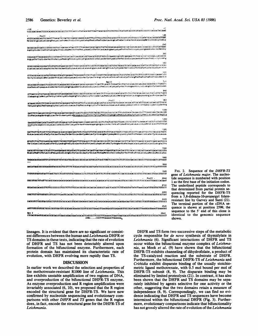
(21). (The histidine at position 13 within the peptide wasidentified as glycine by these authors. DNA sequencing ofboth strands in this region unambiguously predicts histi-dine.) The predicted DHFR-TS has a molecular weight of58,684, somewhat larger than the 57-kDa subunit molecularsize reported for Leishmania and other trypanosomatids(6-8). This may reflect the accuracy of gel electrophoresismethods for estimating protein molecular weights. Alterna-tively, the DHFR-TS may be processed in vivo to a smallerfinal product. Alignment of the Leishmania DHFR-TS withDHFRs of diverse origin indicates that the Leishmania genebears a hydrophilic amino-terminal extension of 22 aminoacids (Fig. 3). The amino terminus of the mature DHFR-TShas been reported to be blocked (9, 21), and the preciseamino terminus of the DHFR-TS is currently unknown.Another 1040 bp of sequence follow the open reading
frame, at which point the sequence of the cDNA and genomicsequences diverge, corresponding to the poly(A) tract of themRNA. The region between the long open reading frame andthe poly(A) tract contains no long open reading frames. Asreported previously for genes of the related parasite Trypan-osoma brucei, we can find no eukaryotic consensus poly(A)addition signal (AATAAA) prior to the poly(A) tract (32, 33).Furthermore, we cannot find a consensus homology in thisregion with the corresponding regions in other trypanosomegenes (33, 34).Comparison of the Predicted Leishmania DHFR-TS Se-
quence with Other DHFR and TS Sequences. The amino acidsequence of the DHFR-TS enzyme was derived from thenucleotide sequence and compared with published DHFRand TS sequences (Fig. 3, Table 1). It is evident that theamino-terminal region of the Leishmania DHFR-TS exhibitssignificant sequence homology (up to 37%) with DHFRs ofdiverse origin, whereas the carboxyl-terminal region of theleishmanial protein exhibits homology (up to 63%) with TSsequences. The DHFR-TS also exhibits a hydrophilic 22-amino acid extension prior to the DHFR domain. The homol-ogies with both DHFR and TS sequences include amino acidsknown to be important in ligand binding and/or located at thecatalytic site (discussed below). We, therefore, infer that theleishmanial protein is encoded as a fused polypeptide chain oftwo separate domains, an amino-terminal DHFR domain fol-lowed by a carboxyl-terminal TS domain. Our genomic DNAsequence data indicate the Leishmania DHFR gene does notcontain intervening sequences, an observation confirmed by S1nuclease analysis oftheDHFR-coding region ofthe Leishmaniagene (data not shown).
There is a nonrandom utilization of the four nucleotidebases at degenerate codon positions of the DHFR-TS. Withinthe coding region the first codon position is 62% G+C, thesecond position 41% G+C, and the third position 86% G+C.Neither the 3'- nor the 5'-untranslated regions show apositional bias. In contrast, genes of the intracellular parasitePlasmodiumfalciparum exhibit an extreme A+T bias at thirdpositions as well as within the genome as a whole (35).
Conservation of Amino Acid Sequence. The amino acidsequence alignment presented in Fig. 3 indicates that certainresidues are highly conserved among DHFR and TS se-quences. In the DHFR domain these residues include manyof those implicated in the interactions of DHFR with sub-strates, cofactors, and/or inhibitors (31). Of the 18 residuesshared in all eukaryotic DHFRs, the Leishmania DHFRdomain shares 15 (positions 32, 39, 40, 46, 47, 56, 60, 80, 83,94, 97, 102, 157, 158, and 180). For TS two separate regionshave been identified in ligand binding studies: a 5-FdUMPbinding site, located at the conserved tripeptide Pro-Cys-Hisofthe TS in many species (5) (residues 399-401 in Fig. 3), anda folylpolyglutamate binding site in Lactobacillus casei (36)(residues 279-292). Both of these sites are conserved in theLeishmania TS domain. There are additionally several otherregions within the TS domain that are highly conservedamong all species and that presumably represent functionallyimportant residues. The Leishmania TS domain shares aninsertion of 12 amino acids with the human sequence (resi-dues 322-333), the site ofwhich has been shown to be a targetfor protease inactivation (21).
Relationships of the Bifunctional DHFR-TS Enzyme toOther Species. Table 1 shows that that Leishmania TS andDHFR are both more closely related to vertebrate enzymesthan to the prokaryotic or phage T4 enzymes. The DHFRdomain of the Leishmania DHFR-TS exhibits 37% aminoacid sequence homology with vertebrate DHFRs, 29% ho-mology with prokaryotes, and 18% homology with phage T4.Similarly, the TS domain of the DHFR-TS exhibits 63%homology with vertebrates, 48% homology with prokaryotes,and 45% homology with phage T4. These findings are inaccordance with the current view of the evolutionary rela-tionships among metazoa, protists, and prokaryotes (37).Examination of the evolutionary insertions of amino acid
sequence within both the TS and DHFR genes confirms thatthe Leishmania DHFR-TS is more closely related to thevertebrate genes than to either prokaryotic or phage T4genes. Within the TS genes there are six introduced gapsamong the five sequences. Two ofthese are specific for phageT4 (positions 306 and 498) and two are specific for Leishma-nia (positions 275 and 411). The remaining two (positions 322and 349) are complex, in that the insertion boundaries arevariable among species. Examination of these insertionsreveals that the Leishmania and human TS sequences areprecisely the same length and homologous at the amino acidsequence level, in contrast to the comparisons involving theother prokaryotic and phage sequences. Similar findings areevident in the insertions found within the DHFR sequencealignments; however, as the degree of sequence homologyamong these genes is considerably less than for TS, there isless certainty about the exact placement of the sites ofinsertion.
Relative Rates of Evolution of Bifunctional vs. Monofunc-tional DHFRs and TS. It is possible that new properties of thebifunctional DHFR-TS relative to monofunctional DHFRand TS may have given rise to new evolutionary pressures,resulting in a change in the rate of evolution of the DHFR-TSgenes. We compared the Leishmania and human genesseparately with the more distantly related prokaryote andphage genes (Table 2). Alterations in evolutionary rate wouldbe revealed by differences in the distance between theoutside reference group and the Leishmania or human
S P R PA I
B
300 bp
Genetics: Beverley et al.
2586 Genetics: Beverley et al.
-1-120
1 PvuII 120AiTGTCCAGGGCAGCTGCGAGGTTTAAGATTCCGATGCCGGAGACGCAACGWAGACTTTCTTTCCCCTCCCTGCGCGCCTTCTCCATCGTCGTCGCCTCGrATATGCAGCACGCCATCGOCMetSerArgAloAlaAlaArgPheLysI leProMetPro0luThrLySAlaAspPheAl1PheProSerLeuArgAlaPheSerI1eValValAlILeuAspMet~lnHisGlyIleGly
240
GACGGCGAGTCGATCCCGTGGCGGGTGCCCCGAGGACATGACG'TTC'rTC AAGAACCAGACGACCCGCTGTGWAACAAGAAGCCGCCGACGCACA AGAAGCGCAACGCCGTCCTGATGGGCAspGlyGluSerI leProTrpArgVal1ProGl1uAspMetTh rP hePheLysAsnGlnThrThrL~euLeuArgAsnLY3LysProP roThrC 1uLysLysA rgAsnAl aVal1V alMet~ly
360
CGCAAGACTTOGGAGAGCGTCCCGGTAA AGTTCCGACCACTCAACCGGACWT;GCAACATCGTGTTATCCTCGAAGGCCACCGTC(;AGGAWCTTCTGCGCCGCI'GlCCGGA~tibGAC ACC(;CArgLysThrTrpGl uSerVa1ProVa LysPheA rgProLeu LyslyArgLeuAsnIleVaLeuSerSerLysAaThrVa1CuGluLeuLeuA1aProLeuProGlu1yG1nArg
480
GCGGCGGCGGCGCAGGATGTGGTGGrGGTGAACGGCGGTCTGCGCCACGCGCTCCGCCTC CTCGCACGCCCGCTGCTACTGCAGTC CATCGAGCAtC~sGT ATTWcGTCGGTG( T¢GCWAAleAlsAaArlaGlnAspVlValVaIVylAsnGlyGlyyLeuAlaluAlaLeuArgLeuLeuA1ArgProLeuTyrCysSerSerIleC1uThrAlaTyrCysVal~ly~lyA!aGin
bOOGSTTTACGCGGACGCCATGCTGTCGCCGTGCATCGAGAAACTGCAGGAAGTGTACCTCACCCGCATCTACCGCCAUCGtCCCTGCGCTCTACGCGCTTCTTTCCCTTTCCGCCCCGAG^AACGtCGValTyrAlaAspAlaIetIeUSerProCY3IleGluLY3LeuGflnAOI TyrLeuThrArgl leTyrAlaThrAlaProAlaCysThrArgPhePheProPheProProGluAsnAla
BglII 7zoG(CCACGG;CGTGGGAWCTGGCGTCGTCTCAGGGACGCCWAAGAWCGAGGCGGAGGGCCCTCGAGTTCGAGATCTCCAAG'TACCTGCCGCGCAACCAC;AC(;GAGCCCAGTACC rTGAGCTGtlaThrAlsTrpAspLeuAlaSerSerGlnGlyArgArtLysSerGluAlaOluGl yLeuGluPheGCuIl eCysLysTyrVal ProArgAsnHisul us;uArgGlnryrLeuGluLeu
840
ATTGACCGCATCATGACACGGGGATCGTGAAGGAGGACCGCACCGGC GTGCGCACCATCAGCCTCTTCCG;CWCCAGATcGCWT'rCTC( CTA CGCGACA ACcCUt,tI'CCXuTGCTGiACG
PavII 960
A^CGAA^GCGrGTCTTCTGGCGCGGCGTGTGCGAGGACCGCTCXTCC~jTTCCTUCeCC;GGC#^AAC GAGTUCCCAGCTGCTGC;CAG;AC AAGCAACATTCACATCTGGGACWCAACCGTTCGCGCThrLysArgValPheTrp~r5GlyValCysluGl uLeu LeuTrpPheLeuA rgGlyl uThrSerA1al nLeu LeuA laAspLysAspI eHisI leTrpAspulyAsnblySerArg
1080
GAGTTTCTCGA^CAGCCGCGC;CTTGAsCAGA^GAATAAGGAGATGGUACCTC CGCCCTGTCTACGGCTTCC AC rvCcCCACTTCUGGiGCACA rTACAAGCGGG1TTGA AGCGAA(;TACGACGOCGluPheLuspSerAr&GlyLevThrGluAsnLysGlUeApLeuGlyProVa lTyr~lyPheGl n'rrp~rlis Phe~lyA1AspTy rLysGl y PheG1uA1aAsnTyrAspGly
1200
G^AAGOGT~GGACCAGAkTCAAGCTbCATCGTGGAGACCATCAAGACGAACCCAA^CGACCGCCGCCTC'T AtiTCACTGCCTGC;AACCCG rGCGCGCT(;CAAAAG;ATG;CGCTUCCCCCCCTCCG1uGlyValAspGlnIleLysLeuIleValGluThrIleLysThrAfsnProAsnAspArgArgLeuLeuVai rhrAiaTrpAsnProCysAlaLeuo;l!nLysMetAlaLeuProProCys
1320CACCTTGCTTCCAGTTCTACGTGAACACAGACACGAGCGAGCTATCCTGCATGTTGTACCAGCGCT'CGTGTGACATGGGTC'rTGGCGTCCCCTTCAACATTGCCTCCTACGCGCTGCTCHisL~LeuAl&Gl nPheTyrValAsnThrA pThrbrGlu Leu~rCys tLeuTyrGl nArrCysAs pMe tGl yLeuGl yVal ProPheAsn I leAlaSerTyrAlaLeuLeu
1440ACCATCCTCATT~OCAAGGCGACGGCTCTGCGOCTGGTGAGCTTGTGCACACCCTCGGCGACGCCCACGTCTACCGCAACCACGTTGATGCCCTCAAGGCGCA GCTCGAGCGAGTCCCGThlrIleLouIle~llLysAloThrly L uArgPr~o~lyGl uLeuValHlsThrL u~ly~spAlaHilsV lTyrArgAsnHi sVa1AspA 1aLeuLysAlaGl nLeuCluArgValPro
1560CWCCCGfTCCCGACCCTCATCrTCAAGCGAGGGCGGCCGTACCTCGAGGACTACGAGTTGACGGACATGGAGCTGATCGACTACGTTCCACACCCCGGCGATCAAGATGGAGATGGCCGTANl&AloPbe~roThrLe~ll PheL~ys~luGl uArgGlnTyrLeuGlu~spTyr~l uL uThrAsp~et~luVel I eAspTyrVa 1Pr~oHl ProAl1aIle LysMetGl uMetA laVal
*.. ~~~~~~~~~~~~~~~~~~~~~~~1680TAGAGAGAGGGAGGGTGTCATGTCCGsTTGTATGCATGCAGCCACCGCCGC'GACGCTGCC;CTCACCTGliCTCCCACCTCCTCGTCGCACGACGACCGCCCCTTGCGCAGACTCGTTGGT
1800AACCATGAGCGGCGCGCGAGGGTACGCGCCT&CGTTTCTCGCACATGGCTGCGGCTGATATCCCGCCCGCACCCGCGCGCGCTCAGGCCGCGrGTCG;TCG;TWTCTTTCCATTTrTTTTT
1920GATTTGG^GCTGCTCTCCGTTGTGTGCTGGGGACCCTCCrTCCC TCGATCTCCTC GTGCGGGGTCTCCGACWCGCAGACGCGGTGCGTGCGAGCGTGCACGTGCTGT
PvuII 2040CCCMMVOCTOTAOTTGCGAGCGaGAGGAGAGAGAGAGGGAGGGGGGAGGGCAGAGGGCAGAGGACATC;CGGGTGGGAACGTGCACCGGCCTCGTCTCACGCAGCTGGAGCCCACGAAT
2160WCCACCACCACACCCTcTsTCCCCCCCCCTCCCTCTTCTTCCCACGGCGGCGACGACGACGGGGGCTCAGCTCACGCTTCTGTAGGGTATTATATTAAAGCACATGTGCC;TGCTGTGCTT
2280CCMC~rCTCTTMaGGcGTTcGCrTTCAAGTCCACGACTCCTCCCGTTGCTCACCGCCGGGCTGTCTTCTTTCGCCCCTCCCTCGGTGCCTCTTCCGCCGCCGCACOCGTGCGCTGAT
2400CACO=tCTTOCGTGATGTGCCTGTC;TGTATACACACCGTGCACGGAGAGAGCGAGCGAGCGAGAGAGAGAGAGGCCGAGAGGAAGAATGWCGGTGCCTCGCGTGGGCAAGCGTGCGCG
2520OmGrffOTGTCGTOCOCCGCGTG^GATCGTCGCCAGCOCAAGACCCCCGTGCGCCTGAAACC;TGAAGGGAGG;TCGAGAirGCGTGCCGCA'TGAGGCCTTAAGGCAGGlkA'AGTGAAAAGAG3st I , , 2640
CTCGACG~OTGACCAGCACGCGCGACATCG^AcACGACGGAATAGACACTCAOCCCCTTCCCCTTTCAAAAACTGAATGGACGGACATCGTAACGCGCTCTCTCCCCTCCACTCCCCCTCDNA:.. CCCCTTTCAAAAACTGAAAAAAn
lineages. It is evident that there are no significant or consist-ent differences between the human and Leishmania DHFR orTS domains in these tests, indicating that the rate ofevolutionof DHFR and TS has not been detectably altered uponformation of the bifunctional enzyme. Furthermore, eachprotein domain has maintained its characteristic rate ofevolution, with DHFR evolving more rapidly than TS.
DISCUSSIONIn earlier work we described the selection and properties ofthe methotrexate-resistant R1000 line of Leishmania. Thisline exhibits unstable amplification of two regions of DNA,and overproduction of the bifunctional DHFR-TS enzyme.As enzyme overproduction and R region amplification were
invariably associated (6, 10), we proposed that the R regionencoded the structural gene for DHFR-TS. We have now
confirmed by nucleotide sequencing and evolutionary com-
parisons with other DHFR and TS genes that the R regiondoes, in fact, encode the structural gene for the DHFR-TS ofLeishmania.
FIG. 2. Sequence of the DHFR-TSgene of Leishmania major. The nucleo-tide sequence is numbered with position1 as the first base of the initiation codon.The underlined peptide corresponds tothat determined from partial protein se-quencing reported for the DHFR-TSfrom a 5,8-dideaza-lO-propargyl folate-resistant line by Garvey and Santi (21).The terminal portion of the cDNA se-quence is shown at position 2598; thesequence to the 5' end of this clone isidentical to the genomic sequenceshown.
DHFR and TS form two successive steps of the metaboliccycle responsible for de novo synthesis of thymidylate inLeishmania (6). Significant interactions of DHFR and TSoccur within the bifunctional enzyme complex of Leishma-nia, as Meek et al. (9) have shown that the bifunctionalDHFR-TS exhibits channeling of dihydrofolate, a product ofthe TS-catalyzed reaction and the substrate of DHFR.Furthermore, the bifunctional DHFR-TS of Leishmania andCrithidia exhibit disparate binding of the usually stoichio-metric ligand methotrexate, with 0.5 mol bound per mol ofDHFR-TS subunit (8, 9). The disparate binding may beeliminated by limited proteolysis (21). In contrast, it has alsobeen shown that the DHFR and TS domains may be sepa-rately inhibited by agents selective for one activity or theother, suggesting that the two domains retain a measure ofindependence (8, 9). Correspondingly, we can find no evi-dence indicating that DHFR and TS sequences have becomeintermixed within the bifunctional DHFR (Fig. 3). Further-more, evolutionary comparisons indicate that bifunctionalityhas not grossly altered the rate ofevolution ofthe Leishmania
Proc. Natl. Acad. Sci. USA 83 (1986)
TGGCGGACGAAACTCGCACACAGGCACGCCGCCTCCTTTCACCCGTCATAGArAGTTGAArrAGACGCCCTCCTCCTCCCTCATCATCGCCGTCGTCATCCGGGTCCGAGCACTACGAAG

Proc. Natl. Acad. Sci. USA 83 (1986) 258720
A.DHFRdomain ~~~~~~~~~~~~~~~~LMM<SRAAARFKIPMPETKADFAFPA. DHFR domain
40 60 80 iQ0 120* * ** .* * *. . * * . ***t ** *t. **
LM SLRAFSIVVALDMQH-GIG-DGES.------IPW-RVPEDMTFFKNQTTLLRNKKPPTE4KRNAVVMGRK FWESVPVKFRPLKGR LNIVLSSKATVEELLAPLPE00RAAAAODVVVVNOGH MVGSLNCI VA VSQNM-GIG-KNGD----LPWPPLRNEFRYFORMTTTSSV-----EGKQNLV MGKKTTF2IPEKNRPLKRINLV LRE----------K---LKEPPOGAHFLSRSEC MISLIAALAVDR-VIG-MENA.------MPW-NLPADLAWFKRNTL.------------ NKPVIMGRHTWESIG---RPLPGRKNII LSOP --------------- TDDR-VTWl KSLC TAFLWAQNRDG-LIG-KDGH.----.LPW-HLPDDLHYFRAQV.------------GKlMVVGRRTYESFP--KRPLPERTNVVLTHO.----------.----EYQAG-AVVVHDT4 MIKLVFRYSPTKTVDGFNELAFGLGDDGPWRVKKDLQNFKARTE.------------GTIMIMGAKTFQSLP---TLLPGRSHIVVCDL.---------.------RYPVTKLGDLAH
140 160 180 200 220*1*. . * .* *
LM LAEALRLLARPLYCSS.IETAYCVGGAQVYADAMLSPCIEKLQEVYLTR IY ATAPACTRFFPF'PPENAATAWDLASSOGRRKSEAEGLEFEICKYVPRNHEH LODALKLTEQPELANKVDMVWIVGGSSVYKEAMNHPGHLKL---FVTRIMQDFESDT-FFPEIDLEKYKLLPEYPGV LSDVQEEKGIKYKFEVYEKONEC VDEA IAACGDVP ------EIMVIGGGRVYEQFL--PKAOKL---YLTHIDAEVEGDT-HFPDYEPDDWESVFS ----- EFHDADAONSHSYCFEIO'ERLVC VAAVFAYAKOHLDQ----ELVIAGGAQIFTAFK--DDVDTL---LVTRLAGSFEGDT-KMIPLNWDDFTKVSS------RTVEDrN-PALTHTYEVWQKKAT4 FYITWEQYT.-----------. YISGGEQVSSPNAPFETML---DQNSKVSVIGGPA-LLYAALPYADEVVVS.------.RIVHR---VNSTVOLDASLDDeILKRLIIVETHWYKIDEV
T4 TTLTESVYK
B. TS domain H MPVAGSELPHRPLPPAAQERDAEPHPPHGLC ML
240 260 280 300 320***tf * * .*f * * ** , * * t** . *** *.* ** ***.tttttf**tf *t ***
LM ERQYLELIDRIMKTGIVKEDRTCVGTISLFGAQM8FSLRDNRLPLLTTKRVFWR5VCEELLWFLRGETSAQLL--- 00--ADK3IHIWDGNGSBEFLDShGLTENKE-------------H ELQYLGQIQHILRCCVRKDDRTGTGTL.SVFGMQARYSLRDE-FPLLTTKRVFWKGVLEELLWFI KGSTiAKEL -------SSK5VKI'OWDANOORDFLDSLOiFS'OTREE -------------EC MKQYLELMQKVLDEGTQKND8 IGTGTLSIFHOMRFNLQDG-FFL CLVTTKRCHLRSIIHELLWFLQ)TN IA --L.---.HENNV DEWADEN.-------------------------LC EUPY LD LAKKVLDEGHFKPDRTHTGTYS IFGHOMRFDLSKG-FPLL'rTK4VPFGLI KSELLWFLHGDTNI RF L------- LQHRNH IWDEWAFE KWV KSOLY H'iPDM'I'iDF '8HFS KDP EFT4 MKOYQDLIKDIFENGYETDDRTGTGTIALFGSKLRWDLTKG-FPAVTTKKLAWKACI AE LIWFLSGSTNVNDL8LIQHDSLIQGKTVWDENYENAKDLGYh.S-.--.--------------
340 360 380 400 420
LM ------------------------- MDLGPVYGFQWRHFGAADYKGFEANYDG EGVDQIKLIVETIKT NPNDRRLLVTAWNPCALQKMALPPCHLLAQFYVNTDi'SE LSCMLYORSCDIIGLH -------------------------GDLGPVYGFQWHHFGAEYRDMESDYSGQGVDQLORV IDTIKTNPDDRR II MCAWNP RD LPLMA LPPCHA LCQFYVV' 1--SELSCQOYUHSGDMGLEC ------L-------------GDLPVYGKaWRAWP-------- TPDGRHIDQOTTVLNQLKNDFDSFFIVSAWN VGELDKIA LAPCAFFUFYVAD--GKL2CQLYQSCDV FLLC AAVYHEEMAKFDD8VLHDDAFAAKYGDLGLVOYGSQWRAWH---------TSKGDTIDQLGDVIEQIKTHPYSRRLIVSAWNPEDVPTMA LPPCHTLYQFYVND--0'iKLL'L( LYURSADIFLT4-------------A------------GELGPIYGKQWRDFG0------------ GVDQI I EV I DRIKKLPNDRRUQISA WN PAELKYMALP PCHMFYQFNV RN --VOY LL LOWY L0S0VDV FL
440 460 480 500
LM GV PFNIASYA LLTILIAKATGLRPGELVHTLGDAHVYRNHVDALKAQLER VPHAF PT LIFKEERQY LEDY0------------ ELIDMEVIDY VPHPAIKMEMAVH GVPFNIASYALLTYMIAHITOLKPGDFIHTLGDAHIYLNHIEPLKIQLQREPRPFPKLRLRKVEKIDDF------------KAEDFQIESYNPHPTIKMEMAJEC GLPFNIASYALLVHMMAQQCDLEVGDFVWTGGD'EHLYSNHMD Q'rHLQLSREPRPLPKLI IKKPEIFDY------------ RFEDFEIEGYDPHPGIKAPVAILC GVPFNIASYALLTHLVAHECGLEVGEFIHTFGDALYVNHLDOKEQLSTPRPAPTLQLNiDKoHDIFDF------------DMKDIKLLNYDPYPAI4A0AVT4 GLPFN IASYA TLVHIVAKCN LIPGD LIFSGGNTH IYMNHVEQCKE ILRREPKELYE LV ISGLPYKF RYLSTKEQLKYVLK LRPKDFV LNNYVSHPP IKKMAV
FIG. 3. Alignment of the DHFR-TS of Leishmania with DHFRs and TSs of other species. Amino acid residues are abbreviated using thesingle-letter code. Stars above amino acid positions indicate residues shared by four or more of the protein sequences. Abbreviations andreferences for the species source of enzyme are: LM, Leishmania major; H, human (22-24); EC, Escherichia coli (25, 26); LC, L. casei (27,28); and T4, phage T4 (29, 30). The initiator methionine of the Leishmania DHFR-TS is assigned position 1; the carboxyl-terminal extensionof T4 DHFR and the amino-terminal extensions of human and L. casei TS lacking homology with Leishmania sequence are not numbered inthis figure. (A) DHFR domain. The amino-terminal region of the DHFR-TS of Leishmania shown in Fig. 2 was aligned with other DHFRs,following the alignments based upon crystallographic data (31). The carboxyl-terminal extension of the phage T4 DHFR protein is shownseparately beneath the five aligned sequences. (B) TS domain. The carboxyl-terminal region of the DHFR-TS of Leishmania shown in Fig. 2was aligned with the indicated TSs. The alignment follows that presented in ref. 24, to which the Leishmania sequence was added. The underlinedregions at positions 399-401 and 279-292 correspond to the 5-FdUMP and folyl polyglutamate binding sites, respectively. The amino-terminalextensions of the human and L. casei enzymes are shown preceding the alignment of the five species TS.
DHFR-TS relative to the monofunctional DHFR and TS ofvertebrates (Table 2).
Origin of the Bifunctional Enzyme. Our data allow a test ofthe hypothesis advanced by several laboratories (21, 29, 30)that the bacteriophage T4 DHFR and TS genes (or somerelative thereof) were an evolutionary precursor of thebifunctional DHFR-TS found in Leishmania and all protists
Table 1. Amino acid similarity of Leishmania DHFR-TS withDHFRs and TSs of other species
Amino acidsshared withLeishmaniadomain,* %
Species DHFR TS
Vertebrates 37t 63*Prokaryotes
E. coli 32 47L. casei 28 50S. faecium 26
Phage T4 18 45
*For those DHFRs not shown in Fig. 3, sequences were aligned asin ref. 31. Ifa gap was inserted into one or more sequences, residuesat these positions were not included in these calculations. Theliterature references for the protein sequences utilized are found inthe legend to Fig. 3.
tIncludes human, cow, mouse, pig, and chicken, all which are 37%similar.tHuman only.
characterized to date (6-8). Within this phage genome theDHFR and TS coding sequences are adjacent and arranged sothat the termination codon of the DHFR gene (located on the5' side of the TS gene) overlaps the initiation codon of the TSgene. Were the T4-related hypothesis correct, we wouldexpect the bifunctional DHFR-TS to be most closely relatedto the T4 genes. In fact, the Leishmania DHFR-TS gene ismore similar to the unlinked monofunctional DHFR and TSgenes of eukaryotes than to the unlinked genes of prokary-otes, and least similar to the adjacent phage T4 genes (Table1). Furthermore, the DHFR-TS enzyme apparently lackssequences related to the carboxyl-terminal extension of theT4 DHFR (Fig. 3). Thus, the model invoking an DHFR-TSintermediate related to phage T4 receives no support fromcomparison of amino acid sequences. It is possible that theevolutionary precursor of DHFR-TS independently resem-bled the T4 gene arrangement, though a simple fusion of two
Table 2. Sequence comparisons among DHFRs and TSs
Outside Amino acid sequencereference similarity, %
Domain group* Leishmania Human
DHFR Prokaryotes (3) 29 29T4 18 15
TS Prokaryotes (2) 48 54T4 45 49
*The number in parentheses refers to the number of sequencecomparisons averaged in columns 3 and 4.
Genetics: Beverley et A

Proc. Natl. Acad. Sci. USA 83 (1986)
independent genes is equally plausible. An alternative pro-posal would be that the T4 genes have for some reasonevolved so rapidly that we cannot discern the true evolution-ary history, a possibility that currently can neither beexcluded nor tested.
It is intriguing that two enzymes that exhibit metabolicchanneling and that form part of a metabolic cycle necessaryfor the de novo synthesis of thymidylate are joined into asingle polypeptide chain. Several laboratories have proposedthat a multienzyme complex is responsible for the in vivosynthesis of DNA (38-40), which in mammalian cells mayinclude both TS and DHFR (38). It is possible that thebifunctional DHFR-TS of Leishmania and all protists repre-sents a covalent stabilization of an interaction that is nor-mally noncovalent in most organisms. The bifunctionalarrangement may also offer advantages for the joint regula-tion of the two enzymatic activities.
Intron Evolution. Our data indicate that the bifunctionalDHFR-TS gene of Leishmania contains no intervening se-quences. Several genes of protists contain intervening se-quences (41-43), and Leishmania and other trypanosomatidsexhibit an unusual form of RNA processing (44-47). ForDHFR, only vertebrate genes appear to contain interveningsequences (23, 48). One intervening sequence has beendescribed within the phage T4 TS gene (29,49), and mouse TSprobably has several (50). The distribution of interveningsequences is evolutionarily complex and is relevant to modelsconcerning the evolutionary origin of intervening sequences.As the intervening sequence structure of additional DHFRand TS genes in phylogenetically diverse organisms such asarchaebacteria, plants, fungi, and invertebrates becomesavailable, we may anticipate being able to examine theevolutionary origin of introns within the DHFR and TS genesin more detail.We thank Dale Sinclair for technical assistance, and members of
our laboratory for discussions. This work was supported by grantsfrom the National Institutes of Health (Al 21903-02), the Pharma-ceutical Manufacturers Association, and the Milton Fund. J.S.C.was supported by The Wellington Fund of Harvard Medical Schooland a Charles King Trust Postdoctoral Fellowship. T.E.E. wassupported by a National Institutes of Health training grant inPharmacological Sciences (GM 07306-11A1). S.M.B. is a BurroughsWellcome Scholar in Molecular Parasitology.
1. Calabresi, P. & Parks, R. E., Jr. (1985) in The Pharmacolog-ical Basis of Therapeutics, eds. Gilman, A. G., Goodman,L. S., Rall, T. W. & Murad, F. (Macmillan, New York), pp.1263-1267.
2. Calabresi, P. & Parks, R. E., Jr. (1985) in The Pharmacolog-ical Basis of Therapeutics, eds. Gilman, A. G., Goodman,L. S., Rall, T. W. & Murad, F. (MacMillan, New York), pp.1267-1271.
3. Roth, B. (1983) in Inhibition of Folate Metabolism in Chemo-therapy, Handbook of Experimental Pharmacology, ed. Hitch-ings, G. H. (Springer, New York), Vol. 64, pp. 102-128.
4. Rollo, I. M. (1983) in Inhibition of Folate Metabolism inChemotherapy, Handbook of Experimental Pharmacology, ed.Hitchings, G. H. (Springer, New York), Vol. 64, pp. 252-287.
5. Santi, D. V. & Danenberg, P. V. (1984) in Folates and Pterins:Chemistry and Biochemistry of Folates, eds. Blakley, R. L. &Benkovic, S. J. (Wiley, New York), Vol. 1, pp. 298-345.
6. Coderre, J. A., Beverley, S. M., Schimke, R. T. & Santi,D. V. (1983) Proc. Natl. Acad. Sci. USA 80, 2132-2136.
7. Garrett, C. E., Coderre, J. A., Meek, T. D., Garvey, E. P.,Claman, D. M., Beverley, S. M. & Santi, D. V. (1983) Mol.Biochem. Parasitol. 11, 257-265.
8. Ferone, R. & Roland, S. (1980) Proc. Natl. Acad. Sci. USA 77,5802-5806.
9. Meek, T. D., Garvey, E. P. & Santi, D. V. (1985) Biochemis-try 24, 678-686.
10. Beverley, S. M., Coderre, J. A., Santi, D. V. & Schimke,R. T. (1984) Cell 38, 431-439.
11. Ebrahimzadeh, A. & Jones, T. C. (1983) Am. J. Trop. Med.Hyg. 32, 694-702.
12. Vieira, J. & Messing, J. (1982) Gene 19, 259-268.13. Huynh, T. V., Young, R. A. & Davis, R. W. (1985) in DNA
Cloning Techniques: A Practical Approach, ed. Glover, D.(IRL, Oxford), pp. 49-78.
14. Young, R. A. & Davis, R. W. (1983) Proc. Natl. Acad. Sci.USA 80, 1194-1198.
15. Maniatis, T., Fritsch, E. F. & Sambrook, J. (1982) MolecularCloning: A Laboratory Manual (Cold Spring Harbor Labora-tory, Cold Spring Harbor, NY).
16. Sanger, F., Nicklen, S. & Coulson, A. R. (1977) Proc. Natl.Acad. Sci. USA 74, 5463-5467.
17. Biggin, M. D., Gibson, T. J. & Hong, G. F. (1983) Proc. Natl.Acad. Sci. USA 80, 3%3-3965.
18. Messing, J. (1983) Methods Enzymol. 101, 40-98.19. Kapler, G. & Beverley, S. M. (1985) Genetics 110, s28.20. Washtien, W. L., Grumont, R. & Santi, D. V. (1985) J. Biol.
Chem. 260, 7809-7812.21. Garvey, E. P. & Santi, D. V. (1985) Proc. Natl. Acad. Sci.
USA 82, 7188-7192.22. Masters, J. N. & Attardi, G. (1983) Gene 21, 59-63.23. Chen, M.-J., Shimada, T., Moulton, A. D., Cline, A.,
Humphries, R. K., Maizel, J. & Nienhuis, A. W. (1984) J.Biol. Chem. 259, 3933-3943.
24. Takeishi, K., Kaneda, S., Ayusawa, D., Shimizu, K., Gotoh,0. & Seno, T. (1985) Nucleic Acids. Res. 13, 2035-2043.
25. Belfort, M., Maley, G., Pedersen-Lane, J. & Maley, F. (1983)Proc. Natl. Acad. Sci. USA 80, 4914-4918.
26. Smith, D. R. & Calvo, J. M. (1980) Nucleic Acids Res. 8,2255-2274.
27. Maley, G. F., Bellisario, R. L., Guarino, D. U. & Maley, F.(1979) J. Biol. Chem. 254, 1301-1304.
28. Freisheim, J. H., Bitar, K. G., Reddy, A. V. & Blankenship,D. T. (1978) J. Biol. Chem. 253, 6437.
29. Chu, F. K., Maley, G. F., Maley, F. & Belfort, M. (1984)Proc. Natl. Acad. Sci. USA 81, 3049-3052.
30. Purohit, S. & Mathews, C. K. (1984) J. Biol. Chem. 259,6261-6266.
31. Blakley, R. L. (1984) in Folates and Pteridines: Chemistry andBiochemistry ofFolates, eds. Blakely, R. L. & Benkovic, S. J.(Wiley, New York), Vol. 1, pp. 191-254.
32. Borst, P. & Cross, G. A. M. (1982) Cell 29, 291-303.33. Sather, S. & Agabian, N. (1985) Proc. Natl. Acad. Sci. USA
82, 5695-5699.34. Hasan, G., Turner, M. J. & Cordingley, J. S. (1984) Cell 37,
333-341.35. McCutchan, F. T., Dame, J. B., Miller, L. H. & Barnwell, J.
(1984) Science 225, 808-811.36. Maley, G. F., Maley, F. & Baugh, C. M. (1982) Arch.
Biochem. Biophys. 216, 551-558.37. Whittaker, R. H. (1977) Parasitic Protozoa 1, 1-33.38. Reddy, G. P. V. & Pardee, A. B. (1980) Proc. Natl. Acad. Sci.
USA 77, 3312-3216.39. Reddy, G. P. V., Singh, A., Stafford, M. E. & Mathews,
C. K. (1977) Proc. Natl. Acad. Sci. USA 74, 3152-3156.40. Liu, C. C,, Burke, R. L., Hibner, U., Barry, J. & Alberts, B.
(1978) Cold Spring Harbor Symp. Quant. Biol. 43, 469-487.41. Unnasch, T. R. & Wirth, D. F. (1983) Nicleic Acids Res. 11,
8461-8472.42. Ravetch, J. V., Feder, R., Pavlovec, A. & Blobel, G. (1984)
Nature (London) 312, 616-620.43. Nellen, W. & Gallwitz, D. (1982) J. Mol. Biol. 159, 1-18.44. Landfear, S. M. & Wirth, D. F. (1985) Mol. Biochem. Parasi-
tol. 15, 61-82.45. Boothroyd, J. C. & Cross, G. A. M. (1982) Gene 20, 281-289.46. Van derPloeg, L. H. T., Liu, A. Y. C., Michels, P. A. M.,De
Lange, T., Borst, P., Majumder, H. K., Veeneman, G. H. &Van Boom, J. (1982) Nucleic Acids Res. 10, 3591-3604.
47. Parsons, M., Nelson, R. G., Watkins, K. D. & Agabian, N.(1984) Cell 34, 901-909.
48. Crouse, G. F., Simonsen, C. C., McEwan, R. N. & Schimke,R. T. (1982) J. Biol. Chem. 257, 7887-7897.
49. Belfort, M., Pedersen-Lane, J., West, D., Ehrenman, K.,Maley, G., Chu, F. & Maley, F. (1985) Cell 41, 375-382.
50. Jenh, C.-H., Geyer, P. K., Baskin, F. & Johnson, L. F. (1985)Mol. Pharmacol. 28, 80-85.
2588 Genetics: Beverley et al.