direct and fast detection of neuronal activation in the ... · direct and fast detection of...
TRANSCRIPT
Direct and fast detection of neuronal activationin the human brain with diffusion MRIDenis Le Bihan†‡§, Shin-ichi Urayama†, Toshihiko Aso†, Takashi Hanakawa†¶, and Hidenao Fukuyama†
†Human Brain Research Center, Kyoto University Graduate School of Medicine, Yoshida-Konoe-cho, Sakyo-ku, Kyoto 606-8501, Japan; ‡Laboratory ofAnatomical and Functional Neuroimaging, Service Hospitalier Frederic Joliot, Commissariat a l’Energie Atomique, 4 Place du General Leclerc,91491 Orsay, France; and ¶National Center of Neurology and Psychiatry, 4-1-1 Ogawahigashi, Kodaira, Tokyo 187-8502, Japan
Edited by Marcus E. Raichle, Washington University School of Medicine, St. Louis, MO, and approved April 12, 2006 (received for review January 25, 2006)
Using MRI, we found that a slowly diffusing water pool wasexpanding (1.7 � 0.3%) upon activation on the human visual cortexat the detriment of a faster diffusing pool. The time course of thiswater phase transition preceded the activation-triggered vascularresponse detected by usual functional MRI by several seconds. Theobserved changes in water diffusion likely reflect early biophysicalevents that take place in the activated cells, such as cell swellingand membrane expansion. Although the exact mechanisms remainto clarify, access to such an early and direct physiological marker ofcortical activation with MRI will provide opportunities for func-tional neuroimaging of the human brain.
biophysics � brain activation � functional MRI � membrane � cell
Our current model of neuronal activation places great impor-tance on transient electrical and biochemical events associated
with the excitation processes. However, there is compelling evi-dence that activation is accompanied by other important physicalphenomena. Microstructural changes in excited tissues have beenobserved, first from optical birefringence measurements (1, 2) andlater more directly by using piezoelectric transducers (3). Thosestudies have revealed, for instance, that in the brain cell swelling isone of the physiological responses associated with neuronal acti-vation (4, 5).
However, such biophysical events have been monitored at themicroscopic level by using invasive techniques in neuronal cellcultures or slices, and do not necessarily reflect physiologicalconditions (6, 7). Observing changes in cortical cell configurationin animals or humans would, thus, have a tremendous impact,because they would be directly linked to neuronal events, such asmembrane expansion, and bridge the gap with current approachesto obtain images of human brain activation. Those approaches, suchas blood oxygen level-dependent (BOLD) MRI, are based on bloodflow changes and only indirectly and remotely related to corticalactivation (8, 9).
A small decrease of the water diffusion coefficient during acti-vation of human visual cortex has been previously reported by usingdiffusion MRI (10). Diffusion MRI provides valuable informationon the microscopic obstacles which hinder diffusing molecules, suchas membranes or macromolecules, and in turn, on the tissue cellularstructure (11). Based on other MRI reports of water diffusivitydecrease during intense neuronal activation (12, 13) or during otherphysiological or pathological conditions inducing cell swelling (14–17), the observed diffusion findings have been putatively ascribedto a transient swelling of cortical cells and a shrinking of theextracellular space, increasing its tortuosity (18) for diffusing mol-ecules. However, no confirmation has been found so far for thismechanism. The aim of this report is to demonstrate that a decreasein water mobility can readily been observed in the human brain withdiffusion MRI upon activation, and to provide evidence that thiseffect results from an early neuronal activation event that precedesthe blood flow response by a sizeable time amount. Results areinterpreted in terms of a physical water phase transition in activatedcells, and links with the physiological changes accompanying brainactivation are discussed.
ResultsDiffusion MRI Activation Maps. Diffusion MRI images from healthyvolunteers were collected during visual stimulation with differentdegrees of diffusion sensitization. In all subjects, the activation mapsdirectly calculated from the raw diffusion-sensitized MRI signalsclearly showed activation of primary visual areas, as well as sec-ondary visual areas, such as visual motion area MT�V5 which isoften activated with flickering stimuli (19) (Fig. 1A). Higher-resolution images suggested that voxels detected as activated fromdiffusion MRI were well located along the cortical ribbon, whereasthe BOLD functional MRI (fMRI) activated foci were muchbroader, encompassing large subcortical areas beyond the cortex(Fig. 1B). However, an in-depth comparison between diffusion andBOLD fMRI activation patterns was not the scope of this studywhich was solely focused on the characterization of the diffusionMRI signal source. Similarly, activated regions lying beyond earlyvisual areas were not investigated.
As expected, the signal time course in a volume of interest (VOI)centered on the calcarine fissures exhibited an increase in thediffusion-sensitized signal (Fig. 2, see Fig. 3, which is published assupporting information on the PNAS web site). The results werenoticeably better than in an earlier study (10), because of severalcritical improvements in the methods: (i) The study was performedat 3T by using an eight-channel phased-array coil, instead of a headcoil at 1.5T, which provides a higher signal-to-noise ratio. (ii) As animportant improvement to Darquie et al. (10), the diffusion MRIsequence was carefully chosen to be immune to background meso-scopic field gradients produced by local magnetic susceptibilityinhomogeneities in the cortical tissue (20, 21). Such susceptibilityeffects can, in particular, be induced by paramagnetic blood de-oxyhemoglobin and result in an artifactual underestimation of thediffusion coefficient measured with most diffusion MRI sequences(22, 23). With those sequences, as the concentration of deoxyhe-moglobin decreases during cortical activation (24), a slight decreaseof the diffusion-sensitized MRI signal is expected to mimic theBOLD signal time course (22) and to oppose the expected truediffusion-induced signal increase.
Functional Diffusion MRI. Although an increase in the diffusion-weighted MRI signal means an overall slowdown of the diffusionprocess, it cannot be converted to a drop of a water diffusioncoefficient per se, as proposed in a simplified approach (10).Previous experimental studies (25) have well established that thewater diffusion-sensitized MRI signal attenuation in brain tissue asa function of the degree of sensitization expressed is not linear, aswould be expected for a free (Gaussian) diffusion behavior, but
Conflict of interest statement: No conflicts declared.
This paper was submitted directly (Track II) to the PNAS office.
Abbreviations: BOLD, blood oxygen level-dependent; fMRI, functional MRI; VOI, volume ofinterest; SDP, slow diffusion phase; FDP, fast diffusion phase; nRMS, normalized rmsdifference.
§To whom correspondence should be addressed. E-mail: [email protected].
© 2006 by The National Academy of Sciences of the USA
www.pnas.org�cgi�doi�10.1073�pnas.0600644103 PNAS � May 23, 2006 � vol. 103 � no. 21 � 8263–8268
NEU
ROSC
IEN
CE
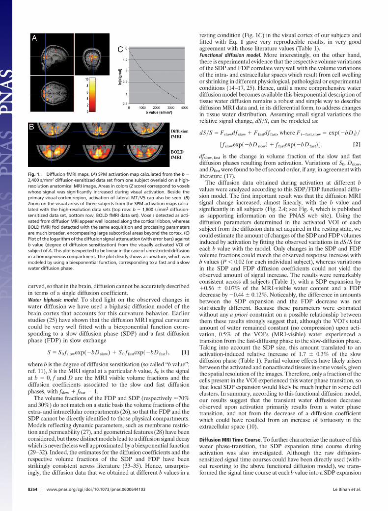
curved, so that in the brain, diffusion cannot be accurately describedin terms of a single diffusion coefficient.Water biphasic model. To shed light on the observed changes inwater diffusion we have used a biphasic diffusion model of thebrain cortex that accounts for this curvature behavior. Earlierstudies (25) have shown that the diffusion MRI signal curvaturecould be very well fitted with a biexponential function corre-sponding to a slow diffusion phase (SDP) and a fast diffusionphase (FDP) in slow exchange
S � S0 fslowexp(�bD slow) � S0 f fastexp��bD fast� , [1]
where b is the degree of diffusion sensitisation (so called ‘‘b value’’;ref. 11), S is the MRI signal at a particular b value, S0 is the signalat b � 0, f and D are the MRI visible volume fractions and thediffusion coefficients associated to the slow and fast diffusionphases, with fslow � ffast � 1.
The volume fractions of the FDP and SDP (respectively �70%and 30%) do not match on a static basis the volume fractions of theextra- and intracellular compartments (26), so that the FDP and theSDP cannot be directly identified to those physical compartments.Models reflecting dynamic parameters, such as membrane restric-tion and permeability (27), and geometrical features (28) have beenconsidered, but those distinct models lead to a diffusion signal decaywhich is nevertheless well approximated by a biexponential function(29–32). Indeed, the estimates for the diffusion coefficients and therespective volume fractions of the SDP and FDP have beenstrikingly consistent across literature (33–35). Hence, unsurpris-ingly, the diffusion data that we obtained at different b values in a
resting condition (Fig. 1C) in the visual cortex of our subjects andfitted with Eq. 1 gave very reproducible results, in very goodagreement with those literature values (Table 1).Functional diffusion model. More interestingly, on the other hand,there is experimental evidence that the respective volume variationsof the SDP and FDP correlate very well with the volume variationsof the intra- and extracellular spaces which result from cell swellingor shrinking in different physiological, pathological or experimentalconditions (14–17, 25). Hence, until a more comprehensive waterdiffusion model becomes available this biexponential description oftissue water diffusion remains a robust and simple way to describediffusion MRI data and, in its differential form, to address changesin tissue water distribution. Assuming small signal variations therelative signal change, dS�S, can be modeled as:
dS�S � Fslowdf slow � F fastdf fast, where F i�fast,slow � exp��bD i��
�f slowexp��bD slow� � f fastexp��bD fast�� . [2]
dfslow, fast is the change in volume fraction of the slow and fastdiffusion phases resulting from activation. Variations of S0, Dslow,and Dfast were found to be of second order, if any, in agreement withliterature (17).
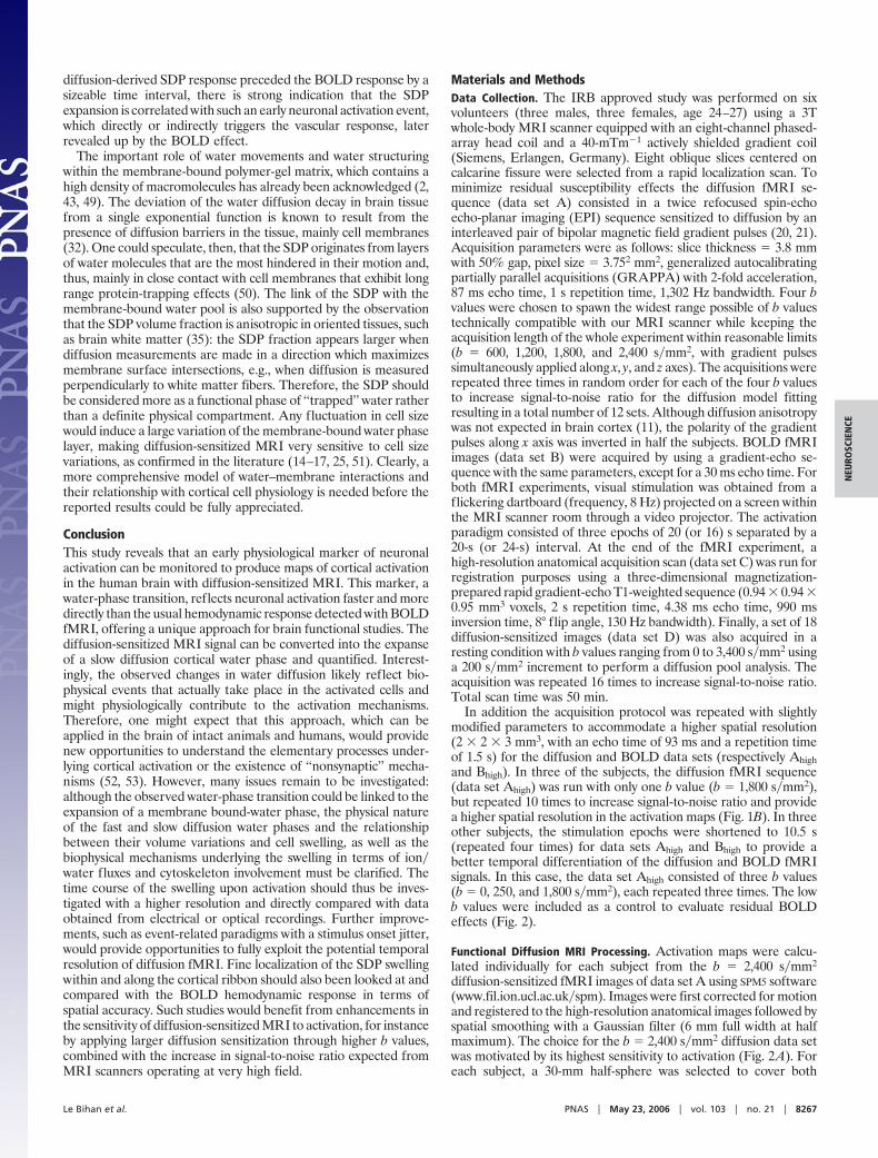
The diffusion data obtained during activation at different bvalues were analyzed according to this SDP�FDP functional diffu-sion model. The first important result was that the diffusion MRIsignal change increased, almost linearly, with the b value andsignificantly in all subjects (Fig. 2A; see Fig. 4, which is publishedas supporting information on the PNAS web site). Using thediffusion parameters determined in the activated VOI of eachsubject from the diffusion data set acquired in the resting state, wecould estimate the amount of changes of the SDP and FDP volumesinduced by activation by fitting the observed variations in dS�S foreach b value with the model. Only changes in the SDP and FDPvolume fractions could match the observed response increase withb values (P 0.02 for each individual subject), whereas variationsin the SDP and FDP diffusion coefficients could not yield theobserved amount of signal increase. The results were remarkablyconsistent across all subjects (Table 1), with a SDP expansion by�0.56 0.07% of the MRI-visible water content and a FDPdecrease by �0.44 0.12%. Noticeably, the difference in amountsbetween the SDP expansion and the FDP decrease was notstatistically different. Because these parameters were estimatedwithout any a priori constraint on a possible relationship betweenthem these results strongly suggest that, although the VOI’s totalamount of water remained constant (no compression) upon acti-vation, 0.5% of the VOI’s (MRI-visible) water experienced atransition from the fast-diffusing phase to the slow-diffusion phase.Taking into account the SDP size, this amount translated to anactivation-induced relative increase of 1.7 0.3% of the slowdiffusion phase (Table 1). Partial volume effects have likely arisenbetween the activated and nonactivated tissues in some voxels, giventhe spatial resolution of the images. Therefore, only a fraction of thecells present in the VOI experienced this water phase transition, sothat local SDP expansion would likely be much higher in some cellclusters. In summary, according to this functional diffusion model,our results suggest that the transient water diffusion decreaseobserved upon activation primarily results from a water phasetransition, and not from the decrease of a diffusion coefficientwhich could have resulted from an increase of tortuosity in theextracellular space (10).
Diffusion MRI Time Course. To further characterize the nature of thiswater phase-transition, the SDP expansion time course duringactivation was also investigated. Although the raw diffusion-sensitized signal time courses could have been directly used (with-out resorting to the above functional diffusion model), we trans-formed the signal time course at each b value into a SDP expansion
Fig. 1. Diffusion fMRI maps. (A) SPM activation map calculated from the b �2,400 s�mm2 diffusion-sensitized data set from one subject overlaid on a high-resolution anatomical MRI image. Areas in colors (Z score) correspond to voxelswhose signal was significantly increased during visual activation. Beside theprimary visual cortex region, activation of lateral MT�V5 can also be seen. (B)Zoom on the visual areas of three subjects from the SPM activation maps calcu-lated with the high-resolution data sets (top row: b � 1,800 s�mm2 diffusion-sensitized data set, bottom row, BOLD fMRI data set). Voxels detected as acti-vated from diffusion MRI appear well located along the cortical ribbon, whereasBOLD fMRI foci detected with the same acquisition and processing parametersare much broader, encompassing large subcortical areas beyond the cortex. (C)Plot of the logarithm of the diffusion signal attenuation (with error bars) againstb value (degree of diffusion sensitization) from the visually activated VOI ofsubject of A. This plot is expected to be linear in the case of unrestricted diffusionin a homogeneous compartment. The plot clearly shows a curvature, which wasmodeled by using a biexponential function, corresponding to a fast and a slowwater diffusion phase.
8264 � www.pnas.org�cgi�doi�10.1073�pnas.0600644103 Le Bihan et al.

time course. According to the functional diffusion model, theexpansion amount no longer depends on b values, and the VOIsignal from all data sets can be merged into a single SDP expansiontime course to provide a better signal-to-noise ratio. In line with theabove results (phase-transition), the decrease in the fast volumefraction was assumed to balance the increase in the slow volumefraction, i.e., dffast � �dfslow, so that the relative change of the slowdiffusion phase volume, dfslow�fslow, could be directly calculatedfrom the signal change, dS�S:
dfslow�f slow � K dS�S , where K � �f slowexp��bD slow�
� �1 � f slow�exp��bD fast���
�f slow�exp��bD slow� � exp��bD fast��� . [3]
A comparison was made with the usual BOLD fMRI signal timecourse extracted from the VOI of each subject. In agreement with
literature (36), the BOLD signal time course exhibited a shallow‘‘initial dip’’ and a poststimulus undershoot (Fig. 2A; see Fig. 5,which is published as supporting information on the PNAS website). By contrast, the diffusion-derived SDP expansion time courseshowed a sharp increase at stimulus onset, but neither any initial dipnor any clear poststimulus undershoot. Overall, the BOLD andSDP expansion time courses were very similar in shape. However,the most striking feature was that the SDP time course was alwaysahead of the BOLD time course by several seconds, most noticeablyat the beginning of the activation period, but often also at its end.To quantify the advance of the SDP response, the BOLD timecourse was shifted in time, and a normalized rms difference(nRMS) was calculated between the SDP and the time-shiftedBOLD time course (Fig. 2A; see Fig. 6, which is published assupporting information on the PNAS web site). The nRMS mini-mum value provided an estimate of the time delay, which made bothtime courses mostly similar. This delay was very consistent across all
Fig. 2. Amplitude and time course analysis. (A Left) Plots of the relative signal increase dS�S � f(b) for the VOI of one representative subject (SD for each datapoint, statistical t test comparing pairs of b values; ns, not significant; *, P 0.05; **, P 0.01; ***, P 0.001). The red curve corresponds to the theoretical signalincrease expected from the biphasic diffusion model with parameters adjusted to the data. (A Center) Time courses of the SDP expansion amount (red) and ofthe normalized BOLD fMRI signal (blue) of one subject. The BOLD signal time course exhibits a shallow ‘‘initial dip’’ and a poststimulus undershoot. By contrast,the SDP time course presents a steeper onset without any initial dip. Although the BOLD and SDP time courses are very similar in shape, the SPD time course isalways ahead of the BOLD time course. This diffusion advance is mostly noticeable at the beginning of the activation response. (A Right) Normalized rmsdifference, nRMS( t), between the diffusion-derived SDP swelling time course and the BOLD signal time course shifted by a time interval t (U-shape curve, nRMS;bottom curve, temporal derivative). nRMS is minimized for t 0, as best seen from the zero-crossing of the nRMS( t) temporal derivative. (B Left) Time coursesof the diffusion MRI (b � 0, 250 and 1,800 s�mm2) and the BOLD fMRI signals with the box-car activation paradigm of one representative subject (left, raw signalchange; right, normalized signal change). A large advance of the MRI signal with high diffusion sensitization (b � 1,800 s�mm2) over the BOLD response can beseen, both at onset and offset. The diffusion MRI response appears well locked to the activation paradigm, whereas the BOLD fMRI response is shifted by severalseconds. On the other hand, the increase signal observed without diffusion sensitization (b � 0 s�mm2) shows a time course perfectly synchronized with the BOLDresponse. The signal change observed at low diffusion-sensitization (b � 250 s�mm2) is significantly lower than that seen at high b value. At low diffusion-sensitization, the signal time course mixes the diffusion time course observed at higher b value (early onset) with that of the vascular, BOLD component (lateoffset) which is totally removed at b � 1,800 s�mm2. (B Right) Normalized rms difference, nRMS( t), between the BOLD signal time course shifted by a timeinterval t and the b � 1,800 s�mm2 diffusion MRI signal time course (left) or the b � 0 s�mm2 diffusion MRI signal time course (right). Although the b � 0 s�mm2
and BOLD time course exactly coincide ( t � 0), the nRMS between b � 1,800 s�mm2 diffusion MRI and BOLD time courses is clearly minimized for t 0.
Le Bihan et al. PNAS � May 23, 2006 � vol. 103 � no. 21 � 8265
NEU
ROSC
IEN
CE

our subjects, with the diffusion-derived SDP response preceding theBOLD response by an average of 2.4 0.7 s (Table 1).
This major result clearly demonstrates that the observed diffu-sion changes and the BOLD effect refer to rather different phys-iological events underlying cortical activation. The BOLD effect hasbeen shown to originate from local changes in the blood deoxyhe-moglobin concentration, which mainly result from the increase inblood flow and blood volume induced by neuronal activation (24,36). Therefore, it is a vascular event and, although BOLD fMRI hasbeen extremely successful for the functional neuroimaging com-munity, presents well known limitations. Although the existence ofa coupling between neuronal activation, metabolism, and bloodflow has been verified in most instances including BOLD fMRI (9),the degree and the mechanism of coupling remain largely notunderstood (37). This is an important concern for the applicationof fMRI in the clinical field, because this coupling might fail in somedisease cases (38). Also, it has been pointed out that the spatialfunctional resolution of BOLD fMRI might be limited, becausevessels responsible for the increase of blood flow and blood volumefeed or drain somewhat large territories, whereas the physiologicaldelay necessary for the mechanisms triggering the vascular responseto work intrinsically limits the temporal resolution of BOLD fMRI.
Origin of the Observed MRI Signal Changes. Water diffusion origin. Thesignificant variations of the observed signal responses upon activa-tion with the amount of diffusion sensitization (b values) sign theirdiffusion origin. Residual BOLD effects could also mimic diffusioneffects through variations in the magnetic susceptibility-inducedlocal field gradients, as noted above, but such effects should becompletely negligible given the particular diffusion sequence usedin this study, which is confirmed by the clear time differencebetween the diffusion and the BOLD time courses. However, itremains that small BOLD induced signal variations due to changesin R2�R2* relaxation in intravascular blood have been reportedwhen using spin-echo sequences with very low b values (39). Toevaluate such residual contribution, and to further provide evidenceof the nonvascular origin of the diffusion MRI changes, controlacquisitions were obtained with a higher spatial resolution and ashorter stimulation duration to enhance the time differences be-tween the diffusion and BOLD effects. The results confirm thelarge advance of the MRI signal with high diffusion sensitization(b � 1,800 s�mm2) (up to 4 s) over the BOLD response (Fig. 2B,Table 2, and Fig. 7, which is published as supporting information on
the PNAS web site), even larger than with a longer stimulus (Fig.2A). On the other hand, the signal increase, which is seen with thespin-echo sequence without diffusion sensitization (b � 0 s�mm2),has a time course that perfectly matches the BOLD response, sothat one may reasonably think it shares its vascular origin. Thisfinding is further confirmed by the significant decrease in signalresponse observed when adding a small amount of diffusion-sensitization (b � 250 s�mm2). The magnetic field gradient pulsesused for diffusion sensitization also induce velocity-dependentphase shifts in the presence of flow and suppress signal fromflowing blood due to the intravoxel incoherent motion effect (40).Adding a small degree of diffusion sensitization, therefore,‘‘crushes’’ the vascular component of the BOLD signal (39). Veryinterestingly, at b � 250 s�mm2, the signal temporal profile mixedthe diffusion time course observed at higher b values (early onset)with a residual vascular (BOLD) component (late offset), which istotally removed at higher b values. The signal change at b � 1,800s�mm2 was significantly larger than that observed at b � 250 s�mm2
(Table 2), in line with the other results of this report. In addition,there was no correlation (Table 1, r � �0.11) between theamplitude of the SDP expansion and that of the BOLD signal, asmeasured before normalization, further supporting the fact that thediffusion-derived signal and the BOLD response have differentorigins. In summary, those results confirm that (i) the observedsignal changes have a true diffusion origin and (ii) the mechanismof the diffusion response is not vascular.Link with cell physiology. In the brain, cell swelling is an importantphysiological response associated with neuronal activation (4, 7,41–43). Such swelling not only involves neuronal soma, but alsoaxons and focal areas along dendrites (42, 44), and probably alsoglial cells (45). The amount of swelling cannot be ascribed to asimple translocation of water from the extra to the intracellularspace to compensate for fluctuations in intracellular osmolarity dueto transient fluxes of ions (46), and further studies have underlinedthe importance of the cytoskeleton, a dense polymer-gel matrixrunning contiguously to the cell membrane (43, 47). Therefore,cortical cell swelling and its active regulation appear of fundamentalimportance to neuronal function. Interestingly, swelling and mem-brane expansion have been shown to start simultaneously with theelectric response and the peak of the mechanical response tocoincide accurately with the action potential peak. The response isasymmetric, as the swelling presents a sharp increase, whereas thereturn to baseline is smooth and monotonic (43, 48). Because the
Table 1. Data from all subjects
SubjectVOI size,
voxelsSDP
fraction, % dfslow, %SDP swellingamount, %*
BOLDsignal
increase, % dffast, %[SDP swelling-BOLD]
time shift, s
Ta . . . 72 35.2 0.50 0.06 1.4 2.3 �0.35 0.23 �1.9 0.5Na . . . 127 34.1 0.65 0.05 1.9 2.8 �0.40 0.22 �2.2 0.5Hi . . . 127 34.0 0.49 0.06 1.4 2.4 �0.52 0.23 �2.2 0.5Ba . . . 90 29.9 0.62 0.10 2.1 2.3 �0.64 0.33 �1.9 0.5Ya . . . 146 32.2 0.50 0.06 1.5 2.8 �0.34 0.30 �3.2 0.5Ch . . . 175 34.0 0.60 0.10 1.8 1.7 �0.37 0.36 �3.2 0.5Mean 123 38 33.1 2.1 0.56 0.07 1.7 0.3 2.4 0.4 �0.44 0.12 �2.4 0.7
*Slow diffusion pool swelling amount was calculated as dfslow�fslow
Table 2. High-resolution and control data
Subject
b � 0signal
change, %
b � 250signal
change, %
b � 1,800signal
change, %
b � 1,800�b � 250(t test)
BOLDsignal
change, %[b � 1,800-BOLD]
time shift, s[b � 0-BOLD]time shift, s
1 0.96 0.06 0.69 0.09 2.02 0.18 P 5.10�5 2.07 0.07 �2.7 0.8 �0.0 0.82 1.15 0.07 0.64 0.11 1.73 0.27 P 2.10�4 2.02 0.18 �3.0 0.8 �0.0 0.83 0.54 0.13 0.33 0.08 1.91 0.28 P 5.10�5 1.53 0.12 �4.0 0.8 �0.1 0.8
8266 � www.pnas.org�cgi�doi�10.1073�pnas.0600644103 Le Bihan et al.
diffusion-derived SDP response preceded the BOLD response by asizeable time interval, there is strong indication that the SDPexpansion is correlated with such an early neuronal activation event,which directly or indirectly triggers the vascular response, laterrevealed up by the BOLD effect.
The important role of water movements and water structuringwithin the membrane-bound polymer-gel matrix, which contains ahigh density of macromolecules has already been acknowledged (2,43, 49). The deviation of the water diffusion decay in brain tissuefrom a single exponential function is known to result from thepresence of diffusion barriers in the tissue, mainly cell membranes(32). One could speculate, then, that the SDP originates from layersof water molecules that are the most hindered in their motion and,thus, mainly in close contact with cell membranes that exhibit longrange protein-trapping effects (50). The link of the SDP with themembrane-bound water pool is also supported by the observationthat the SDP volume fraction is anisotropic in oriented tissues, suchas brain white matter (35): the SDP fraction appears larger whendiffusion measurements are made in a direction which maximizesmembrane surface intersections, e.g., when diffusion is measuredperpendicularly to white matter fibers. Therefore, the SDP shouldbe considered more as a functional phase of ‘‘trapped’’ water ratherthan a definite physical compartment. Any fluctuation in cell sizewould induce a large variation of the membrane-bound water phaselayer, making diffusion-sensitized MRI very sensitive to cell sizevariations, as confirmed in the literature (14–17, 25, 51). Clearly, amore comprehensive model of water–membrane interactions andtheir relationship with cortical cell physiology is needed before thereported results could be fully appreciated.
ConclusionThis study reveals that an early physiological marker of neuronalactivation can be monitored to produce maps of cortical activationin the human brain with diffusion-sensitized MRI. This marker, awater-phase transition, reflects neuronal activation faster and moredirectly than the usual hemodynamic response detected with BOLDfMRI, offering a unique approach for brain functional studies. Thediffusion-sensitized MRI signal can be converted into the expanseof a slow diffusion cortical water phase and quantified. Interest-ingly, the observed changes in water diffusion likely reflect bio-physical events that actually take place in the activated cells andmight physiologically contribute to the activation mechanisms.Therefore, one might expect that this approach, which can beapplied in the brain of intact animals and humans, would providenew opportunities to understand the elementary processes under-lying cortical activation or the existence of ‘‘nonsynaptic’’ mecha-nisms (52, 53). However, many issues remain to be investigated:although the observed water-phase transition could be linked to theexpansion of a membrane bound-water phase, the physical natureof the fast and slow diffusion water phases and the relationshipbetween their volume variations and cell swelling, as well as thebiophysical mechanisms underlying the swelling in terms of ion�water fluxes and cytoskeleton involvement must be clarified. Thetime course of the swelling upon activation should thus be inves-tigated with a higher resolution and directly compared with dataobtained from electrical or optical recordings. Further improve-ments, such as event-related paradigms with a stimulus onset jitter,would provide opportunities to fully exploit the potential temporalresolution of diffusion fMRI. Fine localization of the SDP swellingwithin and along the cortical ribbon should also been looked at andcompared with the BOLD hemodynamic response in terms ofspatial accuracy. Such studies would benefit from enhancements inthe sensitivity of diffusion-sensitized MRI to activation, for instanceby applying larger diffusion sensitization through higher b values,combined with the increase in signal-to-noise ratio expected fromMRI scanners operating at very high field.
Materials and MethodsData Collection. The IRB approved study was performed on sixvolunteers (three males, three females, age 24–27) using a 3Twhole-body MRI scanner equipped with an eight-channel phased-array head coil and a 40-mTm�1 actively shielded gradient coil(Siemens, Erlangen, Germany). Eight oblique slices centered oncalcarine fissure were selected from a rapid localization scan. Tominimize residual susceptibility effects the diffusion fMRI se-quence (data set A) consisted in a twice refocused spin-echoecho-planar imaging (EPI) sequence sensitized to diffusion by aninterleaved pair of bipolar magnetic field gradient pulses (20, 21).Acquisition parameters were as follows: slice thickness � 3.8 mmwith 50% gap, pixel size � 3.752 mm2, generalized autocalibratingpartially parallel acquisitions (GRAPPA) with 2-fold acceleration,87 ms echo time, 1 s repetition time, 1,302 Hz bandwidth. Four bvalues were chosen to spawn the widest range possible of b valuestechnically compatible with our MRI scanner while keeping theacquisition length of the whole experiment within reasonable limits(b � 600, 1,200, 1,800, and 2,400 s�mm2, with gradient pulsessimultaneously applied along x, y, and z axes). The acquisitions wererepeated three times in random order for each of the four b valuesto increase signal-to-noise ratio for the diffusion model fittingresulting in a total number of 12 sets. Although diffusion anisotropywas not expected in brain cortex (11), the polarity of the gradientpulses along x axis was inverted in half the subjects. BOLD fMRIimages (data set B) were acquired by using a gradient-echo se-quence with the same parameters, except for a 30 ms echo time. Forboth fMRI experiments, visual stimulation was obtained from aflickering dartboard (frequency, 8 Hz) projected on a screen withinthe MRI scanner room through a video projector. The activationparadigm consisted of three epochs of 20 (or 16) s separated by a20-s (or 24-s) interval. At the end of the fMRI experiment, ahigh-resolution anatomical acquisition scan (data set C) was run forregistration purposes using a three-dimensional magnetization-prepared rapid gradient-echo T1-weighted sequence (0.94 � 0.94 �0.95 mm3 voxels, 2 s repetition time, 4.38 ms echo time, 990 msinversion time, 8° flip angle, 130 Hz bandwidth). Finally, a set of 18diffusion-sensitized images (data set D) was also acquired in aresting condition with b values ranging from 0 to 3,400 s�mm2 usinga 200 s�mm2 increment to perform a diffusion pool analysis. Theacquisition was repeated 16 times to increase signal-to-noise ratio.Total scan time was 50 min.
In addition the acquisition protocol was repeated with slightlymodified parameters to accommodate a higher spatial resolution(2 � 2 � 3 mm3, with an echo time of 93 ms and a repetition timeof 1.5 s) for the diffusion and BOLD data sets (respectively Ahighand Bhigh). In three of the subjects, the diffusion fMRI sequence(data set Ahigh) was run with only one b value (b � 1,800 s�mm2),but repeated 10 times to increase signal-to-noise ratio and providea higher spatial resolution in the activation maps (Fig. 1B). In threeother subjects, the stimulation epochs were shortened to 10.5 s(repeated four times) for data sets Ahigh and Bhigh to provide abetter temporal differentiation of the diffusion and BOLD fMRIsignals. In this case, the data set Ahigh consisted of three b values(b � 0, 250, and 1,800 s�mm2), each repeated three times. The lowb values were included as a control to evaluate residual BOLDeffects (Fig. 2).
Functional Diffusion MRI Processing. Activation maps were calcu-lated individually for each subject from the b � 2,400 s�mm2
diffusion-sensitized fMRI images of data set A using SPM5 software(www.fil.ion.ucl.ac.uk�spm). Images were first corrected for motionand registered to the high-resolution anatomical images followed byspatial smoothing with a Gaussian filter (6 mm full width at halfmaximum). The choice for the b � 2,400 s�mm2 diffusion data setwas motivated by its highest sensitivity to activation (Fig. 2A). Foreach subject, a 30-mm half-sphere was selected to cover both
Le Bihan et al. PNAS � May 23, 2006 � vol. 103 � no. 21 � 8267
NEU
ROSC
IEN
CE
calcarine fissures. Within this sphere, a VOI was defined from thevoxels classified as activated from the b � 2,400 s�mm2 activationmap (P 0.001, uncorrected for multiple comparisons). Thisdiffusion-defined VOI was then used subsequently to extract datafrom all diffusion and BOLD sets (data sets A, B, and D). The sizeof the VOI for each subject is given in Table 1. The same SPManalysis was performed on the b � 1,800 s�mm2 diffusion-sensitizedfMRI images of data set Ahigh and the BOLD fMRI images of dataset Bhigh (Fig. 2), but with a boxcar basis function to avoid any bias.
Diffusion Pool Analysis. The VOI averaged signal from the imagesof data set D was extracted for each of the 18 b values and for eachindividual subject. The resulting signal, S, was averaged over the 16diffusion acquisitions and plotted against the b value for eachsubject (Fig. 1C) (35). The first three points were discarded to avoidpossible partial volume effects from cerebrospinal fluid. A nonlin-ear Marquardt algorithm was used to estimate from the diffusion-sensitized signal at each b value the diffusion parameters, f and D,of the slow and fast diffusion phases in resting condition for eachsubject VOI according to the biexponential model (Eq. 1).
Diffusion Functional Model Processing. The VOI averaged signaltime course from data set A was extracted for each individual bvalue (600, 1,200, 1,800, and 2,400 s�mm2). The resulting signal wasaveraged over the three acquisitions acquired at each b value,linearly corrected for any baseline drift for each b value (Fig. 1C).The signal from the resting and activated conditions were thenpooled separately to estimate the signal change (dS�S) induced byactivation for each b value. A least-squares bilinear fitting algorithmwas used to estimate the respective changes, dfslow and dffast, fromthe signal changes, dS�S, observed for each b value (Eq. 2) and thediffusion parameters previously determined for the VOI of eachsubject. Both parameters were considered a priori as free (withoutthe fslow � ffast � 1 constraint). The observed and the predictedsignal changes, dS�S, were plotted against b values for each subjectVOI (Fig. 2).
Time Course Analysis. The VOI averaged, b value averaged, baselinedrift corrected signal time course for each b value of data set A was
then folded into a single [activation � rest] epoch by averaging thethree subsequent epochs of the paradigm. The [dS�S](t) time coursefor each b value was transformed into a [dfslow�dfslow](t) time course(Eq. 3), with [dS�S](t) defined as S(t)�S(baseline) � 1. S(baseline)was obtained by averaging signal during the resting condition.Because dfslow�dfslow is now a physiological parameter that does notdepend on the b value, the [dfslow�dfslow](t) time courses wereaveraged over the four b values.
For comparison, the averaged BOLD fMRI signal time coursewas extracted from data set B using the diffusion-defined VOI ofeach subject and linearly corrected for any baseline drift. The rawBOLD fMRI signal time course was expressed as a relative changeto baseline, [dSBOLD�SBOLD](t), defined as SBOLD(t)�SBOLD(base-line) � 1. SBOLD(baseline) was obtained by averaging signal duringthe resting condition. The BOLD fMRI time course was thenfolded into a single [activation � rest] epoch by averaging the threesubsequent epochs of the paradigm. Finally, as the BOLD fMRIsignal intensity is arbitrary the [dSBOLD�SBOLD](t) time course wasnormalized, so that, for timing comparison only, the amplitude ofits envelope matched that of the [dfslow�dfslow](t) time course. Thenormalized [dSBOLD�SBOLD](t) and the [dfslow�dfslow](t) timecourses were finally plotted together for the VOI of each subject(Fig. 2A).
To obtain a quantitative estimation of the time shift between the[dfslow�dfslow](t) and the [dSBOLD�SBOLD](t) time courses, theBOLD fMRI time course was shifted by a time interval t from �6to �4 s. The resulting [dSBOLD�SBOLD](t � t) time course wascompared with the [dfslow�dfslow](t) time course by calculating thenormalized rms difference, nRMS( t), between the activated ep-ochs of the two time courses, which was plotted against t (Fig. 2A).The time shift minimizing the differences between the [dfslow�dfslow](t) and [dSBOLD�SBOLD](t) time courses was estimated fromthe lowest nRMS value, as derived from the zero-crossing of its timederivative. The same processing was performed on the Ahigh andBhigh data sets to compare the time courses of the BOLD and b �1,800 s�mm2 diffusion data sets on one hand, and of the BOLD andb � 0 s�mm2 diffusion data sets on the other hand (Fig. 2B).
We thank Stanislas Dehaene and Andre Syrota for insightful commentsabout the manuscript.
1. Cohen, L. B., Keynes, R. D. & Hille, B. (1968) Nature 218, 438–441.2. Tasaki, I. & Byrne, P. M. (1993) Jpn. J. Physiol. 43, Suppl. 1, S67–S75.3. Tasaki, I. & Iwasa, K. (1982) Jpn. J Physiol. 32, 69–81.4. Andrew, R. D. & Macvicar, B. A. (1994) Neuroscience 62, 371–383.5. Schwartzkroin, P. A., Baraban, S. C. & Hochman, D. W. (1998) Epilepsy Res. 32, 275–285.6. Sykova, E., Vargova, L., Kubinova, D., Jendelova, P. & Chvatal, A. (2003) NeuroImage 18, 214–230.7. Holthoff, K. & Witte, O. W. (1996) J. Neurosci. 16, 2740–2749.8. Ogawa, S., Tank, D. W., Menon, R. S., Ellerman, J. M., Kim, S. G., Merkle, H. & Ugurbil,
K. (1992) Proc. Natl. Acad. Sci. USA 89, 5951–5955.9. Logothetis, N. K., Pauls, J., Augath, M., Trinath, T. & Oeltermann, A. (2001) Nature 412,
150–157.10. Darquie, A., Poline, J. B., Poupon, C., Saint-Jalmes, H. & Le Bihan, D. (2001) Proc. Natl.
Acad. Sci. USA 98, 9391–9395.11. Le Bihan, D. (2003) Nat. Rev. Neurosci. 4, 469–480.12. Zhong, J., Petroff, O. A. C., Pleban, L. A., Gore, J. C. & Prichard, J. W. (1997) Magn. Reson.
Med. 37, 1–6.13. Latour, L. L., Hasegawa, Y., Formato, J. E., Fisher, M. & Sotak, C. H. (1994) Magn. Reson.
Med. 32, 189–198.14. O’Shea, J. M., Williams, S. R., van Bruggen, N. & Gardner-Medwin, A. R. (2000) Magn.
Reson. Med. 44, 427–432.15. Hasegawa, Y., Formato, J. E., Latour, L. L., Gutierrez, J. A., Liu, K. F., Garcia, J. H., Sotak,
C. H. & Fisher, M. (1996) Stroke 27, 1648–1655.16. Van Der Toorn, A., Sykova, E., Dijkhuizen, R. M., Vorisek, I., Vargova, L., Skobisova, E.,
Van Lookeren Campagne, M., Reese, T. & Nicolay, K. (1996) Magn. Reson. Med. 36, 52–60.17. Buckley, D. L., Bui, J. D., Phillips, M. I., Zelles, T., Inglis, B. A., Plant, H. D. & Blackband,
S. J. (1999) Magn. Reson. Med. 41, 137–142.18. Chen, K. C. & Nicholson, C. (2000) Proc. Natl. Acad. Sci. USA 97, 8306–8311.19. Kubova, Z., Kuba, M., Spekreijse, H. & Blakemore, C. (1995) Vision Res. 35, 197–205.20. Reese, T. G., Heid, O., Weisskoff, R. M. & Wedeen, V. J. (2003) Magn. Reson. Med. 49, 177–182.21. Karlicek, R. F. & Lowe, I. J. (1980) J. Magn. Reson. 37, 75–91.22. Does, M. D., Zhong, J. & Gore, J. C. (1999) Magn. Reson. Med. 41, 236–240.23. Kiselev, V. G. (2004) J. Magn. Reson. 170, 228–235.24. Ogawa, S., Menon, R. S., Tank, D. W., Kim, S. G., Merkle, H., Ellermann, J. M. & Ugurbil,
K. (1993) Biophys. J. 64, 803–808.25. Niendorf, T., Dijkhuizen, R. M., Norris, D. G., Van Lookeren Campagne, M. & Nicolay,
K. (1996) Magn. Reson. Med. 36, 847–857.26. Sehy, J. V., Ackerman, J. J. H. & Neil, J. J. (2002) Magn. Reson. Med. 48, 765–770.
27. Novikov, E. G., Van Dusschoten, D. & VanAs, H. (1998) J. Magn. Reson. 135, 522–528.28. vanderWeerd, L., Melnikov, S. M., Vergeldt, F. J., Novikov, E. G. & VanAs, H. (2002) J.
Magn. Reson. 156, 213–221.29. Kroenke, C. D., Ackerman, J. J. H. & Yablonskiy, D. A. (2004) Magn. Reson. Med. 52, 1052–1059.30. Chin, C. L., Wehrli, F. W., Fan, Y. L., Hwang, S. N., Schwartz, E. D., Nissanov, J. & Hackney,
D. B. (2004) Magn. Reson. Med. 52, 733–740.31. Yablonskiy, D. A., Bretthorst, G. L. & Ackerman, J. J. H. (2003) Magn. Reson. Med. 50, 664–669.32. Sukstanskii, A. L., Yablonskiy, D. A. & Ackerman, J. J. H. (2004) J. Magn. Reson. 170, 56–66.33. Mulkern, R. V., Gudbjartsson, H., Westin, C. F., Zengingonul, H. P., Gartner, W.,
Guttmann, C. R. G., Robertson, R. L., Kyriakos, W., Schwartz, R., Holtzman, D. et al. (1999)NMR Biol. 12, 51–62.
34. Maier, S. E., Bogner, P., Bajzik, G., Mamata, H., Mamata, Y., Repa, I., Jolesz, F. A. &Mulkern, R. V. (2001) Radiology 219, 842–849.
35. Clark, C. A. & Le Bihan, D. (2000) Magn. Reson. Med. 44, 852–859.36. Buxton, R. B., Wong, E. C. & Frank, L. R. (1998) Magn. Reson. Med. 39, 855–864.37. Mangia, S., Giove, F., Bianciardi, M., Di Salle, F., Garreffa, G. & Maraviglia, B. (2003)
J. Neurosci. Res. 71, 463–467.38. Lehericy, S., Biondi, A., Sourour, N., Vlaicu, M., du Montcel, S. T., Cohen, L., Vivas, E.,
Capelle, L., Faillot, T., Casasco, A., et al. (2002) Radiology 223, 672–682.39. Duong, T. Q., Yacoub, E., Adriany, G., Hu, X. P., Ugurbil, K. & Kim, S. G. (2003) Magn.
Reson. Med. 49, 1019–1027.40. Le Bihan, D., Breton, E., Lallemand, D., Aubin, M. L., Vignaud, J. & Laval Jeantet, M.
(1988) Radiology 168, 497–505.41. Inglefield, J. R. & Schwartz-Bloom, R. D. (1998) J. Neurochem. 71, 1396–1404.42. Takagi, S., Obata, K. & Tsubokawa, H. (2002) Neurosci. Res. 44, 315–324.43. Tasaki, I. (1999) Jpn. J. Physiol. 49, 125–138.44. Inoue, H., Mori, S. i., Morishima, S. & Okada, Y. (2005) Eur. J. Neurosci. 21, 1648–1658.45. Macvicar, B. A., Feighan, D., Brown, A. & Ransom, B. (2002) Glia 37, 114–123.46. Tasaki, I. & Byrne, P. M. (1990) Biophys. J. 57, 633–635.47. Metuzals, J. & Tasaki, I. (1978) J. Cell. Biol. 78, 597–621.48. Tasaki, I. & Byrne, P. M. (1992) Biochem. Biophys. Res. Commun. 188, 559–564.49. Tsukita, S., Tsukita, S., Kobayashi, T. & Matsumoto, G. (1986) J. Cell. Biol. 102, 1710–1725.50. Xu, X. H. N. & Yeung, E. S. (1998) Science 281, 1650–1653.51. Anderson, A. W., Xie, J., Pizzonia, J., Bronen, R. A., Spencer, D. D. & Gore, J. C. (2000)
Magn. Reson. Imaging 18, 689–695.52. Jefferys, J. G. R. (1995) Physiol. Rev. 75, 689–715.53. Muller, V., Birbaumer, N., Preissl, H., Braun, C. & Lang, F. (2002) Eur. J. Neurosci. 15, 528–538.
8268 � www.pnas.org�cgi�doi�10.1073�pnas.0600644103 Le Bihan et al.