direct optical sensors: principles and selected applications
TRANSCRIPT

REVIEW
Guenter Gauglitz
Direct optical sensors: principles and selected applications
Received: 14 July 2004 / Revised: 11 October 2004 / Accepted: 11 October 2004 / Published online: 11 November 2004
� Springer-Verlag 2004
Abstract In the field of bio and chemosensors a largenumber of detection principles has been publishedwithin the last decade. These detection principles arebased either on the observation of fluorescence-labelledsystems or on direct optical detection in the heteroge-neous phase. Direct optical detection can be measuredby remission (absorption of reflected radiation, op-t(r)odes), by measuring micro-refractivity, or measuringinterference. In the last case either Mach–Zehnderinterferometers or measurement of changes in thephysical thickness of the layer (measuring micro-reflec-tivity) caused, e.g., by swelling effects in polymers (dueto interaction with analytes) or in bioassays (due toaffinity reactions) also play an important role. Here, anoverview of methods of microrefractometric and mi-croreflectometric principles is given and benefits anddrawbacks of the various approaches are demonstratedusing samples from the chemo and biosensor field. Thequality of sensors does not just depend on transductionprinciples but on the total sensor system defined by thistransduction, the sensitive layer, data acquisition elec-tronics, and evaluation software. The intention of thisarticle is, therefore, to demonstrate the essentials of theinteraction of these parts within the system, and thefocus is on optical sensing using planar transducers,because fibre optical sensors have been reviewed in thisjournal only recently. Lack of selectivity of chemosen-sors can be compensated either by the use of sensorarrays or by evaluating time-resolved measurements ofanalyte/sensitive layer interaction. In both caseschemometrics enables the quantification of analytemixtures. These data-processing methods have also beensuccessfully applied to antibody/antigen interactionseven using cross-reactive antibodies. Because miniaturi-
sation and parallelisation are essential approaches inrecent years, some aspects and current trends, especiallyfor bio-applications, will be discussed. Miniaturisation isespecially well covered in the literature.
Keywords Optical Æ Chemosensing Æ Biosensing ÆTransducer Æ Application
Introduction
Because fibre optical sensors have recently been re-viewed in this journal [1], this article covers the princi-ples and applications of planar-type optical sensors, andcovers the essentials of optical sensing in recent researchand development. In parallel to recent key developmentsin conventional analytical techniques, some focus ofresearch has been on biochemical and chemical sensors.The combination of physical sensors (transducers) withmore or less analyte-selective layers of biochemical orchemical substrates has introduced selectivity to thesesystems. For this reason such arrangements have to beconsidered as complete sensor systems containingtransduction principles, the sensitive layer, the signalprocessing, and evaluation strategies. Of the huge vari-ety of transduction principles, this paper concentrates onoptical techniques which provide many possibilities ofapplication of optical principles, either using directmonitoring of the interaction between an analyte andthis sensitive layer, or including an indicator dye or a so-called labelled system, especially for fluorescence detec-tion.
Because this paper is a review based on a lecture, alarge number of optical principles will be classified, asurvey on sensitive layers that differ in sensitivity,selectivity, stability, and reversibility will be given, andthe applicability of multivariate data evaluation will bediscussed. Although this classification of the opticalprinciples attempts to cover most of the optical tech-niques used and discussion of the sensitive layers intends
G. GauglitzInstitute of Physical and Theoretical Chemistry,University of Tuebingen, Auf der Morgenstelle 8,72076 Tuebingen, GermanyE-mail: [email protected].: +49-7071-2976927Fax: +49-7071-295490
Anal Bioanal Chem (2005) 381: 141–155DOI 10.1007/s00216-004-2895-4
to give a survey of potential materials, the applicationmust focus on the work of the author’s group, becauseotherwise the paper would have exceeded the appropri-ate length. To cover part of the wide field [2] recentreview articles have been included. However, because ofthe numerous publications in this field, this article con-centrates on the author’s work.
Optical methods have recently attracted interestespecially for detection of biomolecular interactions andin highly parallelised systems. When discussing sensortechniques, it is usually necessary to restrict the scope tothe original definition of a sensor (reversible, allowcontinuous monitoring, simple and cheap devices).However, especially in the modern application of opticaltechniques in high-throughput screening and biomolec-ular interaction studies, the original definition is nolonger valid. Most biochemical interactions are some-how irreversible, and the sensitive layer has to beregenerated. Nevertheless, the original definition is notapproved any longer by groups doing research work inthis field, but rather implies that modern techniquesbased on optical spectroscopy will be applied to amolecular or biomolecular interaction using detectionprinciples in combination with the sensitive layer anddata evaluation. A restriction to the original definitionand discussing only ‘‘sensors’’ would limit this article toconventional applications only and would not enableintroduction of the interesting field of upcoming appli-cations [3, 4].
In accordance with the different components of asensor system, in addition to discussion of opticalprinciples and a survey of sensitive layers, especiallyinteresting assays in the area of biosensing will bementioned; the influence of fluidics will be discussed,especially when flow-injection analysis is used to support
the sensor system, and finally applications from the widefields of chemosensing and biosensing will be listed, andoccasionally discussed in detail. In this context, modernapproaches such as miniaturisation and parallelisationwill be included. Because it has been mentioned that thesensitive layer must be characterised, some aspects ofspectral ellipsometry and AFM will be added. Thenumerous publications in the area of optical sensingonly enable citation of some related publications andreviews.
Optical principles
The properties of electromagnetic radiation can becharacterised by amplitude, frequency (wavelength),phase, polarisation state, and time dependence. Inoptical spectroscopy absorbance (or transmittance) isusually monitored, although fluorescence and reflectancehave gained increasing interest. At the beginning of thedevelopment of optical sensors, opt(r)odes were intro-duced by Lubbers, who used fluorescence measurementto determine O2, CO2, and pH [5]. Also only simplecolour changes were measured, for example in thedetection of ammonia via pH changes in a urea sensor[6]. Both approaches used fibres coated at the end withthe polymer in which either a pH-sensitive dye wasembedded or a fluorophore with its fluorescence quen-ched by, e.g., a gas. By analogy with ion-selective elec-trodes with doped electrode material this sensitive layershould bring selectivity and the system was named op-t(r)ode. Modern developments of opt(r)odes have beenreviewed elsewhere [1, 7]. The fibre was used just as atransport system for remote sensing using electromag-netic radiation between the transducer (in this case aphoto diode, the physical sensor) and the sensitive layer(the polymer film with embedded dyes). This combina-tion of sensitive layer and transduction is called achemical sensor. Nowadays, this principle is used inmany applications with typical sensor properties, such asbeing reversible and enabling continuous measurementof gases or liquids [2]. The change in amplitude of theradiation is monitored in absorbance. Fluorescenceintensity can be measured simply as its dependence on
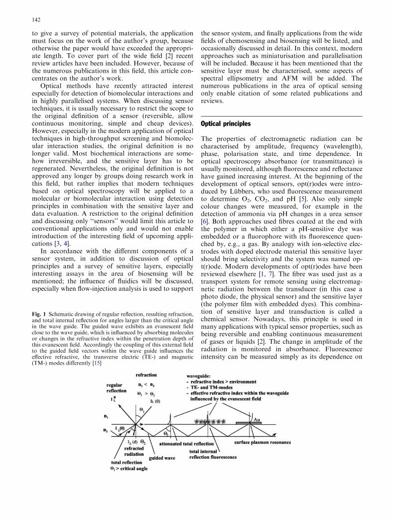
Fig. 1 Schematic drawing of regular reflection, resulting refraction,and total internal reflection for angles larger than the critical anglein the wave guide. The guided wave exhibits an evanescent fieldclose to the wave guide, which is influenced by absorbing moleculesor changes in the refractive index within the penetration depth ofthis evanescent field. Accordingly the coupling of this external fieldto the guided field vectors within the wave guide influences theeffective refractive, the transverse electric (TE-) and magnetic(TM-) modes differently [15]
142
quenching effects or, better, lifetime measurements canbe used [8, 9]. Modern lock-in technologies and phase-sensitive measurements to determine the lifetime providetools for rapid and inexpensive electronic components[10]. Therefore fluorescence enables use of simple andcost-saving devices.
In optodes using absorbance measurements, in prin-ciple diffuse reflectance is applied (sometimes calledremission) [5, 11]. Besides the measurement of quench-ing of fluorescence intensity of a fluorophore by theanalyte or variation of its lifetime, fluorescence anisot-ropy [12] can be used to determine structural changes ororientations, a characteristic which is frequently used formonitoring rotational diffusion processes [13]. Fluores-cence correlation spectroscopy gives information onlateral diffusion and, indirectly, on the increased size ofmolecules after interaction even at the single-moleculelevel [14].
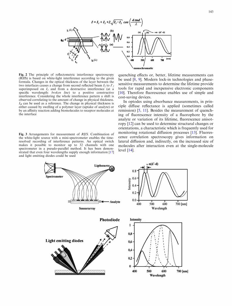
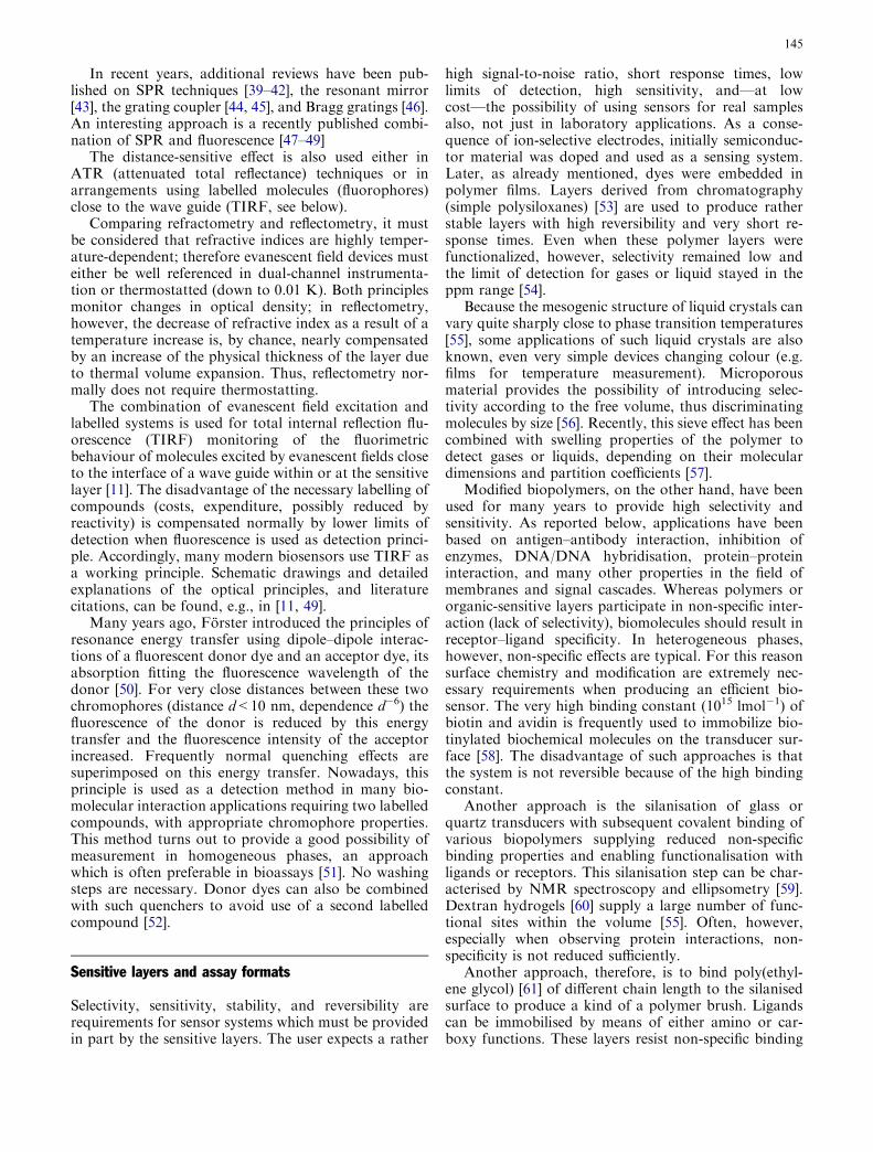
Fig. 2 The principle of reflectometric interference spectroscopy(RIfS) is based on white-light interference according to the givenformula. Changes in the optical thickness of the layer between thetwo interfaces causes a change from second reflected beam I2 to I’2superimposed on I1 and from a destructive interference (at aspecific wavelength: broken line) to a positive constructiveinterference. Considering the whole interference pattern a shift isobserved correlating to the amount of change in physical thickness.IR can be used as a reference. The change in physical thickness iseither caused by swelling of a polymer layer (uptake of analyte) orby an affinity reaction adding biomolecules to receptor molecules atthe interface
Fig. 3 Arrangements for measurement of RIfS. Combination ofthe white-light source with a mini-spectrometer enables the time-resolved recording of interference patterns. An optical switchmakes it possible to monitor up to 32 channels with onespectrometer in a pseudo-parallel method. It has been demon-strated that even four wavelengths supply enough information [17]and light emitting diodes could be used
143

The use of regular reflectance introduced a hugevariety of detection principles based on reflectometryand refractometry. Both use the dependency on thethickness of the layer and/or the refractive index, whichinfluences the phase and/or amplitude of the electro-magnetic radiation penetrating this layer or being re-flected. The use of reflected electromagnetic radiation isrepresented schematically in Fig. 1 [15].
Because one part of the radiation is usually reflectedat the interface of a thin layer, whereas the other pene-trates the layer and is there reflected at the other inter-face, these two partial reflected beams can becomesuperimposed and form an interference pattern, result-ing in constructive or destructive interference dependingon the angle of incidence, wavelength, and optical den-sity of the layer, which is given by the product ofrefractive index and physical thickness of the layer. Themodulation of this interference pattern, as demonstratedin Fig. 2, depends on these properties of the layer andchanges sensitively in response to changes in or at thislayer. This simplified version of ellipsometry, called re-flectometric interference spectroscopy (RIfS) [16], pro-vides a simple and robust technique in chemosensingand biosensing as demonstrated later in the applicationssection. The principle of the arrangement is given inFig. 3.
If polarised light is introduced the information con-tent becomes even larger, because ellipsometry enablesseparation of the refractive index and the physicalthickness when using many wavelengths. Ellipsometrywas introduced as far back as in the 1940s [18, 19], andhas regained interest in modern semiconductor andwafer technology as a simple control technique. On theother hand, it enables characterisation of sensitive layersand is used not only to characterise simple polymerfilms, but also biopolymers [20, 21]. In refractometry,minimum changes in the refractive index or transmit-tance of a medium close to a wave guide influenceradiation guided in the wave guide, because its evanes-cent field probes this medium resulting in an effectiverefractive index. Thus the transverse electrical (TE) andtransverse magnetic (TM) modes of the wave propa-gating in the wave guide are influenced differently. Theseevanescent field techniques open a wide variety of opti-cal detection principles reviewed in [11], such as Mach–Zehnder [22] interferometer, Young interferometer [23],grating coupler [24, 25], resonant mirror [26], and sur-face plasmon resonance devices [27–29]]. Bragg gratings[30] even enable the set up of arrays or remote sensitivedetection along a fibre.
Because the evanescent field penetrates (with expo-nential decay) the medium at the interface of the waveguide by just half the wavelength of the guided radia-tion, these devices have the advantage of detecting onlyeffects within this penetration depth (just a few 100 nm)(Fig. 1).
The various detection principles mentioned for eva-nescent fields all interrogate the change in effectiverefractive index in the wave guide. Interferometer-type
wave guides (e.g. Mach–Zehnder chips) determine thedifference between the phase of two waves travelling intwo arms of the wave guide [22]. The Young interfer-ometer is a similar type of interferometer, the wave guidearms not reunifying but rather imaging the interferencepattern produced by the two open ends of the waveguide arms on a CCD [23]. Using both TE and TMmodes enables internal referencing. This has been im-proved by Lukosz [31] in its mode beat interferometer,measuring amplitudes and phases of both polarisationstates.
The grating coupler is frequently used to monitorchanges in refractive index [24]. A wave guide layer iscombined with a layer in which a grating is embedded.The grating constant is influenced by the refractive indexwithin the adjacent medium. As for interference filters,this grating condition determines the preferred wave-lengths or varies with the angle of incidence. Thus theguided wave will depend on the gradient in the mediumnext to the wave guide. Radiation incident on the grat-ing will be reflected or coupled into the wave guide,depending on refractive index, wavelength, and angle ofincidence [32]. Either an angle-resolving arrangement ora CCD camera (avoiding mechanical parts) is used tomonitor the reflected radiation [23]. Bi-diffractive cou-plers [33] have two gratings with different grating con-stants superimposed. The outcoupled wave has an angledifferent from that of the directly reflected wave. Rathertricky are gratings embossed in polycarbonate whichtake advantage of non-parallel grooves of the grating ora thickness gradient of the wave guide [34].
Another type of interrogation of the polarisationstatus is applied in prism couplers [26]. The radiationcouples out of a prism via the frustrated total internalreflection of a low refractive index layer (with a thicknessof �1000 nm) into the high-refractive-index wave guide.Forty-five degree polarisation is chosen, and TM andTE modes travel in the resonant layer (wave guide:thickness �100 nm), differently influenced via the eva-nescent field by changes in the adjacent medium. Thus,the polarisation state changes in this ‘‘resonant mirror’’[35].
The best examined evanescent field technique is sur-face plasmon resonance; the theory and application tochemo- and biosensing have been reviewed in manyarticles [36, 37]. A prism is coated on its base by anapprox. 50-nm metal film. The prism takes care of totalreflectance of incident radiation, which excites this filmto an extent depending on angle of incidence andwavelength plasmons in resonance at the surface oppo-site to the wave guide interface adjacent to the mediumof interest. The resonance condition of these plasmonsdepends on the refractive index of this medium. Reso-nance of plasmons reduces the reflected intensity of thep-polarised light resulting in a ‘‘dip’’ in the reflectancediagram. Either a prism or a wave guide with a bufferlayer [28] to the metal film is used for achieving totalinternal reflectance. This direct optical detection methodhas been commercialised the longest [38].
144
In recent years, additional reviews have been pub-lished on SPR techniques [39–42], the resonant mirror[43], the grating coupler [44, 45], and Bragg gratings [46].An interesting approach is a recently published combi-nation of SPR and fluorescence [47–49]
The distance-sensitive effect is also used either inATR (attenuated total reflectance) techniques or inarrangements using labelled molecules (fluorophores)close to the wave guide (TIRF, see below).
Comparing refractometry and reflectometry, it mustbe considered that refractive indices are highly temper-ature-dependent; therefore evanescent field devices musteither be well referenced in dual-channel instrumenta-tion or thermostatted (down to 0.01 K). Both principlesmonitor changes in optical density; in reflectometry,however, the decrease of refractive index as a result of atemperature increase is, by chance, nearly compensatedby an increase of the physical thickness of the layer dueto thermal volume expansion. Thus, reflectometry nor-mally does not require thermostatting.
The combination of evanescent field excitation andlabelled systems is used for total internal reflection flu-orescence (TIRF) monitoring of the fluorimetricbehaviour of molecules excited by evanescent fields closeto the interface of a wave guide within or at the sensitivelayer [11]. The disadvantage of the necessary labelling ofcompounds (costs, expenditure, possibly reduced byreactivity) is compensated normally by lower limits ofdetection when fluorescence is used as detection princi-ple. Accordingly, many modern biosensors use TIRF asa working principle. Schematic drawings and detailedexplanations of the optical principles, and literaturecitations, can be found, e.g., in [11, 49].
Many years ago, Forster introduced the principles ofresonance energy transfer using dipole–dipole interac-tions of a fluorescent donor dye and an acceptor dye, itsabsorption fitting the fluorescence wavelength of thedonor [50]. For very close distances between these twochromophores (distance d<10 nm, dependence d�6) thefluorescence of the donor is reduced by this energytransfer and the fluorescence intensity of the acceptorincreased. Frequently normal quenching effects aresuperimposed on this energy transfer. Nowadays, thisprinciple is used as a detection method in many bio-molecular interaction applications requiring two labelledcompounds, with appropriate chromophore properties.This method turns out to provide a good possibility ofmeasurement in homogeneous phases, an approachwhich is often preferable in bioassays [51]. No washingsteps are necessary. Donor dyes can also be combinedwith such quenchers to avoid use of a second labelledcompound [52].
Sensitive layers and assay formats
Selectivity, sensitivity, stability, and reversibility arerequirements for sensor systems which must be providedin part by the sensitive layers. The user expects a rather
high signal-to-noise ratio, short response times, lowlimits of detection, high sensitivity, and—at lowcost—the possibility of using sensors for real samplesalso, not just in laboratory applications. As a conse-quence of ion-selective electrodes, initially semiconduc-tor material was doped and used as a sensing system.Later, as already mentioned, dyes were embedded inpolymer films. Layers derived from chromatography(simple polysiloxanes) [53] are used to produce ratherstable layers with high reversibility and very short re-sponse times. Even when these polymer layers werefunctionalized, however, selectivity remained low andthe limit of detection for gases or liquid stayed in theppm range [54].
Because the mesogenic structure of liquid crystals canvary quite sharply close to phase transition temperatures[55], some applications of such liquid crystals are alsoknown, even very simple devices changing colour (e.g.films for temperature measurement). Microporousmaterial provides the possibility of introducing selec-tivity according to the free volume, thus discriminatingmolecules by size [56]. Recently, this sieve effect has beencombined with swelling properties of the polymer todetect gases or liquids, depending on their moleculardimensions and partition coefficients [57].
Modified biopolymers, on the other hand, have beenused for many years to provide high selectivity andsensitivity. As reported below, applications have beenbased on antigen–antibody interaction, inhibition ofenzymes, DNA/DNA hybridisation, protein–proteininteraction, and many other properties in the field ofmembranes and signal cascades. Whereas polymers ororganic-sensitive layers participate in non-specific inter-action (lack of selectivity), biomolecules should result inreceptor–ligand specificity. In heterogeneous phases,however, non-specific effects are typical. For this reasonsurface chemistry and modification are extremely nec-essary requirements when producing an efficient bio-sensor. The very high binding constant (1015 lmol�1) ofbiotin and avidin is frequently used to immobilize bio-tinylated biochemical molecules on the transducer sur-face [58]. The disadvantage of such approaches is thatthe system is not reversible because of the high bindingconstant.
Another approach is the silanisation of glass orquartz transducers with subsequent covalent binding ofvarious biopolymers supplying reduced non-specificbinding properties and enabling functionalisation withligands or receptors. This silanisation step can be char-acterised by NMR spectroscopy and ellipsometry [59].Dextran hydrogels [60] supply a large number of func-tional sites within the volume [55]. Often, however,especially when observing protein interactions, non-specificity is not reduced sufficiently.
Another approach, therefore, is to bind poly(ethyl-ene glycol) [61] of different chain length to the silanisedsurface to produce a kind of a polymer brush. Ligandscan be immobilised by means of either amino or car-boxy functions. These layers resist non-specific binding
145

[62], but have a reduced number of interaction sites,because they are restricted to the surface and not in thevolume.
Besides these principal approaches, many other ideashave been realised, e.g. the use of His-tags [63] or theimmobilisation of membrane structures of lipid doublelayers [64] to the transducer, to model cell walls. Allthese different approaches are intended to reduce non-specific binding, enable a large number of specificbinding sites, increase the stability of the layer, which isessential for regeneration strategies, and increase selec-tivity and sensitivity.
Because, in contrast with simple polymer films, thesesensor types are not reversible, reusability has to beintroduced by use of regeneration strategies. As men-tioned in the Introduction, these biosensors cannot becalled ‘‘sensors’’ by definition. However, being reusable,this regeneration is often regarded as a substitute for therequired reversibility.
Although these biopolymers should increase the sta-bility of biolayers, their stability is not comparable withthat of, e.g., polysiloxane films or microporous systems[65]. For this reason, for many years supramolecularstructures [66] and biomimetic layers [67] have been awide field of research as attempts have been made tocombine the advantages of stability and reversibilitywith sensitivity and selectivity. Artificial layers are syn-thesised with recognition structures comparable withthose of natural biomolecules. Supramolecular struc-tures such as calixarene [68, 69] were tried first to in-crease selectivity of simple chemosensors. They wereused to separate, e.g., various chlorinated compounds.
Another approach was the use of cyclodextrins [70] orcyclohexapeptide [71] structures. Both could be func-tionalised to supply either dependence on ionic strengthor even discrimination of enantiomers. Such modifiedcyclodextrins or polysiloxanes modified with opticallyactive amino acids [72], prove the capability of sensors tomeasure enantiomers. Similar separation coefficients asfor gas or liquid chromatography have been demon-strated [73].
One of the first approaches used to introduce selec-tivity by immobilising polynucleotide or peptide se-quences to the surface were hybridisation studies.Meanwhile, PNA (with peptides as a backbone) [74] wasproven to be a better complementary binding systemthan DNA because of less repulsion by charges andbetter backbone stability; this resulted in stabilityagainst DNases and nucleases. Locked polynucleotideshave recently been demonstrated to be another suc-cessful approach [75]. The polynucleotides were com-pared with PNA and DNA layers [76]. Further interesthas concentrated on lipid membranes as biopolymers,enabling the construction of a variety of functionalsystems (e.g. for transport of proteins [77, 78]).
Learning from nature, another approach is the syn-thesis of layers of molecular imprinted polymers [79].The problem of these layers is that stability and selec-tivity are in reverse proportion to response times. Ad-vances in this field have been reviewed in [80]. Theirpotential optical sensing approaches for environmentaland industrial applications have been reviewed in [81].Because of growing public concern with regard to healthand environmental problems, researchers are usingmolecularly imprinted polymers in food and agriculture[82]. Molecular imprinting technology has recentlyemerged to produce biomimetic receptors that challengetheir unnatural counterparts. An overview of thismethod and its application can be found in [83]. Despiteall these promising numerous applications and devel-opments, their future perspectives have yet to be vali-dated. Imprinting of the surface instead of the volume isused in the so-called spreader-bar technique [84], inwhich template molecules cause immobilised brushes atthe surface to rearrange around the template to form akind of ‘‘well’’ in which only molecules similar to the
Fig. 4 Assays using interaction between biomolecules in homoge-neous phase and/or at heterogeneous interfaces. In both assaysthermodynamics (equilibrium constant) and kinetics (associationand dissociation rate constants) determine the interaction. Directassays immobilise the receptor at the surface to measure theanalyte, here a binding inhibition assay is demonstrated wherederivatives of the analyte or ligand to be detected are immobilised.In the preincubation phase receptor and ligand are mixed in thehomogeneous phase and the concentration of non-blocked receptormolecules is detected via the heterogeneous phase. Large numbersof interaction sites at the transducer make this process diffusion-controlled; at low ‘‘loading’’ the kinetics at the heterogeneousphase can be measured
146

Table
1Survey
ofopticalprinciples,assayform
ats,andselected
relatedapplications
Sensortype
Opticalprinciple
Sensitivelayer
Application
Absorption[11]
Indicatordye
Ureaconcentration[6]
Labeled
system
sFluorescence
[8–10]
TIR
F[11,87,88]
Biopolymer/antigen-derivative
inheterogeneousphase
Environmentalanalysis:pesticides,
EDCs[87,89,90]
FRET
[50,51]
miniaturisation
Homogeneousassay,
phase-separationassay[91]
Pesticides
[88],nuclease
assay[91],SNPs
Directopticaldetection
atheterogeneousphase
Evanescentfield
Mach–Zehnder
Interferometer
[22]
Polymer
film
s[53],
liquid
crystals[55]
Hydrocarbons,aromaticcompounds
Biopolymer/antigen
derivative
Pesticides
Refractometry
Grating
coupler[24,25]
Biopolymer/antigen
derivative
Resonant
mirror[26,35]
Biopolymer/antigen
derivative
Thrombineinhibitors
Biopolymer/inihibitorderivative
Surface
plasm
on
resonance
[27,28,36,37]
Polymer
film
sEnantiomers[73],homologous
series
ofalcohols[92,93]
Microporouspolymers[56]liquid
crystals
BiomolecularinteractionDNA
[47]
Biopolymer/antigen
derivative
Hybridisation
Mem
brane–peptideinteractions[94]
Parallelisation
Biomolecularinteraction,screening
Reflectometry
Reflectometricinterference
spectroscopy[16]
Polysiloxanefilm
s[95]
Enantiomers[72],Process
monitoring
Microporouspolymers[56]
Hydrocarbons[96],aromaticcompounds[97]
Hyperbranched
polyesters
[98]
Volatile
organic
compounds(V
OC),
Alcohols[99],freons[98,100]
Cyclodextrins[70]
Enantiomers
Cyclohexapeptides
[71]
Aminoacids[101]
MIPs[79]
Enantiomers[102]
Biomolecularinteraction(dextran[60],
PEG
[61],biotin[58])
Pesticides
[103],triazines
[104]
DNA
intercalation
Low
molecularweightanalytes[104,105]
Fermentationcontrol[106]
Parallelisation
Screening[107]
Combinatorialsynthesis[108],
triazines
synthesis[105]
Epitopemapping[109]
Thrombin
inhibition[110]
Phosphorylation
Signalcascade
Miniaturisation
Antigen/antibodyinteraction,
DNA
hybridisation[111]
Wateranalysis[112]
Additionalreview
articles
Braggsensors
[113]
Biomedical[114,115]
Usingquantum
dots
[116]
interferometricsensors
[117]
147
template will later ‘‘bind’’. All these approaches areinteresting for heterogeneous phase assay.
In chemical sensors simple arrangements are usuallyused for gas flow or fluidics. Flow controls are used forthe calibration procedure, for mixing different analyteconcentrations. Simple pumps which enable a changebetween solvent and analyte/solvent mixtures at differ-ent concentrations are sufficient for the fluidics. Incontrast, biosensor systems use more sophisticated flu-idics which enable changes of flow rates, preincubationtimes, and different injection cycles between analyte,analyte derivative, and reagent. These systems must alsoprovide the possibility of regeneration cycles.
As Fig. 4 shows, various possible assay formats areknown. Preferable is a homogeneous assay which en-ables direct interaction between the analyte and the re-agent. The problem, however, especially in opticaldetection is that this interaction must change opticalproperties. Colour changes can occasionally be detected,but with some difficulty. For this reason labelled systemsare used in homogeneous assays. In previous times,radioactive labelling enabled very low limits of detec-tion, but is nowadays not preferable. Therefore fluo-rescence labelling is normally used, taking advantage ofeither quenching effects or (most often) fluorescenceresonance energy transfer.
For measurements in a heterogeneous phase it wouldbe preferable to immobilise the reagent in the biopoly-mer and detect the analyte directly. This is certainlypossible with labelled compounds using quenching ef-fects; for direct optical detection without labelled com-pounds, however, preferably large analyte moleculeshave to be examined. The sensitivity of any kind ofoptical detection can be improved by increasing theanalyte mass or volume. Therefore, either a competitivetest scheme (labelled competing with non-labelled ana-lyte) or a so-called binding inhibition test scheme is used.This represents the following assay type: an analytederivative is immobilised in the biopolymer; in a prein-cubation step the analyte and the reagent are mixedtogether; the analyte as a ligand blocks receptor sites,which can no longer react in this blocked state with theanalyte derivative immobilised to the surface. This ap-proach can be realised with a labelled reagent or directoptical applications. A large number of interaction sitesin the biopolymer causes transport-limited (diffusion-limited) interaction between non-blocked receptor andanalyte derivative at the biopolymer. The slope of time-resolved measurements of the interaction process be-comes linear and can be calibrated to the concentrationof the free receptor in equilibrium in the homogeneousphase. If one measures in dependence on time during thepreincubation step, even association and dissociationrate constants in the homogeneous phase can be ob-tained by simulation and taking account of the super-imposition of the various processes [85].
A small number of immobilised analyte derivativeson the biopolymer enables determination of the kineticsin the heterogeneous phase by use of association and
dissociation rate constants. This biomolecular interac-tion analysis (BIA) is well discussed in literature [86].Equilibrium and rate constants normally differ forhomogeneous and heterogeneous phases, and thekinetics and thermodynamics are influenced by labellingby a fluorophore [85]. This must be well consideredwhen discussing and comparing different biomolecularinteractions. Optical principles and assay types aresummarised in Table 1, with selected typical applica-tions.
Applications
Remission measurements (changes in the absorbance ofreflected light) and fluorescence effects are the mostsimple sensing methods. They enable very simplearrangements which can sometimes cause artefacts.They do not use the advantages of optics, namelyspectral detection. For this reason, occasionally, e.g. formeasurement of urea, in which the ammonia producedcauses a spectral shift of an indicator dye because ofchanges in pH, a spectrum or at least at two wavelengthshave been recorded [6] to overcome artefacts. Intensityquenching of fluorophores, measurement of anisotropy,or even fluorescence correlation spectroscopy for single-molecule detection, result in a wide variety of applica-tions which cannot be reviewed in detail here but whichare the topics of quite a number of review articles [8,113, 114]. A new approach is the use of quantum dots[116].
This review concentrates on selected applications byour group as given in the lecture focussing on sometypical examples. Beginning with chemical sensors anddetection in the gaseous phase, most of the results areinfluenced by the humidity of the gaseous phase. This isalso true for polysiloxane as a polymer using the swellingeffect with RIfS. However, modification of these poly-siloxane layers can reduce effects of humidity even forchlorinated hydrocarbons as analytes [97]. As was pro-ven for RIfS, a whole spectrum does not have to berecorded—even four wavelengths are sufficient fordetermination of the swelling effect [17] and perspectivesfor highly parallelised detection were opened.
This uptake of analytes from the carrier gas into apolymer film coated on the transducer or covalentlybound results in swelling of the polymer film which canbe detected by use of interference spectroscopy. Nor-mally, Henry’s law is valid at low concentrations, andthe limit of detection is down to a few ppm. Higherspecific interaction results in Langmuir-type calibrationcurves given by saturation effects at increasing concen-tration. These effects can be classified by separate mea-surement of refractive index and physical thicknesschanges, by use of spectro ellipsometry [118, 119]. Thesemeasurements demonstrate that most effects in non-selective polymer films are caused by changes in physicalthickness which result from swelling, whereas the effectof changes in the refractive index is negligible. The
148

polymer films used are reviewed in [95]. Various types ofpolymer, for example functionalised polysiloxanes [97],esters, hyperbranched polyesters [98], or dendrimershave been used for a variety of applications. Mach–Zehnder chips have also been used to measure volatileorganic compounds (VOC) at low limits of detection[120–122]. Chemical sensors based on polysiloxane lay-ers have been compared with quartz microbalance,calorimetric, and capacitance sensors [123]. Applicationsof optical detection to chemo gas sensors have been re-viewed [54].
The same films can be applied to the detection ofanalytes in liquids using RIfS [96]; for this, however, thepolymer must be covalently bound to the transducer viasilanisation and peptide-like binding approaches. By thismeans the stability of the films is increased from a fewdays up to 3 months, because water can no longer leveroff the polymer film from the transducer [124].
Although the type of binding of the silanes to thesurface has been discussed, NMR studies have alsoshown that normally even a tripoid immobilisation ispossible [59]. The properties of the polymers depend onthe cross-linking, therefore not all polymers can be used.However, so-called microporous systems also have verygood, sensitive layer properties. In this case, microsieveeffects predetermine the feasibility of detection of mol-
ecules, which depends on their volume. The uptake ofgases or liquids increases the pore volume, however, thusalso causing swelling effects. However, saturation effects(Langmuir-type curves) and observed effects on thechanges of refractive index demonstrate the limitednumber of interaction sites.
These films are particularly useful in environmentalchemistry and process control; typical applications in-clude monitoring of chlorinated compounds in the wastewater of chemical company production processes,determination of concentrations of air-conditionerrefrigerants in, e.g., cars, or measurement of otherchlorinated and non-chlorinated hydrocarbons. Thesemicroporous systems even enable separation of twodifferent refrigerants, R22 and R134a, down to ratherlow limits of detection [98] and with a high reproduc-ibility even in mixtures [100, 125].
These microporous systems also enable determina-tion of the concentration of, e.g., homologous alcoholsin mixtures either by using a sensor array or evenapplying chemometrics [92, 93]. It turns out that highselectivity of each element of the sensor arrays for spe-cific compounds in the mixture will not reduce problemsof cross-reactivity, but that this can be achieved byapplication of modern tools of chemometrics, forexample neural networks or neural networks in combi-nation with evolutionary strategies. Normally, either thearea or the slope of a signal is measured in equilibrium.However, the modern tools of direct optical detectionenable the application of time-resolved measurements.These have the advantage that the signal is measuredduring the interaction process at as many time-points asnecessary. An example is given for a mixture methanol,ethanol, propanol, and butanol. Microsieve effects of
Fig. 5 Time-resolved measurements of the interaction between amircoporous layer and a homologous series of alcohols (gaseousmethanol, ethanol, propanol, butanol): SPR signal/shift in reso-nance wavelength) versus alcohol concentration (relative partialpressure mixed in a gas mixing station) and time. The predicted/true graph represents the quality of the neural network basedevaluation. Good standard deviation is obtained for the smallalcohols [118]
149

microporous systems support the discrimination. Anarray of sensors (RIfS) or even a single element (usingsurface plasmon resonance) causes a minor percentageof errors. This has been published for ternary mixtures[93]. Even a quaternary mixture can be handled, as isdemonstrated for the signals given in Fig. 5 [99]. Evenbetter results could be achieved by applying chemo-metrics to the refrigerants, for which the uncertainty isclose to 2% [100].
Functionalisation of polysiloxanes has even enabledseparation of enantiomers. The feasibility was proven ina comparison of mass-sensitive and reflectometricmethods with Chirasil-Val [72]. Another application isthe use of biomimetic structures based on cyclodextrinto monitor the anaesthetic sevoflurane. In an alkalinerebreathing circuit the inhalation anaesthetic degradesinto a least two products; one of these is a chiral halo-diether and one of the enantiomers has a different,narcotic action [126]. By use of a modified Lipodex E(cyclodextrin derivative) column, the chiral separationfactor obtained at 30�C was larger than nine, the sameas in capillary gas chromatography. Comparable valueswere obtained in a comparative study of enantioselectiverecognition by use of thickness shear mode resonators,surface acoustic wave sensors, surface plasmon reso-nance, and reflectometric interference spectroscopy [73],thus proving that chemosensors using modified cyclo-dextrins enable enantioselective detection of a haloge-nated diether.
In this work a far higher separation factor wasachieved than for some molecular imprinted polymers(MIP) [102]. With MIP, however, rather high separationfactors could also be achieved [127].
In other approaches, surface-bound cyclohexapep-tides have been used as elements for molecular recog-nition of amino acids [101]. The chiral cyclopeptidelibraries can be used as chiral receptors. The cyclo-hexapeptide was immobilised on the transducer bymeans of three lysines; the other three positions werevaried.
Biomimetic structures are also used for label-free,product-specific monitoring of biotechnological pro-cesses used to manufacture the glycopeptide antibioticvancomycin. Usually, the pH or oxygen content and thetemperature are controlled during the fermentationprocess; only glycose [128] is measured, however,whereas normally process control is performed by use ofHPLC. A lysine-D-alanine-D-alanine sequence has beenimmobilised on the transducer. Use of RIfS enablesdetection of vancomycin even during the fermentationprocess [106]. This approach led to the thought ofcombining a parallel RIfS system with MALDI-TOF[129], because the measurement of biomolecular inter-action by RIfS even includes degradation productsinteracting with the peptide sequence immobilised on thesurface.
A typical requirement, especially in biomolecularinteraction, is to measure as small molecules as possible.As mentioned in the section on optical principles all
label-free detection methods have problems detectingsmall ligands at low concentration and with smallbinding constants. Therefore SPR technology and RIfSwere tested for such detection. Two results can be re-ferred to for RIfS, first the interaction measurement ofbiotin with avidin [104] and the on-line monitoring ofsolid-phase peptide synthesis on glass-type surfaces[105]. Label-free detection is reviewed in [130].
The applications already mentioned all used single-channel detection. However, considering future detec-tion requirements for combinatorial libraries, parallelsensing has extreme advantages over other methods[107]. Also, determination of binding constants togetherwith association and dissociation rate constants forantibody–triazine-derivative interactions, with a largenumber of calibration steps and replica measurements,made the need for parallelisation obvious [131]. Com-bining the idea of using single wavelengths instead ofwhite light interference, as in the four wavelength set-upused for cost-effective chemosensing, a parallelised sys-tem was developed [132]. Some of the applications ofthis will be mentioned here.
Primary screening for lead compounds requirescombinatorial synthesis which should be closely linkedto the drug-discovery process [133]. Until now, the usualscreening procedure uses radioactive or fluorescence-la-belled detection techniques directly on the resin beadsafter a split-and-combine library synthesis [134]. Becausedirect optical detection without labelling is an interestingalternative, direct label-free RIfS detection on a mic-rotitre plate format using simultaneous imaging by aCCD camera was applied to the synthesis of a triacinelibrary [108]. The selectivity of an anti-simazine anti-body was evaluated by measuring biomolecular inter-action processes with a variety of immobilised triazinederivatives with different substituents in two positions.Thus, in a single measurement cycle a library of 36derivatives was examined. Single-channel RIfS also en-abled direct control of the synthesis in one well, provingthe feasibility of this using method to monitor theinteraction of small molecules [105].
Another approach which proves the benefits ofscreening is the use of a parallel affinity assay forthrombin inhibitors in label-free screening HTS detec-tion. By using a binding inhibition assay, the screeningof 384 substances for thrombin activity can be per-formed within less than 15 min. The optical reproduc-ibility is high enough to support a data quality whichenables parallel quantification of the IC50 values (half ofthe receptor sites are blocked) of the library substances[110]. Screening could also be performed with 5%DMSO added to the samples; this is relevant in high-throughput screening (HTS) applications in practice inwhich pure water cannot be used as a solvent.
Finally epitope mapping of transglutaminase (tTG-ase) is given as an example for HTS. The enzyme tissuetTGase has been identified as the major autoantigen incoeliac disease and an antibody has been developed. Tolearn the sequence of amino acids a binding assay with
150

various peptides would be helpful. Therefore by com-binatorial chemistry 21 peptides were synthesised asdepicted in Fig 6. A binding inhibition assay is set up. Alead structure (known from enzyme-linked immunosor-bent assay, ELISA) is immobilised in all the wells of amicrotitre plate arrangement. First, buffer is spilled overthe plate and, as can be seen in Fig. 6, no binding signalis measured using RIfS. If antibody alone is in contactwith the immobilised lead structure a high signal is ob-tained. Now antibody and the sequences are preincu-bated and using three replicates these different solutionsare dispensed into the different wells of the microtitreplate. According to the complementarity of peptide andantibody sequences the binding pattern in Fig. 6 isachieved, demonstrating that the lead sequence is not thebest matching sequence. Using the binding curves andrepeating just the interesting measurements in a single-channel device binding constants can be discriminated[109]. Grating-coupled surface plasmon resonance
equipment is on the way to being commercialised byHTS Biosystems [135].
Modern spotting technology rather than pipettingwas used for these parallelised measurements [136].They have advantages over micro arrays and not usingwell plates.
Parallelisation is common in fluorescence methods.Either simple fluorescence techniques with a reader[137], TIRF [138, 139] or FRET [45] are used. A typicalapplication is in environmental analysis [89]. Classicalanalytical methods to determine pesticides in ground orwaste water need enrichment steps but can discriminatebetween different analytes [140]. Therefore ELISA as-says have been introduced. These assays do not enableautomated monitoring at low concentrations, however.TIRF can overcome this problem. At present six spotson a wave guide are used to set up a binding-inhibitionassay in combination with a flow-injection analysissystem. Analyte derivatives are immobilised, a differentone at each spot. Antibodies are added to the pre-incubation solution. Missing analyte (the pollution)causes fluorescence at a specific spot which is read bymeans of fibre optics [49]. Some data are given inTable 2 [87, 141].
Another possibility is the use of FRET in environ-mental analysis using micro or nanotitre plates [88].Both methods have been referenced for real watersamples to classical GC–MS results. TIRF and FREThave been compared [103]. Chemometrics have beenapplied to overcome the problem with non-specificantibodies. For endocrine-disrupting compounds (Ta-ble 2) recovery rates could be improved and limits ofdetection reduced [90].
To enable the change of analyte derivatives within theflow-injection system (FIA) without removal of thetransducer, DNA sequences have been immobilised onthe TIRF transducer (auxiliary system). Matchingstrands carry the analyte derivative. However, DNA isnot stable enough and reduces the number of interactionsites by charge repulsion. Therefore instead of DNA-strands the PNA (peptide backbone) was used to im-prove the system [142].
Miniaturisation is the final topic to be covered [143].Over the last decade many papers have been publisheddealing with miniaturisation, and lab-on-chip techniquesin particular. Micro total analysis systems were devel-
Fig. 6 Epitope mapping to determine the sequence motif oftransglutaminase (tTGase). Twenty-one peptide sequences aresynthesised by combinatorial chemistry and peptide 6 is found tobe a lead structure. When this is immobilised on the transducer,antibody produces a large increase in layer thickness and buffersolution results in very low signal. The better the peptide sequencefits the antibody the smaller the increase in layer thickness andsignal. The measured binding curves are evaluated to givecalibration curves and binding constants (right)
Table 2 Results from measurement of pesticides and endocrinedisruptors by use of TIRF
Analyte IC50
(lg L�1)SD (%)a LOD
(lg L�1)
Atrazine 0.082 2.74 0.006Atrazine-diset. 1.337 2.74 0.039Simazine 0.54 1.7 0.03Isoproturon 1.211 1.11 0.016Alachlor 0.53 3.98 0.072,4D 0.89 2.15 0.07pcp 22.680 1.45 1.120Carbofuran 6.409 1.28 0.024Estradiol 0.590 2.98 0.060Ethinylestradiol 1.070 3.20 0.070Estrone 0.145 1.18 0.006Bisphenol A 0.670 1.73 0.007
aStandard deviation of blank measurements
151

oped many years ago at Ciba Analytical Research. Re-cently, a new review has been published by one of theinventors in this field [144]. Miniaturisation, integration,and systemisation of various types of sensor have beenconsidered with regard to modern technologies in microfabrication; these techniques have found considerableinterest in Japan [145]. The development of miniaturisedimmunosensing devices has been reviewed as a smalltechnology with a large future [146]. Pocket-size ana-lytical equipment based on the lab-on-chip approach hasbecome available and is intended for use in biomedicaland environmental monitoring [147]. This chip tech-nology is also of interest for any type of DNA chip, andwill enable improved analysis in proteomics; it providesa type of proteomics-on-a-chip and is a challenge tocouple lab-on-chip with microfluidics and detectionplatforms [148]. Although all these approaches facelimits of detection problems, they are the prerequisite ofanother very interesting approach in optical sensingdedicated to enable multi-parameter or multi-analytemonitoring in parallel. Therefore, miniaturisation usu-ally appears together with parallelisation. Parallelisationcan be increased by reducing spot or well size. Asmentioned above, the use of nanotitre plates takesadvantage of reduced sample volume and the reducedamount of reagent necessary. An interesting approach iscombination of homogeneous and heterogeneous phaseassays as realised in a so-called phase separation assay.The wells are coated with gold and the derivative isimmobilised via thiol groups. By this means the
dynamics of the fluorescence signal are increased [91]. Inmany applications, however, an FIA system is prefera-ble. Thus, for some time, new approaches for microfluidics have been discussed.
So-called hybrid systems supply miniaturised chipsand flow channels and overcome problems with pumpsand valves, which cause some problems in the handlingof biomolecules. The transport equations have to besolved and diffusion processes simulated to guaranteeperfect mixing [149].
The results of a miniaturised set-up [111] for directoptical detection with RIfS are shown in Fig. 7. Thetime-resolved binding curves of 96 spots for antigen–antibody interaction are given. Kinetic evaluation ispossible. Currently, however, monitoring of up to 384binding events is restricted to large molecules.
Conclusions
Optical sensors have proven in the past to be either verysimple and cost-effective devices or enable rathersophisticated multisensor applications. Because of theexistence of many different optical principles which canbe classified into use of direct optical detection or takingadvantage of labelled compounds, in principle many ofthese methods can be applied to a huge number ofapplications. It is becoming evident that of the differentsensor principles—electronic, electrochemical, mass-sensitive, or optical devices—none is generally superior,but rather the feasibility depends on the application. Thesame holds true for the different optical sensor princi-ples. This became obvious when comparing variousrefractometric and reflectometric methods in the samebiomolecular interaction study using antibodies andantigens, and setting up the surface chemistry by the
Fig. 7 Highly parallelised RIfS set-up for study of interactionsbetween antibodies and antigens in a binding inhibition assay. Aminiaturised system is used with up to 384 spots on 12·18 mm2. Onthe left the real-time binding curves are drawn from the data array;on the right one of these curves is enlarged
152

same person. In both cases of two studies the limits ofdetection for all the methods examined ranged withinone order of magnitude. Discrimination was achievedonly at the cost of expenditure on apparatus and bysophistication of the fluidics used [150, 151].
Reliable results can be obtained with either chemo-sensors or biosensors. The selectivity and limit ofdetection are usually better for biosensors. Chemosen-sors, on the other hand, provide reversibility and greaterstability of the sensitive layer. It turns out that thequality of the set-up depends not only on the opticalmethod but also, especially, on this sensitive layer. Forthis reason, most of the improvements that can be ex-pected in optical detection methods are in the area ofsensitive layers. This becomes obvious from looking atdevelopments in biometics or functionalised polymers[47, 48, 78].
The essential result of these considerations is certainlythat research in sensing requires interdisciplinaryunderstanding of the detection principles, of the sensitivelayer, of the kinetics and thermodynamics of interactionprocesses, and of the fluidics. Thus fundamental researchmust be performed to characterise these layers and theinteraction processes to improve the understandingwhich is the prerequisite of any optimisation approach.
Whereas laboratory systems often give very goodresults and even enable separate determination of con-centrations in multianalyte mixtures, the quality of theoverall sensing system normally becomes obvious whenthese systems are applied either to real environmentalsamples, e.g. waste water, to saline solutions, or, on theother hand, to blood or sera [152–155]. Sensors turn outto be a typical modern example of interdisciplinary re-search, considering multivariate parameter arrays.
Acknowledgements For long years of support of his research theauthor has to thank the Deutschen Forschungsgemeinschaft, theFond der Chemischen Industrie, the BMBF, the Arbeitsgemeins-chaft Industrieller Forschung, the Deutsche Bundesstiftung Um-welt, some European funding and much industrial cooperation.Details of the funding is acknowledged in the different publicationscited. The author also wants to thank all his coworkers, cited andnot cited, for work achieved, and, especially, Dr Martin Mehlmannfor checking the manuscript.
References
1. Marazela MD, Morreno-Bondi MC (2002) Anal BioanalChem 372:664
2. Gopel W, Hesse J, Zemel JN (1992) Sensors, a comprehensivesurvey, vol I–VIII. VCH, Weinheim
3. Bilitewski U, Turner A (eds) (2000) Biosensors in environ-mental monitoring. Harwood Academic Publishers, Amster-dam
4. Scheller FW, Schubert F, Fedrowitz J (eds) (1996) Frontiersof biosensors I+II. Birkhauser Verlag, Basel
5. Lubbers DW, Opitz N (1983) Sens Actuators B 4:6416. Gauglitz G, Reichert M (1992) Sens Actuators B 6:837. Wolfbeis O (2004) Anal Chem 76:32698. Wolfbeis OS (ed) (1992) Fluorescence spectroscopy: new
methods and applications. Springer, Berlin Heidelberg NewYork
9. Wolfbeis OS, Boisde GE, Gauglitz G (1996) Sensors, vol II,part I. In: Baltes H, Goepel W, Hesse J (eds) Weinheim, p 573
10. Draxler S, Lippitsch ME (1996) Appl Optics 35:411711. Gauglitz G (1996) Sensors, update vol I. In: Baltes H, Goepel
W, Hesse J (eds) Weinheim, p 112. Lakowicz JR (1999) Principles of fluorescence spectroscopy.
Kluwer/Plenum, New York, p 29813. Weber G (1966) Hercules DM (ed) Fluorescence and phos-
phorescence analysis. Wiley, New York, p 21714. Rigler R (1993) Eur Biophys 22:16915. Hecht E, Zajak A (2003) Optics. Addison-Wesley, Reading16. Brecht A, Gauglitz G, Kraus G, Nahm W (1993) Sens Actu-
ators B 11:2117. Reichl D, Krage R, Krummel C, Gauglitz G (2000) Appl
Spectrosc 54:58318. Azzam RMA, Bahara NM (1998) Ellipsometry and polarized
light. North Holland19. Arwin H, Aspnes DE (1986) Thin Solid Films 138:19520. Mutschler T, Kieser B, Frank R, Gauglitz G (2002) Anal
Bioanal Chem 374:65821. Heideman RG, Kooyman RPH, Greve J (1993) Sens Actua-
tors B B:20922. Brandenburg A, Henninger R (1994) Appl Optics 33:594123. Brandenburg A, Hinkov V, Konz W (1992) Sensors, vol. 6. In:
Gopel W, Hesse J, Zemel JN (eds) VCH, Weinheim, p 39924. Clerc D, Lukosz W (1994) Sens Actuators B 19:58125. Kunz RE, Edlinger J, Curtis BJ, Gale MT, Kempen LU,
Rudigier H, Schuetz H (1994) Proc SPIE Int Soc Opt Eng2068:313
26. Cush R, Cronin JM, Stewart WJ, Maule CH, Molloy J,Goddard NJ (1993) Biosens Bioelectron 8:347
27. Liedberg B, Nylander C, Lundstrom I (1983) Sens ActuatorsB 4:299
28. Piraud C, Mwarania E, Wylangowski G, Wilkinson J,O’Dwyer K, Schiffrin DJ (1992) Anal Chem 64:651
29. Lakowicz JR (2004) Anal Biochem 324(2):15330. Othonos A (1997) Rev Sci Instr 68:430931. Lukosz W, Stamm C (1991) Sens Actuators A 25:18532. Nellen PhM, Lukosz W (1993) Biosens Bioelectron 8:12933. Fattinger C, Mangold C, Gale MT, Schuetz H (1995) Opt Eng
34:274434. Kunz RE (1991) Proc SPIE Int Soc Opt Eng 1587:9835. http://www.affinity-sensors.co.uk/iasys.htm36. Homola J, Yee S, Myszka D (2002) In: Ligler FS, Rowe T,
Chris A (eds) Optical biosensors present and future. Elsevier,Amsterdam, p 207
37. Homola J, Yee SS, Gauglitz G (1999) Sens Actuators B 54:338. http://www.biacore.com/home.lasso39. Rich RL, Myszka DG (2000) J Mol Recognit 13:38840. Van Der Merwe, Anton P (2001) Surface plasmon resonance,
in protein-ligand interactions: hydrodynamics and calorime-try. Oxford, London, p 137
41. Davis TM, Wilson WD (2001) Methods Enzymol 340:2242. Sadana A (2001) Biotech Genetic Eng Rev 18:2943. Kinning T, Edwards P, In: Ligler FS, Rowe T, Chris A (eds)
Optical biosensors. Elsevier, Amsterdam, p 25344. Voros J, Ramsden JJ, Scucs G, Szendro I, De Paul SM,
Textor M, Spencer ND (2002) Biomaterials 23(17):369945. Kuhlmeier D, Rodda E, Kolarik LO, Furlong DN, Bilitewski
U (2003) Biosens Bioelectron 18:92546. Santos JL, Ferreira LA (2002) Fibre Bragg grating interro-
gation techniques. In: Handbook of optical fibre sensingtechnology. Wiley, Chichester, p 379
47. Knoll W (2004) Bunsenmagazin 3:6948. Liebermann T, Knoll W (2000) Colloids Surfaces A 171:11549. Klotz A, Brecht A, Barzen C, Gauglitz G, Harris RD, Quigley
QR, Wilkinson JS (1998) Sens Actuators B 51:18150. Forster Th (1951) Fluoreszenz Organischer Verbindungen.
Vandenhoek und Ruprecht, Gottingen51. Mere L, Bennett T, Coassin P, England P, Hamman B, Rink
T, Zimmerman S, Negeulescu P (1999) Drug Discovery Today4:363
153
52. Seidel M, Dankbar D (2004) Anal Bioanal Chem 379:90453. Baldini F, Bracci S (2000) Polymers for optical fiber sensors.
In: Osada Y, De Rossi DE (eds) Polymer sensors and actua-tors. Springer, Berlin Heidelberg New York, p 91
54. Rathgeb F, Gauglitz G (2000) In: Meyers RA (ed) Encyclo-pedia of analytical chemistry. Wiley, Chichester, p 2189
55. Tang K, Garetz BA, Green MM, Herman FM (2002) Polymerpreprints 43(2):538
56. Lehner MD (1996) Macromolecular chemistry: a textbook forchemists, physicists, material scientists, and process techni-cians. Birkhauser Verlag, Basel
57. Dieterle F, Belge G, Betsch C, Gauglitz G (2002) Anal Bio-anal Chem 374:858
58. Birkert O, Haake H-M, Schutz A, Mack J, Brecht A, Jung G,Gauglitz G (2000) Anal Biochem 282:200
59. Raitza M, Herold M, Ellwanger A, Gauglitz G, Albert K(2000) Macromol Chem Phys 201:825
60. Lofas L, Johnsson B (1990) J Chem Soc Chem Commun 152661. Feldmann K, Hahner G, Spencer ND, Harder P, Grunze M
(1999) J Am Chem Soc 121:1013462. Piehler J, Brecht A, Valiokas R, Liedberg B, Gauglitz G
(2000) Biosens Bioelectron 15:47363. Gershon PD, Khilko S (1995) J Immun Methods 183:6564. Tien HT (1985) Prog Surf Sci 19:16965. Park J, Groves WA, Zellers ET (1999) Anal Chem 71:387766. Lehn J-M, Ball P (2000) Supramolecular chemistry. In: Hall N
(ed) New chemistry. Cambridge University Press, London,p 300
67. Garnier F (2000) Biomed chem. Wiley, New York, p 34968. Dickert FL, Schuster O (1995) Mikrochim Acta 119:5569. Dominik A, Roth HJ, Schierbaum KD, Goepel W (1994)
Supramol Sci 1:1170. Schurig V, Grosenick H (1994) J Chromatogr A 666:61771. Jung, Hofstetter H, Feiertag S, Stoll D, Hofstetter O, Wie-
muller K-H (1996) Angew Chem Int Ed Engl 35:214872. Bodenhofer K, Hierlemann A, Seemann J, Gauglitz G,
Koppenhoefer B, Gopel W (1997) Nature 577:57773. Kieser B, Fietzek C, Schmidt R, Belge G, Weimar U, Schuring
V, Gauglitz G (2002) Anal Chem 74:300574. Wang J (1999) Curr Issue Mol Biol 1(2):11775. Koch T (2003) J Phys Condensed Matter 15(18):S186176. Demidov VV (2002) Trends Biotechnol 21(1):477. Sinner E, Knoll W (2001) Curr Opin Chem Biol 5:70578. Richter R, Brisson A Langmuir 19:163279. Haupt K, Mosbach K (2000) Chem Rev 100:249580. Haupt K (2003) Anal Chem 75(17):376A81. Diaz-Garcia ME, Badia R (2004) Molecularly imprinted
polymers for optical sensing devices. In: Springer series onchemical sensors and biosensors (optical sensors), p 35
82. Kindschy LM, Alocilja EC (2004) Trans ASAE 47(4):137583. Ye L, Haupt K (2004) Anal Bioanal Chem 378(8):188784. Mirsky VM, Hirsch T, Piletsky S, Wolfbeis OS (1999) Angew
Chem Int Ed Engl 38:110885. Kumpf M, Gauglitz G (2003) Bestimmung der Asso-
ziationsratenkonstanten in homogener Phase mittels reflek-tometrischer Interferenzspektroskopie. In: Proceedings of thebiosensorsymposium, Potsdam
86. Eddowes MJ (1987) Biosens 3:187. Willard D, Proll G, Reder S, Gauglitz G (2003) Environ Sci
Pollut Res 10:18888. Schobel U, Coille I, Brecht A, Steinwand M, Gauglitz G
(2001) Anal Chem 73:517289. Schobel U, Barzen C, Gauglitz G (2000) Fresenius J Anal
Chem 366:64690. Reder S, Dieterle F, Jansen H, Alcock S, Gauglitz G (2003)
Biosens Bioelectron 19:44791. Seidel M, Gauglitz G (2003) TrAC Trend Anal Chem
22:38592. Kieser B, Dieterle F, Gauglitz G (2002) Anal Chem 74:478193. Dieterle F, Kieser B, Gauglitz G (2003) Chemometr Intell Lab
65:6794. Mozsolits H, Aguilar MI (2002) Biopolymers 66(1):3
95. Kaspar S (2000) Dissertation, Tubingen. pdf-file: http://w210.ub.uni-tuebingen. de/dbt/volltexte/2000/197/pdf/Disser-tation_kaspar.pdf
96. Yan HM, Kraus G, Gauglitz G (1995) Anal Chim Acta 312:197. Kaspar S, Rathgeb F, Nopper N, Gauglitz G (1999) Fresenius
J Anal Chem 363:19398. Belge G, Beyerlein D, Betsch C, Eichhorn K-J, Gauglitz G,
Grundke K, Voit B (2002) Anal Bioanal Chem 374:40399. Busche S, Kasper M, Belge G, Dieterle F, Gauglitz G (2004)
Meas Sci Technol 15(3):540100. Busche S, Dieterle F, Kieser B, Gauglitz G (2003) Sensor
Actuators B 89:192101. Leipert D, Nopper D, Bauser M, Gauglitz G, Jung G (1998)
Angew Chem Int Ed 37:3308102. Nopper D, Lammershop O, Wulff G, Gauglitz G (2003) Anal
Bioanal Chem 377(4):608103. Coille I, Reder S, Bucher S, Gauglitz G (2002) Biomol Eng
18:273104. Piehler J, Brecht A, Gauglitz G (1996) Anal Chem 68:139105. Haake H-M, Tunnemann R, Brecht A, Austel V, Jung G,
Gauglitz G (2002) Anal Biochem 300:107106. Tunnemann R, Mehlmann M, Sussmuth RD, Buhler B, Pelzer
S, Wohlleben W, Fiedler H-P, Wiesmuller K-H, Gauglitz G,Jung G (2001) Anal Chem 73:4313
107. Gauglitz G (2000) Curr Opin Chem Biol 4:351108. Birkert O, Tunnemann R, Jung G, Gauglitz G (2002) Anal
Chem 74:834109. Kroger K, Bauer J, Fleckenstein F, Rademann J, Jung G,
Gauglitz G (2002) Biosens Bioelectron 17:937110. Birkert O, Gauglitz G (2002) Anal Bioanal Chem 372:141111. Sauer M, Brecht A, Charisse K, Stemmler I, Gauglitz G,
Bayer E (1999) Anal Chem 71:2850112. Tschmelak J, Proll G, Riedt J, Kaiser J, Kraemmer P, Barzaga
L, Wilkinson JS, Hua P, Hole JP, Nudd R, Jackson M,Abuknesha R, Barcelo D, Rodriguez-Mozaz S, Lopez de AldaMJ, Sacher F, Stien J, Slobodnık J, Oswald P, Kozmenko H,Korenkova E, Tothova L, Krascsenits Z, Gauglitz G (2004)Biosens Bioelectron (accepted)
113. Jones JDC (2002) In: Lopez-Giguera JM (ed) Handbook ofoptical fibre sensing technology. Wiley, Chichester, p 227
114. Baldini F, Mignani AG. In: Lopez-Giguera JM (ed) Hand-book of optical fibre sensing technology. Wiley, Chichester, p705
115. Barker SLR, Clark HA, Kopelman R (2002) In: Law WT,Akmal N, Usmani AM (eds) Biomedical diagnostic scienceand technology. Marcel Dekker Inc., New York, p 139
116. Murphy CJ (2002) Anal Chem 74:520A117. Campbell DP, McCloskey CJ (2002) In: Ligler FS, Rowe T,
Chris A (eds) Optical biosensors. Elsevier, Amsterdam, p 277118. Spaeth K, Kraus G, Gauglitz G (1997) Fresenius Anal Chem
357:292119. Spaeth K, Gauglitz G (1998) Mat Sci Eng C5:187120. Gauglitz G, Ingenhoff J (1991) Ber Bunsen Phys Chem
95:1558121. Fabricius N, Gauglitz G, Ingenhoff J (1992) Sens Actuators B
7:672122. Gauglitz G, Ingenhoff J (1993) Sens Actuators B 11:207123. Haug M, Schierbaum KD, Gauglitz G, Gopel W (1993) Sens
Actuators B 11:383124. Nopper D, Gauglitz G (1998) Fresenius J Anal Chem 362:114125. Kieser B, Pauluth D, Gauglitz G (2001) Anal Chim Acta
434:231126. Franks NP, Lieb WR (1994) Nature 367:607127. Dickert FL, Zwissler GK (1993) Bunsenges Phys Chem
97(2):184128. Filippini C, Sonnleitner B, Fiechter A, Bradley J, Schmid R
(1991) J Biotechnol 18:153129. Mehlmann M, Garvin A, Steinwand M, Gauglitz G (2004)
Coupling of reflectometric interference spectroscopy withMALDI-MS. Anal Bioanal Chem (submitted)
130. Haake H-M, Schutz A, Gauglitz G (2000) Fresenius J AnalChem 366:576
154
131. Piehler J, Brecht A, Giersch T, Kramer K, Hock B, GauglitzG (1997) Sens Actuators B 39:432
132. Rothmund M, Schutz A, Brecht A, Gauglitz G, Berthel G,Graefe D (1997) Fresenius J Anal Chem 359:15
133. Ganesan A (1998) Angew Chem 110:2989134. Rademann J, Groetli M, Meldal M, Bock K (1999) J Am
Chem Soc 121:5459135. http://www.htsbiosystems.com/products/flex_chip.htm136. de Heij B, Steinert C, Sandmaier H, Zengerle R (2003) Sensor
Actuators A 103:88137. Houston JG, Banks M (1997) Curr Opin Biotechnol 8:734138. Peter R, Meusel M, Grawe F, Katerkamp A, Cammann K,
Borchers T (2001) Fresenius J Anal Chem 371:120139. Duveneck G, Pawlak M, Neuschafer D, Baer E, Budach W,
Pieles U, Ehrat M (1997) Sens Actuators B 38:88140. Sherma J (1995) Anal Chem 67:1R141. Barzen C, Brecht A, Gauglitz G (2002) Biosens Bioelectron
17:289142. Kroger K, Jung A, Reder S, Gauglitz G (2002) Anal Chim
Acta 469:37143. Ramsay M (1996) Anal Methods Instrum Spec Issue 24144. Vilkner T, Janasek D, Manz A (2004) Anal Chem 76(12):3373
145. Suzuki H (2004) Chem Sens 20:121146. Sheehan AD, Quinn J, Daly S, Dillon P, O’Kennedy R (2003)
Anal Lett 36(3):511147. Gardeniers JGE, van den Berg A (2004) Anal Bioanal Chem
378(7):1700148. Schasfoort RBM (2004) Expert Rev Proteomics 1(1):123149. Buhler B, Frohlich D, Haake H-M, Brecht A, Gauglitz G
(2001) TRAC Trend Anal Chem 20(4):186150. Piehler J, Brandenburg A, Brecht A, Wagner E, Gauglitz G
(1997) Appl Opt 36:6554151. Hanel C, Gauglitz G (2002) Anal Bioanal Chem 372:91152. Ligler FS, Taitt CR, Shriver-Lake LC, Sapsford KE, Shubin
Y, Golden JP (2003) Anal Bioanal Chem 377(3):469153. Starodub NF, Rebriev AV, Starodub VM (2002) NATO sci-
ence series, series I: life and behavioural sciences, vol 346, p391
154. Ahmad A, Zong Q, Rock M, McLean M, Breau A (2004)Anal Biochem 324(2):304
155. Haasnoot W, Bienenmann-ooum M, Kohen F (2003) AnalChim Acta 483(1-2):171
156. Piehler J, Brecht A, Geckeler KE, Gauglitz G (1996) BiosensBioelectron 11:579
155