disclaimer: to the extent allowed by local … · • evaluation of lymphadenopathy above and below...
TRANSCRIPT
Adult Routine Chest –Abdomen-Pelvis CT Protocols Version 1.0 02/20/14
The disclaimer on page 1 is an integral part of this document.
1 Copyright © February 20, 2014 by AAPM. All rights reserved.
DISCLAIMER: TO THE EXTENT ALLOWED BY LOCAL LAW, THIS INFORMATION IS PROVIDED TO YOU BY THE AMERICAN ASSOCIATION OF PHYSICISTS IN MEDICINE, A NON-PROFIT ORGANIZATION ORGANIZED TO PROMOTE THE APPLICATION OF PHYSICS TO MEDICINE AND BIOLOGY, ENCOURAGE INTEREST AND TRAINING IN MEDICAL PHYSICS AND RELATED FIELDS ("AAPM"), 'AS IS' WITHOUT WARRANTIES OR CONDITIONS OF ANY KIND, WHETHER ORAL OR WRITTEN, EXPRESS OR IMPLIED. AAPM SPECIFICALLY DISCLAIMS ANY IMPLIED WARRANTIES OR CONDITIONS OF MERCHANTABILITY, SATISFACTORY QUALITY, NONINFRINGEMENT AND FITNESS FOR A PARTICULAR PURPOSE. SOME JURISDICTIONS DO NOT ALLOW EXCLUSIONS OF IMPLIED WARRANTIES OR CONDITIONS, SO THE ABOVE EXCLUSION MAY NOT APPLY TO YOU. YOU MAY HAVE OTHER RIGHTS THAT VARY ACCORDING TO LOCAL LAW.
TO THE EXTENT ALLOWED BY LOCAL LAW, IN NO EVENT WILL AAPM OR ITS SUBSIDIARIES, AFFILIATES OR VENDORS BE LIABLE FOR DIRECT, SPECIAL, INCIDENTAL, CONSEQUENTIAL OR OTHER DAMAGES (INCLUDING LOST PROFIT, LOST DATA, OR DOWNTIME COSTS), ARISING OUT OF THE USE, INABILITY TO USE, OR THE RESULTS OF USE OF THE PROVIDED INFORMATION, WHETHER BASED IN WARRANTY, CONTRACT, TORT OR OTHER LEGAL THEORY, AND WHETHER OR NOT ADVISED OF THE POSSIBILITY OF SUCH DAMAGES. YOUR USE OF THE INFORMATION IS ENTIRELY AT YOUR OWN RISK. THIS INFORMATION IS NOT MEANT TO BE USED AS A SUBSTITUTE FOR THE REVIEW OF SCAN PROTOCOL PARAMETERS BY A QUALIFIED AND CERTIFIED PROFESSIONAL. USERS ARE CAUTIONED TO SEEK THE ADVICE OF A QUALIFIED AND CERTIFIED PROFESSIONAL BEFORE USING ANY PROTOCOL BASED ON THE PROVIDED INFORMATION. AAPM IS NOT RESPONSIBLE FOR A USER'S FAILURE TO VERIFY OR CONFIRM APPROPRIATE PERFORMANCE OF THE PROVIDED SCAN PARAMETERS. SOME JURISDICTIONS DO NOT ALLOW THE EXCLUSION OR LIMITATION OF LIABILITY FOR DAMAGES, SO THE ABOVE LIMITATION MAY NOT APPLY TO YOU.
Adult Routine Chest –Abdomen-Pelvis CT Protocols Version 1.0 02/20/14
The disclaimer on page 1 is an integral part of this document.
2 Copyright © February 20, 2014 by AAPM. All rights reserved.
Routine Adult Chest-Abdomen-Pelvis CT Protocol Indications (include but are not limited to) • Evaluation of known or suspected masses or fluid collections in chest, abdomen and pelvis • Diagnosis of malignancies, primary, or metastatic, including melanoma • Trauma • Evaluation of inflammatory processes in chest, abdomen, and pelvis • Evaluation of lymphadenopathy above and below the diaphragm • Work up of fever or suspected abscess • Evaluation of ascites or effusions • Evaluation of vascular abnormalities such as aortic aneurysm • Identification of locations of shunts/tubes/catheters that may cross the diaphragm For reference, see ACR–SPR Practice Guideline for the Performance of Computed Tomography (CT) of the Abdomen and Computed Tomography (CT) of the Pelvis Diagnostic Tasks (include but are not limited to) • Detect nodules or soft tissue masses and determine sizes and shape and relationships to organs • Identify air outside the normal lung anatomy or intestinal tract • Detect nodules or soft tissue masses adjacent to vascular structures • Detect calcifications in abnormal locations or in organs • Characterize soft tissue edema, fluid collections, or ascites • Detect abnormalities that extend above and below the diaphragm Key Elements
Contrast • Oral: Per radiologist. • Injected: Certain indications require administration of intravenous contrast media. • Intravenous contrast enhancement should be performed as directed by the supervising radiologist
using appropriate injection protocols and in accordance with the ACR-SPR Practice Guideline for the Use of Intravascular Contrast Media and the ACR Manual on Contrast Media.
• In some circumstances, an additional scan may be needed after a delay period in order to visualize contrast enhancement that, due to physiological reasons, appears somewhat later. Sites should consider performing this delayed phase scan with reduced scan parameters (lower dose).
Scan mode & Patient Positioning • Scanning should be performed in helical mode. • Patient supine, arms above head. CT Localizer Radiograph • Center the patient within the gantry; this is critical for proper functioning of AEC systems. • If the patient is not sufficiently centered in the vertical direction, substantial magnification or
minification can affect the appearance of the anterior-posterior or posterior-anterior CT localizer radiograph, which in turn can affect the accuracy of the AEC system performance (the scanner may be fooled into thinking the patient is either larger or smaller than he/she actually is).
• Confirming that the table height is correct for each patient should be part of the routine workflow, whenever feasible (e.g. checking vertical height using a lateral CT localizer radiograph).
• When vertically centering a patient with an unusual body habitus in the gantry, it is recommended that the liver region be positioned at the gantry center, as this is typically the most challenging region for diagnostic interpretation.
Adult Routine Chest –Abdomen-Pelvis CT Protocols Version 1.0 02/20/14
The disclaimer on page 1 is an integral part of this document.
3 Copyright © February 20, 2014 by AAPM. All rights reserved.
• If the patient table height is adjusted, the CT localizer radiograph should be repeated so that the AEC system will use an accurate representation of the patient position and size.
• Users need to know whether or not the order in which the CT localizer radiographs are acquired will affect the technique factors, and hence radiation dose, in subsequent scans. In most scanners, only the CT localizer radiograph acquired immediately preceding the scan is used for AEC technique calculations (whether anterior-posterior/posterior-anterior or lateral). In other scanners, both the anterior-posterior/posterior-anterior and lateral CT localizer radiographs are considered, if both are present. Of particular importance, the orientation in which the CT localizer radiograph was acquired (anterior-posterior vs. posterior-anterior vs. lateral) will affect the AEC technique on some scanners. Thus, AEC settings may need to be adjusted based on the orientation of the CT localizer radiograph in order to achieve consistent levels of image quality or noise.
• Each manufacturer has unique nomenclature and operating characteristics for their AEC system(s). Users must be very familiar with how the AEC systems on their particular scanners operate. See the Educational Slides tab at http://www.aapm.org/pubs/CTProtocols/ for additional information regarding the specific details of each manufacturer’s AEC system.
Scan Range – CT localizer radiograph and longitudinal scan range • The CT localizer radiograph should extend beyond the anticipated scan range in the superior-
inferior direction. If a helical (or axial) scan acquisition extends beyond the superior or inferior edge of the CT localizer radiograph, the AEC system may use unexpected technique values in that region due to missing patient information. This may compromise image quality or use unnecessarily high doses.
• A typical scan range extends from the top of the lungs to either the iliac crest or pubic symphysis, depending on the clinical indications.
• Scan only the indicated regions. Images of the neck region or below the pubic symphysis are typically NOT required and can add substantially to the patient’s exposure.
Suspension of Respiration • Patient should be instructed to hold his/her breath at end of inspiration during both the CT
localizer radiograph and during the entire scan, recognizing that for some patients this may not be feasible. When the patient is unable to hold his/her breath over the entire scan duration, the scan should be performed in the cranial to caudal direction such that the chest region is scanned first. Breathing motion is less problematic below the diaphram.
Additional Image Reconstructions • Certain indications may require that images be reconstructed in coronal and/or sagittal planes. • High-resolution (i.e. thin) images may be reconstructed from the helical data set to provide an
additional set of thin images. A sharp reconstruction filter is often used for these images, especially for the chest region.
• Very thin axial images (approximately ≤ 1 mm) may need to be reconstructed to serve as source images for the sagittal and/or coronal reformatted images.
• Creation, use, and archival of these (very thin) additional images are at the discretion of the supervising radiologist and/or departmental policy. Very large datasets may result from these additional reconstructions.
Radiation Dose Management • Automatic Exposure Control (AEC) should be used whenever possible. • Users need to pay careful attention to the values selected to define the desired level of
image quality (e.g., Noise Index, Quality Reference mAs, Standard Deviation).
Adult Routine Chest –Abdomen-Pelvis CT Protocols Version 1.0 02/20/14
The disclaimer on page 1 is an integral part of this document.
4 Copyright © February 20, 2014 by AAPM. All rights reserved.
• Each manufacturer will have recommendations unique to their systems and system features. Be sure to work with your CT equipment manufacturer and a qualified medical physicist to ensure safe and appropriate operation of AEC systems.
CTDI measurements and calculations • Some manufacturers utilize a z-axis “flying focal spot”, in which two unique projections are
acquired at the same z-axis table position. The CTDIvol displayed on the scanner console accurately accounts for use of this feature.
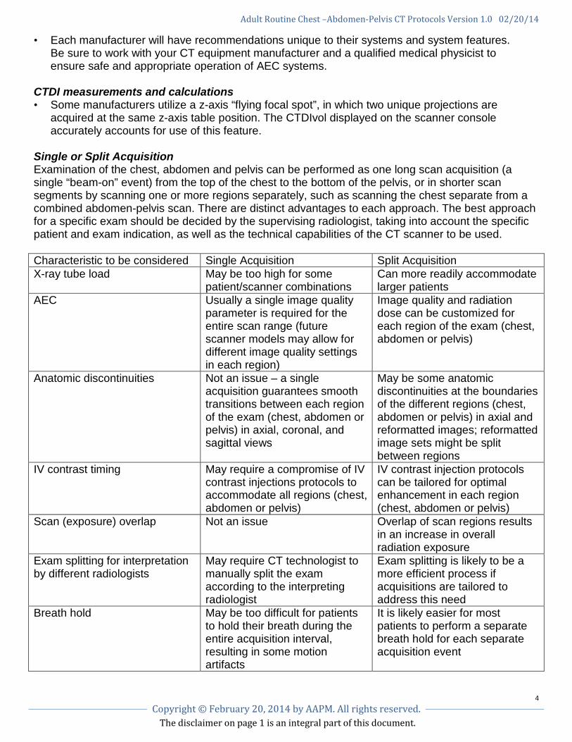
Single or Split Acquisition Examination of the chest, abdomen and pelvis can be performed as one long scan acquisition (a single “beam-on” event) from the top of the chest to the bottom of the pelvis, or in shorter scan segments by scanning one or more regions separately, such as scanning the chest separate from a combined abdomen-pelvis scan. There are distinct advantages to each approach. The best approach for a specific exam should be decided by the supervising radiologist, taking into account the specific patient and exam indication, as well as the technical capabilities of the CT scanner to be used. Characteristic to be considered Single Acquisition Split Acquisition X-ray tube load May be too high for some
patient/scanner combinations Can more readily accommodate larger patients
AEC Usually a single image quality parameter is required for the entire scan range (future scanner models may allow for different image quality settings in each region)
Image quality and radiation dose can be customized for each region of the exam (chest, abdomen or pelvis)
Anatomic discontinuities Not an issue – a single acquisition guarantees smooth transitions between each region of the exam (chest, abdomen or pelvis) in axial, coronal, and sagittal views
May be some anatomic discontinuities at the boundaries of the different regions (chest, abdomen or pelvis) in axial and reformatted images; reformatted image sets might be split between regions
IV contrast timing May require a compromise of IV contrast injections protocols to accommodate all regions (chest, abdomen or pelvis)
IV contrast injection protocols can be tailored for optimal enhancement in each region (chest, abdomen or pelvis)
Scan (exposure) overlap Not an issue Overlap of scan regions results in an increase in overall radiation exposure
Exam splitting for interpretation by different radiologists
May require CT technologist to manually split the exam according to the interpreting radiologist
Exam splitting is likely to be a more efficient process if acquisitions are tailored to address this need
Breath hold May be too difficult for patients to hold their breath during the entire acquisition interval, resulting in some motion artifacts
It is likely easier for most patients to perform a separate breath hold for each separate acquisition event
Adult Routine Chest –Abdomen-Pelvis CT Protocols Version 1.0 02/20/14
The disclaimer on page 1 is an integral part of this document.
5 Copyright © February 20, 2014 by AAPM. All rights reserved.
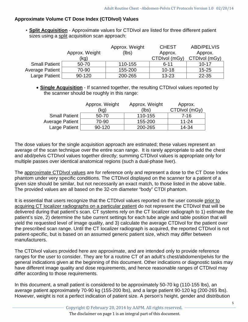
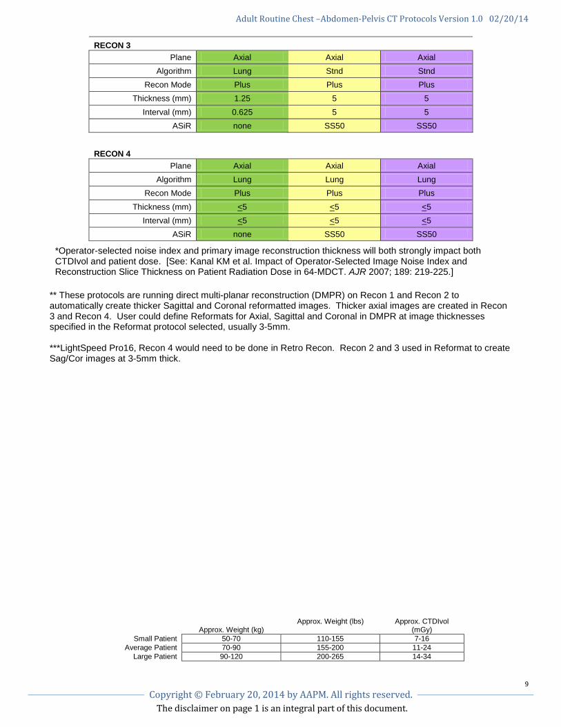
Approximate Volume CT Dose Index (CTDIvol) Values
• Split Acquisition - Approximate values for CTDIvol are listed for three different patient sizes using a split acquisition scan approach:
Approx. Weight (kg)
Approx. Weight (lbs)
CHEST Approx.
CTDIvol (mGy)
ABD/PELVIS Approx.
CTDIvol (mGy) Small Patient 50-70 110-155 6-11 10-17
Average Patient 70-90 155-200 10-18 15-25 Large Patient 90-120 200-265 13-23 22-35
• Single Acquisition - If scanned together, the resulting CTDIvol values reported by
the scanner should be roughly in this range:
Approx. Weight (kg)
Approx. Weight (lbs)
Approx. CTDIvol (mGy)
Small Patient 50-70 110-155 7-16 Average Patient 70-90 155-200 11-24
Large Patient 90-120 200-265 14-34
The dose values for the single acquisition approach are estimated; these values represent an average of the scan technique over the entire scan range. It is rarely appropriate to add the chest and abd/pelvis CTDIvol values together directly; summing CTDIvol values is appropriate only for multiple passes over identical anatomical regions (such a dual-phase liver). The approximate CTDIvol values are for reference only and represent a dose to the CT Dose Index phantom under very specific conditions. The CTDIvol displayed on the scanner for a patient of a given size should be similar, but not necessarily an exact match, to those listed in the above table. The provided values are all based on the 32-cm diameter “body” CTDI phantom. It is essential that users recognize that the CTDIvol values reported on the user console prior to acquiring CT localizer radiographs on a particular patient do not represent the CTDIvol that will be delivered during that patient’s scan. CT systems rely on the CT localizer radiograph to 1) estimate the patient’s size, 2) determine the tube current settings for each tube angle and table position that will yield the requested level of image quality, and 3) calculate the average CTDIvol for the patient over the prescribed scan range. Until the CT localizer radiograph is acquired, the reported CTDIvol is not patient-specific, but is based on an assumed generic patient size, which may differ between manufacturers. The CTDIvol values provided here are approximate, and are intended only to provide reference ranges for the user to consider. They are for a routine CT of an adult’s chest/abdomen/pelvis for the general indications given at the beginning of this document. Other indications or diagnostic tasks may have different image quality and dose requirements, and hence reasonable ranges of CTDIvol may differ according to those requirements. In this document, a small patient is considered to be approximately 50-70 kg (110-155 lbs), an average patient approximately 70-90 kg (155-200 lbs), and a large patient 90-120 kg (200-265 lbs). However, weight is not a perfect indication of patient size. A person’s height, gender and distribution
Adult Routine Chest –Abdomen-Pelvis CT Protocols Version 1.0 02/20/14
The disclaimer on page 1 is an integral part of this document.
6 Copyright © February 20, 2014 by AAPM. All rights reserved.
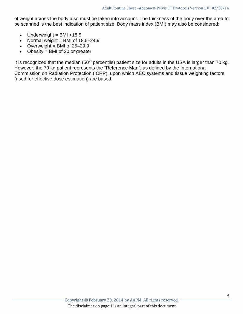
of weight across the body also must be taken into account. The thickness of the body over the area to be scanned is the best indication of patient size. Body mass index (BMI) may also be considered:
• Underweight = BMI <18.5 • Normal weight = BMI of 18.5–24.9 • Overweight = BMI of 25–29.9 • Obesity = BMI of 30 or greater
It is recognized that the median (50th percentile) patient size for adults in the USA is larger than 70 kg. However, the 70 kg patient represents the “Reference Man”, as defined by the International Commission on Radiation Protection (ICRP), upon which AEC systems and tissue weighting factors (used for effective dose estimation) are based.
Adult Routine Chest –Abdomen-Pelvis CT Protocols Version 1.0 02/20/14
The disclaimer on page 1 is an integral part of this document.
7 Copyright © February 20, 2014 by AAPM. All rights reserved.
INDEX OF ADULT ROUTINE ABDOMEN-PELVIS PROTOCOLS (by manufacturer)
GE
Hitachi
Neusoft
Philips
Siemens
Toshiba
Adult Routine Chest –Abdomen-Pelvis CT Protocols Version 1.0 02/20/14
The disclaimer on page 1 is an integral part of this document.
8 Copyright © February 20, 2014 by AAPM. All rights reserved.
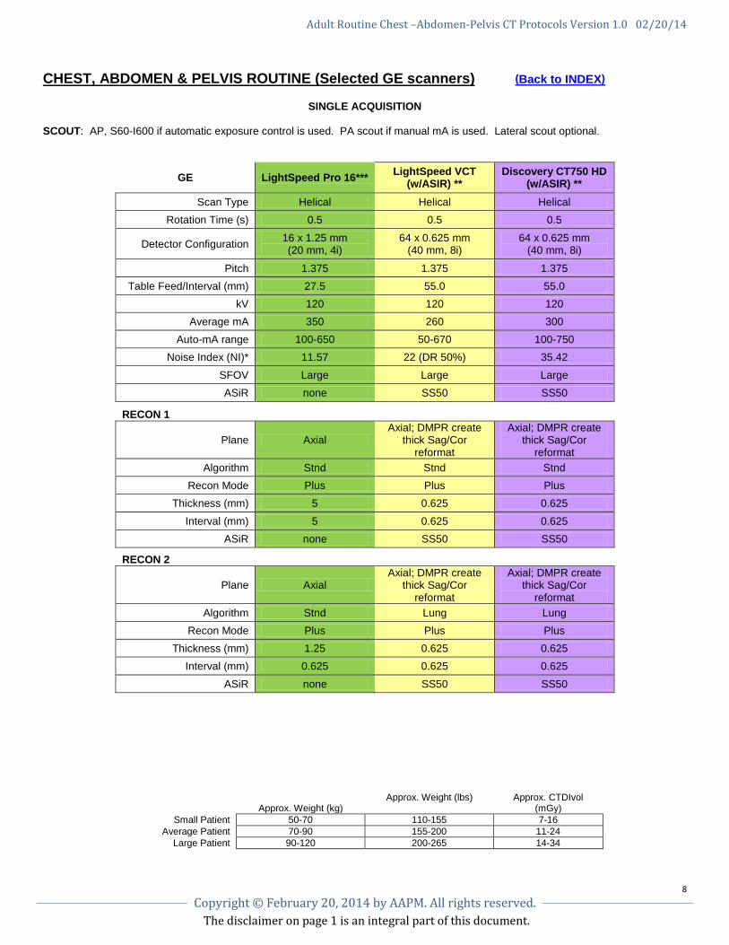
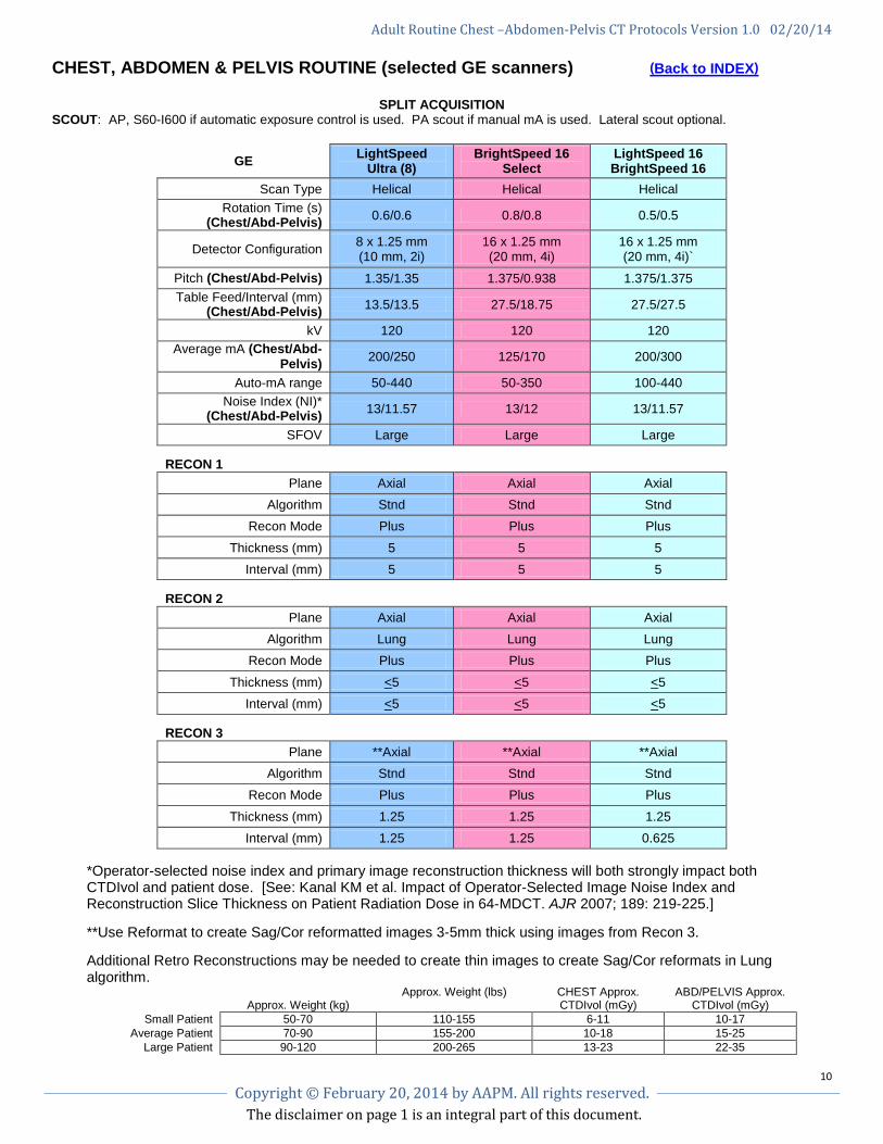
CHEST, ABDOMEN & PELVIS ROUTINE (Selected GE scanners) (Back to INDEX)
SINGLE ACQUISITION SCOUT: AP, S60-I600 if automatic exposure control is used. PA scout if manual mA is used. Lateral scout optional.
GE LightSpeed Pro 16*** LightSpeed VCT (w/ASIR) **
Discovery CT750 HD (w/ASIR) **
Scan Type Helical Helical Helical Rotation Time (s) 0.5 0.5 0.5
Detector Configuration 16 x 1.25 mm (20 mm, 4i)
64 x 0.625 mm (40 mm, 8i)
64 x 0.625 mm (40 mm, 8i)
Pitch 1.375 1.375 1.375 Table Feed/Interval (mm) 27.5 55.0 55.0
kV 120 120 120 Average mA 350 260 300
Auto-mA range 100-650 50-670 100-750 Noise Index (NI)* 11.57 22 (DR 50%) 35.42
SFOV Large Large Large ASiR none SS50 SS50
RECON 1
Plane Axial Axial; DMPR create
thick Sag/Cor reformat
Axial; DMPR create thick Sag/Cor
reformat Algorithm Stnd Stnd Stnd
Recon Mode Plus Plus Plus Thickness (mm) 5 0.625 0.625
Interval (mm) 5 0.625 0.625 ASiR none SS50 SS50
RECON 2
Plane Axial Axial; DMPR create
thick Sag/Cor reformat
Axial; DMPR create thick Sag/Cor
reformat Algorithm Stnd Lung Lung
Recon Mode Plus Plus Plus Thickness (mm) 1.25 0.625 0.625
Interval (mm) 0.625 0.625 0.625
ASiR none SS50 SS50
Approx. Weight (kg)
Approx. Weight (lbs) Approx. CTDIvol (mGy)
Small Patient 50-70 110-155 7-16 Average Patient 70-90 155-200 11-24
Large Patient 90-120 200-265 14-34
Adult Routine Chest –Abdomen-Pelvis CT Protocols Version 1.0 02/20/14
The disclaimer on page 1 is an integral part of this document.
9 Copyright © February 20, 2014 by AAPM. All rights reserved.
RECON 3 Plane Axial Axial Axial
Algorithm Lung Stnd Stnd Recon Mode Plus Plus Plus
Thickness (mm) 1.25 5 5
Interval (mm) 0.625 5 5 ASiR none SS50 SS50
RECON 4
Plane Axial Axial Axial Algorithm Lung Lung Lung
Recon Mode Plus Plus Plus
Thickness (mm) <5 <5 <5 Interval (mm) <5 <5 <5
ASiR none SS50 SS50
*Operator-selected noise index and primary image reconstruction thickness will both strongly impact both CTDIvol and patient dose. [See: Kanal KM et al. Impact of Operator-Selected Image Noise Index and Reconstruction Slice Thickness on Patient Radiation Dose in 64-MDCT. AJR 2007; 189: 219-225.]
** These protocols are running direct multi-planar reconstruction (DMPR) on Recon 1 and Recon 2 to automatically create thicker Sagittal and Coronal reformatted images. Thicker axial images are created in Recon 3 and Recon 4. User could define Reformats for Axial, Sagittal and Coronal in DMPR at image thicknesses specified in the Reformat protocol selected, usually 3-5mm. ***LightSpeed Pro16, Recon 4 would need to be done in Retro Recon. Recon 2 and 3 used in Reformat to create Sag/Cor images at 3-5mm thick.
Approx. Weight (kg)
Approx. Weight (lbs) Approx. CTDIvol (mGy)
Small Patient 50-70 110-155 7-16 Average Patient 70-90 155-200 11-24
Large Patient 90-120 200-265 14-34
Adult Routine Chest –Abdomen-Pelvis CT Protocols Version 1.0 02/20/14
The disclaimer on page 1 is an integral part of this document.
10 Copyright © February 20, 2014 by AAPM. All rights reserved.
CHEST, ABDOMEN & PELVIS ROUTINE (selected GE scanners) (Back to INDEX)
SPLIT ACQUISITION SCOUT: AP, S60-I600 if automatic exposure control is used. PA scout if manual mA is used. Lateral scout optional.
GE LightSpeed Ultra (8)
BrightSpeed 16 Select
LightSpeed 16 BrightSpeed 16
Scan Type Helical Helical Helical Rotation Time (s)
(Chest/Abd-Pelvis) 0.6/0.6 0.8/0.8 0.5/0.5
Detector Configuration 8 x 1.25 mm (10 mm, 2i)
16 x 1.25 mm (20 mm, 4i)
16 x 1.25 mm (20 mm, 4i)`
Pitch (Chest/Abd-Pelvis) 1.35/1.35 1.375/0.938 1.375/1.375 Table Feed/Interval (mm)
(Chest/Abd-Pelvis) 13.5/13.5 27.5/18.75 27.5/27.5
kV 120 120 120 Average mA (Chest/Abd-
Pelvis) 200/250 125/170 200/300
Auto-mA range 50-440 50-350 100-440 Noise Index (NI)*
(Chest/Abd-Pelvis) 13/11.57 13/12 13/11.57
SFOV Large Large Large
RECON 1
Plane Axial Axial Axial Algorithm Stnd Stnd Stnd
Recon Mode Plus Plus Plus
Thickness (mm) 5 5 5 Interval (mm) 5 5 5
RECON 2
Plane Axial Axial Axial Algorithm Lung Lung Lung
Recon Mode Plus Plus Plus
Thickness (mm) <5 <5 <5 Interval (mm) <5 <5 <5
RECON 3
Plane **Axial **Axial **Axial Algorithm Stnd Stnd Stnd
Recon Mode Plus Plus Plus
Thickness (mm) 1.25 1.25 1.25 Interval (mm) 1.25 1.25 0.625
*Operator-selected noise index and primary image reconstruction thickness will both strongly impact both CTDIvol and patient dose. [See: Kanal KM et al. Impact of Operator-Selected Image Noise Index and Reconstruction Slice Thickness on Patient Radiation Dose in 64-MDCT. AJR 2007; 189: 219-225.] **Use Reformat to create Sag/Cor reformatted images 3-5mm thick using images from Recon 3. Additional Retro Reconstructions may be needed to create thin images to create Sag/Cor reformats in Lung algorithm.
Approx. Weight (kg)
Approx. Weight (lbs) CHEST Approx. CTDIvol (mGy)
ABD/PELVIS Approx. CTDIvol (mGy)
Small Patient 50-70 110-155 6-11 10-17 Average Patient 70-90 155-200 10-18 15-25
Large Patient 90-120 200-265 13-23 22-35

Adult Routine Chest –Abdomen-Pelvis CT Protocols Version 1.0 02/20/14
The disclaimer on page 1 is an integral part of this document.
11 Copyright © February 20, 2014 by AAPM. All rights reserved.
CHEST, ABDOMEN & PELVIS ROUTINE (selected GE scanners) (Back to INDEX)
SPLIT ACQUISITION SCOUT: AP, S60-I600 if automatic exposure control is used. PA scout if manual mA is used. Lateral scout optional. SPLIT ACQUISITION– group 1 Chest, group 2 Abdomen/Pelvis These protocols are built with direct multi-planar reconstruction (DMPR) running on Recon 2 and Recon 3 to automatically create
Sagittal and Coronal reformats 3-5mm thick.
GE LightSpeed VCT** Discovery CT750 HD**
LightSpeed VCT (w/ASIR)**
Discovery CT750 HD (w/ASIR)**
Scan Type Helical Helical Helical Helical Rotation Time (s)
(Chest/Abd-Pelvis) 0.5/0.5 0/5/0.5 0.5/0.5 0.4/0.5
Detector Configuration 64 x 0.625 mm (40 mm, 8i)
64 x 0.625 mm (40 mm, 8i)
64 x 0.625 mm (40 mm, 8i)
64 x 0.625 mm (40 mm, 8i)
Pitch (Chest/Abd-Pelvis) 0.984/1.375 0.984/1.375 0.984/1.375 0.984/1.375 Table Feed/Interval (mm)
(Chest/Abd-Pelvis) 39.37/55 39.37/55 39.37/55.0 39.37/55.0
kV 120 120 120 120 Average mA (Chest/Abd-
Pelvis) 300/370 300/400 260/300 250/300
Auto-mA range 100-650 100-750 50-670 100-500 Noise Index (NI)*
(Chest/Abd-Pelvis) 13.0/13.0 13.80/12.65 22 (DR 50%)/ 18 (DR 50%)
22.93/ 19.32
SFOV Large Large Large Large ASiR None None SS50 SS50
RECON 1 – group 1 & 2
Plane Axial Axial Axial Axial
Algorithm Stnd Stnd Stnd Stnd Recon Mode Plus Plus Plus Plus
Thickness (mm) 5 5 5 5 Interval (mm) 5 5 5 5
ASiR None None SS50 SS50
RECON 2
Plane Axial DMPR create
thicker Sag/Cor reformats–group 1 & 2
Axial DMPR create thicker Sag/Cor
reformats-group 1 & 2
Axial DMPR create thicker Sag/Cor
reformats-group 1 & 2
Axial DMPR create thicker Sag/Cor
reformats-group 1 & 2 Algorithm Stnd Stnd Stnd Stnd
Recon Mode Plus Plus Plus Plus Thickness (mm) 0.625 0.625 0.625 0.625
Interval (mm) 0.625 0.625 0.625 0.625 ASiR None None SS50 SS50
Approx. Weight (kg)
Approx. Weight (lbs) CHEST Approx. CTDIvol (mGy)
ABD/PELVIS Approx. CTDIvol (mGy)
Small Patient 50-70 110-155 6-11 10-17 Average Patient 70-90 155-200 10-18 15-25
Large Patient 90-120 200-265 13-23 22-35
Adult Routine Chest –Abdomen-Pelvis CT Protocols Version 1.0 02/20/14
The disclaimer on page 1 is an integral part of this document.
12 Copyright © February 20, 2014 by AAPM. All rights reserved.
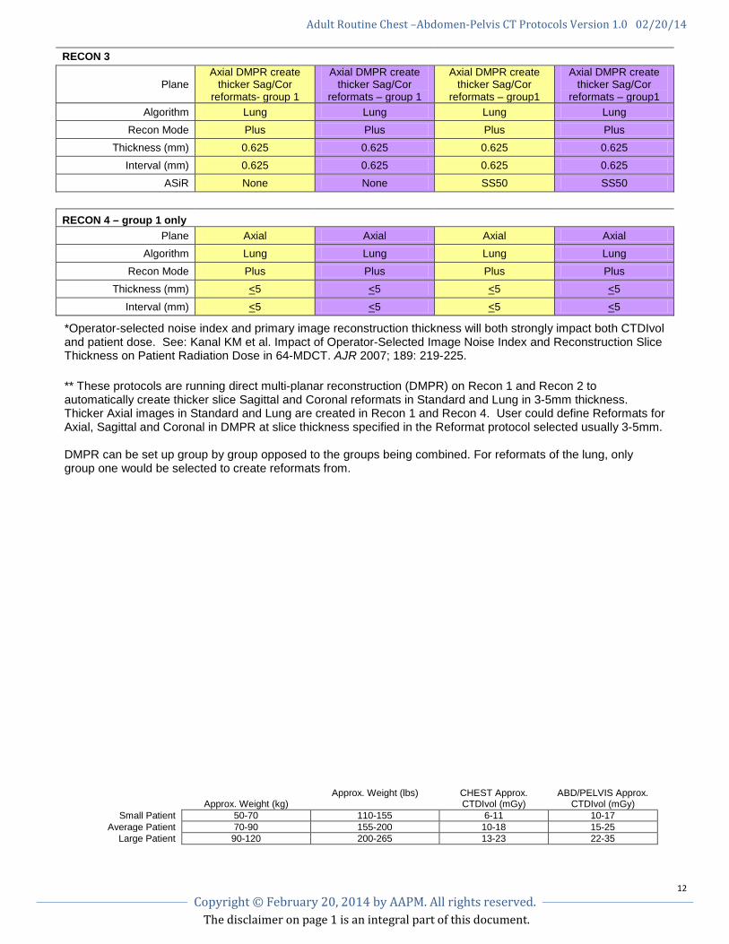
RECON 3
Plane Axial DMPR create
thicker Sag/Cor reformats- group 1
Axial DMPR create thicker Sag/Cor
reformats – group 1
Axial DMPR create thicker Sag/Cor
reformats – group1
Axial DMPR create thicker Sag/Cor
reformats – group1 Algorithm Lung Lung Lung Lung
Recon Mode Plus Plus Plus Plus Thickness (mm) 0.625 0.625 0.625 0.625
Interval (mm) 0.625 0.625 0.625 0.625 ASiR None None SS50 SS50
RECON 4 – group 1 only
Plane Axial Axial Axial Axial Algorithm Lung Lung Lung Lung
Recon Mode Plus Plus Plus Plus Thickness (mm) <5 <5 <5 <5
Interval (mm) <5 <5 <5 <5
*Operator-selected noise index and primary image reconstruction thickness will both strongly impact both CTDIvol and patient dose. See: Kanal KM et al. Impact of Operator-Selected Image Noise Index and Reconstruction Slice Thickness on Patient Radiation Dose in 64-MDCT. AJR 2007; 189: 219-225.
** These protocols are running direct multi-planar reconstruction (DMPR) on Recon 1 and Recon 2 to automatically create thicker slice Sagittal and Coronal reformats in Standard and Lung in 3-5mm thickness. Thicker Axial images in Standard and Lung are created in Recon 1 and Recon 4. User could define Reformats for Axial, Sagittal and Coronal in DMPR at slice thickness specified in the Reformat protocol selected usually 3-5mm. DMPR can be set up group by group opposed to the groups being combined. For reformats of the lung, only group one would be selected to create reformats from.
Approx. Weight (kg)
Approx. Weight (lbs) CHEST Approx. CTDIvol (mGy)
ABD/PELVIS Approx. CTDIvol (mGy)
Small Patient 50-70 110-155 6-11 10-17 Average Patient 70-90 155-200 10-18 15-25
Large Patient 90-120 200-265 13-23 22-35
Adult Routine Chest –Abdomen-Pelvis CT Protocols Version 1.0 02/20/14
The disclaimer on page 1 is an integral part of this document.
13 Copyright © February 20, 2014 by AAPM. All rights reserved.
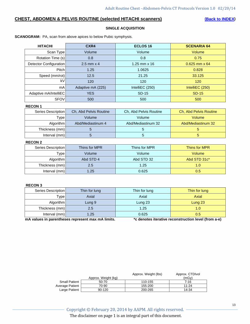
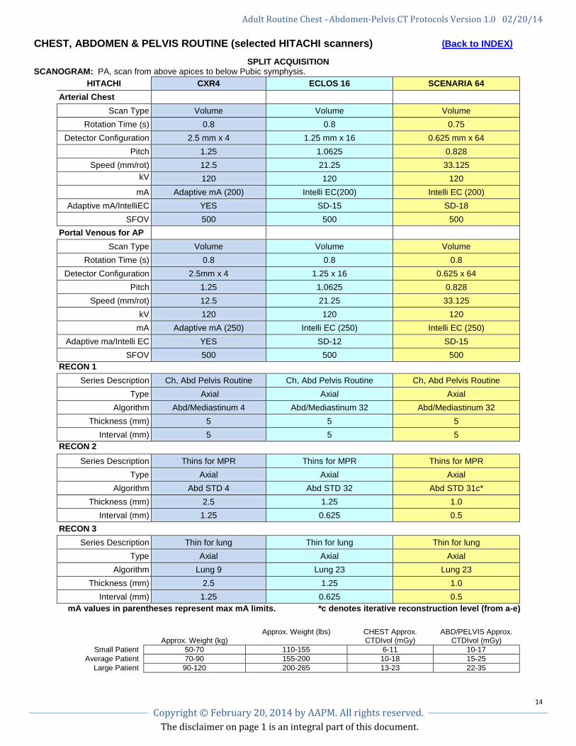
CHEST, ABDOMEN & PELVIS ROUTINE (selected HITACHI scanners) (Back to INDEX)
SINGLE ACQUISITION
SCANOGRAM: PA, scan from above apices to below Pubic symphysis.
HITACHI CXR4 ECLOS 16 SCENARIA 64 Scan Type Volume Volume Volume
Rotation Time (s) 0.8 0.8 0.75 Detector Configuration 2.5 mm x 4 1.25 mm x 16 0.625 mm x 64
Pitch 1.25 1.0625 0.828 Speed (mm/rot) 12.5 21.25 33.125
kV
120 120 120 mA Adaptive mA (225) IntelliEC (250) IntelliEC (250)
Adaptive mA/IntelliEC YES SD-15 SD-15 SFOV 500 500 500
RECON 1
Series Description Ch, Abd Pelvis Routine Ch, Abd Pelvis Routine Ch, Abd Pelvis Routine Type Volume Volume Volume
Algorithm Abd/Mediastinum 4 Abd/Mediastinum 32 Abd/Mediastinum 32 Thickness (mm) 5 5 5
Interval (mm) 5 5 5
RECON 2
Series Description Thins for MPR Thins for MPR Thins for MPR Type Volume Volume Volume
Algorithm Abd STD 4 Abd STD 32 Abd STD 31c* Thickness (mm) 2.5 1.25 1.0
Interval (mm) 1.25 0.625 0.5
RECON 3 Series Description Thin for lung Thin for lung Thin for lung
Type Axial Axial Axial Algorithm Lung 9 Lung 23 Lung 23
Thickness (mm) 2.5 1.25 1.0 Interval (mm) 1.25 0.625 0.5
mA values in parentheses represent max mA limits. *c denotes iterative reconstruction level (from a-e)
Approx. Weight (kg)
Approx. Weight (lbs) Approx. CTDIvol (mGy)
Small Patient 50-70 110-155 7-16 Average Patient 70-90 155-200 11-24
Large Patient 90-120 200-265 14-34
Adult Routine Chest –Abdomen-Pelvis CT Protocols Version 1.0 02/20/14
The disclaimer on page 1 is an integral part of this document.
14 Copyright © February 20, 2014 by AAPM. All rights reserved.
CHEST, ABDOMEN & PELVIS ROUTINE (selected HITACHI scanners) (Back to INDEX)
SPLIT ACQUISITION
SCANOGRAM: PA, scan from above apices to below Pubic symphysis. HITACHI CXR4 ECLOS 16 SCENARIA 64
Arterial Chest Scan Type Volume Volume Volume
Rotation Time (s) 0.8 0.8 0.75 Detector Configuration 2.5 mm x 4 1.25 mm x 16 0.625 mm x 64
Pitch 1.25 1.0625 0.828 Speed (mm/rot) 12.5 21.25 33.125
kV
120 120 120 mA Adaptive mA (200) Intelli EC(200) Intelli EC (200)
Adaptive mA/IntelliEC YES SD-15 SD-18 SFOV 500 500 500
Portal Venous for AP Scan Type Volume Volume Volume
Rotation Time (s) 0.8 0.8 0.8 Detector Configuration 2.5mm x 4 1.25 x 16 0.625 x 64
Pitch 1.25 1.0625 0.828 Speed (mm/rot) 12.5 21.25 33.125
kV 120 120 120 mA Adaptive mA (250) Intelli EC (250) Intelli EC (250)
Adaptive ma/Intelli EC YES SD-12 SD-15 SFOV 500 500 500
RECON 1 Series Description Ch, Abd Pelvis Routine Ch, Abd Pelvis Routine Ch, Abd Pelvis Routine
Type Axial Axial Axial Algorithm Abd/Mediastinum 4 Abd/Mediastinum 32 Abd/Mediastinum 32
Thickness (mm) 5 5 5 Interval (mm) 5 5 5
RECON 2
Series Description Thins for MPR Thins for MPR Thins for MPR Type Axial Axial Axial
Algorithm Abd STD 4 Abd STD 32 Abd STD 31c* Thickness (mm) 2.5 1.25 1.0
Interval (mm) 1.25 0.625 0.5 RECON 3
Series Description Thin for lung Thin for lung Thin for lung Type Axial Axial Axial
Algorithm Lung 9 Lung 23 Lung 23 Thickness (mm) 2.5 1.25 1.0
Interval (mm) 1.25 0.625 0.5 mA values in parentheses represent max mA limits. *c denotes iterative reconstruction level (from a-e)
Approx. Weight (kg) Approx. Weight (lbs) CHEST Approx.
CTDIvol (mGy) ABD/PELVIS Approx.
CTDIvol (mGy) Small Patient 50-70 110-155 6-11 10-17
Average Patient 70-90 155-200 10-18 15-25 Large Patient 90-120 200-265 13-23 22-35
Adult Routine Chest –Abdomen-Pelvis CT Protocols Version 1.0 02/20/14
The disclaimer on page 1 is an integral part of this document.
15 Copyright © February 20, 2014 by AAPM. All rights reserved.
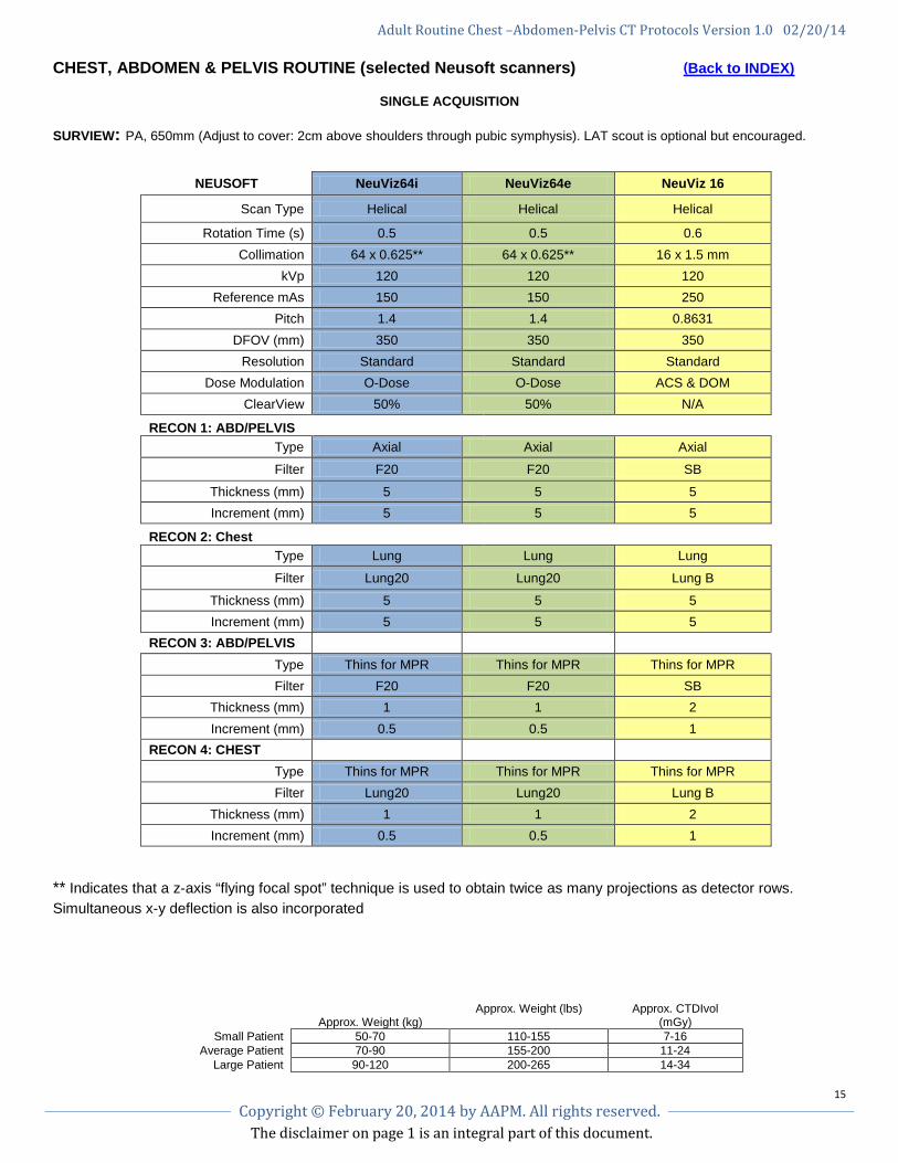
CHEST, ABDOMEN & PELVIS ROUTINE (selected Neusoft scanners) (Back to INDEX)
SINGLE ACQUISITION
SURVIEW: PA, 650mm (Adjust to cover: 2cm above shoulders through pubic symphysis). LAT scout is optional but encouraged.
** Indicates that a z-axis “flying focal spot” technique is used to obtain twice as many projections as detector rows. Simultaneous x-y deflection is also incorporated
Approx. Weight (kg)
Approx. Weight (lbs) Approx. CTDIvol (mGy)
Small Patient 50-70 110-155 7-16 Average Patient 70-90 155-200 11-24
Large Patient 90-120 200-265 14-34
NEUSOFT NeuViz64i NeuViz64e NeuViz 16
Scan Type Helical Helical Helical
Rotation Time (s) 0.5 0.5 0.6 Collimation 64 x 0.625** 64 x 0.625** 16 x 1.5 mm
kVp 120 120 120 Reference mAs 150 150 250
Pitch 1.4 1.4 0.8631 DFOV (mm) 350 350 350
Resolution Standard Standard Standard Dose Modulation O-Dose O-Dose ACS & DOM
ClearView 50% 50% N/A
RECON 1: ABD/PELVIS Type Axial Axial Axial
Filter F20 F20 SB
Thickness (mm) 5 5 5 Increment (mm) 5 5 5
RECON 2: Chest Type Lung Lung Lung
Filter Lung20 Lung20 Lung B
Thickness (mm) 5 5 5 Increment (mm) 5 5 5
RECON 3: ABD/PELVIS Type Thins for MPR Thins for MPR Thins for MPR Filter F20 F20 SB
Thickness (mm) 1 1 2 Increment (mm) 0.5 0.5 1
RECON 4: CHEST Type Thins for MPR Thins for MPR Thins for MPR Filter Lung20 Lung20 Lung B
Thickness (mm) 1 1 2 Increment (mm) 0.5 0.5 1
Adult Routine Chest –Abdomen-Pelvis CT Protocols Version 1.0 02/20/14
The disclaimer on page 1 is an integral part of this document.
16 Copyright © February 20, 2014 by AAPM. All rights reserved.
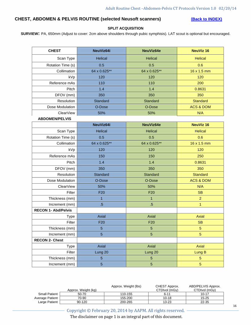
CHEST, ABDOMEN & PELVIS ROUTINE (selected Neusoft scanners) (Back to INDEX)
SPLIT ACQUISITION SURVIEW: PA, 650mm (Adjust to cover: 2cm above shoulders through pubic symphisis). LAT scout is optional but encouraged.
CHEST NeuViz64i NeuViz64e NeuViz 16
Scan Type Helical Helical Helical
Rotation Time (s) 0.5 0.5 0.6 Collimation 64 x 0.625** 64 x 0.625** 16 x 1.5 mm
kVp 120 120 120 Reference mAs 110 110 200
Pitch 1.4 1.4 0.8631 DFOV (mm) 350 350 350
Resolution Standard Standard Standard Dose Modulation O-Dose O-Dose ACS & DOM
ClearView 50% 50% N/A ABDOMEN/PELVIS
NeuViz64i NeuViz64e NeuViz 16 Scan Type Helical Helical Helical
Rotation Time (s) 0.5 0.5 0.6 Collimation 64 x 0.625** 64 x 0.625** 16 x 1.5 mm
kVp 120 120 120
Reference mAs 150 150 250
Pitch 1.4 1.4 0.8631
DFOV (mm) 350 350 350 Resolution Standard Standard Standard
Dose Modulation O-Dose O-Dose ACS & DOM ClearView 50% 50% N/A
Filter F20 F20 SB Thickness (mm) 1 1 2 Increment (mm) .5 .5 1
RECON 1- Abd/Pelvis Type Axial Axial Axial Filter F20 F20 SB
Thickness (mm) 5 5 5 Increment (mm) 5 5 5
RECON 2- Chest Type Axial Axial Axial Filter Lung 20 Lung 20 Lung B
Thickness (mm) 5 5 5 Increment (mm) 5 5 5
Approx. Weight (kg)
Approx. Weight (lbs) CHEST Approx. CTDIvol (mGy)
ABD/PELVIS Approx. CTDIvol (mGy)
Small Patient 50-70 110-155 6-11 10-17 Average Patient 70-90 155-200 10-18 15-25
Large Patient 90-120 200-265 13-23 22-35
Adult Routine Chest –Abdomen-Pelvis CT Protocols Version 1.0 02/20/14
The disclaimer on page 1 is an integral part of this document.
17 Copyright © February 20, 2014 by AAPM. All rights reserved.
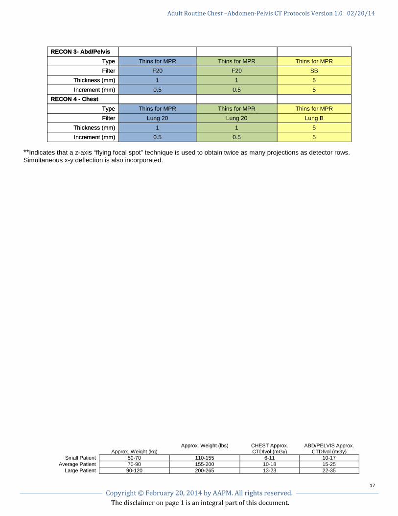
**Indicates that a z-axis “flying focal spot” technique is used to obtain twice as many projections as detector rows. Simultaneous x-y deflection is also incorporated.
Approx. Weight (kg)
Approx. Weight (lbs) CHEST Approx. CTDIvol (mGy)
ABD/PELVIS Approx. CTDIvol (mGy)
Small Patient 50-70 110-155 6-11 10-17 Average Patient 70-90 155-200 10-18 15-25
Large Patient 90-120 200-265 13-23 22-35
RECON 3- Abd/Pelvis Type Thins for MPR Thins for MPR Thins for MPR Filter F20 F20 SB
Thickness (mm) 1 1 5 Increment (mm) 0.5 0.5 5
RECON 4 - Chest Type Thins for MPR Thins for MPR Thins for MPR Filter Lung 20 Lung 20 Lung B
Thickness (mm) 1 1 5 Increment (mm) 0.5 0.5 5
RECON 3- Abd/Pelvis Type Thins for MPR Thins for MPR Thins for MPR Filter F20 F20 SB
Thickness (mm) 1 1 5 Increment (mm) 0.5 0.5 5
RECON 4 - Chest Type Thins for MPR Thins for MPR Thins for MPR Filter Lung 20 Lung 20 Lung B
Thickness (mm) 1 1 5 Increment (mm) 0.5 0.5 5
Adult Routine Chest –Abdomen-Pelvis CT Protocols Version 1.0 02/20/14
The disclaimer on page 1 is an integral part of this document.
18 Copyright © February 20, 2014 by AAPM. All rights reserved.
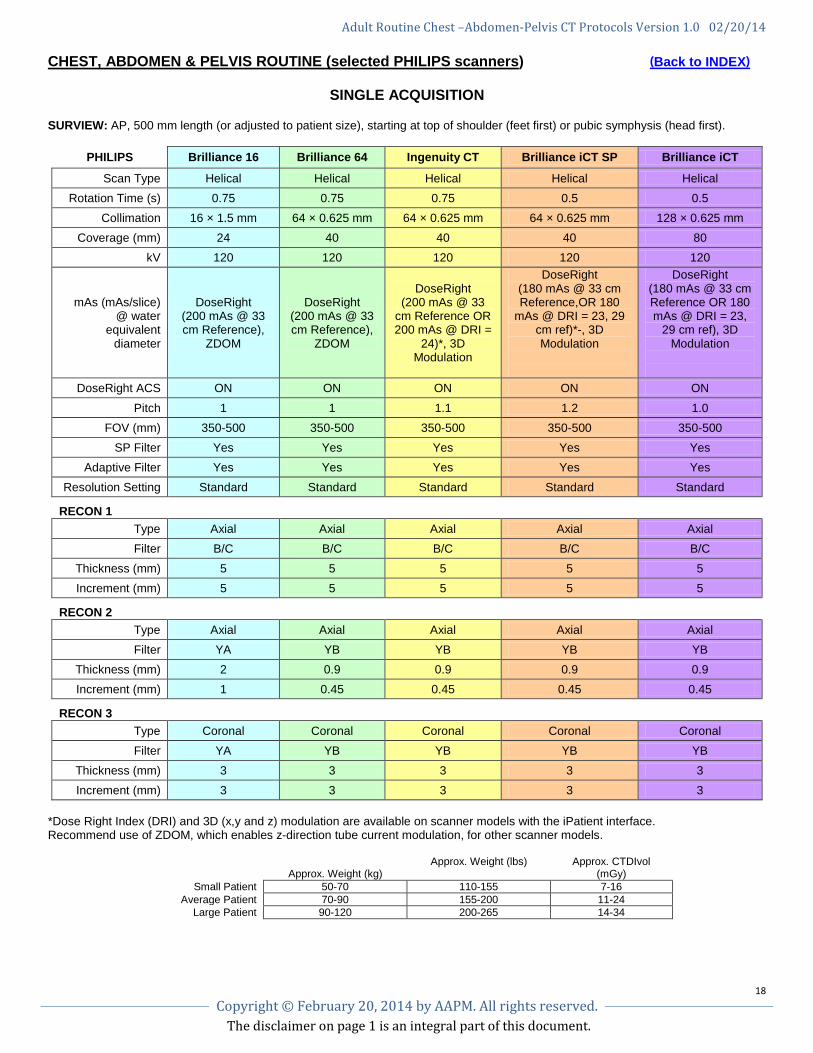
CHEST, ABDOMEN & PELVIS ROUTINE (selected PHILIPS scanners) (Back to INDEX)
SINGLE ACQUISITION
SURVIEW: AP, 500 mm length (or adjusted to patient size), starting at top of shoulder (feet first) or pubic symphysis (head first).
PHILIPS Brilliance 16 Brilliance 64 Ingenuity CT Brilliance iCT SP Brilliance iCT
Scan Type Helical Helical Helical Helical Helical Rotation Time (s) 0.75 0.75 0.75 0.5 0.5
Collimation 16 × 1.5 mm 64 × 0.625 mm 64 × 0.625 mm 64 × 0.625 mm 128 × 0.625 mm Coverage (mm) 24 40 40 40 80
kV 120 120 120 120 120
mAs (mAs/slice) @ water
equivalent diameter
DoseRight (200 mAs @ 33 cm Reference),
ZDOM
DoseRight (200 mAs @ 33 cm Reference),
ZDOM
DoseRight (200 mAs @ 33
cm Reference OR 200 mAs @ DRI =
24)*, 3D Modulation
DoseRight (180 mAs @ 33 cm Reference,OR 180
mAs @ DRI = 23, 29 cm ref)*-, 3D Modulation
DoseRight (180 mAs @ 33 cm Reference OR 180 mAs @ DRI = 23,
29 cm ref), 3D Modulation
DoseRight ACS ON ON ON ON ON
Pitch 1 1 1.1 1.2 1.0 FOV (mm) 350-500 350-500 350-500 350-500 350-500
SP Filter Yes Yes Yes Yes Yes Adaptive Filter Yes Yes Yes Yes Yes
Resolution Setting Standard Standard Standard Standard Standard
RECON 1 Type Axial Axial Axial Axial Axial Filter B/C B/C B/C B/C B/C
Thickness (mm) 5 5 5 5 5 Increment (mm) 5 5 5 5 5
RECON 2 Type Axial Axial Axial Axial Axial Filter YA YB YB YB YB
Thickness (mm) 2 0.9 0.9 0.9 0.9 Increment (mm) 1 0.45 0.45 0.45 0.45
RECON 3 Type Coronal Coronal Coronal Coronal Coronal Filter YA YB YB YB YB
Thickness (mm) 3 3 3 3 3 Increment (mm) 3 3 3 3 3
*Dose Right Index (DRI) and 3D (x,y and z) modulation are available on scanner models with the iPatient interface. Recommend use of ZDOM, which enables z-direction tube current modulation, for other scanner models.
Approx. Weight (kg)
Approx. Weight (lbs) Approx. CTDIvol (mGy)
Small Patient 50-70 110-155 7-16 Average Patient 70-90 155-200 11-24
Large Patient 90-120 200-265 14-34
Adult Routine Chest –Abdomen-Pelvis CT Protocols Version 1.0 02/20/14
The disclaimer on page 1 is an integral part of this document.
19 Copyright © February 20, 2014 by AAPM. All rights reserved.
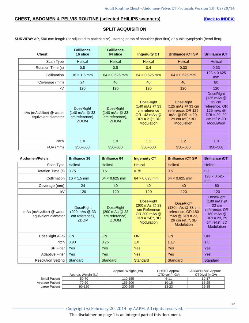
CHEST, ABDOMEN & PELVIS ROUTINE (selected PHILIPS scanners) (Back to INDEX)
SPLIT ACQUISITION
SURVIEW: AP, 500 mm length (or adjusted to patient size), starting at top of shoulder (feet first) or pubic symphysis (head first).
Chest Brilliance 16 slice
Brilliance 64 slice Ingenuity CT Brilliance iCT SP Brilliance iCT
Scan Type Helical Helical Helical Helical Helical Rotation Time (s) 0.5 0.5 0.4 0.33 0.33
Collimation 16 × 1.5 mm 64 × 0.625 mm 64 × 0.625 mm 64 × 0.625 mm 128 × 0.625 mm
Coverage (mm) 24 40 40 40 80 kV 120 120 120 120 120
mAs (mAs/slice) @ water equivalent diameter
DoseRight (140 mAs @ 33 cm reference),
ZDOM
DoseRight (140 mAs @ 33 cm reference),
ZDOM
DoseRight (140 mAs @ 33
cm reference OR 143 mAs @ DRI = 21)*, 3D
Modulation
DoseRight (125 mAs @ 33 cm reference, OR 125 mAs @ DRI = 20, 29 cm ref.)* 3D
Modulation
DoseRight (125 mAs @
33 cm reference, OR 125 mAs @ DRI = 20, 29 cm ref.)* 3D Modulation
Pitch 1.0 1.0 1.1 1.2 1.0 FOV (mm) 350–500 350–500 350–500 350–500 350–500
Abdomen/Pelvis Brilliance 16 Brilliance 64 Ingenuity CT Brilliance iCT SP Brilliance iCT
Scan Type Helical Helical Helical Helical Helical Rotation Time (s) 0.75 0.5 0.75 0.5 0.5
Collimation 16 × 1.5 mm 64 × 0.625 mm 64 × 0.625 mm 64 × 0.625 mm 128 × 0.625 mm
Coverage (mm) 24 40 40 40 80 kV 120 120 120 120 120
mAs (mAs/slice) @ water equivalent diameter
DoseRight (200 mAs @ 33 cm reference),
ZDOM
DoseRight (200 mAs @ 33 cm reference),
ZDOM
DoseRight (200 mAs @ 33 cm Reference
OR 200 mAs @ DRI = 24)*, 3D
Modulation
DoseRight (180 mAs @ 33 cm reference, OR 180 mAs @ DRI = 23, 29 cm ref.)*, 3D
Modulation
DoseRight (180 mAs @
33 cm reference, OR 180 mAs @ DRI = 23, 29 cm ref.)*, 3D Modulation
DoseRight ACS ON ON ON ON ON
Pitch 0.93 0.75 1.0 1.17 1.0 SP Filter Yes Yes Yes Yes Yes
Adaptive Filter Yes Yes Yes Yes Yes Resolution Setting Standard Standard Standard Standard Standard
Approx. Weight (kg) Approx. Weight (lbs) CHEST Approx.
CTDIvol (mGy) ABD/PELVIS Approx.
CTDIvol (mGy) Small Patient 50-70 110-155 6-11 10-17
Average Patient 70-90 155-200 10-18 15-25 Large Patient 90-120 200-265 13-23 22-35
Adult Routine Chest –Abdomen-Pelvis CT Protocols Version 1.0 02/20/14
The disclaimer on page 1 is an integral part of this document.
20 Copyright © February 20, 2014 by AAPM. All rights reserved.
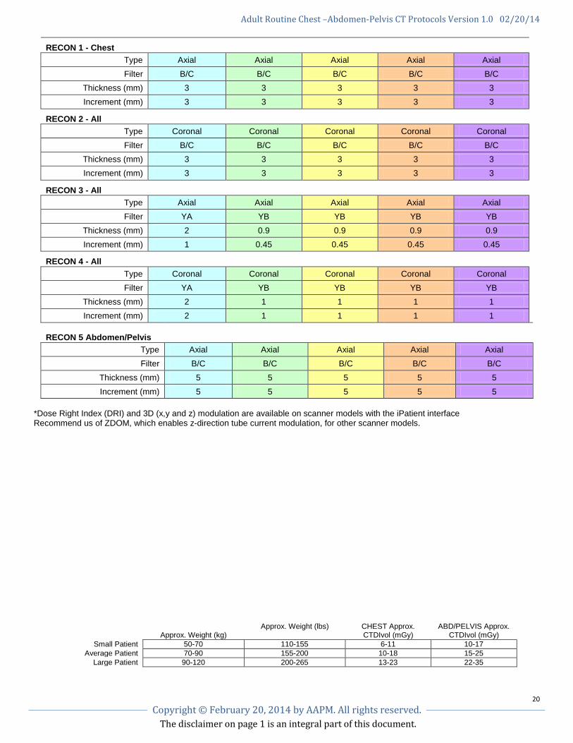
RECON 1 - Chest
Type Axial Axial Axial Axial Axial Filter B/C B/C B/C B/C B/C
Thickness (mm) 3 3 3 3 3 Increment (mm) 3 3 3 3 3
RECON 2 - All
Type Coronal Coronal Coronal Coronal Coronal Filter B/C B/C B/C B/C B/C
Thickness (mm) 3 3 3 3 3 Increment (mm) 3 3 3 3 3
RECON 3 - All
Type Axial Axial Axial Axial Axial Filter YA YB YB YB YB
Thickness (mm) 2 0.9 0.9 0.9 0.9 Increment (mm) 1 0.45 0.45 0.45 0.45
RECON 4 - All
Type Coronal Coronal Coronal Coronal Coronal Filter YA YB YB YB YB
Thickness (mm) 2 1 1 1 1 Increment (mm) 2 1 1 1 1
RECON 5 Abdomen/Pelvis
Type Axial Axial Axial Axial Axial Filter B/C B/C B/C B/C B/C
Thickness (mm) 5 5 5 5 5
Increment (mm) 5 5 5 5 5 *Dose Right Index (DRI) and 3D (x,y and z) modulation are available on scanner models with the iPatient interface Recommend us of ZDOM, which enables z-direction tube current modulation, for other scanner models.
Approx. Weight (kg)
Approx. Weight (lbs) CHEST Approx. CTDIvol (mGy)
ABD/PELVIS Approx. CTDIvol (mGy)
Small Patient 50-70 110-155 6-11 10-17 Average Patient 70-90 155-200 10-18 15-25
Large Patient 90-120 200-265 13-23 22-35
Adult Routine Chest –Abdomen-Pelvis CT Protocols Version 1.0 02/20/14
The disclaimer on page 1 is an integral part of this document.
21 Copyright © February 20, 2014 by AAPM. All rights reserved.
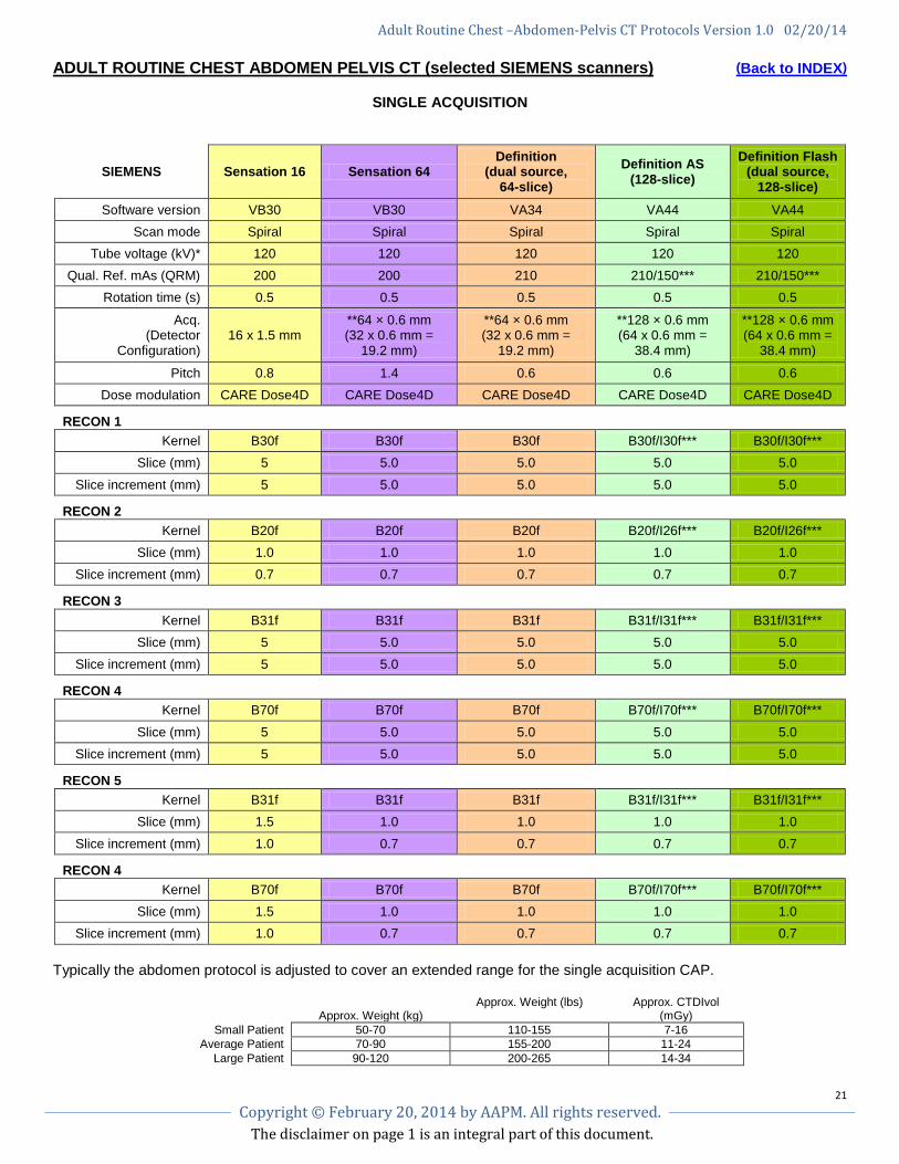
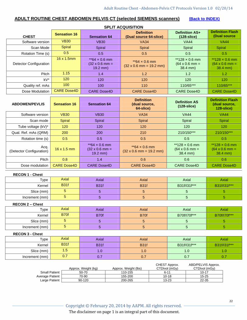
ADULT ROUTINE CHEST ABDOMEN PELVIS CT (selected SIEMENS scanners) (Back to INDEX)
SINGLE ACQUISITION
SIEMENS Sensation 16 Sensation 64 Definition
(dual source, 64-slice)
Definition AS (128-slice)
Definition Flash (dual source,
128-slice) Software version VB30 VB30 VA34 VA44 VA44
Scan mode Spiral Spiral Spiral Spiral Spiral Tube voltage (kV)* 120 120 120 120 120
Qual. Ref. mAs (QRM) 200 200 210 210/150*** 210/150*** Rotation time (s) 0.5 0.5 0.5 0.5 0.5
Acq. (Detector
Configuration) 16 x 1.5 mm
**64 × 0.6 mm (32 x 0.6 mm =
19.2 mm)
**64 × 0.6 mm (32 x 0.6 mm =
19.2 mm)
**128 × 0.6 mm (64 x 0.6 mm =
38.4 mm)
**128 × 0.6 mm (64 x 0.6 mm =
38.4 mm)
Pitch 0.8 1.4 0.6 0.6 0.6 Dose modulation CARE Dose4D CARE Dose4D CARE Dose4D CARE Dose4D CARE Dose4D
RECON 1
Kernel B30f B30f B30f B30f/I30f*** B30f/I30f*** Slice (mm) 5 5.0 5.0 5.0 5.0
Slice increment (mm) 5 5.0 5.0 5.0 5.0
RECON 2
Kernel B20f B20f B20f B20f/I26f*** B20f/I26f*** Slice (mm) 1.0 1.0 1.0 1.0 1.0
Slice increment (mm) 0.7 0.7 0.7 0.7 0.7
RECON 3
Kernel B31f B31f B31f B31f/I31f*** B31f/I31f*** Slice (mm) 5 5.0 5.0 5.0 5.0
Slice increment (mm) 5 5.0 5.0 5.0 5.0
RECON 4
Kernel B70f B70f B70f B70f/I70f*** B70f/I70f***
Slice (mm) 5 5.0 5.0 5.0 5.0 Slice increment (mm) 5 5.0 5.0 5.0 5.0
RECON 5
Kernel B31f B31f B31f B31f/I31f*** B31f/I31f*** Slice (mm) 1.5 1.0 1.0 1.0 1.0
Slice increment (mm) 1.0 0.7 0.7 0.7 0.7
RECON 4
Kernel B70f B70f B70f B70f/I70f*** B70f/I70f*** Slice (mm) 1.5 1.0 1.0 1.0 1.0
Slice increment (mm) 1.0 0.7 0.7 0.7 0.7 Typically the abdomen protocol is adjusted to cover an extended range for the single acquisition CAP.
Approx. Weight (kg) Approx. Weight (lbs) Approx. CTDIvol
(mGy) Small Patient 50-70 110-155 7-16
Average Patient 70-90 155-200 11-24 Large Patient 90-120 200-265 14-34
Adult Routine Chest –Abdomen-Pelvis CT Protocols Version 1.0 02/20/14
The disclaimer on page 1 is an integral part of this document.
22 Copyright © February 20, 2014 by AAPM. All rights reserved.
ADULT ROUTINE CHEST ABDOMEN PELVIS CT (selected SIEMENS scanners) (Back to INDEX)
SPLIT ACQUISITION
CHEST Sensation 16 Sensation 64 Definition
(Dual source 64-slice) Definition AS+
(128-slice) Definition Flash
(Dual source 128 li ) Software version VB30 VB30 VA34 VA44 VA44
Scan Mode Spiral Spiral Spiral Spiral Spiral Rotation Time (s) 0.5 0.5 0.5 0.5 0.5
Detector Configuration 16 x 1.5mm **64 × 0.6 mm
(32 x 0.6 mm = 19.2 mm)
**64 × 0.6 mm (32 x 0.6 mm = 19.2 mm)
**128 × 0.6 mm (64 x 0.6 mm =
38.4 mm)
**128 × 0.6 mm (64 x 0.6 mm =
38.4 mm) Pitch 1.15 1.4 1.2 1.2 1.2
kV* 120 120 120 120 120 Quality ref. mAs 100 100 110 110/65*** 110/65***
Dose Modulation CARE Dose4D CARE Dose4D CARE Dose4D CARE Dose4D CARE Dose4D
ABDOMEN/PEVLIS Sensation 16 Sensation 64 Definition
(dual source, 64-slice)
Definition AS (128-slice)
Definition Flash (dual source,
128-slice) Software version VB30 VB30 VA34 VA44 VA44
Scan mode Spiral Spiral Spiral Spiral Spiral Tube voltage (kV)* 120 120 120 120 120
Qual. Ref. mAs (QRM) 200 200 210 210/150*** 210/150*** Rotation time (s) 0.5 0.5 0.5 0.5 0.5
Acq. (Detector Configuration) 16 x 1.5 mm
**64 × 0.6 mm (32 x 0.6 mm =
19.2 mm)
**64 × 0.6 mm (32 x 0.6 mm = 19.2 mm)
**128 × 0.6 mm (64 x 0.6 mm =
38.4 mm)
**128 × 0.6 mm (64 x 0.6 mm =
38.4 mm)
Pitch 0.8 1.4 0.6 0.6 0.6 Dose modulation CARE Dose4D CARE Dose4D CARE Dose4D CARE Dose4D CARE Dose4D
RECON 1 - Chest
Type Axial Axial Axial Axial Axial Kernel B31f B31f B31f B31f/I31f*** B31f/I31f***
Slice (mm) 5 5 5 5 5 Increment (mm) 5 5 5 5 5
RECON 2 – Chest Type Axial Axial Axial Axial Axial
Kernel B70f B70f B70f B70f/I70f*** B70f/I70f*** Slice (mm) 5 5 5 5 5
Increment (mm) 5 5 5 5 5
RECON 3 - Chest Type Axial Axial Axial Axial Axial
Kernel B31f B31f B31f B31f/I31f*** B31f/I31f*** Slice (mm) 1.5 1.0 1.0 1.0 1.0
Increment (mm) 0.7 0.7 0.7 0.7 0.7
Approx. Weight (kg)
Approx. Weight (lbs)
CHEST Approx. CTDIvol (mGy)
ABD/PELVIS Approx. CTDIvol (mGy)
Small Patient 50-70 110-155 6-11 10-17 Average Patient 70-90 155-200 10-18 15-25
Large Patient 90-120 200-265 13-23 22-35
Adult Routine Chest –Abdomen-Pelvis CT Protocols Version 1.0 02/20/14
The disclaimer on page 1 is an integral part of this document.
23 Copyright © February 20, 2014 by AAPM. All rights reserved.
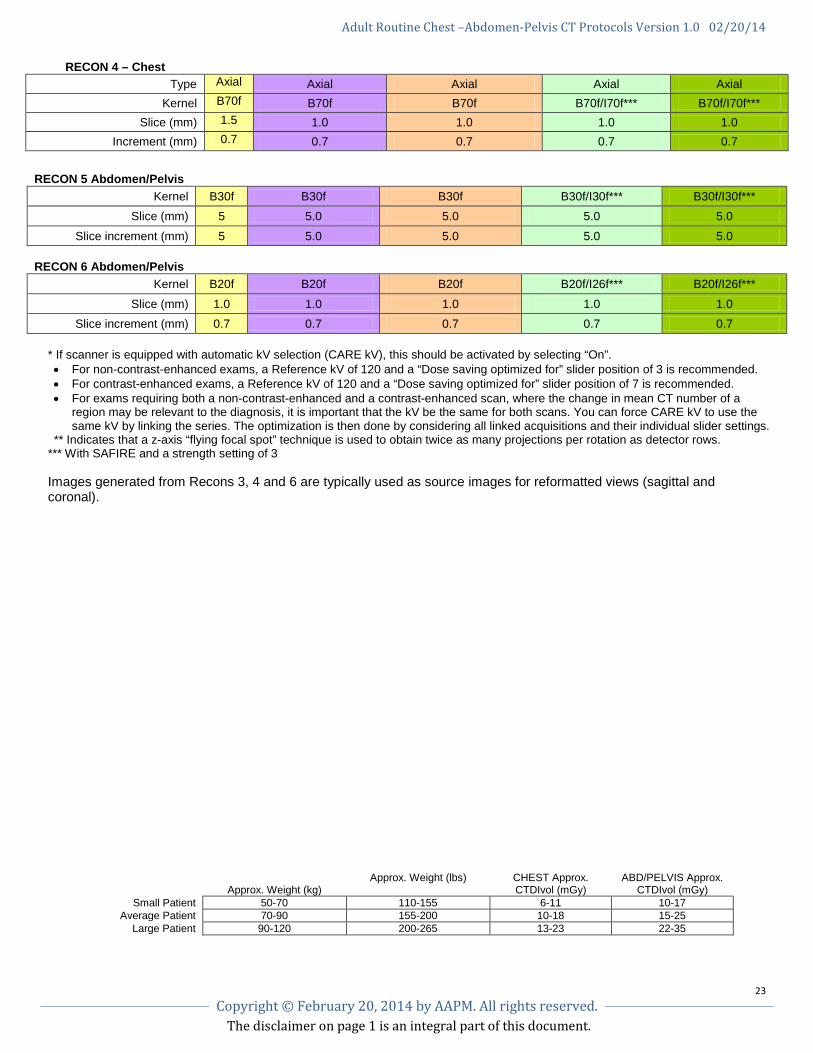
RECON 4 – Chest Type Axial Axial Axial Axial Axial
Kernel B70f B70f B70f B70f/I70f*** B70f/I70f*** Slice (mm) 1.5 1.0 1.0 1.0 1.0
Increment (mm) 0.7 0.7 0.7 0.7 0.7
RECON 5 Abdomen/Pelvis
Kernel B30f B30f B30f B30f/I30f*** B30f/I30f*** Slice (mm) 5 5.0 5.0 5.0 5.0
Slice increment (mm) 5 5.0 5.0 5.0 5.0 RECON 6 Abdomen/Pelvis
Kernel B20f B20f B20f B20f/I26f*** B20f/I26f***
Slice (mm) 1.0 1.0 1.0 1.0 1.0 Slice increment (mm) 0.7 0.7 0.7 0.7 0.7
* If scanner is equipped with automatic kV selection (CARE kV), this should be activated by selecting “On”. • For non-contrast-enhanced exams, a Reference kV of 120 and a “Dose saving optimized for” slider position of 3 is recommended. • For contrast-enhanced exams, a Reference kV of 120 and a “Dose saving optimized for” slider position of 7 is recommended. • For exams requiring both a non-contrast-enhanced and a contrast-enhanced scan, where the change in mean CT number of a
region may be relevant to the diagnosis, it is important that the kV be the same for both scans. You can force CARE kV to use the same kV by linking the series. The optimization is then done by considering all linked acquisitions and their individual slider settings.
** Indicates that a z-axis “flying focal spot” technique is used to obtain twice as many projections per rotation as detector rows. *** With SAFIRE and a strength setting of 3 Images generated from Recons 3, 4 and 6 are typically used as source images for reformatted views (sagittal and coronal).
Approx. Weight (kg)
Approx. Weight (lbs) CHEST Approx. CTDIvol (mGy)
ABD/PELVIS Approx. CTDIvol (mGy)
Small Patient 50-70 110-155 6-11 10-17 Average Patient 70-90 155-200 10-18 15-25
Large Patient 90-120 200-265 13-23 22-35
Adult Routine Chest –Abdomen-Pelvis CT Protocols Version 1.0 02/20/14
The disclaimer on page 1 is an integral part of this document.
24 Copyright © February 20, 2014 by AAPM. All rights reserved.
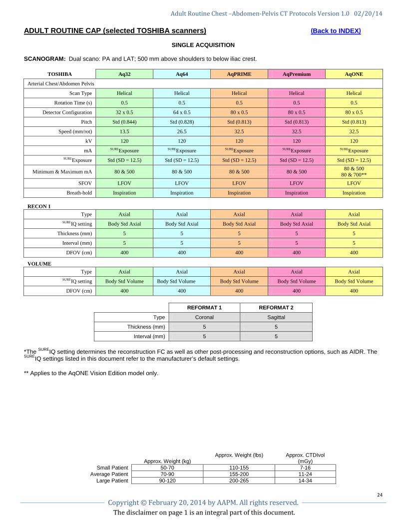
ADULT ROUTINE CAP (selected TOSHIBA scanners) (Back to INDEX)
SINGLE ACQUISITION SCANOGRAM: Dual scano: PA and LAT; 500 mm above shoulders to below iliac crest.
TOSHIBA Aq32 Aq64 AqPRIME AqPremium AqONE
Arterial Chest/Abdomen Pelvis
Scan Type Helical Helical Helical Helical Helical
Rotation Time (s) 0.5 0.5 0.5 0.5 0.5
Detector Configuration 32 x 0.5 64 x 0.5 80 x 0.5 80 x 0.5 80 x 0.5
Pitch Std (0.844) Std (0.828) Std (0.813) Std (0.813) Std (0.813)
Speed (mm/rot) 13.5 26.5 32.5 32.5 32.5
kV 120 120 120 120 120
mA SUREExposure SUREExposure SUREExposure SUREExposure SUREExposure SUREExposure Std (SD = 12.5) Std (SD = 12.5) Std (SD = 12.5) Std (SD = 12.5) Std (SD = 12.5)
Minimum & Maximum mA 80 & 500 80 & 500 80 & 500 80 & 500 80 & 500 80 & 700**
SFOV LFOV LFOV LFOV LFOV LFOV
Breath-hold Inspiration Inspiration Inspiration Inspiration Inspiration
RECON 1
Type Axial Axial Axial Axial Axial SUREIQ setting Body Std Axial Body Std Axial Body Std Axial Body Std Axial Body Std Axial
Thickness (mm) 5 5 5 5 5
Interval (mm) 5 5 5 5 5
DFOV (cm) 400 400 400 400 400
VOLUME
Type Axial Axial Axial Axial Axial SUREIQ setting Body Std Volume Body Std Volume Body Std Volume Body Std Volume Body Std Volume
DFOV (cm) 400 400 400 400 400
REFORMAT 1 REFORMAT 2
Type Coronal Sagittal
Thickness (mm) 5 5
Interval (mm) 5 5 *The SUREIQ setting determines the reconstruction FC as well as other post-processing and reconstruction options, such as AIDR. The SUREIQ settings listed in this document refer to the manufacturer’s default settings. ** Applies to the AqONE Vision Edition model only.
Approx. Weight (kg)
Approx. Weight (lbs) Approx. CTDIvol (mGy)
Small Patient 50-70 110-155 7-16 Average Patient 70-90 155-200 11-24
Large Patient 90-120 200-265 14-34
Adult Routine Chest –Abdomen-Pelvis CT Protocols Version 1.0 02/20/14
The disclaimer on page 1 is an integral part of this document.
25 Copyright © February 20, 2014 by AAPM. All rights reserved.
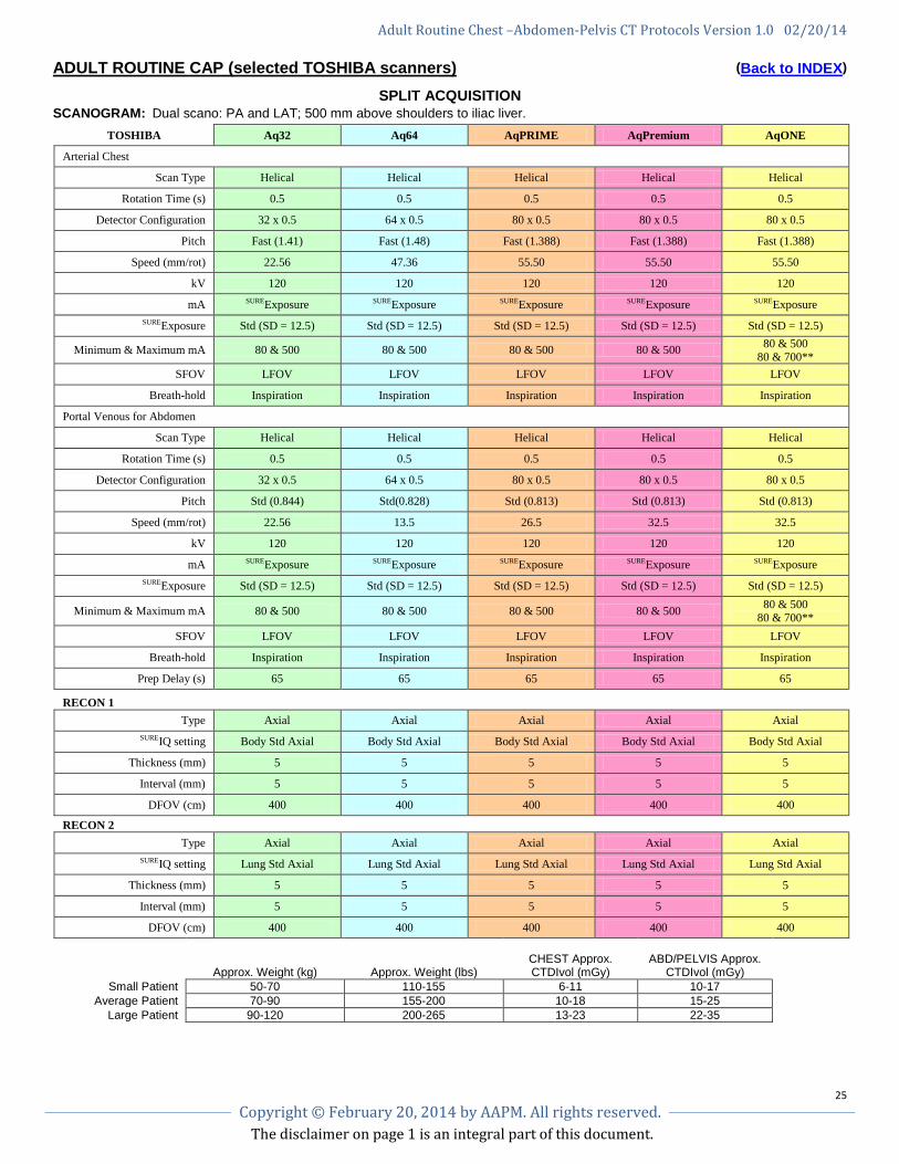
ADULT ROUTINE CAP (selected TOSHIBA scanners) (Back to INDEX)
SPLIT ACQUISITION
SCANOGRAM: Dual scano: PA and LAT; 500 mm above shoulders to iliac liver.
TOSHIBA Aq32 Aq64 AqPRIME AqPremium AqONE
Arterial Chest
Scan Type Helical Helical Helical Helical Helical
Rotation Time (s) 0.5 0.5 0.5 0.5 0.5
Detector Configuration 32 x 0.5 64 x 0.5 80 x 0.5 80 x 0.5 80 x 0.5
Pitch Fast (1.41) Fast (1.48) Fast (1.388) Fast (1.388) Fast (1.388)
Speed (mm/rot) 22.56 47.36 55.50 55.50 55.50
kV 120 120 120 120 120
mA SUREExposure SUREExposure SUREExposure SUREExposure SUREExposure SUREExposure Std (SD = 12.5) Std (SD = 12.5) Std (SD = 12.5) Std (SD = 12.5) Std (SD = 12.5)
Minimum & Maximum mA 80 & 500 80 & 500 80 & 500 80 & 500 80 & 500 80 & 700**
SFOV LFOV LFOV LFOV LFOV LFOV
Breath-hold Inspiration Inspiration Inspiration Inspiration Inspiration
Portal Venous for Abdomen
Scan Type Helical Helical Helical Helical Helical
Rotation Time (s) 0.5 0.5 0.5 0.5 0.5
Detector Configuration 32 x 0.5 64 x 0.5 80 x 0.5 80 x 0.5 80 x 0.5
Pitch Std (0.844) Std(0.828) Std (0.813) Std (0.813) Std (0.813)
Speed (mm/rot) 22.56 13.5 26.5 32.5 32.5
kV 120 120 120 120 120
mA SUREExposure SUREExposure SUREExposure SUREExposure SUREExposure SUREExposure Std (SD = 12.5) Std (SD = 12.5) Std (SD = 12.5) Std (SD = 12.5) Std (SD = 12.5)
Minimum & Maximum mA 80 & 500 80 & 500 80 & 500 80 & 500 80 & 500 80 & 700**
SFOV LFOV LFOV LFOV LFOV LFOV
Breath-hold Inspiration Inspiration Inspiration Inspiration Inspiration
Prep Delay (s) 65 65 65 65 65
RECON 1
Type Axial Axial Axial Axial Axial SUREIQ setting Body Std Axial Body Std Axial Body Std Axial Body Std Axial Body Std Axial
Thickness (mm) 5 5 5 5 5
Interval (mm) 5 5 5 5 5
DFOV (cm) 400 400 400 400 400
RECON 2 Type Axial Axial Axial Axial Axial
SUREIQ setting Lung Std Axial Lung Std Axial Lung Std Axial Lung Std Axial Lung Std Axial
Thickness (mm) 5 5 5 5 5
Interval (mm) 5 5 5 5 5
DFOV (cm) 400 400 400 400 400 Approx. Weight (kg)
Approx. Weight (lbs) CHEST Approx. CTDIvol (mGy)
ABD/PELVIS Approx.
CTDIvol (mGy) Small Patient 50-70 110-155 6-11 10-17
Average Patient 70-90 155-200 10-18 15-25 Large Patient 90-120 200-265 13-23 22-35
Adult Routine Chest –Abdomen-Pelvis CT Protocols Version 1.0 02/20/14
The disclaimer on page 1 is an integral part of this document.
26 Copyright © February 20, 2014 by AAPM. All rights reserved.
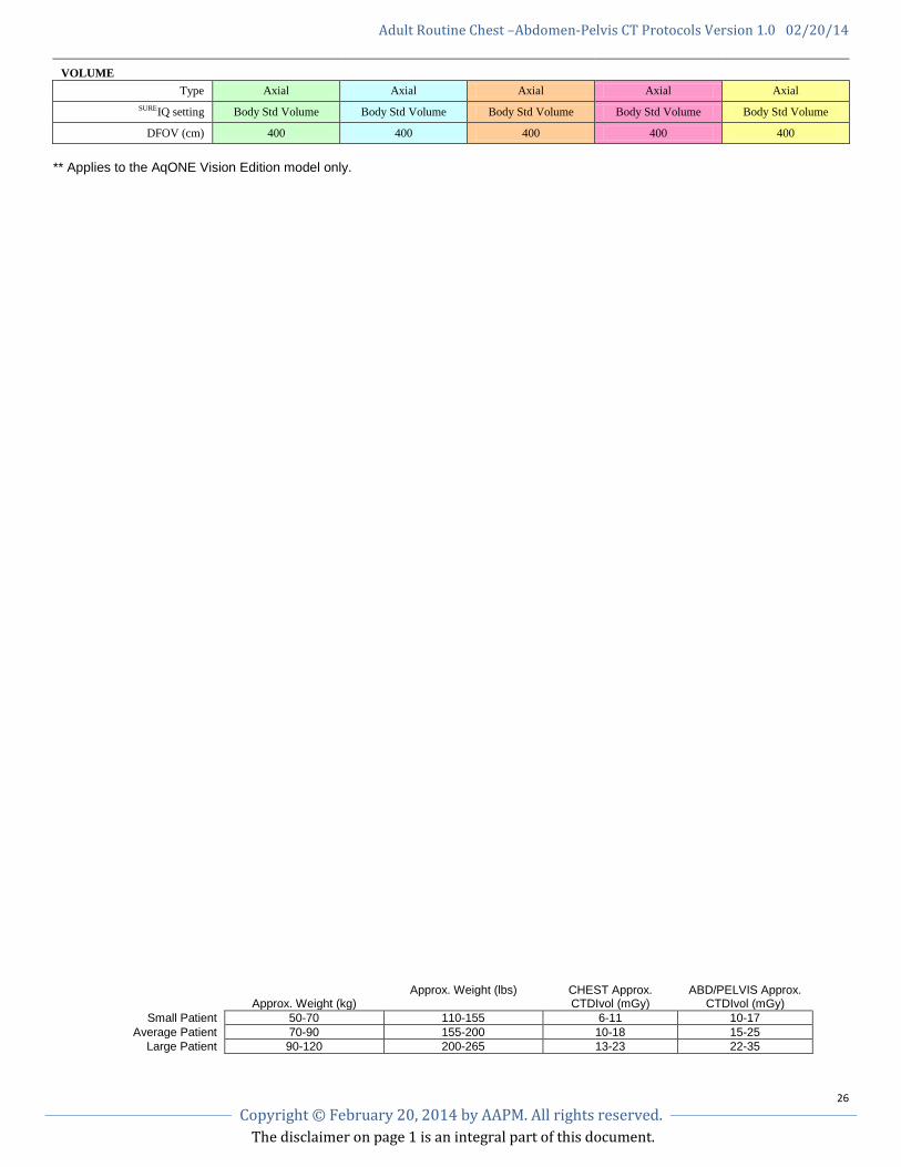
VOLUME
Type Axial Axial Axial Axial Axial SUREIQ setting Body Std Volume Body Std Volume Body Std Volume Body Std Volume Body Std Volume
DFOV (cm) 400 400 400 400 400
** Applies to the AqONE Vision Edition model only.
Approx. Weight (kg)
Approx. Weight (lbs) CHEST Approx. CTDIvol (mGy)
ABD/PELVIS Approx. CTDIvol (mGy)
Small Patient 50-70 110-155 6-11 10-17 Average Patient 70-90 155-200 10-18 15-25
Large Patient 90-120 200-265 13-23 22-35