disclosure cervical spinal disorders - ucsf cme. matsumoto... · intractable neck pain and...
TRANSCRIPT
11/8/2013
1
Pedicle Screw Fixation for
Cervical Spinal Disorders
Morio Matsumoto
Associate Professor
Director of Spine Section
Dept . Of Orthopaedic Surgery, Keio University,
Japan
Disclosure
Morio Matsumoto received honorarium for
lecture from Medtronic Sofamor Danek Japan.
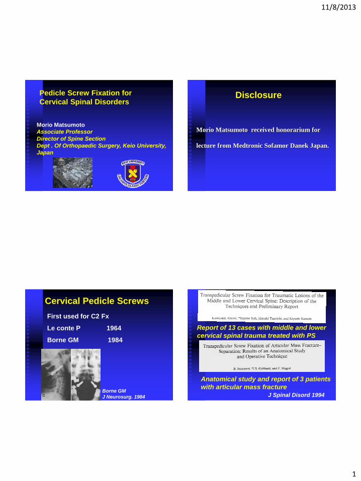
Cervical Pedicle Screws
Borne GM
J Neurosurg. 1984
First used for C2 Fx
Le conte P 1964
Borne GM 1984
J Spinal Disord 1994
Report of 13 cases with middle and lower
cervical spinal trauma treated with PS
Anatomical study and report of 3 patients
with articular mass fracture
11/8/2013
2
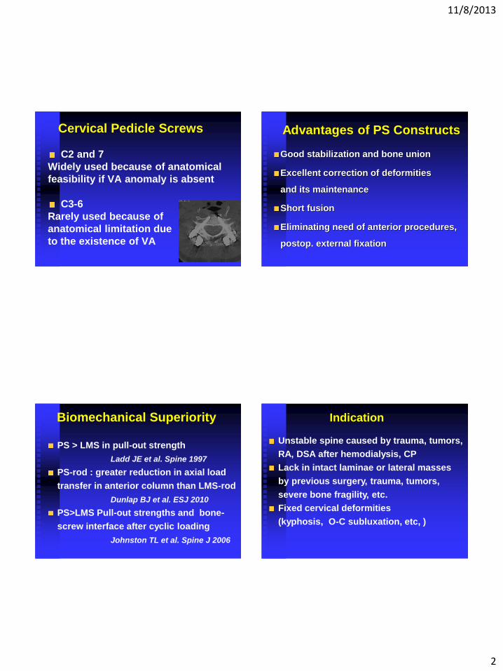
Cervical Pedicle Screws
C2 and 7
Widely used because of anatomical
feasibility if VA anomaly is absent
C3-6
Rarely used because of
anatomical limitation due
to the existence of VA
Advantages of PS Constructs
Good stabilization and bone union
Excellent correction of deformities
and its maintenance
Short fusion
Eliminating need of anterior procedures,
postop. external fixation
Biomechanical Superiority
PS > LMS in pull-out strength
Ladd JE et al. Spine 1997
PS-rod : greater reduction in axial load
transfer in anterior column than LMS-rod
Dunlap BJ et al. ESJ 2010
PS>LMS Pull-out strengths and bone-
screw interface after cyclic loading
Johnston TL et al. Spine J 2006
Indication
Unstable spine caused by trauma, tumors,
RA, DSA after hemodialysis, CP
Lack in intact laminae or lateral masses
by previous surgery, trauma, tumors,
severe bone fragility, etc.
Fixed cervical deformities
(kyphosis, O-C subluxation, etc, )
11/8/2013
3
Contra-indication of PS placement
An absent or extremely small
pedicle(outer diameter<4mm)
A pedicle destroyed by tumors etc.
Anomalies of the vertebral artery
Infection in the posterior elements
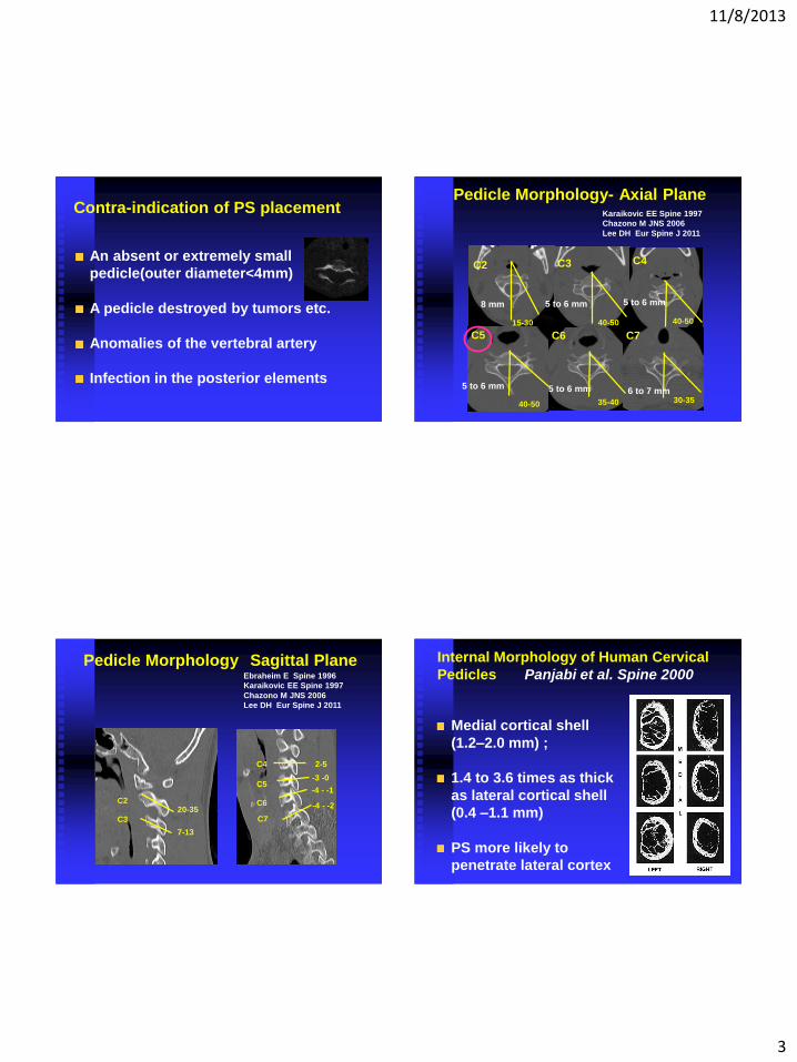
Pedicle Morphology- Axial Plane
C2 C3 C4
C5 C6 C7
5 to 6 mm 8 mm
40-50 15-30 40-50
40-50 35-40 30-35
Karaikovic EE Spine 1997
Chazono M JNS 2006
Lee DH Eur Spine J 2011
5 to 6 mm
5 to 6 mm 5 to 6 mm 6 to 7 mm
Pedicle Morphology Sagittal Plane
C2
C3
C4
C5
C6
C7
7-13
2-5
-3 -0
-4 - -1
-4 - -2 20-35
Ebraheim E Spine 1996
Karaikovic EE Spine 1997
Chazono M JNS 2006
Lee DH Eur Spine J 2011
Internal Morphology of Human Cervical
Pedicles Panjabi et al. Spine 2000
Medial cortical shell
(1.2–2.0 mm) ;
1.4 to 3.6 times as thick
as lateral cortical shell
(0.4 –1.1 mm)
PS more likely to
penetrate lateral cortex
11/8/2013
4
Individual variations++
• Morphology of Pedicles
• Anatomy of Vertebral Artery
Fine cut CT
CT / MR angiography
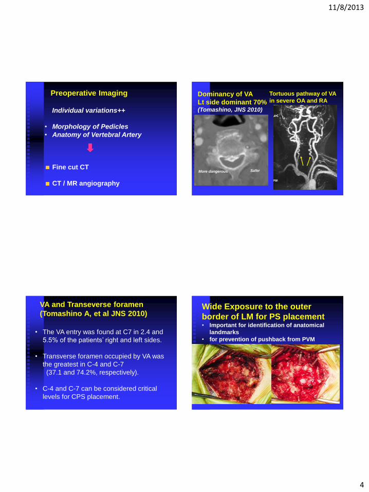
Preoperative Imaging Tortuous pathway of VA
in severe OA and RA Dominancy of VA
Lt side dominant 70% (Tomashino, JNS 2010)
Safer More dangerous
• The VA entry was found at C7 in 2.4 and
5.5% of the patients’ right and left sides.
• Transverse foramen occupied by VA was
the greatest in C-4 and C-7
(37.1 and 74.2%, respectively).
• C-4 and C-7 can be considered critical
levels for CPS placement.
VA and Transeverse foramen
(Tomashino A, et al JNS 2010) Wide Exposure to the outer
border of LM for PS placement • Important for identification of anatomical
landmarks
• for prevention of pushback from PVM
11/8/2013
5
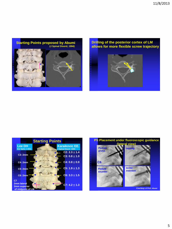
Starting Points proposed by Abumi (J Spinal Disord, 1994)
Drilling of the posterior cortex of LM
allows for more flexible screw trajectory
C2: 2.3 + 1.4
C3: 0.8 + 1.0
C4: 0.8 + 0.9
C5: 1.9 + 1.3
C6: 3.3 + 1.5
C7: 4.2 + 1.3
Starting Points
Lee DH Eur Spine J 2011
C3: 2mm
C4: 2mm
C5: 2mm
C6: 2mm
Karaikovic EE. J Spinal Disord 2000
C7
2mm lateral
2mm superior
of midpoint of LM
A
C6
Pedicle
probe tapping
Pedicle
sounder
screw
insertion
Courtesy of Prof. Abumi
PS Placement under fluoroscopic guidance
(lateral view)
11/8/2013
6
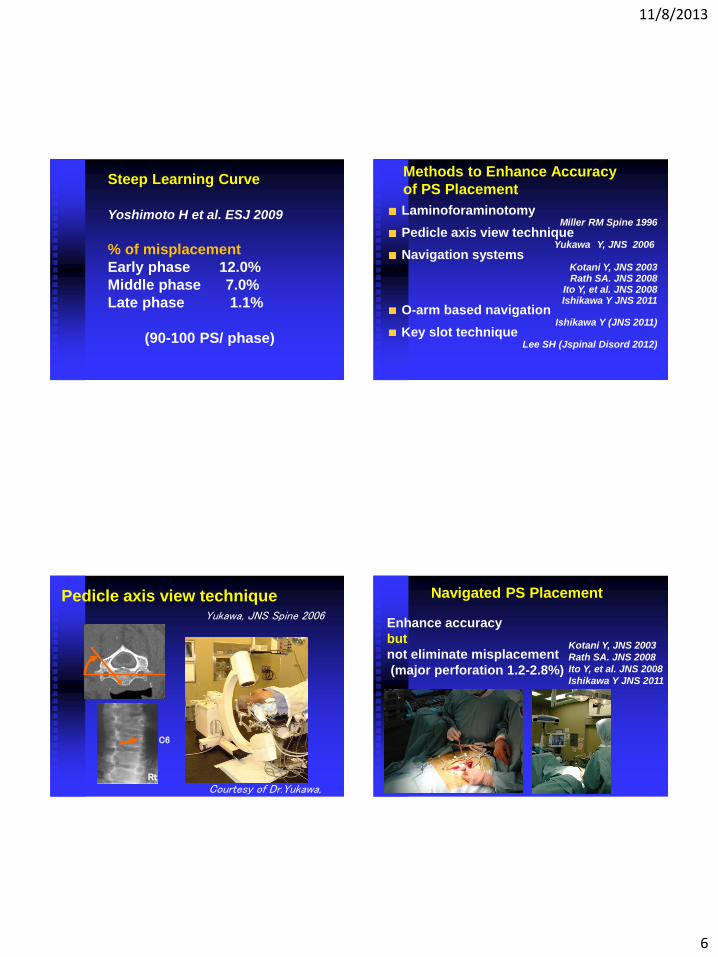
Steep Learning Curve
Yoshimoto H et al. ESJ 2009
% of misplacement
Early phase 12.0%
Middle phase 7.0%
Late phase 1.1%
(90-100 PS/ phase)
Methods to Enhance Accuracy
of PS Placement
Laminoforaminotomy Miller RM Spine 1996
Pedicle axis view technique Yukawa Y, JNS 2006
Navigation systems Kotani Y, JNS 2003 Rath SA. JNS 2008
Ito Y, et al. JNS 2008 Ishikawa Y JNS 2011
O-arm based navigation Ishikawa Y (JNS 2011)
Key slot technique Lee SH (Jspinal Disord 2012)
Pedicle axis view technique
C6
Rt
Yukawa, JNS Spine 2006
Courtesy of Dr.Yukawa,
Navigated PS Placement
Kotani Y, JNS 2003
Rath SA. JNS 2008
Ito Y, et al. JNS 2008
Ishikawa Y JNS 2011
Enhance accuracy
but
not eliminate misplacement
(major perforation 1.2-2.8%)
11/8/2013
7
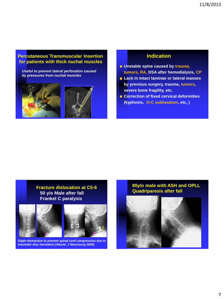
Percutaneous Transmuscular Insertion
for patients with thick nuchal muscles
Useful to prevent lateral perforation caused
by pressures from nuchal muscles
Indication
Unstable spine caused by trauma,
tumors, RA, DSA after hemodialysis, CP
Lack in intact laminae or lateral masses
by previous surgery, trauma, tumors,
severe bone fragility, etc.
Correction of fixed cervical deformities
(kyphosis, O-C subluxation, etc, )
Fracture dislocation at C5-6
50 y/o Male after fall
Frankel C paralysis
Slight distraction to prevent spinal cord compression due to
traumatic disc herniation (Abumi, J Neurosurg 2000)
80y/o male with ASH and OPLL
Quadriparesis after fall
11/8/2013
8
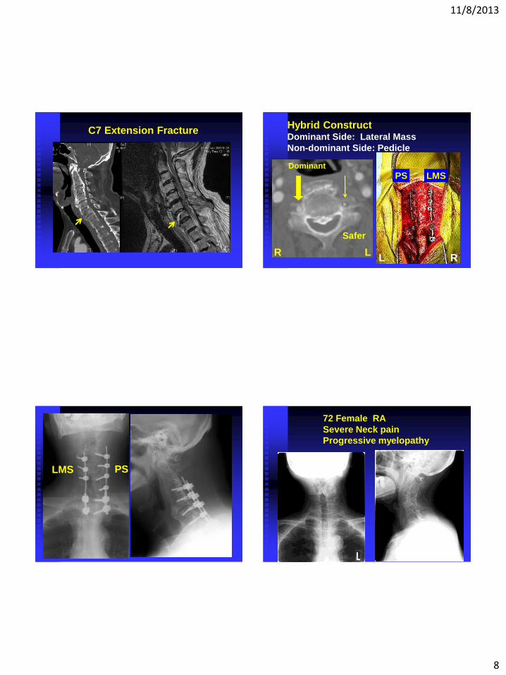
C7 Extension Fracture
Dominant
R L R L
Hybrid Construct Dominant Side: Lateral Mass
Non-dominant Side: Pedicle
Safer
PS LMS
PS LMS
72 Female RA
Severe Neck pain
Progressive myelopathy
11/8/2013
9
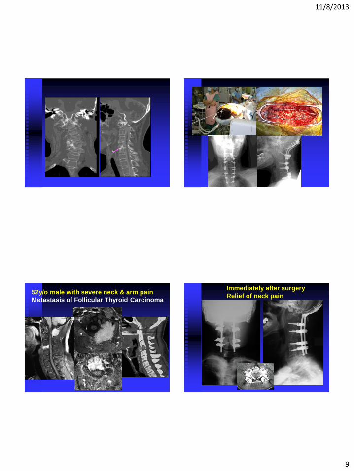
52y/o male with severe neck & arm pain
Metastasis of Follicular Thyroid Carcinoma
Immediately after surgery
Relief of neck pain
11/8/2013
10
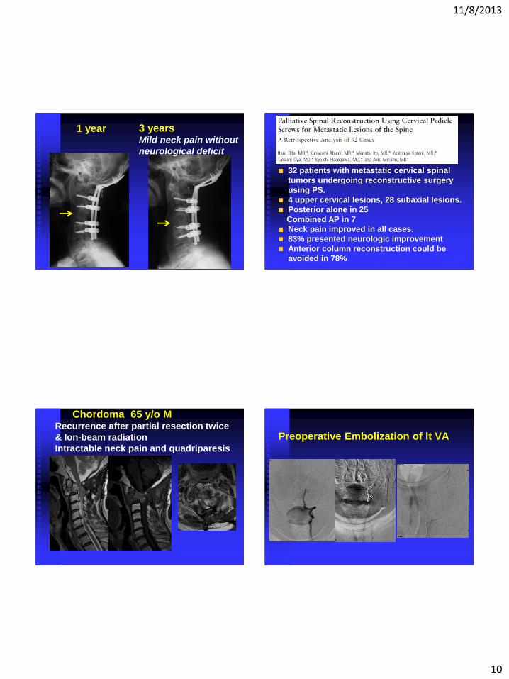
1 year 3 years
Mild neck pain without
neurological deficit
32 patients with metastatic cervical spinal
tumors undergoing reconstructive surgery
using PS.
4 upper cervical lesions, 28 subaxial lesions.
Posterior alone in 25
Combined AP in 7
Neck pain improved in all cases.
83% presented neurologic improvement
Anterior column reconstruction could be
avoided in 78%
Chordoma 65 y/o M Recurrence after partial resection twice
& Ion-beam radiation
Intractable neck pain and quadriparesis
Preoperative Embolization of lt VA
11/8/2013
11
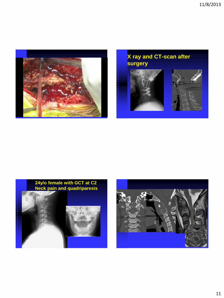
X ray and CT-scan after
surgery
24y/o female with GCT at C2
Neck pain and quadriparesis
11/8/2013
12
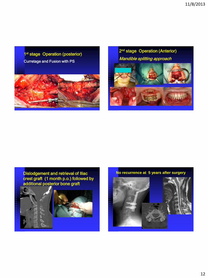
1st stage Operation (posterior)
Curretage and Fusion with PS
2nd stage Operation (Anterior)
Mandible splitting approach
Dislodgement and retrieval of Iliac
crest graft (1 month p.o.) followed by
additional posterior bone graft
No recurrence at 5 years after surgery
11/8/2013
13
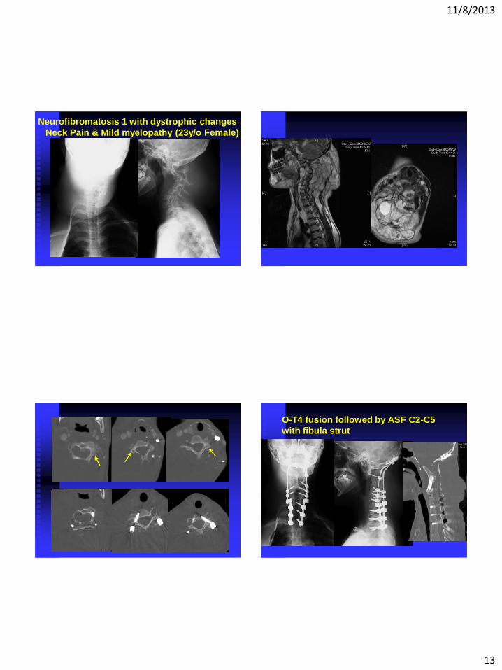
Neurofibromatosis 1 with dystrophic changes
Neck Pain & Mild myelopathy (23y/o Female)
O-T4 fusion followed by ASF C2-C5
with fibula strut
11/8/2013
14
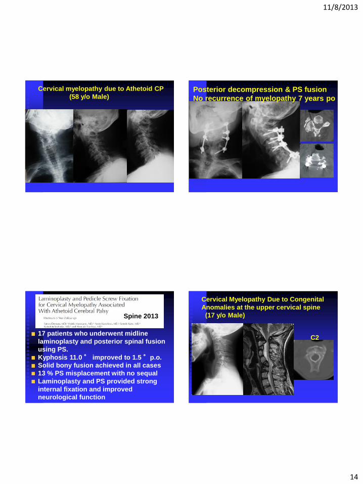
Cervical myelopathy due to Athetoid CP
(58 y/o Male) Posterior decompression & PS fusion
No recurrence of myelopathy 7 years po
17 patients who underwent midline
laminoplasty and posterior spinal fusion
using PS.
Kyphosis 11.0 ° improved to 1.5 °p.o.
Solid bony fusion achieved in all cases
13 % PS misplacement with no sequal
Laminoplasty and PS provided strong
internal fixation and improved
neurological function
Spine 2013
Cervical Myelopathy Due to Congenital
Anomalies at the upper cervical spine
(17 y/o Male)
C2
11/8/2013
15
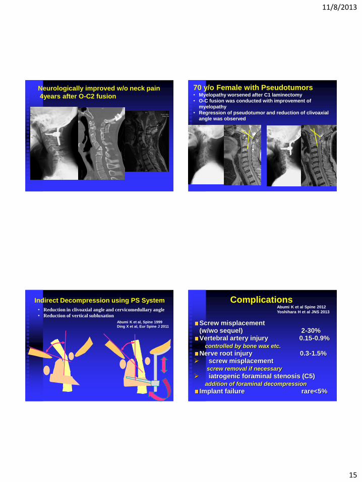
Neurologically improved w/o neck pain
4years after O-C2 fusion
70 y/o Female with Pseudotumors • Myelopathy worsened after C1 laminectomy
• O-C fusion was conducted with improvement of
myelopathy
• Regression of pseudotumor and reduction of clivoaxial
angle was observed
Abumi K et al, Spine 1999
Ding X et al, Eur Spine J 2011
Indirect Decompression using PS System
• Reduction in clivoaxial angle and cervicomedullary angle
• Reduction of vertical subluxation
Complications
Screw misplacement (w/wo sequel) 2-30%
Vertebral artery injury 0.15-0.9% controlled by bone wax etc.
Nerve root injury 0.3-1.5% screw misplacement screw removal if necessary
iatrogenic foraminal stenosis (C5) addition of foraminal decompression
Implant failure rare<5%
Abumi K et al Spine 2012
Yoshihara H et al JNS 2013
11/8/2013
16
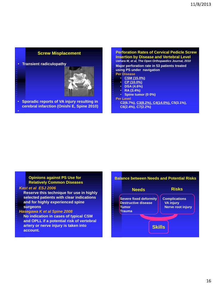
• Transient radiculopathy
Screw Misplacement
• Sporadic reports of VA injury resulting in
cerebral infarction (Onishi E, Spine 2010)
•
Major perforation rate in 53 patients treated
using PS under navigation
Per Disease
• CSM (15.0%)
• CP (10.0%)
• DSA (4.6%)
• RA (3.4%)
• Spine tumor (0 0%)
Per Level
C2(6.7%), C3(8.2%), C4(14.0%), C5(3.1%),
C6(2.4%), C7(2.2%)
Perforation Rates of Cervical Pedicle Screw
Insertion by Disease and Vertebral Level Uehara M, et al, The Open Orthopaedics Journal, 2010
Kast et al ESJ 2006
Reserve this technique for use in highly
selected patients with clear indications
and for highly experienced spine
surgeons
Hasegawa K et al Spine 2008
No indication in cases of typical CSM
and OPLL if a potential risk of vertebral
artery or nerve injury is taken into
account.
Opinions against PS Use for
Relatively Common Diseases Balance between Needs and Potential Risks
Severe fixed deformity
Destructive disease
Tumor
Trauma
Complications
VA injury
Nerve root injury
Needs Risks
Skills
11/8/2013
17
Summary
PS is useful for treatment of trauma,
severely destructive diseases, tumors,
and deformities.
Preoperative precise evaluation of
bony and neurovascular anatomy is
mandatory.
PS is associated with potentially
catastrophic complications
PS use for degenerative diseases is
debatable.
Keio University Hospital