disclosure common eye conditions every primary … family medicine board review chiu 1 common eye...
TRANSCRIPT

UCSF Family Medicine Board Review Chiu
1
Common Eye Conditions Every Primary Care
Clinician Should Know
Cynthia S. Chiu, MD, FACSAssociate Professor
Director, Comprehensive Ophthalmology & OptometryUCSF Department of Ophthalmology
UCSF Family Medicine Board ReviewMarch 18, 2015
http://www.timandjeni.com/images/cookiemonster.jpg
Disclosure
I have no financial interest in any of the products mentioned in this presentation
The Eye Exam
� Eye Vital Sign� Near Vision Card� Held at 14 inches� Glasses as needed
http://www.drbanker.com/images/Nearvision.gif
Pupils
� Look for afferent pupillary defect� Swinging flashlight test� +APD indicates optic nerve or large retinal lesion
http://img.tfd.com/ElMill/thumb/F0P-24-S2958.jpg

UCSF Family Medicine Board Review Chiu
2
Motility
� Six extraocularmuscles� Test cardinal fields of gaze
http://www.opsweb.org/OpPhoto/Extern/Motility/muscle.jpg
http://www.opsweb.org/OpPhoto/Extern/Motility/9gazedwg.jpg
Confrontational Visual Fields
� Cover eyes on same side� Hold fingers midway between� Normal per eye: 60-60-60-90
http://www.ncbi.nlm.nih.gov/bookshelf/br.fcgi?book=cm&part=A3460&rendertype=figure&id=A3464
Penlight Exam
http://cache.heraldinteractive.com/blogs/sports/rap_sheet/wp-content/uploads/2009/11/bionic-eye.jpg
LidsLashes
Conjunctiva/Sclera
CorneaIris/Pupil
Lens Puncta
Anterior Chamber Depth
� Deep chamber: illumination of nasal iris� Shallow chamber: shadow on nasal iris� Dilation: Phenylephrine 2.5%, Tropicamide 1%
http://www.ophthobook.com/wp-content/uploads/2007/12/video-glaucoma-shallowpenlight.jpg
http://iei.ico.edu/images/anatomy.jpg
UCSF Family Medicine Board Review Chiu
3
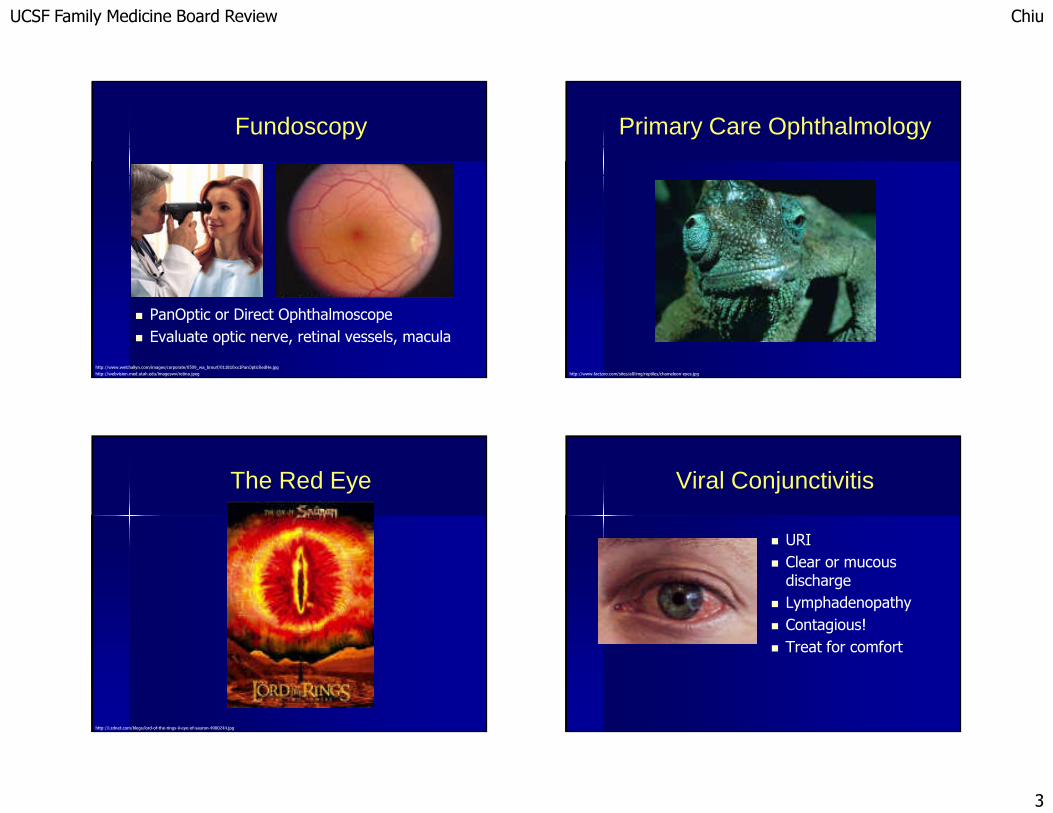
Fundoscopy
� PanOptic or Direct Ophthalmoscope� Evaluate optic nerve, retinal vessels, macula
http://www.welchallyn.com/images/corporate/0509_wa_bmurf/011810xx1PanOpticRedHe.jpghttp://webvision.med.utah.edu/imageswv/retina.jpeg
Primary Care Ophthalmology
http://www.factzoo.com/sites/all/img/reptiles/chameleon-eyes.jpg
The Red Eye
http://i.zdnet.com/blogs/lord-of-the-rings-ii-eye-of-sauron-4900244.jpg
Viral Conjunctivitis
� URI� Clear or mucous discharge� Lymphadenopathy� Contagious!� Treat for comfort

UCSF Family Medicine Board Review Chiu
4
Bacterial Conjunctivitis
� Purulent discharge� Culture� Staph, Strep, Hflu
– Polymixin/Trimethoprim– Fluoroquinolones– NOT Tobra or Gent
� GC, chlamydia– Systemic Rx
Allergic Conjunctivitis
� History of atopy� Conjunctival edema� Itchy!
� Topical antihistamines– Elestat– Zaditor– Patanol
� Visine tachyphylaxis
Blepharitis
� Redness, itching, “grit”, dry eyes� Rosacea, Staph, Demadex� Warm compresses, baby shampoo, artificial tears� Doxycycline, Azithromycin
http://www.stop-rosacea.com/
Herpes Simplex
� When to refer:V1, V2red eyeeye painchange in vision
� Acyclovir

UCSF Family Medicine Board Review Chiu
5
Herpes Zoster
� Hutchinson’s sign: nasociliary nerve� Treat Post-Herpetic Neuralgia: Lyrica, Neurontin, TCA’s
Uveitis
� Inflammation of vascular tissue� Auto-immune� Infectious� Toxic� Masquerade
� May require immunosuppression
Angle Closure Glaucoma
� Headache� Loss of vision� Firm eye
� IV Diamox, Mannitol� Glaucoma gtt’s� Surgical treatment
Subconjunctival Hemorrhage
� Valsalva, HTN, anticoagulants, eye rubbing, spontaneous� In the setting of trauma: refer

UCSF Family Medicine Board Review Chiu
6
Corneal Abrasion
� Pain!� Loss of epithelium� Not infected
� Erythromycin ung� Artificial Tears� Patching
Corneal Foreign Body
� Iron is toxic� Surgical treatment
Hyphema
� Severe eye trauma� Risk of rebleed� Risk of glaucoma
Ruptured Globe
� Peaked pupil� Brown tissue outside the eye� CT scan NOT MRI� Fox shield, NPO

UCSF Family Medicine Board Review Chiu
7
Common Diseases of Aging
http://www.jaredyellin.com/wp-content/uploads/2009/12/old-man-winking1.jpg
Cataract
� Painless progressive loss of vision with age� Also caused by DM, XRT, trauma, medications� Difficulty reading, driving, glare� Outpatient surgery
Glaucoma
� Chronic progressive optic neuropathy� Loss of visual field� Risk factors: age, tobacco, race, family hx� Medications may have systemic interactions
http://img.medscape.com/fullsize/migrated/569/545/569545.fig2.gifhttp://cdn.shopmedvet.com/images/uploads/2640_7780_thumb.jpg
Macular Degeneration
� Risk factors: age, UV, tobacco, Family Hx� Loss of central vision� Dry form: AREDS vitamins, stop smoking� Wet form: anti-VEGF injections

UCSF Family Medicine Board Review Chiu
8
Disorders of theEyelid and Orbit
http://greatpiercingshop.com/blog/wp-content/uploads/2009/12/eyelid-piercing-method-and-aftercare_49.jpg
Chalazion
� Blocked oil gland� Inflammation
� Warm compresses� Incision/curettage
Cellulitis
� Preseptal vs Orbital
� Preseptal: full EOM, no proptosis, quiet eye� Treat PO Abx
� Orbital: proptosis, strabismus, inflamed eye� Treat IV ABx
Thyroid Orbitopathy
� Proptosis� Strabismus/Diplopia� Corneal exposure� Optic nerve compression
� 131-I may aggravate� Surgical treatment
UCSF Family Medicine Board Review Chiu
9
Disorders of the Retina
http://webphysics.davidson.edu/faculty/dmb/EdibleOpticalMaterials/index_files/cyclops.jpg
Retinal Detachment
� Flashes/Floaters� Loss of vision/field� Sudden, painless
� Surgical treatment
http://www.rameshshahmd.com/pictures/retinal%20detach.%201.jpg?nxg_versionuid=published
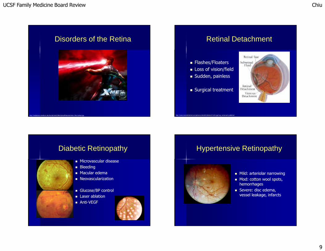
Diabetic Retinopathy
� Microvascular disease� Bleeding� Macular edema� Neovascularization
� Glucose/BP control� Laser ablation� Anti-VEGF
Hypertensive Retinopathy
� Mild: arteriolar narrowing� Mod: cotton wool spots, hemorrhages
� Severe: disc edema, vessel leakage, infarcts

UCSF Family Medicine Board Review Chiu
10
HIV/CMV Retinopathy
� Microvasculardisease� Annual exam if CD4>200� CMV retinitis
http://depts.washington.edu/hivaids/images/oit/oit_c7_d06.jpg
Retinal Artery Occlusion
� Embolic: cardiac echo, carotid doppler� Vasculitic (GCA)
Retinal Vein Occlusion
� Hypertension� Glaucoma� Young patients: hypercoagulable
Neuro-Ophthalmology
http://www.capitalgainsmedia.com/images/Development%20News%20Photos/Dev-Ino-Issues%2002/Dev-Ino%20Issue%200244/DSC01869.jpg

UCSF Family Medicine Board Review Chiu
11
Stroke
� Homonymous field defect� Location of lesion is contralateral+ upside-down
Pituitary Adenoma
� Compression of nasal fibers at optic chiasm� Bitemporal hemianopia
http://www.lfhk.cuni.cz/patfyz/intranet/Figures/58/18.13.jpg
Temporal Arteritis
� Acute vision loss
� Headache, jaw claudication, scalp tenderness, proximal myalgias, constitutional symptoms
� ESR and CRP� Prednisone 100mg QD� Temporal artery biopsy
Optic Neuritis
� Loss of vision� Pain with EOM� +/- disc edema
� Multiple Sclerosis� Steroids
http://www.djo.harvard.edu/files/2982_355.jpg

UCSF Family Medicine Board Review Chiu
12
Pseudotumor Cerebri
� Headache, tinnitus� Papilledema� Vision loss
� Female, overweight� Medication-induced
� LP: opening pressurehttp://www.caleyes.com/images/papilledema-image.jpg
Horner Syndrome
� Anisocoria in the dark� Mild ptosis� Acute and painful: R/O carotid dissection
http://www.mrcophth.com/oculoplasticgallery/traumatichorner/horner.jpg
Third Nerve Palsy
� Aniscoria in the light� Severe Ptosis� EOM paresis� Microvascular� PCA/PComaneurysm
http://www.nature.com/eye/journal/v18/n3/images/6700625f1.jpghttp://www.revophth.com/Images//2008/10/083_RPJ8_F6.gif
Thank You!
http://blog.omy.sg/dingan/files/2009/05/bob.jpg

UCSF Family Medicine Board Review Chiu
13
References
� http://www.drgreene.com/21_1254.html� http://www.bobzyeruncle.com/archives/images/conjunctivitis-GC.html� http://www.ueseyecare.com/images/photos/allergic_conjunctivitis_l.jpg� http://www.ueseyecare.com/images/photos/viral_conjunctivitis_l.jpg� http://www.vigamox.com/images/packaging.jpg� http://dro.hs.columbia.edu/ced3/conjhemb.jpg� http://www.aafp.org/afp/20030401/1481_f3.jpg� http://eyemicrobiology.upmc.com/Images/Sub/PhotoEyeDendrite.jpg� http://www.hkmj.org.hk/skin/images/108-1.jpg� http://www.medicalskincare.be/images/content/herpes03.jpg� http://www.tusalud.com.mx/images/herpes_zoster.jpg� http://eyelearn.med.utoronto.ca/Lectures04-05/RedEye/images/11Kera2.jpg� http://www.nova.edu/~albert/ScleritisWeb.jpg� http://www.mrcophth.com/externaleyediseases/sp.JPG� http://e-learning.studmed.unibe.ch/augenheilkunde/systematik/aderhaut/images/iritis2.jpg� http://www.emedicine.com/oph/images/456cmv7.jpg� http://www.revoptom.com/handbook/images/62a.jpg� http://images.google.com/imgres?imgurl=http://www.asoprs.org/Graphics/gravesct.jpg&imgrefurl=http://www.asoprs.org/Pages/thyroid.html&h=250&w=429&sz=9&tbnid=ul6zL-4JdFYJ:&tbnh=71&tbnw=122&start=1&prev=/images%3Fq%3Dthyroid%2Beye%2Bmuscles%26hl%3Den%26lr%3D� http://www.medscape.com/content/2002/00/44/63/446304/art-mop446304.coat.fig3.jpg� http://www.jeremydiamond.co.uk/graphics/cataract_eye.jpg� http://technology.kingston.ac.uk/dirc/medical/images/catara2.jpg� http://www.drloden.com/Pages/Pics/cataract.gif� http://www.eri.harvard.edu/faculty/peli/Opthalm_Mngt_0803_files/xNat.beauty.jpg� http://www.avclinic.com/images/tonometry.jpg� http://www.avclinic.com/Glaucoma.htm� http://www.lasikinstitute.org/Glaucoma_whatis.html� http://www.neec.com/images/photos/mitrev5.jpg� http://www.rvscny.com/images/Diabetic%20retinopathy.jpg� http://www.tedmontgomery.com/the_eye/eyephotos/pics/DiabeticRetinopathyProliferative.jpg� http://www.neec.com/images/photos/diabetic%20retinopathy%20figure%203.jpg
References
� http://www.gaileyeyeclinic.com/images/dr3.gif� http://www.foxeyecare.com/canon/large/diabetic_retinopathy.jpg� http://hubnet.buffalo.edu/ophthalmology/site/Home/Eye_Disorders/Hypertensive_retinopathy.jpg� http://www.mrcophth.com/retinacases/CMV.gif� http://www.opt.indiana.edu/ce/antseg/graphics/cwsaids.jpg� http://www.ohiovalleyeye.com/images/macdegen_K45_dry.jpg� http://www.opt.pacificu.edu/ce/catalog/10701-PS/4hemorr.jpg� http://www.ttuhsc.edu/eye/Faculty%20Presentations/Visual%20Field%20Testing%20Tech%20Seminar_files/slide0036_image002.jpg� http://webeye.ophth.uiowa.edu/dept/GCA/images/GCAfig1b_.jpg� http://www.ophthalmic.hyperguides.com/tutorials/oculoplastics/temporal_arteritis/slides/Slide1.jpg� http://www.mrcophth.com/pathology/gca2.jpg� http://content.lib.utah.edu/EHSL-WFH/image/icon124.jpg� http://www.atlasophthalmology.com/atlasimg/1396_6_low_thumb.jpg� http://www.tedmontgomery.com/the_eye/eyephotos/pics/CentralRetinalArteryOcclusion.jpg� http://www.omnieyespecialists.com/images/eye_brao.jpg� http://dro.hs.columbia.edu/vr3/crvob.jpg� http://www.scielo.br/img/revistas/abo/v66n6/18991f1.jpg� http://www.drugdigest.org/images/pillimages/A0315100.JPG� http://ec1.images-amazon.com/images/I/31ITYXvQYpL._AA200_.jpg� http://www.aafa.org/images/eye.jpg� http://www.aclens.com/accessoryphotos/visine-advanced-relief-eye-drops-15-6683-v3b.jpg� http://content.answers.com/main/content/wp/en-commons/thumb/6/69/420px-Gray784.png� http://eyelearn.med.utoronto.ca/Lectures02-03/RedEye/images/RedEye_033.jpg� http://www.varga.org/corneal_foreign_body_2.jpg� http://www.permanente.net/homepage/kaiser/pictures/7945.gif� http://www.medscape.com/pi/editorial/conferences/2001/747/art-aad-02.fig1.jpg� http://www.opt.pacificu.edu/ce/catalog/10310-SD/Trauma%20Pictures/Hyphema.jpg� http://www.revoptom.com/handbook/images/25aa.jpg� http://www.tedmontgomery.com/the_eye/eyephotos/pics/Chalazion.jpg� http://medicine.ucsd.edu/clinicalimg/eye-periorbital-cellulitis1.jpg� http://eyelearn.med.utoronto.ca/Lectures05-06/RedEye/images/RedEye_010.jpg