discolosures myeloproliferative neoplasms - ucsf … neoplasms ... – sustained thrombocytosis and...
TRANSCRIPT
1
Robert P Hasserjian, MDAssociate ProfessorMassachusetts General Hospital and Harvard Medical School
Advances in the Diagnosis of Myeloproliferative Neoplasms
Discolosures• Consulting income from Promedior, Inc.
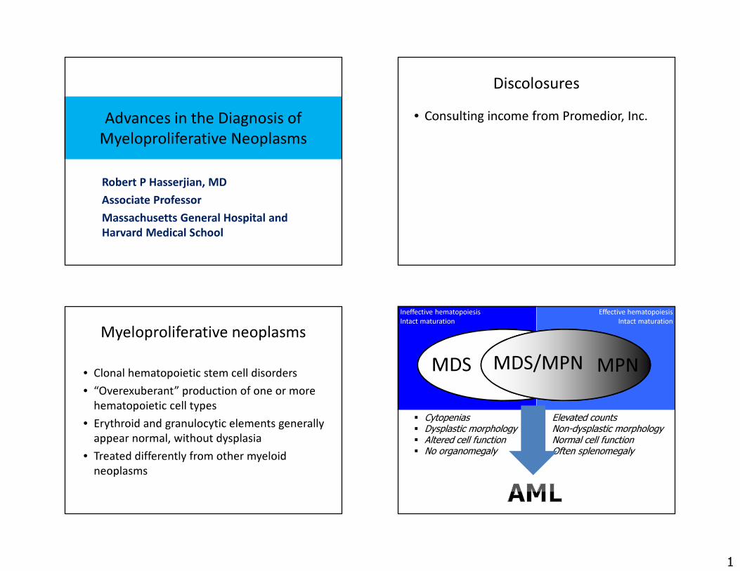
Myeloproliferative neoplasms
• Clonal hematopoietic stem cell disorders• “Overexuberant” production of one or more
hematopoietic cell types• Erythroid and granulocytic elements generally
appear normal, without dysplasia• Treated differently from other myeloid
neoplasms
Ineffective hematopoiesisIntact maturation
MDS
� Cytopenias� Dysplastic morphology� Altered cell function� No organomegaly
Effective hematopoiesisIntact maturation
MDS/MPN MPN
� Elevated counts� Non-dysplastic morphology� Normal cell function� Often splenomegaly
2
Chronic myeloproliferative neoplasms (WHO 2016)
• Chronic myeloid leukemia, Ph+• Polycythemia vera• Essential thrombocythemia• Primary myelofibrosis• Rare entities
– Chronic neutrophilic leukemia– Chronic eosinophilic leukemia/hypereosinophilic syndrome– Myeloproliferative neoplasm, unclassifiable
Genetically defined eosinophilic neoplasms
BCR-ABL
CSF3R
JAK2MPL
CALR
PDGFRAPDGFRBFGFR1
PCM1-JAK2
Diagnostic issues with MPN• Distinguishing MPN from reactive conditions
that can produce elevated counts• Separating MPN from other myeloid neoplasms
(MDS and MDS/MPN)• Providing a specific diagnosis
– Requires integration of clinical and molecular genetic data with morphology
– Important in predicting prognosis and dictating therapy
• Recognizing signs of progression
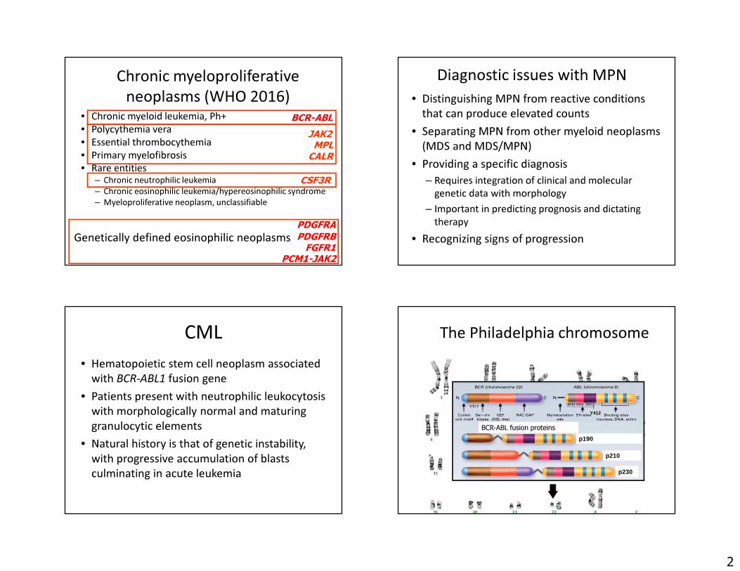
CML• Hematopoietic stem cell neoplasm associated
with BCR-ABL1 fusion gene• Patients present with neutrophilic leukocytosis
with morphologically normal and maturing granulocytic elements
• Natural history is that of genetic instability, with progressive accumulation of blasts culminating in acute leukemia
The Philadelphia chromosome
Y412
p230
p210
p190
BCR-ABL fusion proteins
3
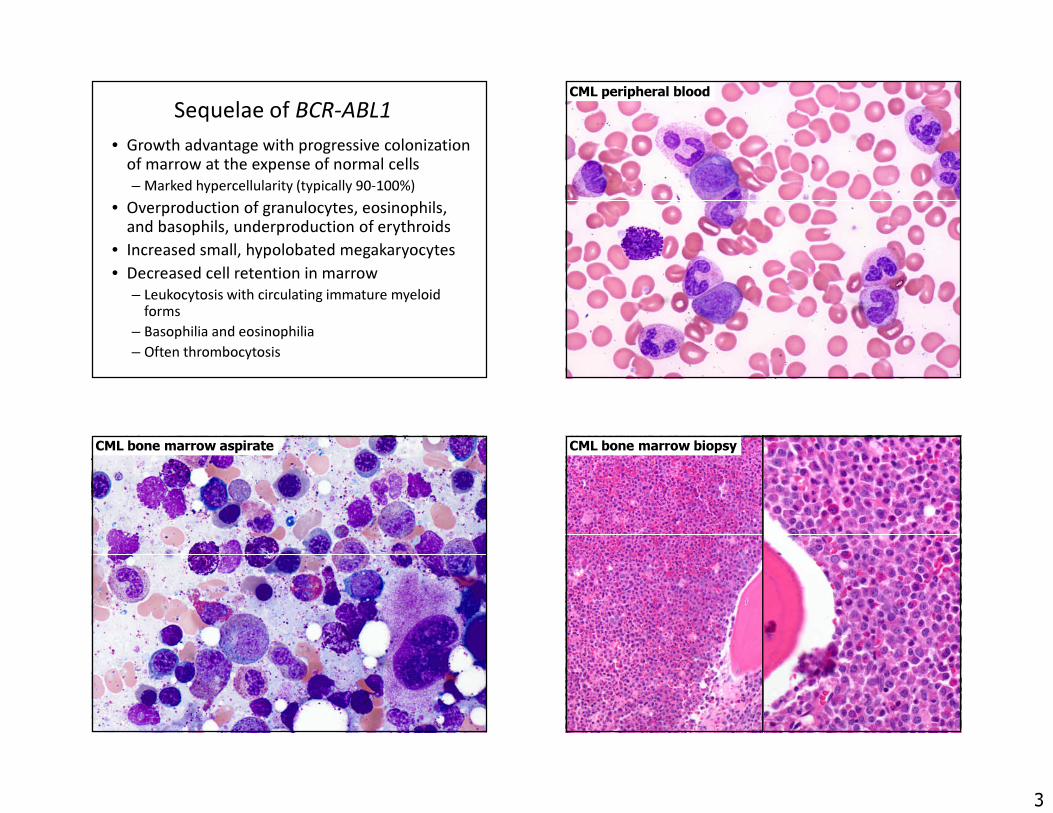
Sequelae of BCR-ABL1• Growth advantage with progressive colonization
of marrow at the expense of normal cells– Marked hypercellularity (typically 90-100%)
• Overproduction of granulocytes, eosinophils, and basophils, underproduction of erythroids
• Increased small, hypolobated megakaryocytes• Decreased cell retention in marrow
– Leukocytosis with circulating immature myeloid forms
– Basophilia and eosinophilia– Often thrombocytosis
CML peripheral blood
CML bone marrow aspirate CML bone marrow biopsy
4
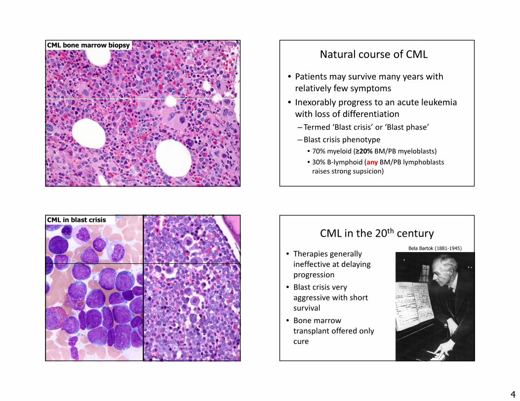
CML bone marrow biopsy Natural course of CML• Patients may survive many years with
relatively few symptoms • Inexorably progress to an acute leukemia
with loss of differentiation– Termed ‘Blast crisis’ or ‘Blast phase’– Blast crisis phenotype
• 70% myeloid (≥20% BM/PB myeloblasts)• 30% B-lymphoid (any BM/PB lymphoblasts
raises strong supsicion)
CML in blast crisisCML in the 20th century
• Therapies generally ineffective at delaying progression
• Blast crisis very aggressive with short survival
• Bone marrow transplant offered only cure
Bela Bartok (1881-1945)

5
Faderl S et al. N Engl J Med 1999;341:164-172, Goldman J and Melo J. N Engl J Med 2003;349:1451-1464
Tyrosine kinase inhibitors CML in the 21st century• Treated very effectively with tyrosine
kinase inhibitors (TKI)– Imatinib mesylate, nilotinib, dasatinib,
bosutinib, ponatinib• Disease progression no longer inevitable• Patterns of disease evolution closely
linked to responsiveness (versus resistance) to TKI therapy
Role of pathology in the current era of CML management
• At the time of initial diagnosis of CML– Get the diagnosis right!– Provide prognostic information
• At later timepoints, determine any progression and evaluate for other pathologic processes while on therapy
Criteria for accelerated phase
**Even small numbers of neoplastic B-lymphoblasts may indicate impending blast crisis

6
Required at diagnosis• Bone marrow biopsy and aspirate
– Reticulin stain to assess baseline fibrosis level
– Blast count (may be higher in marrow than blood)
• Full karyotype of bone marrow– Karyotype/FISH of blood may not pick up all
abnormalities• CBC and review of peripheral smear
– Blast and basophil count
Caveats with CML diagnosis• Relatively low M:E ratio in patients with
hemoglobinopathies• Prominent thrombocytosis mimicking ET• Minimal or no myeloid left-shift in blood• Monocytosis mimicking CMML• Blast crisis mimicking AML or ALL
– Splenomegaly, basophilia, cytogenetic clues help differentiate CML blast crisis from Ph+ AML or B-ALL• +8, double Ph, +19, i(17q)
– Ph+ AML will be a new genetically-defined AML subtype in the 2016 WHO update
Erythroid-rich CML 73 yo manWBC 13.3, HGB 14.0, PLT 1,48356% polys28% lymphs4% monos9% basos2% eos
7
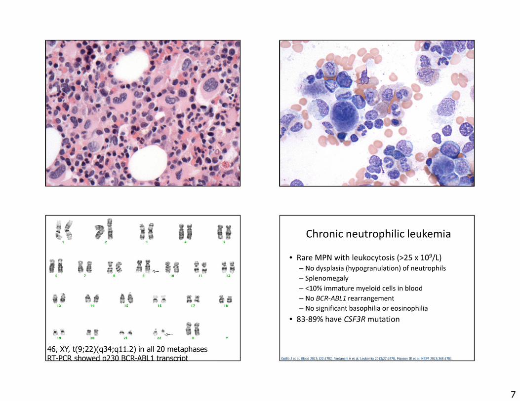
46, XY, t(9;22)(q34;q11.2) in all 20 metaphasesRT-PCR showed p230 BCR-ABL1 transcript
Chronic neutrophilic leukemia• Rare MPN with leukocytosis (>25 x 109/L)
– No dysplasia (hypogranulation) of neutrophils– Splenomegaly– <10% immature myeloid cells in blood– No BCR-ABL1 rearrangement– No significant basophilia or eosinophilia
• 83-89% have CSF3R mutation
Gotlib J et al. Blood 2013;122:1707, Pardanani A et al. Leukemia 2013;27:1870, Maxson JE et al. NEJM 2013;368:1781
8
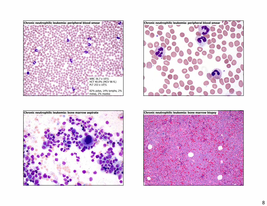
Chronic neutrophilic leukemia: peripheral blood smear
WBC 36.7 x 10⁹/LHCT 40.0% (MCV 98 fL)PLT 253 x 109/L82% polys, 14% lymphs, 2% metas, 2% myelos
Chronic neutrophilic leukemia: peripheral blood smear
Chronic neutrophilic leukemia: bone marrow aspirate Chronic neutrophilic leukemia: bone marrow biopsy
9
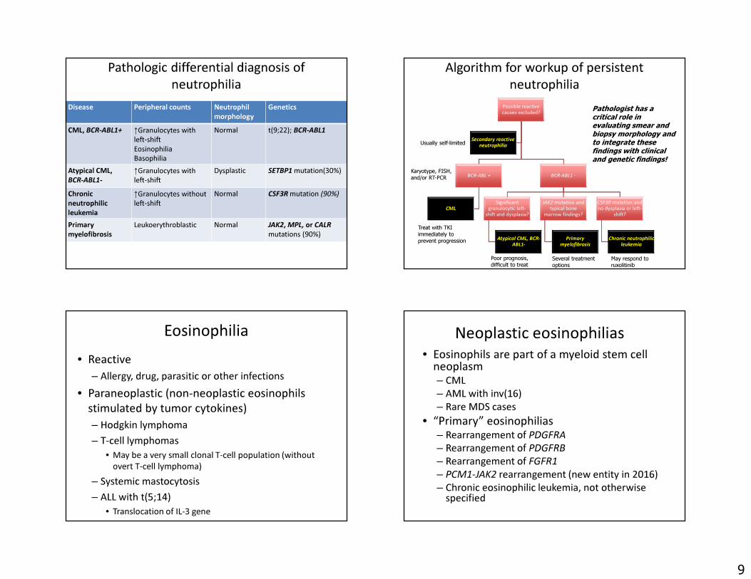
Pathologic differential diagnosis of neutrophilia
Disease Peripheral counts Neutrophilmorphology
Genetics
CML, BCR-ABL1+ ↑Granulocytes with left-shift EosinophiliaBasophilia
Normal t(9;22); BCR-ABL1
Atypical CML, BCR-ABL1-
↑Granulocytes with left-shift
Dysplastic SETBP1 mutation(30%)
Chronicneutrophilic leukemia
↑Granulocytes without left-shift
Normal CSF3R mutation (90%)
Primary myelofibrosis
Leukoerythroblastic Normal JAK2, MPL, or CALRmutations (90%)
Algorithm for workup of persistent neutrophilia
Possible reactive causes excluded?
BCR-ABL +
CML
BCR-ABL1 -
Significant granulocytic left-
shift and dysplasia?
Atypical CML, BCR-ABL1-
JAK2 mutation and typical bone
marrow findings?
Primary myelofibrosis
CSF3R mutation and no dysplasia or left-
shift?
Chronic neutrophilic leukemia
Secondary reactive neutrophiliaUsually self-limited
Treat with TKIimmediately to prevent progression
Poor prognosis, difficult to treat
Several treatment options
May respond to ruxolitinib
Pathologist has a critical role in evaluating smear and biopsy morphology and to integrate these findings with clinical and genetic findings!
Karyotype, FISH, and/or RT-PCR
Eosinophilia• Reactive
– Allergy, drug, parasitic or other infections• Paraneoplastic (non-neoplastic eosinophils
stimulated by tumor cytokines)– Hodgkin lymphoma – T-cell lymphomas
• May be a very small clonal T-cell population (without overt T-cell lymphoma)
– Systemic mastocytosis– ALL with t(5;14)
• Translocation of IL-3 gene
Neoplastic eosinophilias• Eosinophils are part of a myeloid stem cell
neoplasm– CML – AML with inv(16)– Rare MDS cases
• “Primary” eosinophilias – Rearrangement of PDGFRA– Rearrangement of PDGFRB– Rearrangement of FGFR1– PCM1-JAK2 rearrangement (new entity in 2016)– Chronic eosinophilic leukemia, not otherwise
specified
10
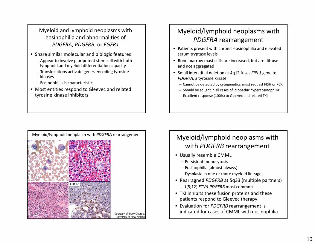
Myeloid and lymphoid neoplasms with eosinophilia and abnormalities of
PDGFRA, PDGFRB, or FGFR1• Share similar molecular and biologic features
– Appear to involve pluripotent stem cell with both lymphoid and myeloid differentiation capacity
– Translocations activate genes encoding tyrosine kinases
– Eosinophilia is characteristic• Most entities respond to Gleevec and related
tyrosine kinase inhibitors
Myeloid/lymphoid neoplasms with PDGFRA rearrangement
• Patients present with chronic eosinophilia and elevated serum tryptase levels
• Bone marrow mast cells are increased, but are diffuse and not aggregated
• Small interstitial deletion at 4q12 fuses FIPL1 gene to PDGRFA, a tyrosine kinase– Cannot be detected by cytogenetics, must request FISH or PCR– Should be sought in all cases of idiopathic hypereosinophilia– Excellent response (100%) to Gleevec and related TKI
39
Myeloid/lymphoid neoplasm with PDGFRA rearrangement
Courtesy of Tracy George, University of New Mexico
CD117
Myeloid/lymphoid neoplasms with with PDGFRB rearrangement
• Usually resemble CMML– Persistent monocytosis– Eosinophilia (almost always)– Dysplasia in one or more myeloid lineages
• Rearragned PDGFRB at 5q33 (multiple partners)– t(5;12) ETV6-PDGFRB most common
• TKI inhibits these fusion proteins and these patients respond to Gleevec therapy
• Evaluation for PDGFRB rearrangement is indicated for cases of CMML with eosinophilia
11
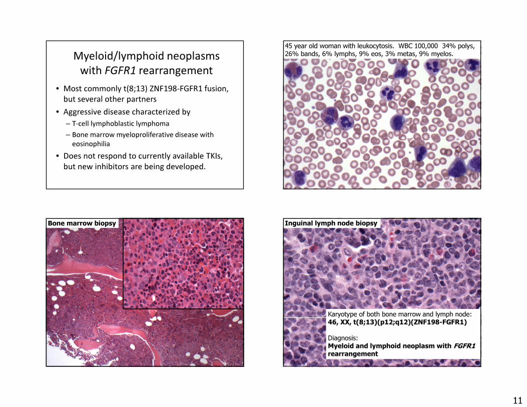
Myeloid/lymphoid neoplasms with FGFR1 rearrangement
• Most commonly t(8;13) ZNF198-FGFR1 fusion, but several other partners
• Aggressive disease characterized by– T-cell lymphoblastic lymphoma– Bone marrow myeloproliferative disease with
eosinophilia• Does not respond to currently available TKIs,
but new inhibitors are being developed.
45 year old woman with leukocytosis. WBC 100,000 34% polys, 26% bands, 6% lymphs, 9% eos, 3% metas, 9% myelos.
Bone marrow biopsy Inguinal lymph node biopsy
Karyotype of both bone marrow and lymph node:46, XX, t(8;13)(p12;q12)(ZNF198-FGFR1)Diagnosis: Myeloid and lymphoid neoplasm with FGFR1rearrangement
12
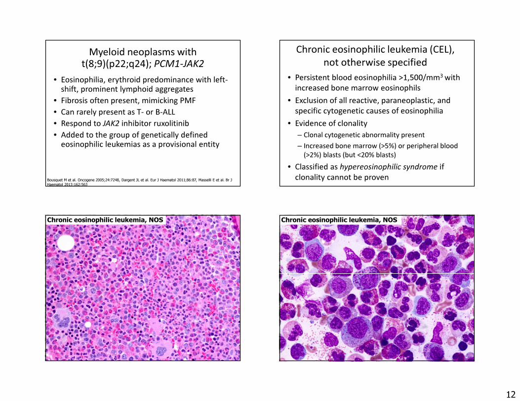
Myeloid neoplasms with t(8;9)(p22;q24); PCM1-JAK2
• Eosinophilia, erythroid predominance with left-shift, prominent lymphoid aggregates
• Fibrosis often present, mimicking PMF• Can rarely present as T- or B-ALL• Respond to JAK2 inhibitor ruxolitinib• Added to the group of genetically defined
eosinophilic leukemias as a provisional entity
Bousquet M et al. Oncogene 2005;24:7248, Dargent JL et al. Eur J Haematol 2011;86:87, Masselli E et al. Br J Haematol 2013;162:563
Chronic eosinophilic leukemia (CEL), not otherwise specified
• Persistent blood eosinophilia >1,500/mm3 with increased bone marrow eosinophils
• Exclusion of all reactive, paraneoplastic, and specific cytogenetic causes of eosinophilia
• Evidence of clonality– Clonal cytogenetic abnormality present – Increased bone marrow (>5%) or peripheral blood
(>2%) blasts (but <20% blasts)• Classified as hypereosinophilic syndrome if
clonality cannot be proven
Chronic eosinophilic leukemia, NOS Chronic eosinophilic leukemia, NOS
13
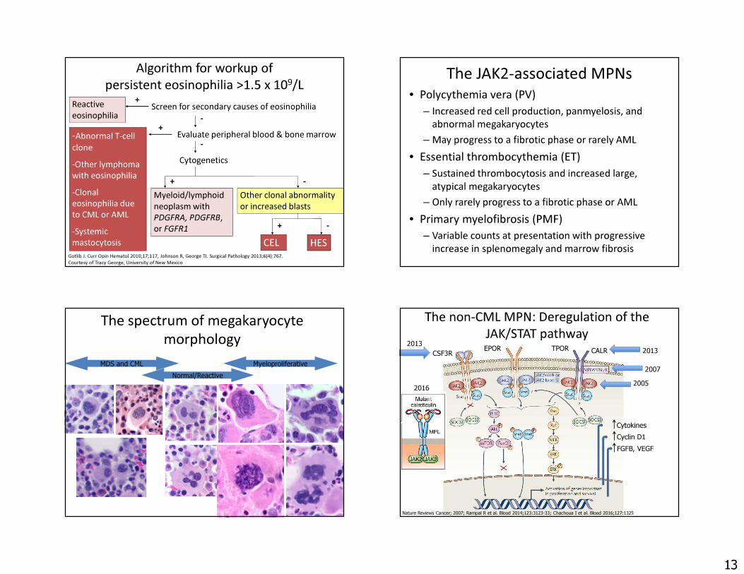
Algorithm for workup of persistent eosinophilia >1.5 x 10⁹/L
Reactive eosinophilia-Abnormal T-cell clone-Other lymphoma with eosinophilia-Clonal eosinophilia due to CML or AML-Systemic mastocytosis
Gotlib J. Curr Opin Hematol 2010;17;117, Johnson R, George TI. Surgical Pathology 2013;6(4):767.Courtesy of Tracy George, University of New Mexico
Screen for secondary causes of eosinophilia+
Evaluate peripheral blood & bone marrow
Myeloid/lymphoid neoplasm with PDGFRA, PDGFRB, or FGFR1
Other clonal abnormality or increased blasts
CEL HES
-
-Cytogenetics
+ -
+ -
+
The JAK2-associated MPNs• Polycythemia vera (PV)
– Increased red cell production, panmyelosis, and abnormal megakaryocytes
– May progress to a fibrotic phase or rarely AML• Essential thrombocythemia (ET)
– Sustained thrombocytosis and increased large, atypical megakaryocytes
– Only rarely progress to a fibrotic phase or AML• Primary myelofibrosis (PMF)
– Variable counts at presentation with progressive increase in splenomegaly and marrow fibrosis
The spectrum of megakaryocyte morphology
Normal/ReactiveMyeloproliferativeMDS and CML
The non-CML MPN: Deregulation of the JAK/STAT pathway
Nature Reviews Cancer; 2007; Rampal R et al. Blood 2014;123:3123-33; Chachoua I et al. Blood 2016;127:1325
CytokinesCyclin D1FGFB, VEGF
20052007
2016
CSF3R CALR 20132013 EPOR TPOR
14
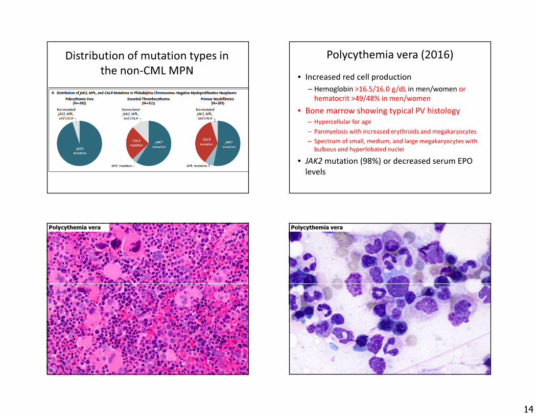
Distribution of mutation types in the non-CML MPN
Klampfl T et al. NEJM 2013;369:2379
Polycythemia vera (2016)• Increased red cell production
– Hemoglobin >16.5/16.0 g/dL in men/women or hematocrit >49/48% in men/women
• Bone marrow showing typical PV histology– Hypercellular for age – Panmyelosis with increased erythroids and megakaryocytes– Spectrum of small, medium, and large megakaryocytes with
bulbous and hyperlobated nuclei• JAK2 mutation (98%) or decreased serum EPO
levels
Polycythemia vera Polycythemia vera

15
“Masked” polycythemia vera• Some patients have bone marrow findings typical
of PV, but do not meet 2008 WHO hemoglobin levels– Male ≥18.5 g/dL, female ≥16.5 g/dL
• These patients appear to behave clinically like typical PV and have PV-like morphology– Often present with thrombocytosis mimicking ET
• Required hemoglobin/hematocrit levels have been reduced in 2016 update to correctly diagnose these patients
Barbui T et al. Am J Hematology 2013;89:52, Gianelli U et al. Am J Clin Pathol 2008;130:336, Thiele J et al. Acta Hematol2005;113:213
Masked polycythemia vera
65 year-old womanWBC 14.2 x 10⁹/LHGB 16 g/dLPLT 744 x 109/L
WHO Essential thrombocythemia criteria (2016)
1. Platelet count ≥450 x 109/uL2. Bone marrow biopsy showing typical morphology of ET and no
or (rarely) minor increase in reticulin fibers.3. Not meeting WHO criteria for CML, PV, PMF, MDS, or other
myeloid neoplasms
AND
Presence of JAK2, CALR or MPL mutationorPresence of another clonal marker orAbsence of evidence for reactive thrombocytosis
Causes of reactive thrombocytosis• Non-neoplastic
hematologic conditions:– Acute blood loss– Acute hemolytic anemia– Iron-deficiency anemia– Treatment of B12
deficiency– Rebound effect after
treatment for ITP or ethanol-induced thrombocytopenia
• Inflammatory conditions:– Rheumatoid disorders– Vasculitides– IBD– Celiac disease– POEMS syndrome
• Tissue damage– Trauma, MI, thermal burns
• Infections• Exercise• Allergic/medication
reactions• Asplenia
16
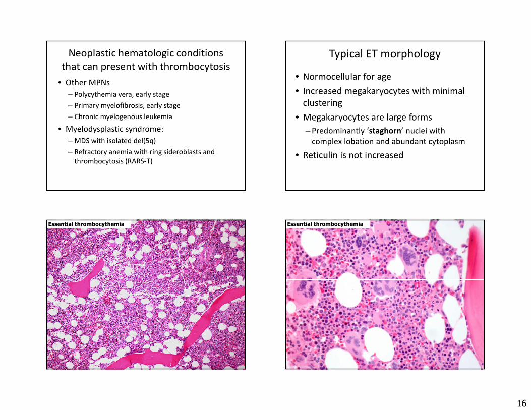
Neoplastic hematologic conditions that can present with thrombocytosis
• Other MPNs– Polycythemia vera, early stage– Primary myelofibrosis, early stage– Chronic myelogenous leukemia
• Myelodysplastic syndrome:– MDS with isolated del(5q)– Refractory anemia with ring sideroblasts and
thrombocytosis (RARS-T)
Typical ET morphology• Normocellular for age• Increased megakaryocytes with minimal
clustering• Megakaryocytes are large forms
– Predominantly ‘staghorn’ nuclei with complex lobation and abundant cytoplasm
• Reticulin is not increased
Essential thrombocythemia Essential thrombocythemia
17
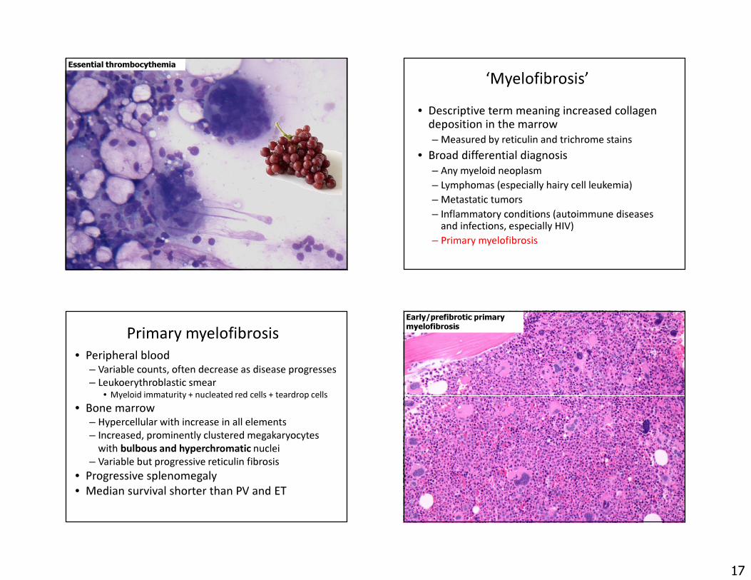
Essential thrombocythemia‘Myelofibrosis’
• Descriptive term meaning increased collagen deposition in the marrow – Measured by reticulin and trichrome stains
• Broad differential diagnosis– Any myeloid neoplasm– Lymphomas (especially hairy cell leukemia)– Metastatic tumors– Inflammatory conditions (autoimmune diseases
and infections, especially HIV)– Primary myelofibrosis
Primary myelofibrosis• Peripheral blood
– Variable counts, often decrease as disease progresses– Leukoerythroblastic smear
• Myeloid immaturity + nucleated red cells + teardrop cells• Bone marrow
– Hypercellular with increase in all elements– Increased, prominently clustered megakaryocytes
with bulbous and hyperchromatic nuclei– Variable but progressive reticulin fibrosis
• Progressive splenomegaly • Median survival shorter than PV and ET
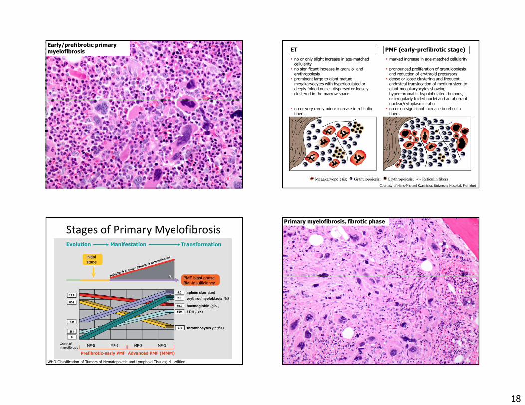
Early/prefibrotic primary myelofibrosis
18
Early/prefibrotic primary myelofibrosis ET PMF (early-prefibrotic stage)
� no or only slight increase in age-matched cellularity
� marked increase in age-matched cellularity� no significant increase in granulo- and
erythropoiesis� pronounced proliferation of granulopoiesis
and reduction of erythroid precursors� prominent large to giant mature
megakaryocytes with hyperlobulated or deeply folded nuclei, dispersed or loosely clustered in the marrow space
� dense or loose clustering and frequent endosteal translocation of medium sized to giant megakaryocytes showing hyperchromatic, hypolobulated, bulbous, or irregularly folded nuclei and an aberrant nuclear/cytoplasmic ratio
� no or very rarely minor increase in reticulinfibers
� no or no significant increase in reticulinfibers
Courtesy of Hans-Michael Kvasnicka, University Hospital, Frankfurt
Stages of Primary Myelofibrosis
WHO Classification of Tumors of Hematopoietic and Lymphoid Tissues; 4th edition
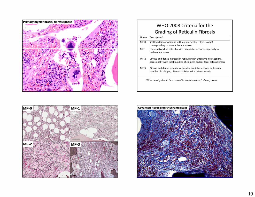
Primary myelofibrosis, fibrotic phase
19
Primary myelofibrosis, fibrotic phase WHO 2008 Criteria for the Grading of Reticulin Fibrosis
Grade Descriptiona
MF-0 Scattered linear reticulin with no intersections (crossovers) corresponding to normal bone marrow
MF-1 Loose network of reticulin with many intersections, especially in perivascular areas
MF-2 Diffuse and dense increase in reticulin with extensive intersections, occasionally with focal bundles of collagen and/or focal osteosclerosis
MF-3 Diffuse and dense reticulin with extensive intersections and coarse bundles of collagen, often associated with osteosclerosis
aFiber density should be assessed in hematopoietic (cellular) areas.
MF-0 MF-1
MF-2 MF-3
Advanced fibrosis on trichrome stain

20
Advanced osteosclerosis in PMF Importance of accurate diagnosis of MPN to inform prognosis and guide
therapy PV ET PMF
Leukemic transformation
3% at 10 years 1% at 10 years 12-30% at 10 years
Fibrosis progression 15-25% Rare 100%Thrombosis,per 100 patients/year
5.5 1-3 2
Initial treatment Phlebotomy +/- HU None , aspirin +/-HU
Allo-SCT, JAKinhibitors,
chemotherapy
Courtesy of Olga Pozdnyakova, BWH
Survival in the non-CML MPN (n=826)ET (n= 292) vs PV (n=267) vs PMF (n=267)
Tefferi et al. Blood 2014;124:2507-2513
ETPV
PMF
Courtesy of Hans-Michael Kvasnicka, University Hospital, Frankfurt
Summary• Myeloproliferative neoplasms have distinctive
morphologies and distinctive genetic aberrations– Important to correctly diagnose the various MPN diseases,
which have different patterns of progression and are treated differently
• The presence of dysplastic features in a patient with cytosis should suggest the possibility of an MDS/MPNoverlap disease– Generally poorer prognosis than MPN diseases
• Eosinophilic myeloid disorders are characterized by recurrent genetic abnormalities and some are amenable to targeted therapies with TKIs