discovery and characterization of thermoproteus spherical
TRANSCRIPT

Discovery and Characterization of Thermoproteus SphericalPiliferous Virus 1: a Spherical Archaeal Virus Decorated withUnusual Filaments
Ross Hartman,a Lieuwe Biewenga,b Jacob Munson-McGee,b Mohammed Refai,a,c Eric S. Boyd,c Brian Bothner,a
C. Martin Lawrence,a Mark Youngc
aDepartment of Chemistry and Biochemistry, Montana State University, Bozeman, Montana, USAbDepartment of Microbiology and Immunology, Montana State University, Bozeman, Montana, USAcDepartment of Plant Sciences and Plant Pathology, Montana State University, Bozeman, Montana, USA
ABSTRACT We describe the discovery of an archaeal virus, one that infects archaea,tentatively named Thermoproteus spherical piliferous virus 1 (TSPV1), which was pu-rified from a Thermoproteales host isolated from a hot spring in Yellowstone Na-tional Park (USA). TSPV1 packages an 18.65-kb linear double-stranded DNA (dsDNA)genome with 31 open reading frames (ORFs), whose predicted gene products showlittle homology to proteins with known functions. A comparison of virus particlemorphologies and gene content demonstrates that TSPV1 is a new member of theGlobuloviridae family of archaeal viruses. However, unlike other Globuloviridae mem-bers, TSPV1 has numerous highly unusual filaments decorating its surface, which canextend hundreds of micrometers from the virion. To our knowledge, similar fila-ments have not been observed in any other archaeal virus. The filaments are re-markably stable, remaining intact across a broad range of temperature and pH val-ues, and they are resistant to chemical denaturation and proteolysis. A majorcomponent of the filaments is a glycosylated 35-kDa TSPV1 protein (TSPV1 GP24).The filament protein lacks detectable homology to structurally or functionally char-acterized proteins. We propose, given the low host cell densities of hot spring envi-ronments, that the TSPV1 filaments serve to increase the probability of virus attach-ment and entry into host cells.
IMPORTANCE High-temperature environments have proven to be an important sourcefor the discovery of new archaeal viruses with unusual particle morphologies and genecontent. Our isolation of Thermoproteus spherical piliferous virus 1 (TSPV1), with numer-ous filaments extending from the virion surface, expands our understanding of viral di-versity and provides new insight into viral replication in high-temperature environments.
KEYWORDS crenarchaea, Globuloviridae, archaeal virus
High-temperature acidic environments, such as those found in the hot springs ofYellowstone National Park (YNP), USA, provide a rich source of archaeal viruses (1).
A range of virion morphologies is observed in archaeal viruses from these environ-ments, including rod-shaped helical viruses, spherical icosahedral viruses, and unusualspindle- and bottle-shaped virions (2–6). Archaeal virus diversity is also reflected at thegenome level, where most genes lack detectable homology to other known genes withknown function (7, 8). The factors driving archaeal virus diversity in acidic hot springsare unknown but likely reflect adaptations to the physical and geochemical nature ofthese environments and the cell biology and biochemistry of their archaeal hosts.
Of the 62 known thermophilic archaeal viruses, 47 viruses infect hosts from theorder Sulfolobales (6, 8–13). The remaining 15 viruses have been isolated from members
Citation Hartman R, Biewenga L, Munson-McGee J, Refai M, Boyd ES, Bothner B, LawrenceCM, YoungM. 2020. Discovery andcharacterization of Thermoproteus sphericalpiliferous virus 1: a spherical archaeal virusdecoratedwith unusual filaments. J Virol 94:e00036-20. https://doi.org/10.1128/JVI.00036-20.
Editor Rebecca Ellis Dutch, University ofKentucky College of Medicine
Copyright © 2020 American Society forMicrobiology. All Rights Reserved.
Address correspondence to Mark Young,[email protected].
Received 7 January 2020Accepted 8 March 2020
Accepted manuscript posted online 25March 2020Published
GENETIC DIVERSITY AND EVOLUTION
crossm
June 2020 Volume 94 Issue 11 e00036-20 jvi.asm.org 1Journal of Virology
18 May 2020
on May 20, 2020 at M
ON
TANA STATE U
NIV AT BO
ZEMAN
http://jvi.asm.org/
Dow
nloaded from

of the Thermoproteales, Desulfurococcales, Methanobacteriales, and Thermococcales (2,14–18). This bias likely reflects the ease with which many Sulfolobales are isolated andcultured under predominantly aerobic, heterotrophic conditions. It is therefore reason-able to suspect that a substantial level of archaeal virus diversity remains unexplored.
In order to expand the diversity of cultured archaeal viruses, we sought to isolatenew viruses infecting the order Thermoproteales. A common genus of this order,Thermoproteus, is found in sulfur sediments of YNP hot springs (19, 20). These anaerobicsulfur-reducing organisms have a global distribution in acidic and near-neutral thermalfeatures (12, 13, 19, 21, 22). They are capable of growing chemolithotrophically usingH2 and CO2 or organoheterotrophically using a wide variety of sugars, organic acids,and alcohols (23, 24).
To date, only 6 viruses have been described from Thermoproteales hosts. Despite thislimited description, their structural and morphological diversity help define 3 newarchaeal virus families (12, 13, 18, 25). Thermoproteus tenax virus 1 (TTV1), Thermopro-teus tenax virus 2 (TTV2), and Thermoproteus tenax virus 3 (TTV3) are members of theLipothrixviridae, a family of flexible helical viruses, whereas Thermoproteus tenax virus 4(TTV4) is a member of the Rudiviridae, a related family of stiff rod-like viruses (18). Theremaining 2 viruses are Pyrobaculum spherical virus (PSV) and Thermoproteus tenaxspherical virus 1 (TTSV1), the only members of the Globuloviridae archaeal virus family(12, 13). We describe here a seventh Thermoproteales virus, Thermoproteus sphericalpiliferous virus 1 (TSPV1), with unusual hair-like filaments extending from its virionsurface. We propose that these filaments facilitate viral attachment to host cells.
RESULTS AND DISCUSSION
Pure cultures of Thermoproteus sp. strain CP80, originally isolated from Cinder Pool(NHSP103) in YNP, were found to be chronically infected with a 75- to 85-nm sphericalvirus (Fig. 1A). There was no evidence of host cell lysis. Cultures remained viable aftermultiple rounds of passaging over a 2-year time period, with no significant change ineither virus production or host cell viability.
Cesium chloride density gradient centrifugation was utilized to purify viral capsidsthat appeared by transmission electron microscopy (TEM) visualization to be intact.Virus particles purified at a density of 1.29 g/cm3, which is typical for enveloped virions.Using both negatively stained and frozen hydrated samples, electron micrographs andtomograms of the virus were acquired. These images revealed a spherical capsidmorphology with an average diameter of 83 nm (Fig. 1C to F; see also Movie S1 in thesupplemental material). Ten- to 20-nm protrusions were often observed extendingfrom the surface of the virion (arrows, Fig. 1C). An external lipid envelope also appearsto be present in the electron micrographs (arrows, Fig. 1F). A suspected 3.4-nm-thick(�0.3 nm) lipid envelope and an envelope surface protein layer extending out to7.4 nm (�0.4 nm) are observed (Fig. 1F inset). The addition of 20% diethyl ether to asample of TSPV1 resulted in the disruption of approximately 70% of the virions, withmost of the remaining virus displaying obvious damage to their capsids, as determinedby TEM analysis. In addition to this, TSPV1 has a buoyant density in CsCl densitygradients that is typical for spherical enveloped viruses. Based on the apparent nonlyticlifestyle of TSPV1 and the likely presence of a lipid envelope, we hypothesize that TSPV1buds from the host cell in a manner similar to that reported for the archaeal Sulfolobusspindle-shaped virus 1 (SSV1) (26).
The most striking and morphologically unique feature of the virion is numerous3-nm-diameter filaments of various lengths that extend from the virion surface (arrows,Fig. 1C to F). Each virion contains an average of 7 filaments, with the typical range beingfrom 0 to 20 filaments per virion. Some of these filaments extend up to 500 nm fromthe capsid surface and are highly flexible. In some micrographs, it appears as if thefilaments pass through the surface protein layer and attach to the underlying envelope.However, higher-resolution microscopy is necessary to confirm this speculation. At thispoint, the nature of the association of the filaments with its virion is unknown.
The morphology of TSPV1 resembles those of PSV and TTSV, both members of the
Hartman et al. Journal of Virology
June 2020 Volume 94 Issue 11 e00036-20 jvi.asm.org 2
on May 20, 2020 at M
ON
TANA STATE U
NIV AT BO
ZEMAN
http://jvi.asm.org/
Dow
nloaded from

Globuloviridae (12, 13). The resemblance includes the presence of protrusions from thecapsid surface, the similar diameters of the virions, and the appearance of a lipid layerin micrographs. However, the filaments present on TSPV1 were not observed on PSV orTTSV1. Subjecting TSPV1 to increased pH from pH 4.0 to 8.0 ruptured the particles,releasing a nucleoprotein material (Fig. 1B) whose diameter matches that of thenucleoprotein observed in ruptured PSV particles (13).
Sequencing and assembly of the TSPV1 genome with 1,200� average coverageyielded a 18,655-bp linear double-stranded DNA (dsDNA) genome with a G�C contentof 56%, which is similar to that of its host (Fig. 2). The TSPV1 genome codes for 31 gene
FIG 1 (A to F) TSPV1 visualized by negative staining (A to D) and cryo TEM (E and F). (A) TSPV1 particlesin association with its Thermoproteus sp. CP80 host. (B) A ruptured TSPV1 particle showing an exposed6-nm-diameter nucleoprotein complex (white arrow) and virion filaments (black arrow). (C) Purifiedparticles showing virion protrusions (white arrows) and virion filaments (black arrows). (D) Enlargednegative stain of TSPV1 with protrusion (white arrow) and filaments (black arrow). (E) Cryo-TEMmicrographs of TSPV1 with filaments (black arrow) associated with a Thermoproteus sp. CP80 cell. (F)Tomographic slice of TSPV1 (2.5-nm diameter) showing a virion filament (black arrow). An enlargedsection showing the suspected 3.4-nm-diameter lipid envelope (LL; white arrow) and a 7.4-nm surfaceenvelope protein layer (PL) are shown (white arrow).
TSPV1, a Spherical Archaeal Virus with Filaments Journal of Virology
June 2020 Volume 94 Issue 11 e00036-20 jvi.asm.org 3
on May 20, 2020 at M
ON
TANA STATE U
NIV AT BO
ZEMAN
http://jvi.asm.org/
Dow
nloaded from

products (GPs), with 85% coding density (Table 1). Most GPs occur on a single strandof the genome, with only GP2 and GP6 found on the complementary strand. Thepredicted masses of the GPs range from 4.7 to 59.2 kDa, with 19 GPs having a mass of�20 kDa. Some of TSPV1 predicted genes lack obvious promoter sequences and somay produce transcripts that are translated as operons. This includes GP3, GP4, GP8,GP9, GP14-GP17, GP19-GP22, and GP28-GP30. We could not detect a distinct origin ofreplication; however, the genome was found to have inverted terminal repeats (ITRs) of102 bp in length which likely play a role in genome replication.
FIG 2 The genome map of the 18,655-bp dsDNA TSPV1 genome and its 31 predicted gene products (GPs). TSPV1 has 102-bp inverted terminal repeats (ITR).GPs conserved across the Globuloviridae are colored.
TABLE 1 Predicted TSPV1 proteins, characteristics, and homologues
TSPV1gene
TTSV1homologue
PSVhomologue
Mol wt(kDa)
Predictedisoelectricpoint
No. of N-linkedglycosylationsites predicted
Predicted signalpeptide(s) Structure/function annotations
GP1 GP1 GP2 59.2 8.80 1 AAA� helicaseGP2 GP4 GP3 33.4 4.70 Na�/Ca2� exchangerGP3 GP5 GP5 16.0 8.63 1 Thioredoxin foldGP4 GP6 GP8 10.2 6.35GP5 GP8 GP15 26.2 7.90GP6 GP27 GP16 26.5 8.98 Yes (FlaFind) Minor virion proteinGP7 GP11 GP21 31.4 8.80GP8 GP13 GP23 13.3 8.62 1 Yes (FlaFind and
SignalP)GP9 GP14 GP24 8.0 7.92 4GP10 GP15 GP25 18.3 7.39 Yes (FlaFind)GP11 GP16 GP26 11.1 6.52 Major virion proteinGP12 GP18 GP28 8.3 7.53GP13 21.0 9.04 7 Yes (SignalP)GP14 7.4 9.17GP15 18.0 9.92GP16 GP22 GP11 24.0 4.37 Armadillo �-solenoid repeat protein familyGP17 11.0 9.37 1GP18 GP19 GP29 10.0 8.46GP19 GP20 vp2 26.0 4.80 Minor virion protein (PSV)GP20 GP21 GP17 31.7 5.83GP21 11.3 5.58GP22 13.4 4.56 1GP23 GP27 27.0 10.83 3GP24 GP24 VP3 33.5 6.50 6 Yes (SignalP) Minor virion protein (PSV), filament protein
(TSPV1)GP25 4.7 9.29GP26 GP32 10.4 8.76 Possible anti-CRISPR proteinGP27 12.7 11.20GP28 15.6 10.45GP29 4.23GP30 14.3 9.38GP31 GP18 12.5 10
Hartman et al. Journal of Virology
June 2020 Volume 94 Issue 11 e00036-20 jvi.asm.org 4
on May 20, 2020 at M
ON
TANA STATE U
NIV AT BO
ZEMAN
http://jvi.asm.org/
Dow
nloaded from

Comparative genomic and virion morphology analyses place TSPV1 as a newmember of the Globuloviridae family of archaeal viruses. The highest similarity of TSPV1GPs was to predicted proteins found in two Globuloviridae members, PSV and TTSV1.Eighteen of the 31 TSPV1 GPs (60%) are conserved across these three Globuloviridaemembers (BLASTp E values � 1 � 10�5). Fifteen of these 31 GPs are exclusively foundwithin Globuloviridae members and are not found in other viral or cellular genomes(Fig. 3A). One block of TSPV1 genes (GP8 to GP12) shows a high degree of synteny inall three viruses. Given that this block contains the major virion protein (MVP; discussedbelow), the other proteins in this gene block could play a role in virion assembly (Fig.3A, boxed region). All three viruses have linear dsDNA genomes with ITRs of �100 bp.TSRV1 and PSV have ITRs of similar length (120 bp). However, no sequence homologycould be detected between the repeats of each virus. TSPV1 has the smallest genomeof the three viruses, which have genomes of 18.6 kb, 21.6 kb, and 28.3 kb for TSPV1,TTSV1, and PSV, respectively. At the nucleotide level, TSPV1 is distinct from the othertwo viruses of the Globuloviridae.
MAUVE whole-genome alignment detected only small segments of the genome thatcould be aligned across the three Globuloviridae members (Fig. 3B). The overall maxi-mum nucleotide identities of the TSPV1 genome with the PSV genome or TTSV1
FIG 3 Genome comparisons of the three Globuloviridae members. (A) Genome maps for PSV, TSPV1, and TTSV1 are shown with conserved genes colored thesame in each map. Eighteen of 31 TSPV1 GPs are shared with the other two Globuloviridae members. A block of genes (boxed regions) is conserved amongthe 3 viruses and contains the predicted major virion protein (TSPV1 GP11). (B) MAUVE alignment of the 3 Globuloviridae genomes showing only limited regionsof sequence similarity at the nucleotide level; however, TSPV1 appears to be more similar to TTSV1 than to PSV.
TSPV1, a Spherical Archaeal Virus with Filaments Journal of Virology
June 2020 Volume 94 Issue 11 e00036-20 jvi.asm.org 5
on May 20, 2020 at M
ON
TANA STATE U
NIV AT BO
ZEMAN
http://jvi.asm.org/
Dow
nloaded from

genome are 2.2% and 1.1%, respectively; however, there are short regions of highidentity. Not surprisingly, TSPV1 and TTSV1 share the highest identity most likelybecause they infect similar host genera.
A combination of analyses using BLAST, HHpred, and Phyre2 was employed toannotate the proteins of TSPV1. Despite this combined annotation approach, we wereonly able to assign tentative annotations for 9 TSPV1 GPs (Table 1). Two PSV GPs forwhich high-resolution structures have been determined by X-ray crystallography haveidentifiable homologs in TSPV1 (27). PSV GP11 (PDB 2X3M) is homologous to TSPV1GP16, and PSV GP32 (PDB 2X5C) is homologous to TSPV1 GP26. Despite crystalstructures, functions for these two proteins have yet to be determined. However, thePSV GP11 structure, and therefore the TSPV1 GP16 structure, suggests that it is a likelymember of the armadillo �-solenoid repeat protein family that is known to orchestrateprotein-protein interactions in a diversity of cellular functions (28). Though it has adifferent fold, the PSV GP32 structure has an overall morphology similar to that of theB116 protein of the Sulfolobus turreted spherical virus (PDB 2J85) (29), which hasrecently been suggested to have potent anti-CRISPR activity (30). It is tempting tospeculate that TSPV1 GP26 also has an anti-CRISPR function.
Both Phyre2 and HHpred analyses suggest that TSPV1 GP2 is similar to the sodium-calcium exchange (NCX) protein (79% of residues modeled with 93% confidence usingPhyre2). The TSPV1 GP3 has a predicted thioredoxin fold, with 81% of residues modeledat 96% confidence with thioredoxin 2 from Pseudomonas aeruginosa (PDB 2LRC); therole GP3 plays in TSPV1 biology remains to be elucidated. We suspect that TSPV1 budsfrom its host cell surface. Given this, we predict that virion envelope proteins would belocated in the host membrane prior to budding, through either the Sec or Tattranslocase pathway, both of which are present in archaea (31). Consistent with this, 5TSPV1 proteins, GP6, GP8, GP10, GP13, and GP24, are predicted to contain N-terminalsecretion signal peptides (Table 1).
SDS-PAGE analysis of the purified TSPV1 virions revealed an intense band migratingwith an estimated molecular weight of 10 kDa, a less-intense band with an estimatedmolecular weight of 15 kDa, minor protein bands at 24, 35, 48, 60, and 115 kDa, and aprotein smear stretching from the top of the gel downward to an estimated molecularweight of 200 kDa (Fig. 4A). In-gel tryptic digestion identified GP11 as the proteinproduct for the 10-kDa band. Four peptide sequences belonging to GP11 were de-tected, resulting in 71% coverage of this protein. Based on band intensity, we assignGP11 as the major virion protein (MVP). This agrees with the previous results that founda homologous protein from TTSV1 (GP16) as the MVP (12). GP11 is predicted to containfour �-helices. Four-helix bundle proteins are commonly used to facilitate nucleic acidcondensation (32). We suspect that GP11 is a major component of the virion nucleo-protein complex that serves to condense and form a superhelical, protein-bound formof the viral DNA. In-gel tryptic digestion of the 15-kDa band produced liquidchromatography-mass spectrometry (LC-MS) spectra matching to four peptides fromTSPV1 GP6. We assign GP6 as a minor capsid protein. The predicted molecular weightof GP6 is 26 kDa, which suggests that this protein may be posttranslationally cleaved.In support of this, the four identified peptides map to the N terminus, with a predictedmolecular weight of 15.6 kDa. Given its predicted secretion signal and presence inpurified TSPV1 capsids, we suspect that GP6 is an envelope protein.
Glycan SDS-PAGE gel staining was used to examine the glycosylation status ofvirion-associated GP6, GP11, and GP24 based on the prediction that they too containmultiple N-linked glycosylation sites (Table 1). The results indicate that all three proteinsare glycosylated (Fig. 4B). One role of glycosylation is to mediate virus-host interactionsduring attachment and entry (33, 34). We propose that TSPV1 glycosylation servesto increase the stability of the filaments and/or to mediate the recognition of hostreceptor proteins.
Also present in CsCl density gradients was a second protein band at a density of 1.35g/cm3. Inspection of this band by negative-stain TEM revealed abundant filaments (Fig.5A). SDS-PAGE analysis of these purified filaments resulted in a series of protein bands
Hartman et al. Journal of Virology
June 2020 Volume 94 Issue 11 e00036-20 jvi.asm.org 6
on May 20, 2020 at M
ON
TANA STATE U
NIV AT BO
ZEMAN
http://jvi.asm.org/
Dow
nloaded from

ranging in estimated mass from 35 kDa to �250 kDa (Fig. 5B). The prominent MVPbands at 10 kDa for GP11 and at 15 kDa for GP6 were absent from the sample. Theseries of protein bands observed in both intact capsids and purified filament samplesindicates that the filaments are likely composed of multimers formed from protein(s)with a subunit mass of �35 kDa. Only TSPV1 GP2 and GP24 have predicted molecularweights close to this value (Table 1). Since GP2 appears to be an NCX type transportprotein, this implicates GP24 as a likely filament constituent.
N-terminal Edman sequencing of purified filaments identified one peptide se-quence, AVFLVAVAIYITYT. This sequence is present near the N terminus of TSPV1 GP24.A nearly identical N-terminal sequence is present in the homologous PSV protein GP16.The sequenced N terminus is recessed 13 amino acids from the initiator methionine ofPSV and 11 amino acids internal for TSPV1. Although the TSPV1 and PSV proteins havelow overall homology (16% identity), the sequence immediately preceding the identi-fied N-terminal match is well conserved in TSPV1, PSV, and TTSV1. This suggests that itmay serve as a cleavable secretion signal.
To further validate the assignment of TSPV1 GP24 to the viral filaments, a variety ofproteases and digestion conditions were explored prior to LC-MS analysis. Treatmentwith proteinase K, pepsin, and thermolysin produced no visible change in filamentstructure by TEM or detectable peptide signatures by LC-MS. Similarly, pH conditionsranging from 1 to 10 had no observable effect on the filament structure. However,exposure of samples to either 6 M guanidinium chloride or 8 M urea, combined withincubation at 98°C for 40 min, completely eliminated filament structures from the 8 Murea-treated samples and substantially reduced the number of filament structures inthe guanidinium chloride-treated samples. The use of saturated urea and extendedthermal denaturation was therefore incorporated into both in-solution and in-geldigestion of the purified viral filaments. LC-MS-based sequencing of both in-solution
FIG 4 (A) Identification of GP11 and GP6 as major and minor virion proteins. An SDS-PAGE gel of purifiedTSPV1 virions showing major protein bands corresponding to a molecular weight (MW) of 10 kDa (whitebox) and 15 kDa (black box). The 15-kDa band produced spectra by LC-MS matching 4 peptides fromGP6. The 10-kDa band produced spectra by LC-MS matching 4 peptides from GP11. (B) Glycosylationstatus of TSPV1 virions and purified filaments. Virion and purified filament proteins dispalyed on anSDS-PAGE gel stained with either Coomassie (lanes 1 to 4) or Pro-Q Emerald 488 glycoprotein stain (lanes5 to 8). Lanes 1, 4, 5, and 8 display virion proteins, lanes 2 and 4 display molecular weight standards ofknown glycosylated proteins, and lanes 3 and 7 display molecular weight standards of known nongly-cosylated proteins.
TSPV1, a Spherical Archaeal Virus with Filaments Journal of Virology
June 2020 Volume 94 Issue 11 e00036-20 jvi.asm.org 7
on May 20, 2020 at M
ON
TANA STATE U
NIV AT BO
ZEMAN
http://jvi.asm.org/
Dow
nloaded from
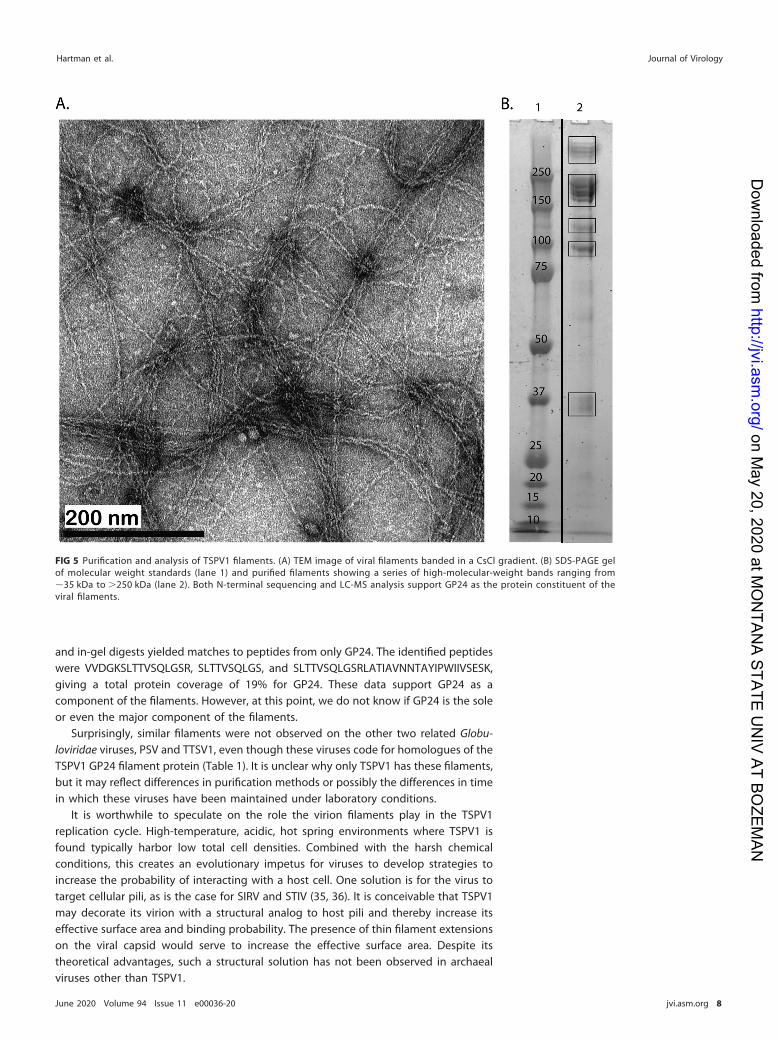
and in-gel digests yielded matches to peptides from only GP24. The identified peptideswere VVDGKSLTTVSQLGSR, SLTTVSQLGS, and SLTTVSQLGSRLATIAVNNTAYIPWIIVSESK,giving a total protein coverage of 19% for GP24. These data support GP24 as acomponent of the filaments. However, at this point, we do not know if GP24 is the soleor even the major component of the filaments.
Surprisingly, similar filaments were not observed on the other two related Globu-loviridae viruses, PSV and TTSV1, even though these viruses code for homologues of theTSPV1 GP24 filament protein (Table 1). It is unclear why only TSPV1 has these filaments,but it may reflect differences in purification methods or possibly the differences in timein which these viruses have been maintained under laboratory conditions.
It is worthwhile to speculate on the role the virion filaments play in the TSPV1replication cycle. High-temperature, acidic, hot spring environments where TSPV1 isfound typically harbor low total cell densities. Combined with the harsh chemicalconditions, this creates an evolutionary impetus for viruses to develop strategies toincrease the probability of interacting with a host cell. One solution is for the virus totarget cellular pili, as is the case for SIRV and STIV (35, 36). It is conceivable that TSPV1may decorate its virion with a structural analog to host pili and thereby increase itseffective surface area and binding probability. The presence of thin filament extensionson the viral capsid would serve to increase the effective surface area. Despite itstheoretical advantages, such a structural solution has not been observed in archaealviruses other than TSPV1.
FIG 5 Purification and analysis of TSPV1 filaments. (A) TEM image of viral filaments banded in a CsCl gradient. (B) SDS-PAGE gelof molecular weight standards (lane 1) and purified filaments showing a series of high-molecular-weight bands ranging from�35 kDa to �250 kDa (lane 2). Both N-terminal sequencing and LC-MS analysis support GP24 as the protein constituent of theviral filaments.
Hartman et al. Journal of Virology
June 2020 Volume 94 Issue 11 e00036-20 jvi.asm.org 8
on May 20, 2020 at M
ON
TANA STATE U
NIV AT BO
ZEMAN
http://jvi.asm.org/
Dow
nloaded from

MATERIALS AND METHODSHost and virus source. Thermoproteus sp. strain CP80 is an anaerobic, sulfur-reducing archaeon
originally isolated from Cinder Pool (44°43=56.9N, 110°42=32.4W) in Yellowstone National Park (20). Thisthermal feature is a high-temperature (80 to 89°C) and low-pH (4.0 to 4.4) hot spring. Strain CP80 wasisolated from sulfur-rich hot spring sediments, and 16S rRNA sequencing (20) and metagenomicsequencing of the sediment and planktonic communities typically show it to be among the mostabundant taxa in these ecological compartments (E. S. Boyd, unpublished data). A pure culture of thisstrain was obtained by dilution to extinction, and purity was confirmed by 16S rRNA gene and genomicsequencing (20).
A pure culture of Thermoproteus sp. CP80 was screened for the presence of virus-like particles (VLPs)by negative-stain electron microscopy. Samples were stained with 2% uranyl acetate prior to TEMimaging using a LEO 912AB with 2K � 2K charge-coupled-device (CCD) camera (Carl Zeiss, Jena,Germany).
Culture conditions. Medium was prepared as described by Boyd et al. (37), with the followingmodifications: in place of peptone, 3 g/liter tryptone and 0.2 g/liter yeast extract were used as thesources of carbon. Anaerobic bottles (0.5 liters) were filled with 150 ml of medium, heated to 80°C,purged for 30 min with nitrogen gas, and inoculated with 5 ml of a log-phase cell culture. Cultures wereincubated for 10 days at 80°C or until peak viral production was observed. Viral production wasmonitored by quantitative PCR (qPCR) amplification of a genome segment using qPCR primers orf14_F(TGGCCGGCCGCTTCCAGATA) and orf14_R (GCGGTCGATAGGTTAGCGGGC). Standards for qPCR consistedof a 255-bp fragment from the predicted major virion protein GP11 cloned into a TOPO TA pCR2.1 vector(Thermo Fisher, Waltham, MA). SsoAdvanced Sybr green supermix was used for qPCRs, according to themanufacturer’s directions (Bio-Rad, Hercules, CA). A Rotor-Gene series Q instrument (Qiagen, Hilden,Germany) was used for qPCR, with an initial denaturation at 98°C for 30 s, followed by 22 cycles at 98°Cfor 5 s and 65°C for 30 s.
Virus isolation and purification of virion filaments. Tangential flow filtration (TFF) was used toconcentrate 12 liters of cell culture to 100 ml using a 10-kDa molecular weight cutoff (MWCO) poly-ethersulfone (PES) column (Amersham, Westborough, MA), followed by centrifugation for 15 min at1,200 � g in a swinging bucket rotor to pellet cells and debris. Viruses present in the supernatant werefurther concentrated to 50 ml by TFF, followed by in-line filtration through 0.8-�m Acrodisc syringe filters(Pall, New York, NY) to remove any remaining cells. The final step in purification was accomplished bybanding viruses on buoyant density gradients created by the addition of CsCl to a final density of 1.286g/cm3, followed by ultracentrifugation for 24 h at 37,000 rpm (175,000 � g) in an SW41 rotor. Gradientswere hand fractionated, dialyzed against 5 mM citric acid (pH 4), and analyzed by both qPCR andnegative-stain electron microscopy. The protein concentrations in fractions were measured using Qubitfluorometric protein quantification (Invitrogen, Eugene, OR). Fractions containing separated filamentswere further purified by 10 cycles of diafiltration with a 0.5-ml Amicon Ultra spin column and a 100-kDaMWCO (Millipore, Burlington, MA).
Cryo-electron tomography. For cryo-electron tomography, purified virus was frozen on QuantifoilR2/1 holey carbon grids (200-mesh copper) in liquid propane-ethane using a Vitrobot Mark III system (FEI,Hillsboro, OR). Tilt series were taken using a Titan Krios TEM (FEI) operated at 300 kV and equipped witha Volta phase plate (38, 39), a Quantum postcolumn energy filter (Gatan, Pleasanton, CA), and a SummitK2 camera (Gatan) operated in electron counting mode at the Max Plank Institute of Biochemistry,Martinsried, Germany. Tilt-series images were collected using SerialEM (40). Individual frames of imagesacquired with the K2 camera were aligned in DigitalMicrograph (Gatan). The acquisition parameters weremagnification, �35,700; tilt range, � 60°; tilt increment, 2°; total dose, �60 e�/Å2; pixel (px) size,0.14 nm/px; and defocus with phase plate, �0.25 �m. The Volta phase plate was operated as previouslydescribed (41, 42).
Tomograms were reconstructed using IMOD 4.7 (43). Contrast transfer function (CTF) correctionswere not performed. Ten-nanometer gold nanoparticles were used as fiducial markers for the alignmentof tilt-series projection images. Local alignment solutions were used to refine rotation, magnification, andtilt angles, while a global solution was employed for distortion. Gold particles were erased from the finalaligned stack. Images were binned 2�, radially filtered with a cutoff of 0.4 and falloff of 0.05, and runthrough 10 simultaneous iterative reconstruction technique (SIRT)-like filter iterations.
Genome sequencing and assembly. Viral nucleic acid was extracted from CsCl fractions using aPureLink viral RNA/DNA extraction kit (Thermo Fisher, Waltham, MA) and was sequenced at theUniversity of Illinois Sequencing Center (Urbana-Champaign, IL) using Illumina HiSeq technology with2 � 250-bp paired-end reads (Illumina, San Diego, CA). Genome assembly was performed using the MIRAassembly program (version 4.0.4). Initial assembly required subsampling 30,000 reads and assemblingthese in order to avoid assembly failure from high sequence coverage. Read recruitment to verify theinverted terminal repeats (ITRs) was performed using the Geneious assembler (Biomatters, Newark, NJ).For this process, the ITR region was trimmed from each end of the genome, and then paired reads wererecruited to the trimmed sequence. Genome alignments between TSPV1, TTSV1, and PSV were con-ducted using MAUVE v.20159226 (44).
Gene prediction and annotation. Glimmer and Prodigal were used to predict viral genes (45, 46).Predicted gene products were compared to the NCBI nr database using BLASTp (47). In addition, proteinsfrom both PSV and TTSV were queried against the TSPV1 genome to identify homologs using BLASTp(47). Protein alignments were generated using MUSCLE (48). Predicted proteins were further analyzedusing the Phyre2 structure prediction server and HHpred to detect distant homologues (49, 50). PSIPRED,
TSPV1, a Spherical Archaeal Virus with Filaments Journal of Virology
June 2020 Volume 94 Issue 11 e00036-20 jvi.asm.org 9
on May 20, 2020 at M
ON
TANA STATE U
NIV AT BO
ZEMAN
http://jvi.asm.org/
Dow
nloaded from

FlaFind, and SignalP 5.0 were used to identify potential signal peptides in the predicted proteins of TSPV1(51–53). PSIPRED was also used for protein secondary structure prediction (53).
N-terminal sequencing and LC-MS sequencing. N-terminal Edman sequencing was performedusing purified filaments (5 �g) that were applied to a Prosorb cartridge, followed by 8 sequencing cyclesconducted using a Shimadzu PPSQ-53A sequencer at the Iowa State University Sequencing Center.
LC-MS analysis was performed on in-solution digests of both purified viral capsids and purifiedfilaments as follows: samples were dialyzed into 12.5 mM NH4HCO3 (pH 8), followed by the addition ofmass spectrometry-grade urea to a final concentration of 8 M. Following this, samples were reduced in5 mM dithiothreitol (DTT), alkylated with 14 mM iodoacetic acid (IAA), and digested for 4 h (37°C) at a 1:20protease/protein ratio using trypsin–Lys-C mix (Promega, Madison, WI) with ProteaseMax (Promega,Madison, WI) addition to 0.03%. Samples were then diluted to a urea concentration of 1 M, followed byovernight digestion (37°C).
For in-gel digestion, 9 �g of purified capsids or 7 �g of purified filaments was dialyzed into 12.5 mMNH4HCO3 (pH 8), and then mass spectrometry-grade urea was added directly to a final concentration of8 M together with SDS-PAGE loading buffer (50 mM Tris-HCl, 2% [wt/vol] SDS, 0.1% [wt/vol] bromophe-nol blue, 10% glycerol, 100 mM �-mercaptoethanol [pH 6.8]). Samples were denatured at 98°C for 40 minor at room temperature for 2 h. Electrophoresis was performed using a 4 to 12% Bis-Tris NuPAGE gel(Thermo Fisher, Waltham, MA) for separation. Gels were stained with colloidal Coomassie and imagedwith a Typhoon Trio laser scanner (GE, Boston, MA). Gel slices were excised and proteolyzed as follows:gel slices were destained in 50 mM NH4HCO3 until clear and then reduced with 100 mM DTT, followedby alkylation with 55 mM iodoacetamide. Digestion was performed overnight in 25 mM NH4HCO3 using0.3 �g sequencing-grade trypsin (Promega). Samples were subjected to reverse-phase chromatographyusing a Dionex nano-ultrahigh-performance liquid chromatography (nano-UHPLC) and then analyzedwith a Bruker MaXis Impact mass spectrometer (Billerica, MA).
Data analysis was performed using MetaMorpheus v. 0.0.300 with precursor and product toleranceof 35 ppm, 3 max missed cleavages, and posttranslational modifications (PTMs) of common fixed,common variable, glycosylation, common biological, less common, common artifact, metal, trypsindigested, UniProt, and Unimod modifications. Searches were performed against a database of viralproteins, cellular proteins, and/or common contaminants. Alternatively, searches were performed withSearchGui v. 3.3.13, with precursor and product tolerance of 35 ppm, 3 max missed cleavages, oxidationof M, and carbamylation of K as variable modifications using X! tandem, MS-GF�, and Tide searchengines (54).
Virion protein glycosylation analysis. The NetNGlyc server was used to predict N-linked glycosy-lation sites (55). For glycan staining, electrophoresis was performed with 9 �g of purified capsids or 7 �gof purified filaments using a 4 to 12% Bis-Tris NuPAGE SDS-PAGE gel (Thermo Fisher, Waltham, MA) forseparation with 4 �l CandyCane ladder (Invitrogen, Eugene, OR) and 4 �l of Precision Plus Protein DualXtra ladder (Bio-Rad, Hercules, CA) as standards. The gel was stained with the Pro-Q Emerald 488glycoprotein gel and blot stain kit (Invitrogen, Eugene, OR), following the manufacturer’s recommenda-tions, and visualized using an AlphaImager 2200 imager (ProteinSimple, San Jose, CA).
Diethyl ether sensitivity assay. To test for the presence of a viral envelope, the method developedby Andrewes and Horstmann was employed. Briefly, diethyl ether was added to viral particles in 5 mMcitric acid buffer (pH 4) (56). The final concentration of diethyl ether was 20% (vol/vol); samples wereincubated at room temperature for 24 h and then analyzed by negative-stain TEM.
Data availability. The genome for TSPV1 is available under GenBank accession number MT047590.
SUPPLEMENTAL MATERIALSupplemental material is available online only.SUPPLEMENTAL FILE 1, MOV file, 14.6 MB.SUPPLEMENTAL FILE 2, PDF file, 0.1 MB.
ACKNOWLEDGMENTSThis work was supported by National Science Foundation grants DEB-1342876 to
M.Y., MCB-1413534 to C.M.L., and EAR-1820658 to E.S.B. Funding of the Proteomics,Metabolomics, and Mass Spectrometry Facility at Montana State University was madepossible in part by the M. J. Murdock Charitable Trust, National Institute of GeneralMedical Sciences, National Institutes of Health, award P20GM103474, the NationalScience Foundation (grant NSF-MRI:DBI-1532078), and the Office of the Vice Presidentfor Research and Economic Development.
REFERENCES1. Bolduc B, Wirth J, Mazurie A, Young M. 2015. Viral assemblage compo-
sition in Yellowstone acidic hot springs assessed by network analysis.ISME J 9:2162–2177. https://doi.org/10.1038/ismej.2015.28.
2. Mochizuki T, Krupovic M, Pehau-Arnaudet G, Sako Y, Forterre P, Prang-ishvili D. 2012. Archaeal virus with exceptional virion architecture and
the largest single-stranded DNA genome. Proc Natl Acad Sci U S A109:13386–13391. https://doi.org/10.1073/pnas.1203668109.
3. Krupovic M, Quemin ER, Bamford DH, Forterre P, Prangishvili D. 2014.Unification of the globally distributed spindle-shaped viruses of theArchaea. J Virol 88:2354–2358. https://doi.org/10.1128/JVI.02941-13.
Hartman et al. Journal of Virology
June 2020 Volume 94 Issue 11 e00036-20 jvi.asm.org 10
on May 20, 2020 at M
ON
TANA STATE U
NIV AT BO
ZEMAN
http://jvi.asm.org/
Dow
nloaded from

4. Prangishvili D, Mochizuki T, Krupovic M, ICTV Report Consortium. 2018.ICTV virus taxonomy profile: Guttaviridae. J Gen Virol 99:290–291.https://doi.org/10.1099/jgv.0.001027.
5. Häring M, Rachel R, Peng X, Garrett RA, Prangishvili D. 2005. Viraldiversity in hot springs of Pozzuoli, Italy, and characterization of aunique archaeal virus, Acidianus bottle-shaped virus, from a new family,the Ampullaviridae. J Virol 79:9904–9911. https://doi.org/10.1128/JVI.79.15.9904-9911.2005.
6. Krupovic M, Cvirkaite-Krupovic V, Iranzo J, Prangishvili D, Koonin EV.2018. Viruses of archaea: structural, functional, environmental and evo-lutionary genomics. Virus Res 244:181–193. https://doi.org/10.1016/j.virusres.2017.11.025.
7. Prangishvili D, Garrett RA, Koonin EV. 2006. Evolutionary genomics ofarchaeal viruses: unique viral genomes in the third domain of life. VirusRes 117:52–67. https://doi.org/10.1016/j.virusres.2006.01.007.
8. Prangishvili D, Bamford DH, Forterre P, Iranzo J, Koonin EV, Krupovic M.2017. The enigmatic archaeal virosphere. Nat Rev Microbiol 15:724–739.https://doi.org/10.1038/nrmicro.2017.125.
9. Sayers EW, Agarwala R, Bolton EE, Brister JR, Canese K, Clark K, ConnorR, Fiorini N, Funk K, Hefferon T, Holmes JB, Kim S, Kimchi A, Kitts PA,Lathrop S, Lu Z, Madden TL, Marchler-Bauer A, Phan L, Schneider VA,Schoch CL, Pruitt KD, Ostell J. 2019. Database resources of the NationalCenter for Biotechnology Information. Nucleic Acids Res 47:D23–D28.https://doi.org/10.1093/nar/gky1069.
10. Munson-McGee JH, Snyder JC, Young MJ. 2018. Archaeal viruses fromhigh-temperature environments. Genes (Basel) 9:128. https://doi.org/10.3390/genes9030128.
11. Wagner C, Reddy V, Asturias F, Khoshouei M, Johnson JE, Manrique P,Munson-McGee J, Baumeister W, Lawrence CM, Young MJ. 2017. Isola-tion and characterization of Metallosphaera turreted icosahedral virus(MTIV), a founding member of a new family of archaeal viruses. J Virol91:e00925-17. https://doi.org/10.1128/JVI.00925-17.
12. Ahn DG, Kim SI, Rhee JK, Kim KP, Pan JG, Oh JW. 2006. TTSV1, a newvirus-like particle isolated from the hyperthermophilic crenarchaeoteThermoproteus tenax. Virology 351:280–290. https://doi.org/10.1016/j.virol.2006.03.039.
13. Häring M, Peng X, Brugger K, Rachel R, Stetter KO, Garrett RA, Prang-ishvili D. 2004. Morphology and genome organization of the virus PSV ofthe hyperthermophil ic archaeal genera Pyrobaculum andThermoproteus: a novel virus family, the Globuloviridae. Virology 323:233–242. https://doi.org/10.1016/j.virol.2004.03.002.
14. Geslin C, Le Romancer M, Erauso G, Gaillard M, Perrot G, Prieur D. 2003.PAV1, the first virus-like particle isolated from a hyperthermophiliceuryarchaeote, “Pyrococcus abyssi.” J Bacteriol 185:3888–38894. https://doi.org/10.1128/jb.185.13.3888-3894.2003.
15. Gorlas A, Koonin EV, Bienvenu N, Prieur D, Geslin C. 2012. TPV1, the firstvirus isolated from the hyperthermophilic genus Thermococcus. EnvironMicrobiol 14:503–516. https://doi.org/10.1111/j.1462-2920.2011.02662.x.
16. Mochizuki T, Sako Y, Prangishvili D. 2011. Provirus induction in hyper-thermophilic archaea: characterization of Aeropyrum pernix spindle-shaped virus 1 and Aeropyrum pernix ovoid virus 1. J Bacteriol 193:5412–5419. https://doi.org/10.1128/JB.05101-11.
17. Mochizuki T, Yoshida T, Tanaka R, Forterre P, Sako Y, Prangishvili D. 2010.Diversity of viruses of the hyperthermophilic archaeal genus Aeropyrum,and isolation of the Aeropyrum pernix bacilliform virus 1, APBV1, thefirst representative of the family Clavaviridae. Virology 402:347–354.https://doi.org/10.1016/j.virol.2010.03.046.
18. Zillig W, Prangishvilli D, Schleper C, Elferink M, Holz I, Albers S, JanekovicD, Gotz D. 1996. Viruses, plasmids and other genetic elements of ther-mophilic and hyperthermophilic Archaea. FEMS Microbiol Rev 18:225–236. https://doi.org/10.1111/j.1574-6976.1996.tb00239.x.
19. Jay ZJ, Beam JP, Kozubal MA, Jennings RD, Rusch DB, InskeepWP. 2016. Thedistribution, diversity and function of predominant Thermoproteales inhigh-temperature environments of Yellowstone National Park. Environ Mi-crobiol 18:4755–4769. https://doi.org/10.1111/1462-2920.13366.
20. Urschel MR, Hamilton TL, Roden EE, Boyd ES. 2016. Substrate preference,uptake kinetics and bioenergetics in a facultatively autotrophic, ther-moacidophilic crenarchaeote. FEMS Microbiol Ecol 92:fiw069. https://doi.org/10.1093/femsec/fiw069.
21. Yim KJ, Song HS, Choi JS, Roh SW. 2015. Thermoproteus thermophilus spnov., a hyperthermophilic crenarchaeon isolated from solfataric soil. IntJ Syst Evol Microbiol 65:2507–2510. https://doi.org/10.1099/ijs.0.000293.
22. Zillig W, Stetter KO, Schafer W, Janekovic D, Wunderl S, Holz I, Palm P.1981. Thermoproteales: a novel type of extremely thermoacidophilic
anaerobic archaebacteria isolated from Icelandic solfataras. ZentralblBakteriol Mikrobiol Hyg 2:205–227. https://doi.org/10.1016/S0721-9571(81)80001-4.
23. Bonch-Osmolovskaya EA, Miroshnichenko ML, Kostrikina NA, Chernych NA,Zavarzin GA. 1990. Thermoproteus uzoniensis sp. nov., a new extremelythermophilic archaebacterium from Kamchatka continental hot springs.Arch Microbiol 154:556–559. https://doi.org/10.1007/BF00248836.
24. Siebers B, Zaparty M, Raddatz G, Tjaden B, Albers SV, Bell SD, BlombachF, Kletzin A, Kyrpides N, Lanz C, Plagens A, Rampp M, Rosinus A, von JanM, Makarova KS, Klenk HP, Schuster SC, Hensel R. 2011. The completegenome sequence of Thermoproteus tenax: a physiologically versatilemember of the Crenarchaeota. PLoS One 6:e24222. https://doi.org/10.1371/journal.pone.0024222.
25. Janekovic D, Wunderl S, Holz I, Zillig W, Gierl A, Neumann H. 1983. Ttv1,Ttv2 and Ttv3, a Family of viruses of the extremely thermophilic, anaer-obic, sulfur reducing archaebacterium Thermoproteus tenax. Mol GenGenet 192:39–45. https://doi.org/10.1007/BF00327644.
26. Quemin ER, Chlanda P, Sachse M, Forterre P, Prangishvili D, Krupovic M.2016. Eukaryotic-like virus budding in Archaea. mBio 7:e01439-16.https://doi.org/10.1128/mBio.01439-16.
27. Oke M, Carter LG, Johnson KA, Liu H, McMahon SA, Yan X, Kerou M,Weikart ND, Kadi N, Sheikh MA, Schmelz S, Dorward M, Zawadzki M,Cozens C, Falconer H, Powers H, Overton IM, van Niekerk CA, Peng X,Patel P, Garrett RA, Prangishvili D, Botting CH, Coote PJ, Dryden DT,Barton GJ, Schwarz-Linek U, Challis GL, Taylor GL, White MF, Naismith JH.2010. The Scottish structural proteomics facility: targets, methods andoutputs. J Struct Funct Genomics 11:167–180. https://doi.org/10.1007/s10969-010-9090-y.
28. Hatzfeld M. 1999. The armadillo family of structural proteins. Int RevCytol 186:179–224. https://doi.org/10.1016/s0074-7696(08)61054-2.
29. Larson ET, Eilers BJ, Reiter D, Ortmann AC, Young MJ, Lawrence CM.2007. A new DNA binding protein highly conserved in diverse crenar-chaeal viruses. Virology 363:387–396. https://doi.org/10.1016/j.virol.2007.01.027.
30. Athukoralage JS, McMahon S, Zhang C, Grüschow S, Graham S, KrupovicM, Whitaker RJ, Gloster T, White MF. 2019. A viral ring nuclease anti-CRISPR subverts type III CRISPR immunity. Nature 577:572–575. https://doi.org/10.1038/s41586-019-1909-5.
31. Ellen AF, Zolghadr B, Driessen AM, Albers SV. 2010. Shaping the archaeal cellenvelope. Archaea 2010:608243. https://doi.org/10.1155/2010/608243.
32. Krupovic M, Koonin EV. 2017. Multiple origins of viral capsid proteinsfrom cellular ancestors. Proc Natl Acad Sci U S A 114:E2401–E2410.https://doi.org/10.1073/pnas.1621061114.
33. Montefiori DC, Robinson WE, Jr, Mitchell WM. 1988. Role of proteinN-glycosylation in pathogenesis of human immunodeficiency virus type1. Proc Natl Acad Sci U S A 85:9248–9252. https://doi.org/10.1073/pnas.85.23.9248.
34. Kandiba L, Aitio O, Helin J, Guan ZQ, Permi P, Bamford DH, Eichler J,Roine E. 2012. Diversity in prokaryotic glycosylation: an archaeal-derivedN-linked glycan contains legionaminic acid. Mol Microbiol 84:578–593.https://doi.org/10.1111/j.1365-2958.2012.08045.x.
35. Quemin ER, Lucas S, Daum B, Quax TE, Kuhlbrandt W, Forterre P, AlbersSV, Prangishvili D, Krupovic M. 2013. First insights into the entry processof hyperthermophilic archaeal viruses. J Virol 87:13379–13385. https://doi.org/10.1128/JVI.02742-13.
36. Hartman R, Eilers BJ, Bollschweiler D, Munson-McGee JH, Engelhardt H,Young MJ, Lawrence CM. 2019. The molecular mechanism of cellularattachment for an archaeal virus. Structure 27:1634–1646. https://doi.org/10.1016/j.str.2019.09.005.
37. Boyd ES, Jackson RA, Encarnacion G, Zahn JA, Beard T, Leavitt WD, PiY, Zhang CL, Pearson A, Geesey GG. 2007. Isolation, characterization,and ecology of sulfur-respiring crenarchaea inhabiting acid-sulfate-chloride-containing geothermal springs in Yellowstone National Park.Appl Environ Microbiol 73:6669–6677. https://doi.org/10.1128/AEM.01321-07.
38. Khoshouei M, Pfeffer S, Baumeister W, Forster F, Danev R. 2017. Subto-mogram analysis using the Volta phase plate. J Struct Biol 197:94–101.https://doi.org/10.1016/j.jsb.2016.05.009.
39. Danev R, Baumeister W. 2017. Expanding the boundaries of cryo-EMwith phase plates. Curr Opin Struct Biol 46:87–94. https://doi.org/10.1016/j.sbi.2017.06.006.
40. Mastronarde DN. 2005. Automated electron microscope tomographyusing robust prediction of specimen movements. J Struct Biol 152:36–51. https://doi.org/10.1016/j.jsb.2005.07.007.
TSPV1, a Spherical Archaeal Virus with Filaments Journal of Virology
June 2020 Volume 94 Issue 11 e00036-20 jvi.asm.org 11
on May 20, 2020 at M
ON
TANA STATE U
NIV AT BO
ZEMAN
http://jvi.asm.org/
Dow
nloaded from
41. Danev R, Buijsse B, Khoshouei M, Plitzko JM, Baumeister W. 2014. Voltapotential phase plate for in-focus phase contrast transmission electronmicroscopy. Proc Natl Acad Sci U S A 111:15635–15640. https://doi.org/10.1073/pnas.1418377111.
42. Fukuda Y, Laugks U, Lucic V, Baumeister W, Danev R. 2015. Electroncryotomography of vitrified cells with a Volta phase plate. J Struct Biol190:143–154. https://doi.org/10.1016/j.jsb.2015.03.004.
43. Kremer JR, Mastronarde DN, McIntosh JR. 1996. Computer visualizationof three-dimensional image data using IMOD. J Struct Biol 116:71–76.https://doi.org/10.1006/jsbi.1996.0013.
44. Darling AC, Mau B, Blattner FR, Perna NT. 2004. Mauve: multiple align-ment of conserved genomic sequence with rearrangements. GenomeRes 14:1394–1403. https://doi.org/10.1101/gr.2289704.
45. Delcher AL, Harmon D, Kasif S, White O, Salzberg SL. 1999. Improvedmicrobial gene identification with GLIMMER. Nucleic Acids Res 27:4636–4641. https://doi.org/10.1093/nar/27.23.4636.
46. Hyatt D, Chen GL, Locascio PF, Land ML, Larimer FW, Hauser LJ. 2010.Prodigal: prokaryotic gene recognition and translation initiation siteidentification. BMC Bioinformatics 11:119. https://doi.org/10.1186/1471-2105-11-119.
47. Camacho C, Coulouris G, Avagyan V, Ma N, Papadopoulos J, Bealer K,Madden TL. 2009. BLAST�: architecture and applications. BMC Bioinfor-matics 10:421. https://doi.org/10.1186/1471-2105-10-421.
48. Edgar RC. 2004. MUSCLE: multiple sequence alignment with high accu-racy and high throughput. Nucleic Acids Res 32:1792–1797. https://doi.org/10.1093/nar/gkh340.
49. Söding J, Biegert A, Lupas AN. 2005. The HHpred interactive server for
protein homology detection and structure prediction. Nucleic Acids Res33:W244–W248. https://doi.org/10.1093/nar/gki408.
50. Kelley LA, Mezulis S, Yates CM, Wass MN, Sternberg MJ. 2015. The Phyre2Web portal for protein modeling, prediction and analysis. Nat Protoc10:845–858. https://doi.org/10.1038/nprot.2015.053.
51. Armenteros JJA, Tsirigos KD, Sonderby CK, Petersen TN, Winther O,Brunak S, von Heijne G, Nielsen H. 2019. SignalP 5.0 improves signalpeptide predictions using deep neural networks. Nat Biotechnol 37:420–423. https://doi.org/10.1038/s41587-019-0036-z.
52. Szabó Z, Stahl AO, Albers SV, Kissinger JC, Driessen AJ, Pohlschroder M.2007. Identification of diverse archaeal proteins with class III signalpeptides cleaved by distinct archaeal prepilin peptidases. J Bacteriol189:772–778. https://doi.org/10.1128/JB.01547-06.
53. Buchan DWA, Jones DT. 2019. The PSIPRED Protein Analysis Workbench:20 years on. Nucleic Acids Res 47:W402–W407. https://doi.org/10.1093/nar/gkz297.
54. Barsnes H, Vaudel M. 2018. SearchGUI: a highly adaptable commoninterface for proteomics search and de novo engines. J Proteome Res17:2552–2555. https://doi.org/10.1021/acs.jproteome.8b00175.
55. Steentoft C, Vakhrushev SY, Joshi HJ, Kong Y, Vester-Christensen MB,Schjoldager KT, Lavrsen K, Dabelsteen S, Pedersen NB, Marcos-Silva L,Gupta R, Bennett EP, Mandel U, Brunak S, Wandall HH, Levery SB,Clausen H. 2013. Precision mapping of the human O-GalNAc glycopro-teome through SimpleCell technology. EMBO J 32:1478–1488. https://doi.org/10.1038/emboj.2013.79.
56. Andrewes CH, Horstmann DM. 1949. The susceptibility of viruses to ethylether. J Gen Microbiol 3:290–297. https://doi.org/10.1099/00221287-3-2-290.
Hartman et al. Journal of Virology
June 2020 Volume 94 Issue 11 e00036-20 jvi.asm.org 12
on May 20, 2020 at M
ON
TANA STATE U
NIV AT BO
ZEMAN
http://jvi.asm.org/
Dow
nloaded from