discovery of novel dengue ns2b/ns3 protease inhibitors...
TRANSCRIPT


OPEN ACCESS International Journal of Pharmacology
ISSN 1811-7775DOI: 10.3923/ijp.2016.621.632
Research ArticleDiscovery of Novel Dengue NS2B/NS3 Protease Inhibitors UsingPharmacophore Modeling and Molecular Docking Based VirtualScreening of the ZINC Database
1,2Muhammad Tahir ul Qamar, 2Saleha Kiran, 2Usman Ali Ashfaq, 2Muhammad Rizwan Javed,3,4Farooq Anwar, 1,5Muhammad Amjad Ali and 6,7Anwar ul Hassan Gilani
1Center of Agricultural Biochemistry and Biotechnology (CABB), University of Agriculture (UAF), 38040 Faisalabad, Pakistan2Department of Bioinformatics and Biotechnology, Government College University Faisalabad (GCUF), 38000 Faisalabad, Pakistan3Department of Pharmaceutical Chemistry, College of Pharmacy, Prince Sattam bin Abdulaziz University, 11942 Al-Kharij, Saudi Arabia4Department of Chemistry, University of Sargodha, 40100 Sargodha, Pakistan5Department of Plant Pathology, University of Agriculture Faisalabad (UAF), 38040 Faisalabad, Pakistan6Pakistan Council for Science and Technology, G-5/2, 44000 Islamabad, Pakistan7Department of Biological and Biomedical Sciences, Aga Khan University Medical College, 74800 Karachi, Pakistan
AbstractBackground: Dengue, a vector borne disease has become a crucial health concern globally. The search for a suitable dengue vaccine has beengoing on for the last few decades due to unavailability of any effective treatment. An efficient medication strategy is required to overcome thedevastating effects of dengue. Now, computational biology has emerged as a novel tool to improve the domain of computer aided drug designing.The present study reports a complex-based pharmacophore computational modeling that elucidates important pharmacophoric features helpful for the inhibition of protease activity of NS2B/NS3 protein of Dengue Virus (DV). Materials and Methods: A seven featured pharmacophoremodel of DV NS2B/NS3 protease has been developed via crystal structure of NS2B/NS3 protease and its inhibitor complex in Molecular OperatingEnvironment (MOE) pharmacophore constructing tool. The developed pharmacophore model was validated by a test database of the publishedinhibitors. Validated pharmacophore model was then used to virtually screen the potential compounds from ZINC database. The screenedcompounds were filtered by Lipinski’s rule of five and further evaluated through molecular docking studies. The results of docking and interactionstudies were validated through binding affinity analysis and ADMET profiling. Results: Six hits (ZINC ID’s: 75163069, 59170698, 06395655,32933073, 13728171 and 65395833) of different scaffolds having interactions with important active site residues (His51, Asp75, Ser135) werepredicted. Conclusion: It can be concluded from the finding of the present study that predicted hits could serve as potential candidates to actas starting point in the development of novel and potent NS2B/NS3 protease inhibitors. The present modeling explores the significant role of thepredicted hits towards blocking the replication of DV.
Key words: Dengue, ZINC database, pharmacophore modeling, molecular docking, drug profiling
Received: February 29, 2016 Accepted: June 21, 2016 Published: July 15, 2016
Citation: Muhammad Tahir ul Qamar, Saleha Kiran, Usman Ali Ashfaq, Muhammad Rizwan Javed, Farooq Anwar, Muhammad Amjad Ali and Anwar ul Hassan Gilani,2016. Discovery of novel dengue NS2B/NS3 protease inhibitors using pharmacophore modeling and molecular docking based virtual screening of the ZINC database. Int. J. Pharmacol., 12: 621-632.
Corresponding Authors: Usman Ali Ashfaq, Department of Bioinformatics and Biotechnology, Government College University Faisalabad (GCUF),38000 Faisalabad, Pakistan Tel: +92-331-4728790Anwar ul Hassan Gilani, Pakistan Council for Science and Technology, G-5/2, 44000 Islamabad, Pakistan Tel: +92-51-9204416
Copyright: © 2016 Muhammad Tahir ul Qamar et al. This is an open access article distributed under the terms of the creative commons attribution License, whichpermits unrestricted use, distribution and reproduction in any medium, provided the original author and source are credited.
Competing Interest: The authors have declared that no competing interest exists.
Data Availability: All relevant data are within the paper and its supporting information files.

Int. J. Pharmacol., 12 (6): 621-632, 2016
INTRODUCTION
Due to unavailability of any effective treatment, Dengue,a vector borne disease has become a crucial health concernglobally. Dengue related health disorders such as fever andacute joint pains are caused as result of mosquito-borne viralinfections1,2. This rapidly spreading and debilitating diseasehas been reported to affect almost 2.5 billion peopleworldwide3 with approximately 25,000 deaths per year4.According to a latest study approximately 50-100 millionpeople from more than 100 countries are suffering from thedevastating effects of this viral infection. Several countries andespecially the tropical regions of Asia, Central America, SouthAmerica and Africa are at a higher risk of dengue infections5,6.As investigated by immunological and virological studies,Dengue infection is caused by Dengue Virus (DV), aFlaviviradae virus with four serotypes (DV-1, DV-2, DV-3 andDV-4) reported7. Dengue virus belongs to viral genusFlavivirus that consists of pathogenic RNA viruses. Theseviruses are mostly colonized in temperate and tropical regionsof the world and cause several infections in humans includingdengue fever, west-nile fever, tick-borne encephalitis andyellow fever8.
In humans, DV infection spreads via the bite of two carriermosquitoes namely Aedes aegypti and Aedes albopictus9.The genomic size of DV is reported to extending over 10.7 kband encodes a total 10 structural and nonstructural proteins10.The three structural proteins include core/capsid protein,membrane associated protein and an envelope protein. Theremaining seven proteins namely NS1, NS2A, NS2B, NS3, NS4A,NS4B and NS5 are non-structural proteins. These proteins arelocated in the order of 5!-C-prM-E-NS1-NS2A-NS2B-NS3-NS4A-NS4B-NS5-3! on viral RNA11,12 (Fig. 1) and play a vibrant role inthe structural organization of virus and viral genome entry intothe host cell, while non-structural proteins help the DV to
control the host synthetic machinery for its own replicationand other cellular functions13. Recently, it has beeninvestigated that the potential of NS3 depends upon itsinteraction with cofactor (NS2B). The NS2B is a serine proteasedomain which is located at the N-terminal region of NS314. Asa result of NS3 and NS2B interaction, NS2B/NS3 pro-complexis formed, which helps to cleave viral proteins to perform theirfunctions. Any disturbance in functional behavior of thisregion is reported to inhibit viral replication. Therefore,NS2B/NS3 complex is believed to be an effective target for thescreening and assessment of the effective antiviral drugcandidates1,4,12,14.
Recent advances in computational biology havenoticeably impacted and improved the domain of computeraided drug designing. Due to high cost and low hit ratespracticed during wet lab testing, computational processes andvirtual screening have largely aided the drug designing.Computational methods are supportive in taking decision andvirtually stimulating various aspects of drug discovery anddevelopment. By the last few decades, computationallygenerated virtual High Throughput Screening (vHTS) hasgained sufficient maturity to becoming an alternative to thecounterpart experimental high-throughput virtual screening(HTS). Being a knowledge driven approach, vHTS has provedto be an integral part of drug designing process15.
Advanced computational approaches such as multifariouspharmacophore modeling, virtual screening and moleculardocking can be applied to identify and recognize new andeffective inhibitors to minimizing the activity of NS2B/NS3protease from DV. A pharmacophore is the 3D arrangementsof structural or chemical features of small organic compoundsof a drug that might be essential for its interaction with thetarget and drug’s bioactivity. In drug designing process, theuse of pharmacophore approach can be supportive in severalways: (a) It could be used as a 3D query in virtual screening to
Fig. 1: Schematic diagram of Dengue Virus (DV) RNA gene organization and their translation
622
5UTR
3UTR
Structural Non-structural
Translation and processing of protein
C prM E NS1 2A 2B NS3 4A 4B NS5

Int. J. Pharmacol., 12 (6): 621-632, 2016
recognize novel and effective hits with an arrangementdifferent from the previously reported structures from thedatabases of drug-like molecules, (b) To identify the possiblemode of actions and (c) To predict the mode of action of newcompounds prior to synthesis16,17. Recent advances in virtualscreening technologies have prompted the researchers torapidly screen drug libraries and inhibitors databases to findout therapeutic activities against different new targets withcost effectiveness18. One of the similar approaches, namelymolecular docking involves the use of multiple computationalmethods to predict the true binding orientations of smallmolecules within their protein targets19.
The plan of the current study was to design and developa complex based pharmacophore model and to find outthe hits which have important pharmacophore features bycomputational screening of effective compounds against allsubsets of a previously developed ZINC database20 coupledwith molecular docking, protein ligand binding interactions,binding affinity predictions and drug likeness predictions.Virtual screening is supposed to provide us an insight ofvarious features of compounds essential for the inhibition ofprotease activity of NS2B/NS3 complex. The ZINC is a freedatabase of commercially available small organic compoundsfor virtual screening and drug discovery. It has over 35 millionpotential compounds in ready-to-dock, 3D formats21. Forexample, compound-1 of natural product like subset ofZINC database is merely the second immunomodulatoryinhibitor to date which has successfully inhibited TLR1-TLR2heterodimerization in vitro and proved as a potential drugcandidate against different fungal and inflammatory/immunedisorders (atherosclerosis, sepsis syndrome, rheumatoidarthritis, asthma) and viral diseases (human cytomegalovirus,lymphocytic choriomeningitis virus, herpex simplex virus)22.Compound-1 also exhibited anti-proliferative activity againstcancer cells in vitro by inhibiting STAT3 dimerization23.Optimization and virtual screening of Janus Kinase 2 (JAK2)Type-II compounds from ZINC database has exploredpotential inhibitors of Hepatitis C Virus (HCV) translation andreplication24. Increased resistance of Plasmodium falciparumagainst most of the available drugs is a challenging tasktowards controlling malaria. According to researchers,previously, 19 potential anti-malarial compounds werereported from ZINC database which successfully inhibitedthe growth of Plasmodium falciparum25. The compound 6,6``-biapigenin has been only identified as the secondinhibitor of NEDD8-activiating enzyme as well as reported tobe anti-cancer agent by virtual screening of ZINC database26.There are also many other examples of different compoundsfrom ZINC database which successfully proved as a potentialdrug compounds against various diseases.
To date, no any other study as such screened whole ZINCdatabase against DV NS2B/NS3 protease. Therefore, in the present study this database have selected for computationalscreening of potent lead inhibitors which could serve as drugcompounds against DV. The identification of six novel andpotent lead compounds as NS2B/NS3 protease inhibitorsclearly reflects the significance of this study. The results of thisstudy provide valuable information about computer aideddrug screening and development of drugs against DVinfection.
MATERIALS AND METHODS
Pharmacophore modeling and molecular docking wasaccomplished via the Molecular Operating Environment(MOE) software package. The MOE is a comprehensivesuite developed by Chemical Computing Group (CCG)Incorporation. It has different tools and computationalmethods that are applied for structure and fragment baseddrug designing, pharmacophore discovery, protein structureanalysis, data processing and molecular docking andsimulations27.
Generation and validation of complex basedpharmacophore model: The assembly of electronic and stericcharacteristics of pharmacophore is compulsory to ensurefinest supra-molecular interactions with a particular biologicaltarget for inhibiting its biological response28,29. If 3D structureof the target protein is available, complex basedpharmacophore system can be used to modify the drugadvancement process. In the present study, the crystalstructure of DV NS2B/NS3 protease in complex with asubstrate based inhibitor benzoyl-norleucine (P4)-lysine(P3)-arginine (P2)-arginine (P1)-aldehyde (Bz-Nle-Lys-Arg-Arg-H) was used (PDB ID: 2FOM) for the creation of complex basedpharmacophore system30. The inhibitor had strong interactionwith catalytic triad residues of DV NS2B/NS3 protease andfurther used as a reference in molecular docking (Fig. 2). TheMOE pharmacophore constructing tool was employed forthe creation and visualization of three dimensional (3D)pharmacophore from protein structure ligand complex. Thegenerated pharmacophore system was validated througha test database of 20 known inhibitors of DV NS2B/NS3protease. These inhibitors of the database and theirinhibitory potential were taken from the reportedliterature31,32. The 3D structures of the test database werecreated in MOE builder tool. Energy minimization of 3Dstructures was achieved by MOE energy minimizationalgorithm with parameters; Gradient: 0.05 and Force Field:MMFF94X.
623

Int. J. Pharmacol., 12 (6): 621-632, 2016
Fig. 2(a-b): Complex structure of reference ligand (Bz-Nle-Lys-Arg-Arg-H) and DV NS2B/NS3 protease. (a) Three dimensionalrepresentation of the interaction of the reference ligand and receptor and (b) Three dimensional pharmacophoremodel generated from DV NS2B/NS3 protease complex map view
Pharmacophore-based database screening: The next stepwas to identify different hits with multiple chemical natures.This was achieved by using in silico pharmacophore basedvisual screening of the ZINC database with 3D query of thevalidated pharmacophore model. The MOE software was usedfor this virtual screening of various hits in the ZINC database20.This type of screening was adopted due to a couple ofreasons: firstly, the validated pharmacophore model wassuccessfully used for appropriate identification of compoundshaving known inhibitory potential against dengue proteaseand secondly, the importance of this technique to recognizenew and effective drug-like items for further assessment29,33.Similarly, Lipinski’s rule of five was used to determine drug-likeattributes of the compounds retrieved from the ZINCdatabase. The rule illustrates molecular properties that aresignificant for a drug’s pharmacokinetics in the human body34.To check the appropriate molecular properties of compounds,their drug scan was executed using the ligand propertieschecking tool molinspiration server35. Drug scan will tell us thepotential and effectiveness of these compounds.
Molecular docking: For further evaluation of hit compounds,all the retrieved compounds were docked into the binding siteof 3D structure of DV NS2B/NS3 protease (PDB ID: 2FOM).Original substrate based inhibitor was also test-docked intothe binding site of DNV NS2B/NS3 protease as referenceligand. Removal of water molecules, 3D protonation andenergy minimization was carried out using MOE27 withparameters, force field: MMFF94X+solvation, Gradient: 0.05,Chiral constraint and current geometry. This minimizedstructure was used as receptor for docking analysis. Bindingpocket with catalytic triad (His51, Asp75, Ser135) was selected
with the help of MOE site finder tool. The selected parametersthat have been used to calculate the score and interaction ofligand molecules with catalytic triad of dengue virusNS2B/NS3 were Rescoring function: London dG, Placement:Triangle matcher, Retain: 10, Refinement: Force field,Rescoring 2: London dG. Most appropriate docked ligandtarget structure was selected on the basis of higher S-scorethan reference inhibitor S-score and Root Mean SquareDeviation (RMSD) values. The S-score is the value calculated bybuilt-in scoring functions of MOE on the basis of ligandbinding affinity with receptor protein after docking. While,RMSD value is generally used to compare the dockedconformation with the reference conformation or with otherdocked conformation. The only compounds that have higherS-score and lower RMSD value than its natural substrates canbe developed as potential inhibitors29. Top hit compoundswere preferred and kept in separate database for furtherevaluation of interactions.
Binding affinity calculations and ADMET profiling: Torecognize the most effective ligands, MOE GeneralizedBorn/Volume Integral (GB/VI) implicit solvent method wasused for the determination of binding affinities of drug-likecompounds with NS2B/NS3 protease complexes. In general,the generalized born interaction energy is the non-bondedinteraction energy connecting the ligand with the receptormolecule. This energy is found between several bindinginteractions like implicit solvent interaction, Coulombelectrostatic interaction and van der Waals29,36. Nonetheless,the strain energies between the hit compounds andreceptor molecules were not considered. According to thecomputations, the receptor atoms were kept stiff which
624
(a) (b)

Int. J. Pharmacol., 12 (6): 621-632, 2016
situated away from the ligand. However, the receptor atomsfalling closely to ligand were kept supple. The selectedcompounds were further studied for the binding pocket. Forthis purpose, in the selected compounds, energy minimizationof binding pocket was carried out in NS2B/NS3 protease-ligand complex before going for the evaluation of theirbinding affinity which was measured in kcal molG1. Theselection criteria for the most capable compounds were: (a)Visualization of binding of each hit inside the catalytic cavityof receptor protein, (b) Compounds having binding energyand affinity equal or higher than that calculated for thereference ligand in the complex structure and (c) Hits showinginteractions with important residues (His51, Asp75, Ser135) inbinding cavity of DV NS2B/NS3 protease29. The basis ofselecting compounds with binding affinities higher than thatof reference ligand is that only compounds that can bind toDV NS2B/NS3 protease with a higher affinity than its naturalsubstrates can be developed as possible inhibitors. Applyingthe above mentioned criteria, most appropriate potentialcompounds were selected and saved in separate databasefor further evaluation of ADMET properties. For in silico screening of the ADMET profiles of the potential compounds,admetSAR server34 was used which predicted theADMET-associated properties of the active compounds forvarious kinds of models, all of which explained significantresults.
RESULTS AND DISCUSSION
Enormous research study has been carried out to developtherapeutic vaccines against DV. However, no any effectivedrug/vaccination is available to contest dengue virus till now.Thus, there is a strong need to search cost-effective treatmentstrategy that can target and control DV3. Different strategieshave been used so far to search out potent inhibitors forDV NS2B/NS3 protease37,38 including rational discovery ofpotential agents based on non-competitive binding39,structure based virtual screening40-42, ligand based virtualscreening43,44, synthesizing rationally designed substrate-based cyclopeptide45 or peptidomimetics46-48, virtual screeningand scaffold hopping49, screening natural products50-52 andsmall compound libraries53. The conventional drug discoverytechniques are mostly inefficient and therefore unproductiveto tackle the emerging threats to public health worldwide54.Thus, there is need to introduce most efficient methods whichcould promptly handle this adverse situation.
However, to date, only few peptide or small molecule(natural, synthetic) inhibitors of DV NS2B/NS3 proteasehave been reported37,38. Although, extensive study has beenperformed and several peptides, cyclopeptides, natural
inhibitors, synthetic small molecules have been reported so farbut undesirable features, weak bonding with receptor proteinand high toxicity may hinder their future development intothe drug compounds46-49. Just recently, Lima et al.55 reporteda novel peptide-hybrids based on 2,4-thiazolidinedionescaffolds containing non-polar groups. The most promisingcompound has an IC50 of 0.75 µM against WNV protease,which represents a seventy fold improvement in activitycompared to previously reported compounds (Kalta B1analogues)45. Idrees and Ashfaq56 designed different cyclicpeptides in combination with positively charged amino acids.They docked their peptides against DV NS2B/NS3 protease.Only seven of them showed potential interactions withDV NS2B/NS3 protease. Their results need further optimizationand in vitro investigation to confirm their efficacy.Tomlinson et al.40 reported 2 compounds with potentialinhibitory activity against the DV protease in vitro.Vernekar et al.57 reported 5'-Silylated 3'-1,2,3-triazolylthymidine analogues as DV inhibitor in vitro. Meanwhile, thisstudy also reported 12 medicinal plant phytochemicals withstrong inhibitory activities against DV NS2B/NS3 proteasein silico4,31. But, all mentioned and other publishedcompounds58 need further optimization to be therapeuticallyuseful.
Keeping in view of above discussion, current studyfocuses on the pharmacophore based virtual screeningfollowed by molecular docking and drug likeness prediction.This is the novel methodology and revealed potentialcompounds which strongly bind with DV NS2B/NS3 proteaseactive site by hindering its replication and can be further usedas drug compounds.
Generation and validation of complex basedpharmacophore model: Complex based pharmacophoremodel of 3D structure of NS2B/NS3 protease in complexwith a reported inhibitor was generated by using MOEpharmacophore constructing tool. Binding interactions inducesignificant chemical features which were taken into accountfor the creation of pharmacophore model. Significant bindinginteractions were observed in the protein ligand complex viaMOE LigPlot tool. By using default factors of MOE, seven keyfeatures including two hydrogen bond donor (Don), twohydrogen bond acceptors (Acc), one donor and accepter(Don and Acc), one aromatic (Aro) and one hydrophobic(Hyd) were generated in the resulting pharmacophore model(Fig. 3). The created pharmacophore system was assessed viaa test database of twenty known inhibitors of DV NS2B/NS3protease. All inhibitors of the test database along with theirmapping modes were evaluated on the basis of sevenfeatured complex pharmacophore model. The evaluation
625

Int. J. Pharmacol., 12 (6): 621-632, 2016
Fig. 3: Three dimensional pharmacophoric features generated from complex structure of DV NS2B/NS3 protease. Green, Orange,Yellow, Purple and Cyan represent hydrophobic, hydrogen bond donor and acceptor, aromatic, hydrogen bond donorand hydrogen bond acceptor features, respectively
resulted in 14 out of 15 most active compounds as hits. Thesehits showed mapping of 100 various conformations of sevenfeatures created pharmacophore system. None of the inactivewas mapped to any feature of complex based pharmacophoresystem. The results from the test database revealed theprecision of generated pharmacophore model.
Pharmacophore-based database screening: The best hitcompounds with similar features and novel structuralconformations were screened from the ZINC database usingthe validated pharmacophore model. In Computer Aided DrugDesigning (CADD), virtual screening is one of the time savingmethods for the discovery of novel, potent and drug-likecompounds29. Pharmacophore model based screeningresulted in 900 structurally diverse hits from ZINC database,strengthening seven featured concept of generatedpharmacophore model. For further evolution of drug ability ofthese hit compounds, Lipinski’s rule of five was used. Theserules state that drug-like molecules should contain log p-value<5, molecular weight <500 Da, hydrogen bond acceptors <10and hydrogen bond donors <5. A deviation from these rulesresults in poor permeation or absorption of the compounds59.Only 400 hits of protease inhibitors were able to pass thecriteria of Lipinski’s rule of five and were further analyzed.
Molecular docking: Algorithms of molecular dockingtechnique as compared to pharmacophore modeling are moreadvanced, composite and computationally challenging. Thus,molecular docking has more potential to accurately predictbinding affinities of screening hits as well as potentiallyelucidate lead structures with novel modes of binding60.Together with the pharmacohore modeling and virtual
screening, it is believed that molecular docking has greatpromise in drug discovery. Hence, for further improvement ofthe hit inhibitors, all the retrieved compounds were dockedinto the binding site of DV NS2B/NS3 protease using theMOE docking tool to discover the binding pattern of smallmolecules against their targets. The best docked results ofcompounds were saved in a separate database. On the basisof docking score, top 100 compounds were taken for furtherassessment. The MOE LigPlot tool was used to visually observe the binding interactions between 100 inhibitor’shits and NS2B/NS3 protease. The molecules having vitalinteractions with most of the significant catalytic triad ofDV NS2B/NS3 protease (His51, Asp75, Ser135) were selectedas capable hits. From docked structures, 40 out of 100 hitsshowed essential interactions with the catalytic triad of target protein. These 40 hits were further subjected to bindingenergy, binding affinity calculation and ADMET profilinganalysis.
Binding affinity calculations and ADMET profiling: Todistinguish the most effective ligands, binding affinities for allthe 40 retrieved compounds were measured with GeneralizedBorn/Volume Integral (GB/VI) feature of MOE. However, beforeexecuting this step, energy minimization of binding pocket inNS2B/NS3 protease-ligand complex was done in each case.The binding affinity was reported in unit of kcal molG1. Aselection criterion of hit compounds was defined and strictly followed. Only those hit compounds were selected that not only visualized in the binding pocket but also showninteractions with catalytic triad residues of DV NS2B/NS3protease. Thirty out of 40 compounds met the abovementioned criteria. Moreover, we also used admetSAR server34
626
F3: Acc
F4: Don
F1: Aro
F5: Don
F6: Don and Acc
F2: Hyd|Pin
F7: Acc
Int. J. Pharmacol., 12 (6): 621-632, 2016
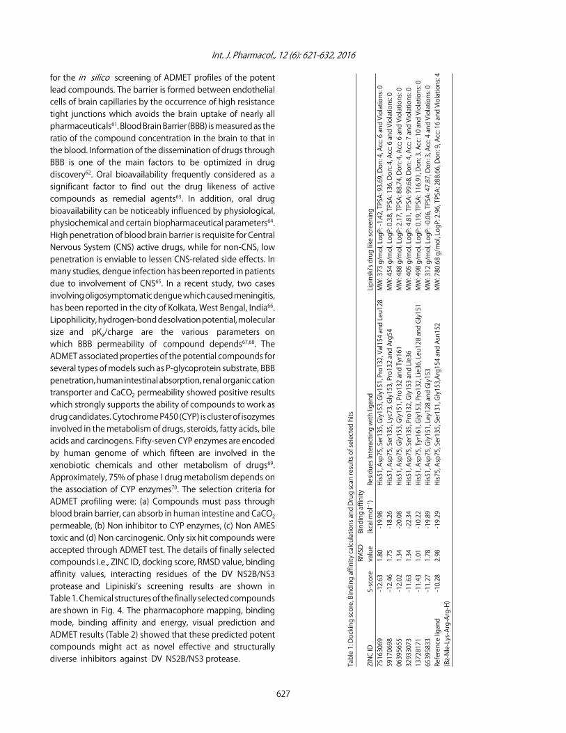
for the in silico screening of ADMET profiles of the potentlead compounds. The barrier is formed between endothelialcells of brain capillaries by the occurrence of high resistancetight junctions which avoids the brain uptake of nearly allpharmaceuticals61. Blood Brain Barrier (BBB) is measured as theratio of the compound concentration in the brain to that inthe blood. Information of the dissemination of drugs throughBBB is one of the main factors to be optimized in drugdiscovery62. Oral bioavailability frequently considered as asignificant factor to find out the drug likeness of activecompounds as remedial agents63. In addition, oral drugbioavailability can be noticeably influenced by physiological,physiochemical and certain biopharmaceutical parameters64.High penetration of blood brain barrier is requisite for CentralNervous System (CNS) active drugs, while for non-CNS, lowpenetration is enviable to lessen CNS-related side effects. Inmany studies, dengue infection has been reported in patientsdue to involvement of CNS65. In a recent study, two casesinvolving oligosymptomatic dengue which caused meningitis,has been reported in the city of Kolkata, West Bengal, India66.Lipophilicity, hydrogen-bond desolvation potential, molecularsize and pKa/charge are the various parameters onwhich BBB permeability of compound depends67,68. TheADMET associated properties of the potential compounds forseveral types of models such as P-glycoprotein substrate, BBBpenetration, human intestinal absorption, renal organic cationtransporter and CaCO2 permeability showed positive resultswhich strongly supports the ability of compounds to work asdrug candidates. Cytochrome P450 (CYP) is cluster of isozymesinvolved in the metabolism of drugs, steroids, fatty acids, bileacids and carcinogens. Fifty-seven CYP enzymes are encodedby human genome of which fifteen are involved in thexenobiotic chemicals and other metabolism of drugs69.Approximately, 75% of phase I drug metabolism depends onthe association of CYP enzymes70. The selection criteria forADMET profiling were: (a) Compounds must pass throughblood brain barrier, can absorb in human intestine and CaCO2permeable, (b) Non inhibitor to CYP enzymes, (c) Non AMEStoxic and (d) Non carcinogenic. Only six hit compounds wereaccepted through ADMET test. The details of finally selectedcompounds i.e., ZINC ID, docking score, RMSD value, bindingaffinity values, interacting residues of the DV NS2B/NS3protease and Lipiniski’s screening results are shown inTable 1. Chemical structures of the finally selected compoundsare shown in Fig. 4. The pharmacophore mapping, bindingmode, binding affinity and energy, visual prediction andADMET results (Table 2) showed that these predicted potentcompounds might act as novel effective and structurally diverse inhibitors against DV NS2B/NS3 protease.
627
Table 1: Docking score, Binding affinity calculations and Drug scan results of selected hits
RMSD
Binding affinity
ZINC ID
S-score
value
(kcal molG1)
Residues Interacting with ligand
Lipinski’s drug like screening
75163069
-12.63
1.80
-19.98
His51, Asp75, Ser135, Gly153, Gly151, Pro132, Val154 and Leu128
MW: 373 g/mol, LogP: -1.42, TPSA: 93.69, Don: 4, Acc: 6 and Violations: 0
59170698
-12.46
1.75
-18.26
His51, Asp75, Ser135, Lyc73, Gly153, Pro132 and Arg54
MW: 454 g/mol, LogP: 0.38, TPSA: 136, Don: 4, Acc: 6 and Violations: 0
06395655
-12.02
1.34
-20.08
His51, Asp75, Gly153, Gly151, Pro132 and Tyr161
MW: 488 g/mol, LogP: 2.17, TPSA: 88.74, Don: 4, Acc: 6 and Violations: 0
32933073
-11.63
1.34
-22.34
His51, Asp75, Ser135, Pro132, Gly153 and Lie36
MW: 405 g/mol, LogP: 4.81, TPSA: 99.68, Don: 4, Acc: 7 and Violations: 0
13728171
-11.43
1.01
-10.22
His51, Asp75, Tyr161, Gly153, Pro132, Lie36, Leu128 and Gly151
MW: 498 g/mol, LogP: 0.19, TPSA: 116.91, Don: 3, Acc: 10 and Violations: 0
65395833
-11.27
1.78
-19.89
His51, Asp75, Gly151, Ley128 and Gly153
MW: 312 g/mol, LogP: -0.06, TPSA: 47.87, Don: 3, Acc: 4 and Violations: 0
Reference ligand
-10.28
2.98
-19.29
His75, Asp75, Ser135, Ser131, Gly153,Arg154 and Asn152
MW: 780.68 g/mol, LogP: 2.96, TPSA: 288.66, Don: 9, Acc: 16 and Violations: 4
(Bz-Nle-Lys-Arg-Arg-H)

Int. J. Pharmacol., 12 (6): 621-632, 2016
59170698
OH
OH
BrMeO
NH
NH
CI
N
06395655
HN+
65395833
N+H
Ma
HO
MaO
HN
13728171
CMa
MaO
Ma
O
HN N
NN
HN+
O
HN
32933073
HN
O
Ma
HN
HO
O
CMa
Ma
Fig. 4: 2D structures of finally retrieved hits from ZINC database. The numbers are representing their ZINC database IDs
Interaction analysis of finally selected hit compounds:The S-score is the mathematical method used to calculatethe strength of interaction between receptor protein andligand after they have been docked27. On the basis of S-score,binding energy and affinity, it is possible to conclude thatthese finally selected compounds have potential interactionwith catalytic triad residues and fulfill the requirements to
be drug-able. The compound having ZINC ID 75163069was placed at the top as it had potential interactionswith His51, Asp75, Ser135, Gly153, Gly151, Pro132, Val154and Leu128 residues of binding pocket and bears bestdocking score. Docking conformations and pharmacophoremapping of top three selected compounds are given inFig. 5.
628

Int. J. Pharmacol., 12 (6): 621-632, 2016
Table 2: ADMET profiling results for selected hit compoundsZINC ID Blood-brain barrier Human intestinal absorption CaCO2 permeability P-glycoprotein inhibitor Renal organic cation transporterAbsorption75163069 BBB+ HIA+ CaCO2- NI T59170698 BBB- HIA- CaCO2- NI T06395655 BBB+ HIA+ CaCO2+ NI T32933073 BBB+ HIA+ CaCO2- NI T13728171 BBB- HIA+ CaCO2- NI T65395833 BBB+ HIA+ CaCO2- NI TZINC ID CYP450 1A2 inhibitor CYP450 2C9 inhibitor CYP450 2D6 inhibitor CYP450 2C19 inhibitor CYP450 3A4 inhibitorMetabolism75163069 NI NI NI NI NI59170698 NI NI NI NI NI06395655 I NI NI I I32933073 NI NI NI NI NI13728171 NI I NI NI I65395833 NI NI NI NI NIZINC ID AMES toxicity CarcinogensToxicity75163069 Non AMES Toxic Non carcinogenic59170698 Non AMES Toxic Non carcinogenic06395655 Non AMES Toxic Non carcinogenic32933073 Non AMES Toxic Non carcinogenic13728171 Non AMES Toxic Non carcinogenic65395833 Non AMES Toxic Non carcinogenicI: Inhibitor, NI: Non inhibitor
Fig. 5(a-c): Docking conformations and pharmacophore mapping of selected hits, (a) A1, B1 and C1 represent 3D interactionsof selected compounds 75163069, 59170698 and 06395655, respectively with DV NS2B/NS3 protease, (b) A2, B2 andC2 represent pharmacophore mapping of selected compounds 75163069, 59170698 and 06395655, respectively,While (c) A3, B3 and C3 represent binding pocket mode of selected compounds 75163069, 59170698 and 06395655,respectively with DV NS2B/NS3 protease
629
A1 A2 A3
B1 B2 B3
C1 C2 C3
(a)
(b)
(c)

Int. J. Pharmacol., 12 (6): 621-632, 2016
CONCLUSION
The present study focuses on structure basedpharmacophore modeling, computational screening andmolecular docking of ZINC database compounds againstDengue Virus (DV) NS2B/NS3 protease. As an outcome ofthe study, six compounds from ZINC database (ZINC IDs:75163069, 59170698, 06395655, 32933073, 13728171 and65395833) have shown strong bindings with catalytic triad ofDV NS2B/NS3 protease. This study also explores that thesecompounds can be utilized as potential and strong drugcandidates against DV NS2B/NS3 protease on the basis ofdrug profiling. The findings will be useful as they provideinsight for the effectiveness of drug before its manufacturingand testing on pilot scale in the pharmaceutical industry(for in vivo drug design and development). Hence, it can beconcluded that in future these compounds can serve as astrong and potential drug leads against DV on the basis ofsignificant binding affinity against DV NS2B/NS3 protease.
REFERENCES
1. Cabarcas-Montalvo, M., W. Maldonado-Rojas,D. Montes-Grajales, A. Bertel-Sevilla andI. Wagner-Dobler et al., 2016. Discovery of antiviral moleculesfor dengue: In silico search and biological evaluation. Eur.J. Med. Chem., 110: 87-97.
2. Guzman, M.G. and G. Kouri, 2002. Dengue: An update. LancetInfect. Dis., 2: 33-42.
3. Idrees, S. and U.A. Ashfaq, 2012. A brief review ondengue molecular virology, diagnosis, treatment andprevalence in Pakistan. Genet. Vaccines Ther.,Vol. 10. 10.1186/1479-0556-10-6
4. Tahir ul Qamar, M., A. Mumtaz, U.A. Ashfaq, M.M. Adeel andT. Fatima, 2014. Potential of plant alkaloids as dengue NS3protease inhibitors: Molecular docking and simulationapproach. Bangladesh J. Pharmacol., 9: 262-267.
5. Gibbons, R.V. and D.W. Vaughn, 2002. Dengue: An escalatingproblem. Br. Med. J., 324: 1563-1566.
6. Das, S., M.R. Pingle, J. Munoz-Jordon, M.S. Rundell andS. Rondini et al., 2008. Detection and serotyping of denguevirus in serum samples by multiplex reverse transcriptasePCR-ligase detection reaction assay. J. Clin. Microbiol.,46: 3276-3284.
7. Weaver, S.C. and N. Vasilakis, 2009. Molecular evolutionof dengue viruses: Contributions of phylogenetics tounderstanding the history and epidemiology of thepreeminent arboviral disease. Infect. Genet. Evol., 9: 523-540.
8. Akey, D.L., W.C. Brown, S. Dutta, J. Konwerski and J. Jose et al.,2014. Flavivirus NS1 structures reveal surfaces for associationswith membranes and the immune system. Science,343: 881-885.
9. Thomas, S.J., D. Strickman and D.W. Vaughn, 2003. Dengueepidemiology: Virus epidemiology, ecology and emergence.Adv. Virus Res., 61: 235-289.
10. Munoz-Jordan, J.L., G.G. Sanchez-Burgos, M. Laurent-Rolleand A. Garcia-Sastre, 2003. Inhibition of interferon signalingby dengue virus. Proc. Natl. Acad. Sci. USA., 100: 14333-14338.
11. Umareddy, I., O. Pluquet, Q.Y. Wang, S.G. Vasudevan,E. Chevet and F. Gu, 2007. Dengue virus serotype infectionspecifies the activation of the unfolded protein response.Virol. J., Vol. 4. 10.1186/1743-422X-4-91
12. Beltramello, M., K.L. Williams, C.P. Simmons, A. Macagnoand L. S imonelli et al., 2010. The human immune responseto Dengue virus is dominated by highly cross-reactiveantibodies endowed with neutralizing and enhancingactivity. Cell Host Microbe, 8: 271-283.
13. Hung, S.L., P.L. Lee, H.W. Chen, L.K. Chen, C.L. Kao andC.C. King, 1999. Analysis of the steps involved in dengue virusentry into host cells. Virology, 257: 156-167.
14. Rothan, H.A., H.C. Han, T.S. Ramasamy, S. Othman,N.A. Rahman and R. Yusof, 2012. Inhibition of dengueNS2B-NS3 protease and viral replication in Vero cellsby recombinant retrocyclin-1. BMC Infect. Dis.,Vol. 12. 10.1186/1471-2334-12-314
15. Lyne, P.D., 2002. Structure-based virtual screening: Anoverview. Drug Discov. Today, 7: 1047-1055.
16. Debnath, A.K., 2003. Generation of predictive pharmacophoremodels for CCR5 antagonists: Study with piperidine- andpiperazine-based compounds as a new class of HIV-1 entryinhibitors. J. Med. Chem., 46: 4501-4515.
17. Wei, J., S. Wang, S. Gao, X. Dai and Q. Gao, 2007.3D-pharmacophore models for selective A2A and A2Badenosine receptor antagonists. J. Chem. Inform. Model.,47: 613-625.
18. Ma, D.L., D.S.H. Chan and C.H. Leung, 2013. Drugrepositioning by structure-based virtual screening. Chem.Soc. Rev., 42: 2130-2141.
19. Lengauer, T. and M. Rarey, 1996. Computational methods forbiomolecular docking. Curr. Opin. Struct. Biol., 6: 402-406.
20. Irwin, J.J., T. Sterling, M.M. Mysinger, E.S. Bolstad andR.G. Coleman, 2012. ZINC: A free tool to discover chemistry forbiology. J. Chem. Inform. Model., 52: 1757-1768.
21. Awale, M. and J.L. Reymond, 2014. A multi-fingerprintbrowser for the ZINC database. Nucl. Acids Res.,42: W234-W239.
22. Zhong, Z., L.J. Liu, Z.Q. Dong, L. Lu and M. Wang et al., 2015.Structure-based discovery of an immunomodulatory inhibitorof TLR1-TLR2 heterodimerization from a natural product-likedatabase. Chem. Commun., 51: 11178-11181.
23. Liu, L.J., K.H. Leung, D.S. Chan, Y.T. Wang, D.L. Ma andC.H. Leung, 2014. Identification of a natural product-likeSTAT3 dimerization inhibitor by structure-based virtualscreening. Cell Death Dis., Vol. 5. 10.1038/cddis.2014.250.
630

Int. J. Pharmacol., 12 (6): 621-632, 2016
24. Ma, D.L., D.S.H. Chan, G. Wei, H.J. Zhong and H. Yang et al.,2014. Virtual screening and optimization of type II inhibitorsof JAK2 from a natural product library. Chem. Commun.,50: 13885-13888.
25. Mugumbate, G., A.S. Newton, P.J. Rosenthal, J. Gut, R. Moreira,K. Chibale and R.C. Guedes, 2013. Novel anti-Plasmodialhits identified by virtual screening of the ZINC database.J. Comput.-Aided Mol. Des., 27: 859-871.
26. Leung, C.H., D.S.H. Chan, H. Yang, R. Abagyan andS.M.Y. Lee et al., 2011. A natural product-like inhibitor ofNEDD8-activating enzyme. Chem. Commun., 47: 2511-2513.
27. Vilar, S., G. Cozza and S. Moro, 2008. Medicinal chemistry andthe molecular operating environment (MOE): Application ofQSAR and molecular docking to drug discovery. Curr. Top.Med. Chem., 8: 1555-1572.
28. Yang, S.Y., 2010. Pharmacophore modeling and applicationsin drug discovery: Challenges and recent advances. DrugDiscov. Today, 15: 444-450.
29. Wadood, A., M. Riaz, R. Uddin and Zaheer-ul-Haq, 2014.In silico identification and evaluation of leads for thesimultaneous inhibition of protease and helicase activitiesof HCV NS3/4A protease using complex basedpharmacophore mapping and virtual screening. PLoS One,Vol. 9. 10.1371/journal.pone.0089109.
30. Erbel, P., N. Schiering, A. D'Arcy, M. Renatus and M. Kroemeret al., 2006. Structural basis for the activation of flaviviralNS3 proteases from dengue and West Nile virus. Nat. Struct.Mol. Biol., 13: 372-373.
31. Tahir ul Qamar, A. Mumtaz, U.A. Ashfaq, S. Azhar andT. Fatima et al., 2014. Computer aided screening ofphytochemicals from garcinia against the dengue NS2B/NS3protease. Bioinformation, 10: 115-118.
32. Ashfaq, U.A., A. Mumtaz, Tahir ul Qamar and T. Fatima, 2013.MAPS database: Medicinal plant activities, phytochemical andstructural database. Bioinformation, 9: 993-995.
33. Kurogi, Y. and O.F. Guner, 2001. Pharmacophore modelingand three-dimensional database searching for drug designusing catalyst. Curr. Med. Chem., 8: 1035-1055.
34. Cheng, F., W. Li, Y. Zhou, J. Shen and Z. Wu et al., 2012.admetSAR: A comprehensive source and free tool forassessment of chemical ADMET properties. J. Chem. Inform.Model., 52: 3099-3105.
35. Jarrahpour, A., J. Fathi, M. Mimouni, T.B. Hadda, J. Sheikh,Z. Chohan and A. Parvez, 2012. Petra, Osiris andMolinspiration (POM) together as a successful support indrug design: Antibacterial activity and biopharmaceuticalcharacterization of some azo Schiff bases. Med. Chem. Res.,21: 1984-1990.
36. Labute, P., 2008. The generalized Born/volume integralimplicit solvent model: Estimation of the free energy ofhydration using London dispersion instead of atomic surfacearea. J. Comput. Chem., 29: 1693-1698.
37. Lescar, J., D. Luo, T. Xu, A. Sampath, S.P. Lim, B. Canard andS.G. Vasudevan, 2008. Towards the design of antiviralinhibitors against flaviviruses: The case for the multifunctionalNS3 protein from dengue virus as a target. Antiviral Res.,80: 94-101.
38. Noble, C.G., Y.L. Chen, H. Dong, F. Gu and S.P. Lim et al., 2010.Strategies for development of dengue virus inhibitors.Antiviral Res., 85: 450-462.
39. Heh, C.H., R. Othman, M.J.C. Buckle, Y. Sharifuddin, R. Yusofand N.A. Rahman, 2013. Rational discovery of dengue type 2non-competitive inhibitors. Chem. Biol. Drug Des., 82: 1-11.
40. Tomlinson, S.M., R.D. Malmstrom, A. Russo, N. Mueller,Y.P. Pang and S.J. Watowich, 2009. Structure-based discoveryof dengue virus protease inhibitors. Antiviral Res.,82: 110-114.
41. Tomlinson, S.M. and S.J. Watowich, 2011. Anthracene-basedinhibitors of dengue virus NS2B-NS3 protease. Antiviral Res.,89: 127-135.
42. Knehans, T., A. Schuller, D.N. Doan, K. Nacro and J. Hill et al.,2011. Structure-guided fragment-based in silico drug designof dengue protease inhibitors. J. Comput.-Aided Mol. Des.,25: 263-274.
43. Lim, S.V., M.B.A. Rahman and B.A. Tejo, 2011. Structure-basedand ligand-based virtual screening of novelmethyltransferase inhibitors of the dengue virus. BMCBioinform., Vol. 12. 10.1186/1471-2105-12-S13-S24.
44. Mishra, V., S. Kashyap and Y. Hasija, 2015. Ligand based virtualscreening for identifying potent inhibitors against viralneuraminidase: An in silico approach. J. Taibah Univ. Sci.,9: 20-26.
45. Gao, Y., T. Cui and Y. Lam, 2010. Synthesis and disulfidebond connectivity-activity studies of a kalata B1-inspiredcyclopeptide against dengue NS2B-NS3 protease. Bioorg.Med. Chem., 18: 1331-1336.
46. Yin, Z., S.J. Patel, W.L. Wang, W.L. Chan andK.R. Ranga Rao et al., 2006. Peptide inhibitors of denguevirus NS3 protease. Part 2: SAR study of tetrapeptide aldehydeinhibitors. Bioorg. Med. Chem. Lett., 16: 40-43.
47. Yin, Z., S.J. Patel, W.L. Wang, G. Wang and W.L. Chan et al.,2006. Peptide inhibitors of dengue virus NS3 protease. Part 1:Warhead. Bioorg. Med. Chem. Lett., 16: 36-39.
48. Chanprapaph, S., P. Saparpakorn, C. Sangma,P. Niyomrattanakit, S. Hannongbua, C. Angsuthanasombatand G. Katzenmeier, 2005. Competitive inhibition of thedengue virus NS3 serine protease by synthetic peptidesrepresenting polyprotein cleavage sites. Biochem. Biophys.Res. Commun., 330: 1237-1246.
49. Deng, J., N. Li, H. Liu, Z. Zuo and O.W. Liew et al., 2012.Discovery of novel small molecule inhibitors of dengue viralNS2B-NS3 protease using virtual screening and scaffoldhopping. J. Med. Chem., 55: 6278-6293.
631

Int. J. Pharmacol., 12 (6): 621-632, 2016
50. Kiat, T.S., R. Pippen, R. Yusof, H. Ibrahim, N. Khalid andN. Abd Rahman, 2006. Inhibitory activity of cyclohexenylchalcone derivatives and flavonoids of fingerroot,Boesenbergia rotunda (L.), towards dengue-2 virus NS3protease. Bioorg. Med. Chem. Lett., 16: 3337-3340.
51. Hidari, K.I.P.J., N. Takahashi, M. Arihara, M. Nagaoka, K. Moritaand T. Suzuki, 2008. Structure and anti-dengue virus activityof sulfated polysaccharide from a marine alga. Biochem.Biophys. Res. Commun., 376: 91-95.
52. Frimayanti, N., C. Chee, S.M. Zain and N.A. Rahman, 2011.Design of new competitive dengue Ns2b/Ns3 proteaseinhibitors-a computational approach. Int. J. Mol. Sci.,12: 1089-1100.
53. Yang, C.C., Y.C. Hsieh, S.J. Lee, S.H. Wu and C.L. Liao et al.,2011. Novel dengue virus-specific NS2B/NS3 proteaseinhibitor, BP2109, discovered by a high-throughput screeningassay. Antimicrobial Agents Chemother., 55: 229-238.
54. Velmurugan, D., U. Mythily and K. Rao, 2014. Design anddocking studies of peptide inhibitors as potential antiviraldrugs for dengue virus Ns2b/Ns3 protease. Protein Pept.Lett., 21: 815-827.
55. Lima, A.B., M.A. Behnam, Y. El Sherif, C. Nitsche, S.M. Vechi andC.D. Klein, 2015. Dual inhibitors of the dengue and West Nilevirus NS2B-NS3 proteases: Synthesis, biological evaluationand docking studies of novel peptide-hybrids. Bioorg. Med.Chem., 23: 5748-5755.
56. Idrees, S. and U.A. Ashfaq, 2014. Discovery and designof cyclic peptides as dengue virus inhibitors throughstructure-based molecular docking. Asian Pac. J. Trop. Med.,7: 513-516.
57. Vernekar, S.K.V., L. Qiu, J. Zhang, J. Kankanala, H. Li,R.J. Geraghty and Z. Wang, 2015. 5'-Silylated 3'-1,2,3-triazolylthymidine analogues as inhibitors of West Nile virus anddengue virus. J. Med. Chem., 58: 4016-4028.
58. Steuer, C., C. Gege, W. Fischl, K.H. Heinonen, R. Bartenschlagerand C.D. Klein, 2011. Synthesis and biological evaluation ofα-ketoamides as inhibitors of the Dengue virus proteasewith antiviral activity in cell-culture. Bioorg. Med. Chem.,19: 4067-4074.
59. Lipinski, C.A., F. Lombardo, B.W. Dominy and P.J. Feeney,2012. Experimental and computational approaches toestimate solubility and permeability in drug discovery anddevelopment settings. Adv. Drug Deliv. Rev., 64: 4-17.
60. Ma, D.L., D.S.H. Chan and C.H. Leung, 2011. Molecular dockingfor virtual screening of natural product databases. Chem. Sci.,2: 1656-1665.
61. Stamatovic, S.M., R.F. Keep and A.V. Andjelkovic, 2008. Brainendothelial cell-cell junctions: How to open the blood brainbarrier. Cur. Neuropharmacol., 6: 179-192.
62. Alavijeh, M.S., M. Chishty, M.Z. Qaiser and A.M. Palmer, 2005.Drug metabolism and pharmacokinetics, the blood-brainbarrier and central nervous system drug discovery. NeuroRX,2: 554-571.
63. Thomas, V.H., S. Bhattachar, L. Hitchingham, P. Zocharski andM. Naath et al., 2006. The road map to oral bioavailability:An industrial perspective. Expert Opin. Drug Metab. Toxicol.,2: 591-608.
64. Varma, M.V.S., R.S. Obach, C. Rotter, H.R. Miller andG. Chang et al., 2010. Physicochemical space for optimumoral bioavailability: Contribution of human intestinalabsorption and first-pass elimination. J. Med. Chem.,53: 1098-1108.
65. Araujo, F., R. Nogueira, M.S. Araujo, A. Perdigao andL. Cavalcanti et al., 2012. Dengue in patients with centralnervous system manifestations, Brazil. Emerg. Infect. Dis.,18: 677-679.
66. Goswami, R.P., A. Mukherjee, T. Biswas, P.S. Karmakar andA. Ghosh, 2012. Two cases of dengue meningitis: A rare firstpresentation. J. Infect. Dev. Countries, 6: 208-211.
67. Abraham, M.H., 2004. The factors that influence permeationacross the blood-brain barrier. Eur. J. Med. Chem.,39: 235-240.
68. Goodwin, J.T. and E.C. David, 2005. In silico predictions ofblood-brain barrier penetration: Considerations to keep inmind. J. Pharmacol. Exp. Ther., 315: 477-483.
69. Guengerich, F.P., 2003. Cytochromes P450, drugs anddiseases. Mol. Interv., 3: 194-204.
70. Bibi, Z., 2008. Role of cytochrome P450 in drug interactions.Nutr. Metab., Vol. 5. 10.1186/1743-7075-5-27.
632