disease^.^ - sljol
TRANSCRIPT

J. Nattt. Sci. Cowr. Sri Larrka 1993 21(1) : 49-62
THE EFFECTS OF COLLETOTRlCHUM LINDEMUTHIANUM PECTIN LYASE ON BEAN TISSUE
R.L.C. WIJESUNDERA Department of Botany, Universily of Colotnbo, Colombo 3.
(Date of receipt : 10 December 1992) (Date of acceptance : 1.5 March 1993)
Abslrack A homogeneous preparation of pectin lyase ( PL 11, isoelectric point.9.7 ) from Collelolrichum Iindernuthlnnum, caused the leakage of potassium ions from hypocotyl tissue discs of Phascolus vulgaris. The enzyme preparation also released unsaturated uronic acids from pectin,.isolaled cell walls and hypocotyl tissue discs. Analysis of degradation products shows that PL I1 is an endo-acting enzyme.
Key words: Collctotrlchum Iindemulhlanum. Phnseollis vulgaris, Pectin lyase
INTRODUCTION
Colletotrickunt lindetnuthiatrwl~ (Sacc et Magn) Bri. d %av. affects the above ground parts of its host Pl~aseolus vulgaris, the common bean. Invasion and growth of the fungus result in anthracnose disease in which large, water soaked necrotic lesions are produced.
C. Lrdemutl~iaratm in culture produces pectic enzymes. These are, two forms of pectin lyase having isoelectric points of 8.2 ( PL 1 ) and 9.7 ( PL I1 ) and a single form of polygalacturonase having a pI of 9.3.1i2 However, in P.vlrlgaris hypocotyl tissue infected with the fungus; PL I1 is the only pectic enzyme Pectic enzymes are known to be toxic to host cells and they play a major role in the development of necrotic symptoms in. many disease^.^ It is possible that the PL I1 of C. litrdemutl~iari~trn also plays a similar role in bean anthracnose disea~e.~ This paper describes the purification of PI. 11 from C. li~~dc~ttrriltiarrirnt and the effect of the purified enzyme on host cells..
METHODS AND hlATERJALS
Growth of the fungus: Race 0 of C. li~tder?t~rtlriartrm~ was grown in an ammonium tartrate medium5 with sodium polypectate (Sigma, St. huis , USA) as the main source of carbon. The medium was buffered with 0.1M NN' - Bis - (-2-hydroxyethy1)-2 amino ethane sulphonic acid (BDH, ~ o i d o n ) and the pH adjusted to 6.5 with 0.1M NaOH. Twenty five ml of the medium was dispensed into 200 ml medicine bottles and autoclaved. Inoculation and incbbation was performed as described by -. - -
Ahbrevinlions - PL : Pectin $me, PC : Polygnlacturonnsc, pl : Isoelectric poinl, TBA : Thiobarbiluric acid.

50 R.L.C. Wijesundera
Wijesundera et al.' Cultures were harvested 15 d after inoculatioh by filtration through Whatman no 1 filter paper. The culture filtrates were dialysed (using Spectrapor-2 membrane tubing) at 4OC for 24 h against distilled water and concentrated by freeze drying.
Ion-exchange chromatography: CM-Sephadex C -50 (Pharmacia, Sweden ) was used for ion-exchange chromatography. The gel, equilibrated in 0.1M sodium acetate buffer, pH 4.5, was packed into a 16.0 x 2.0 cm2 column according to the manufacturer's instructions.
The dry culture filtrate, re-dissolved in 15 ml of 0.1M sodium acetate buffer, pH 4.5, was applied to the gel and the gel was sequentially eluted with 50 ml of different 0.1M sodium acetate buffers of pH 4.5,5.0,55,6.0,6.5 and 7.0 respectively. This was followed by a final wash with 50 ml of 0.1M sodium acetate, pH 7.0 containing 0.3M NaCI. Ten ml fractions were collected using gravity flow and assayed for enzyme activity. Protein content of the fractions were determined as described below.
Isoelectric focusing: Isoelectric focusing was carried out in an LKB 110 ml column using either Ampholine (LKB, Sweden) of pI range 3.5-10.5 or Pharmalyte (Pharmacia, Sweden) of pI range 7.5-10.5 as-the carrier ampholytes in a linear sucrose gradient at 10'~ for 24 h. The column was eluted using a peristaltic pump at 120 ml h" and 3.0 ml fractions were collected. The pH of the fractions was measured immediately.
Isoelectric focusing was also carried out using pre-coated Servalyte precotes (Serva Finebiochemica GMBH, Heidelberg, Germany) as instructed by the manufacturers. The proteins were fured by placing the precotes in a 3% solution of perchloric acid for 30 min to 1 h. To stain the fmed proteins the precotes were then placed in a solution of Coomassie blue G-250 (BDH, London) in 5% perchloric acid for 24 h. This was followed by the removal of excess stain by immersing the gel in a running water bath for 24 h. The washed precotes were dried at room temperature. The following proteins were used as markers: Cytochrome-c (horse heart) pI 10.8; Myoglobin (equine) pI 7.3 and carbonic anhydrase, p16.5 (Sigma, St.Louis, USA).
Growth of seedlings of P. 'v~llgaris and preparation of cell walls: Seedlings of P. ntlgaris cv Kievitsboon Koekoek were grown as described by Wijesundera et al.' Cell walls were prepared from hypocotyls of seedlings that were harvested 10 d after sowing.'
.Determination of cellular permeability changes: Twenty 1.0 mm thick hypocotyl discs (approximately 0.05gl disc) obtained from 7-day old P. vulgaris seedlings were used.in each experiment. Before use,'the hypocotyl discs were rinsed in the bathing solution

Effect of Pectin Lyase on Bean Tissue 51
for 1 min. Leakage of K+ from the hypocotyl discs was used to detect permeability alterations.
The bathing solution used to measure K+ was 0.01M Tris-HCI buffer, pH 8.5. The rinsed discs were transferred to the test solution which included the enzyme sample in 8.0 ml Tris-HC1 buffer. The concentration of the bathing solution was measured soon after the addition of the discs and at specified time intervals using a flame photometer (Model lA, Evans Electro Selenium Ltd., U.K.). Other discs were treated with an enzyme sample inactivated by boiling for 10 min. All experiments were conducted at 20'~. At the end of the study period the discs together with the bathing solutions were boiled for 5 min and after cooling the concentration was measured. This value was taken as the total K+ concentration, and results are expressed as a percentage of the total K+ concentration.
Purification of pectin: A method described by ~ r c h e r ~ was used to purify pectin. Pectin (H.P.Bulmer, Hereford, UK) was washed several times with acidified (0.1M HCl) 80% vlv ethanol until the filtrate was free of pigments and reducing sugars. els son's' method was used to test for reducing sugars. After two more washings with ethanol the residue was collected by suction filltration, and dried using absolute ethano1:ether (1:l v/v). Traces of solvent were removed by storing in a vacuum desiccator followed by heating to 60'~. The resulting solid was ground to a fine powder and stored at 20'~.
Action of PL 11 on different substrates: Action of purified PL I1 preparation on three different substrates - purified pectin (0.002g), isolated hypocotyl cell walls of P. vulgaris (0.05 g), hypocotyl tissue discs of P. vulgaris (20-discs, each 1.0 mm thick) were tested. The tissue discs were from 7-day old seedlings and were washed for 1 h in 8.0 ml of the buffer used in the experiment. During washing the buffer was changed every 20 min.
Purified enzyme preparation' (2.5 units) in 8.0 ml of 0.1M Tris-HC1 buffer, pH 8.5 containing the antibiotic gentamycin sulphate (50 pg ml" Sigma, St. Louis, USA) and 0.01M CaC12 was used to treat each sample. Boiled inactivated enzyme was used as the control.
The reaction mixtures were incubated in a reciprocal shaker (100 strokes rnin", each stroke 1.1 cm) at 25'~. Samples were taken immediately after the addition of the enzyme and at intervals thereafter. The samples were placed immediately in a boiling water bath for 20 min to terminate enzyme activity and the cooled samples were examined for reducing sugars,' uronic acids? and 4-5 unsaturated uronic acids by the thiobarbituric acid method9 and by absorbance at 240 nm! Activities are expressed as arbitrary units based on the absorbance after necessary corrections for substrate
52 R.L. C. Wijesz~ndera
blanks. The cooled samples were also subjected to both thin layer and paper chromatography as described below.
Enzyme assays: Pectin lyase (PL) activity was measured spectrophoto- metrically by the change in absorbance at 240 nm? The standard reaction mixture comprized 3.0 ml0.25% citrus pectin in 0.1M Tris-HC1 buffer of appropriate pH and 0.1 ml of enzyme sample. The absorbance was read immediately afterwards'and after an incubation period at 30°C. One unit of enzyme activity is defined as that amount which produce 1 C( mol unsaturated uronic acid in 1 min based on the molar absorption for the product of 4600. PL activity was also examined using the TBA method.' After specified reaction periods, 1.0 ml samples of the reaction mixture described above were added to 6.0 ml TBA reagent (5.0 ml0.01M TBA and 0.1 ml1M HCI) and kept in a boiling water bath for 60 min. After cooling the absorption was read at 550 nm.
Polygalacturonase activity was measured by the cup-plate method and the viscometric methods described in Wijesundera et d. In the cup-plate- method a sodium polypectate agar gel (Sigma, St.Louis, .USA) was used. Activities are expressed relative to an aqueous solution -(1 mg m1-l) of Pectin01 10M which was defined as having 100 units of PG activity ml-l.
Protease: Protease activity was determined using hide-powder azure blue as the ~ubstrate.~ The reaction mixture had 10 g hide powder azure blue (Sigma, St-Louis, U.S.A.), 5.0 ml Tris-HC1 buffer of pH 8.5 and 0.5 ml of the enzyme sample in 25 ml plastic bottles. Incubation was at 37OC. After incubation for a specified period the reaction was terminated by filtration through Whatman no 41 filter paper. Absorbance of the filtrate was measured' immediately at 595 nm. Necessary corrections were made for substrate blanks.
Estimation of proteins: Protein estimation was based on a dye-binding method?' Bovine-serum albumin (Sigma, St. Louis, USA) was used as the reference standard; To 0.8 ml standards and approximately diluted sample, 0.2.ml of protein dye reagent (Bio-Rad, Germany) diluted as instructed by the manufacturers was added. The absorbance was measured at 595 nm after 30 min incubation at 25OC.
Thin layer' and p a p r chromatography: Awending thin layer chromatography on DF-Fertigplatten Cellulose-F plates (20 x 20 x 0.01 cm3, Merck) developed in.butano1: formic a@ 2 3 (vlv) for 4-5 h. Descending paper chromatography on Whatman no.1 .paper, developed in butanol: acetic acid : water 21:l (vlvlv) for 12-16 h.
The chromatogra@hs werc developed at room temperat'ure @.tanks &uilibrated with respective solvents. Reducing groups were revealed as pink brown spots by

Effect of Pectin Lyase on Bean Tisue 53
spraying with aniline phthalate reagent (0.93 g aniline and 1.66 g phthalate reagent in 100 ml of 1-butanol saturated with water) followed by heating to 100 '~ for 15-30 min. Unsaturated compounds appeared pink when sprayed with a saturated aqueous solution of thiobarbituric acid.''
In all thin layer and paper chromatography experiments polygalacturonic acid; galacturonic acid (Sigma, St.Louis, U.S.A.) and 4- 5 unsaturated digalacturonic acid was used as reference standards. The 4-5 unsaturated digalacturonic acid was prepared according to the method described by Nagel & vaughn.12
RESULTS
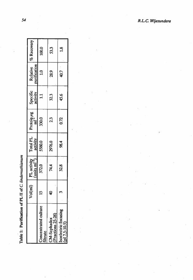
Purification of PL 11: When the fungus was gown in sodium polypectate medium buffered at pH 6.5, the only pectic enzymes detected were the two lyases (PL I and PL 11). No PG activity was detected. The concentrate of the culture filtrate in sodium acetate buffer of pH 4.5, when subjected to ion-exchange chromatography gave the elution pattern shown in Figure 1. The PL resolved into two peaks, peak 1 corresponding to PL I and peak I1 to PL 11. A peak of protease activity occurred just after the highest PL I1 activity, but the fractions having the highest PL activity were devoid of protease activity (Figure 1). The Fractions 25-28 were bulked, dialysed (using Spectrapor-2 membrane tubing) and concentrated by freeze drying. On sewalyte precotes this PL preparation gave only one band when stained for proteins. The position of the protein band indicated a pI value corresponding to the pI of PL 11.
. .
The PL I1 prepared from ion-exchange .chromatography on narrow range isoelectric focussing yielded a well defined peak of pI 9.7. This preparation is referred to as purified PL 11. Details of the purification are given in Table 1.
Effect of PL 11 on permeability of cells: The purified PL IT preparation caused'.a leakage of K+ from tissue discs, and the amount and the rate of the ions leaked increased with the increase in activity of the PL I1 preparation (Figure 2). Statistical analysis of the result; confirms this observation (Tabla 2).
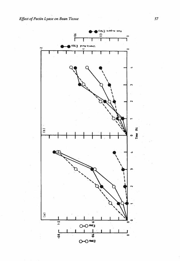
Degradation of substrates: Purified pectin, isolated cell walls and hypocotyl tissue discs when treated with 2.5 units of purified PL I1 released unsaturated uronic acids and reducing sugars (Figures 3 a, b, and c). At the end of the incubation period the hypocotyl tissue discs treated with the enzyme had lost their turgidity. In the controls where boiled, inactive PL I1 was used the release of the above compounds was not detected and the loss of turgidity was not observed.
When the products of purified PL I1 action on pectin were subjected to thin layer and paper chromatography, the presence of a series unsaturated oligomers and very
Tab
le 1
: P
urifi
catio
n of
PL
lI o
f C.
lin&
mut
hian
um
Con
cent
rate
d cu
lture
fil
trat
e C
M-S
epha
dex
(Fra
ctio
ns 25
-28)
Is
oele
ctri
c focusing
(PI
7.5-
105)
Vol
(m1)
15
40 3
PL a
ctiv
q
(uni
ts m
l- )
372.
0
74.4
32.8
Tot
al P
L
activ
ity
5580.0
2976.0
98.4
Pro
teip
pg
ml-
330.
0
23
0.72
Spec
ific
activ
ity
1.1
32.3
45.6
Rel
ativ
e pu
rific
atio
n 1.
0
28.9
40.7
% R
ecov
ery
100.0
53.3
1.8

Eflect of Pectiil Lyase ort Beail Tissue

R.L.C. Wijeszrrtdera
Efect of Pectin Lyase on Bean Tissue

R.L.C. Wijeslrndera
Eflect of Pectin Lyase on Bean 7issue 59
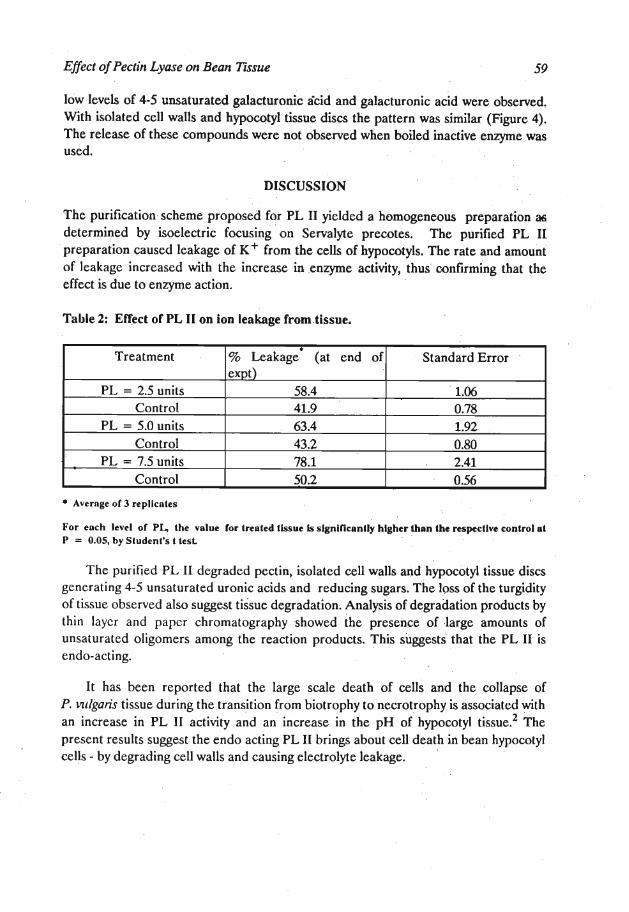
low levels of 4-5 unsaturated galacturonic k i d and galacturonic acid were observed. With isolated cell walls and hypocotyl tissue discs the pattern was similar (Figure 4). The release of these compounds were not observed when boiled inactive enzyme was used.
DISCUSSION
The purification scheme proposed for PL I1 yielded a homogeneous preparation as determined by isoelectric focusing on Servalyte precotes. The purified PL I1 preparation caused leakage of K+ from the cells of hypocotyls. The rate and amount of leakage increased with the increase in enzyme activity, thus confirming that the effect is due to enzyme action.
Table 2: Effect of PL I1 on ion leakage frorn.tissue.
Avernge of 3 replicntes
Treatment
PL = 2.5 units Control
PL = 5.0 units Control
PL = 7.5 units Control
For each level of PL, the value for treated tissue is significantly higher lhan the rtsptcllve control at P = .0.05, by Sludent's t test.
The purified PL II degraded pectin, isolated cell walls and hypocotyl tissue discs generating 4-5 unsaturated uronic acids and reducing sugars. The loss of the turgidity of tissue observed also suggest tissue degradation. Analysis of degradation products by thin layer and paper chromatography showed the presence of large amounts of unsaturated oligomers among the reaction products. This suggests that the PL I1 is endo-acting.
% ~ e a k a ~ e * (at end of expt)
58.4 41.9 63.4 43.2 78.1 50.2
It has been reported that the large scale death of cells and' the collapse of P. vlrlgaris tissue during the transition from biotrophy to necrotrophy is associated with an increase in PL I1 activity and an increase in the pH of'hypocotyl t i ~ s u e . ~ The present results suggest the endo acting PL I1 brings about cell death in bean hypocotyl cells - by degrading cell walls and causing electrolyte leakage.
Standard Error
1.06 0.78 1.92 0.80
. 2.41 0.56

R.L.C. Wijesurtdera
Figure 4: Thin layer chromatographs of reaction products from purified PL action on, i and ii washtd peelin, 111 and Iv Isolated cell walls, v and vi llssue discr
'I, 111 and h. - plaks sprayed with aniline phthalatt to expost reducingsugars ii,iv and vi - plalts sprayed with TBA to expose unsotumltd uronic acids. ,
Inknslfy ofspo& a very high, b - high, c - moderate, d ; low.
GA - galacluronlc acid UdGA - unseturntcd dignlacturonic acid h - hours aRer s h r t of renction.

Effect of Pectin Lyase on Bean T i m e
References
1. Wijesundera R.L.C., Bailey J.A. & Byrde R.J.W. (1984). Production oipectin ' lyase by Colletotrichum lindemuthianum in culture and in infected bean
(Phaseolus vulgaris) tissue. Journal of General Microbiology WO: 285-290.
2. Wijesundera R.L.C., Bailey J.A., Byrde R.J.W. & Fielding A.H. (1989). Cell wall degrading enzymes of Colletotichum lindemuthianum: Their role in the develop ment of bean anthracnose. Physiological and Molecular Plant Pathology 34: 403-413.
3. Bateman D.F. & Basham H.G. (1976). Degradation of plant cell walls and membranes by microbial enzymes. In Encyclopaedia of Plunt Physiology Vol. IV, (Eds. R. Heitefuss & P.H. Williams) pp. 316-355. Springer-Verlag, New York.
4. Rowel1 P.M. & Bailey J.A. (1983). The influence of cotyledons, roots and leaves on the susceptibility of the hypocotyls of bean (Phaseolus - vulgaris) to compatible races of Colletotrichum lindemuthianum: Physiological Plant Pathology 23: 245-256.
5. Byrde R.J.W. & Fielding A.H. (1%8). Pectin methyl-tram- eliminase as the macerating factor of Sclerotinia fmctigena and its significance in. brown rot of apple. Journal of General Microbiology 52: 287-297.
6. Archer S.A. (1973). Pectolytic enzymes and disintegration of pectin associated with breakdown of sulphated strawberries. Journal of the Science of Food and Agriculture 30: 692-703.
7. Nelson N. (1944). A photometric adaption. of the Somogyi method for the determination of glucose. Jorcnlal of Biological Chemistry 153: 375-380.
8. Blumendrantz N. & Asboe-Hansen G. (1973). New method for quantitative determination of uronic acids. Analytical Biochemistry 54: 484-489.
9. Ayres W.A., Papavizas G.C. & Diem A.E (1966). Polygalacturonate- trans-eliminase and polygalacturonase production by Rhizoctonia solani. Phytopathology 56: 1006-1011.
10. Bradford M.M. (1976). A rapid and sensitive method for the quantification of microgram quantities of protein using the principle of protein-dye binding,'
. Analytical Biochemis 72: 248-254.
li. Cooper R.M., Rankin B. & Wood R.K.S. (1978). ~ e h wall degrading enzymes of vascular wilt fungi 11. PropertBs and modes of action of polysaccharidases of. Verlicillium albo-atrum and Fusarium arysponim f.sp. Cycopersica. Physiological Plant Patliology 13: 101-134.

62 RLC Wijesun&m
12. Nagel C.W. & Vaughn R.H. (1961). The degradation of oligogalacturonides by the polygalacturonase of BaciIIuspolymyra Archives of Biochemistty and Bio- physics 94: 328-332.