diseaseofthemonth anca small-vessel vasculitis · pdf filelarge-vessel vasculitis giant-cell...
TRANSCRIPT
DISEASEOF THE MONTH
ANCA Small-Vessel Vasculitis
RONALD J. FALK* and J. CHARLES JENNETTEt
*Department of Medicine and �Department of Pathology and Laboratory Medicine, University of North
Carolina at Chapel Hill, Chapel Hi!!, North Carolina.
Small-vessel vasculitis (SVV) that is injurious to the kidney
includes immune complex-mediated vasculitis, such as He-
noch-Sch#{246}nlein purpura and cryogbobulinemic vasculitis, and
necrotizing vasculitis associated with anti-neutrophil cytoplas-
mic autoantibodies (ANCA), such as microscopic polyangiitis
and Wegener’s granubomatosis. The most frequent renal lesion
caused by SVV is glomerubonephritis, whereas barge-vessel
vasculitis (LVV) and medium-sized-vessel vasculitis (MVV)
do not cause glomerubonephritis but may cause renal dysfunc-
tion secondary to ischemia.
In 1993, the Chapel Hill Consensus Conference for the
Nomenclature of Systemic Vasculitis ( 1 ) agreed on the names
and definitions of many vasculitides that affect the kidneys
(Table 1 ). These names and definitions are used in this review.
LVV, such as giant-cell arteritis and Takayasu arteritis, rarely
cause clinically significant renal disease (2). When a LVV does
cause renal dysfunction, it is usually in the form of the reno-
vascular hypertension secondary to disease in the main renal
arteries or in the aorta at the ostia of the renal arteries. MVV,
including pobyarteritis nodosa and Kawasaki disease, results in
necrotizing inflammation of arteries without inflammation in
vessels other than arteries, including no glomerulonephritis (2).
MVV may cause aneurysmal dilation, thrombosis, and rupture
of renal arteries, resulting in infarction and hemorrhage (1,2).
Patients with pauci-immune SVV, such as microscopic poly-
angiitis, Wegener’s granubomatosis, and Churg-Strauss syn-
drome, have a high frequency of ANCA (1,2). ANCA react
with cytoplasmic constituents of neutrophils and monocytes
(3,4). Approximately 90% of cytoplasmic-staining ANCA
(C-ANCA) react with a serine proteinase called proteinase 3
(PR3-ANCA). In patients with SVV, approximately 90% of
perinuclear-staining ANCA (P-ANCA) react with myeboper-
oxidase (MPO-ANCA). In ANCA-positive patients who do not
have SVV or gbomerubonephritis, such as patients with ulcer-
ative colitis, primary sclerosing choleangiitis, or Felty’s syn-
drome, many P-ANCA have specificity for antigens other than
MPO, such as lactoferrin and elastase.
PR3-ANCA are most common in patients with Wegener’s
granubomatosis, but are not specific for this disease. Our own
data, as well as that of a recent European vascubitis study group
(E. Christiaan Hagen, personal communication), indicate that
Correspondence to Dr. Ronald J. Falk. UNC School of Medicine, Division of
Nephrology and Hypertension, 349 MacNider Bldg./CB 7155, Chapel Hill, NC27599-7155.
1046-6673/0802-03 14$03.00/0
Journal of the American Society of NephrologyCopyright U 1997 by the American Society of Nephrology
approximately 65% of patients with Wegener’s granubomatosis
have PR3-ANCA and approximately 20% have MPO-ANCA
(Table 2). PR3-ANCA are also found in patients with micro-
scopic polyangiitis and necrotizing gbomerulonephritis without
evidence for systemic SVV, although MPO-ANCA are more
common in these diseases. MPO-ANCA and PR3-ANCA oc-
cur in patients with Churg-Strauss syndrome, but relative fre-
quencies are poorly defined because of the small numbers of
patients who have been studied. Thus, although there are
different frequencies of PR3-ANCA and MPO-ANCA among
different types of SVV, neither ANCA subtype provides a
diagnostic test that allows for the diagnostic differentiation
among different phenotypes of ANCA-SVV. However, in a
patient with signs and symptoms of SVV, ANCA positivity
does confirm the presence of some form of ANCA-associated
SVV, which is often useful for directing management even if
the specific type of ANCA-SVV has not yet been determined.
Pathologic FeaturesThe characteristic acute vascular lesion of ANCA-SVV is
focal fibrinoid necrosis of vessels with associated leukocyte
infiltration, frequently with leukocytoclasia. In a given patient,
this lesion may affect any or all of the following vessels:
arteries, arterioles, venules, and capillaries, especially gbomer-
ular capillaries and pulmonary alveolar capillaries (2,5). The
major clinicopathologic categories of systemic ANCA-vascu-
bitis are microscopic polyangiitis, Wegener’s granubomatosis,
and Churg-Strauss syndrome. Crescentic gbomerubonephritis is
a frequent component of systemic ANCA-vasculitis, and also
occurs as a renal-limited form of ANCA-vasculitis (ANCA-
ON). Microscopic polyangiitis, Wegener’ s granulomatosis,
and Churg-Strauss syndrome all share pathologically identical
necrotizing inflammation in small vessels. Wegener’s granu-
bomatosis is distinguished by the presence of necrotizing gran-
ubomatous inflammation, Churg-Strauss syndrome by the pres-
ence of asthma and eosinophilia, and microscopic pobyangiitis
by the absence of granulomatous inflammation and asthma
(Table 1). Severe necrotizing gbomerulonephritis is a frequent
component of the vascular inflammation in Wegener’s granu-
lomatosis and microscopic polyangiitis, but necrotizing gb-
merulonephritis is less frequent and usually less severe in
Churg-Strauss syndrome.
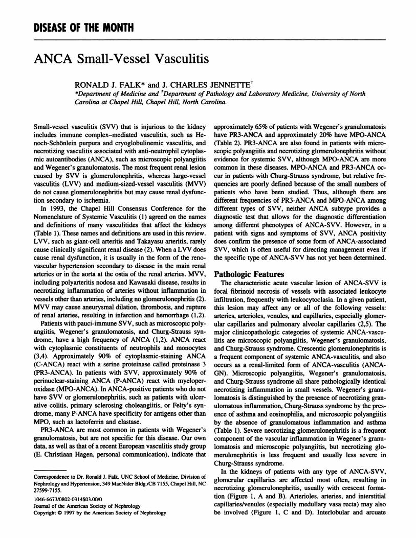
In the kidneys of patients with any type of ANCA-SVV,
gbomerular capillaries are affected most often, resulting in
necrotizing glomerubonephritis, usually with crescent forma-
tion (Figure 1 , A and B). Arterioles, arteries, and interstitial
capillaries/venubes (especially medullary vasa recta) may also
be involved (Figure 1 , C and D). Interlobular and arcuate
Large-vessel vasculitis
giant-cell (temporal) arteritis
Takayasu arteritis
Medium-sized vessel vasculitis
polyarteritis nodosa
Kawasaki disease
Small-vessel vasculitis
Wegener’ 5 granulomatosis
Churg-Strauss syndrome
microscopic polyangiitis
Henoch-Sch#{246}nbein purpura
essential cryoglobubinemic vasculitis
cutaneous leukocytoclastic angiitis
ANCA Small-Vessel Vasculitis 315
ANCA-SVV typically has an absence or paucity of immu-
Table 1. Names and definitions of vasculitis adopted by the Chapel Hill Consensus Conference on the Nomenclature of
Systemic Vasculitisa
Granubomatous arteritis of the aorta and its major branches, with a
predilection for the extracranial branches of the carotid artery. Often
involves the temporal artery. Usually occurs in patients older than 50
and is often associated with polymyalgia rheumatica.
Granulomatous inflammation of the aorta and its major branches. Usually
occurs in patients younger than 50.
Necrotizing inflammation of medium-sized or small arteries without
gbomerulonephritis or vasculitis in arterioles, capillaries, or venules.
Arteritis involving large, medium-sized, and small arteries, and associated
with mucocutaneous lymph node syndrome. Coronary arteries are often
involved. Aorta and veins may be involved. Usually occurs in children.
Granulomatous inflammation involving the respiratory tract, and necrotizing
vasculitis affecting small- to medium-sized vessels, e.g., capillaries,
venules, arterioles, and arteries. Necrotizing glomerulonephritis is
common.
Eosinophib-rich and granubomatous inflammation involving the respiratory
tract and necrotizing vasculitis affecting small- to medium-sized vessels,
and associated with asthma and blood eosinophibia.
Necrotizing vascubitis with few or no immune deposits, affecting small
vessels, i.e., capillaries, venubes, or arterioles. Necrotizing arteritis
involving small- and medium-sized arteries may be present. Necrotizing
glomerulonephritis is very common. Pulmonary capi!laritis often occurs.
Vascubitis with immunogbobubin A-dominant immune deposits, affecting
small vessels, i.e. , capillaries, venules, or arterioles. Typically involves
skin, gut, and glomeruli, and is associated with arthralgias or arthritis.
Vasculitis with cryogbobubin immune deposits, affecting small vessels, i.e.,
capillaries, venules, or arterioles, and associated with cryogbobulins in
serum. Skin and glomeruli are often involved.
Isolated cutaneous beukocytoclastic angiitis without systemic vasculitis or
glomerulonephntis.
a The ANCA-associated vasculitides are Wegener’s granulomatosis, microscopic polyangiitis, and Churg-Strauss syndrome. (Modified
from Jennette et al. [I] with permission.).
Table 2. Approximate frequencies of PR3-ANCA (C-ANCA) and MPO-ANCA (P-ANCA) in patients with
glomerulonephritis caused by Wegener’s granubomatosis (WG), microscopic polyangiitis (MPA), and
pauci-immune necrotizing and crescentic gbomerubonephritis without systemic vascubitis (NCON)
ANCA Result WG MPA NCGN
Positive PR3-ANCAIC-ANCA 65 to 75% 35 to 45% 30 to 40%
Positive
Negative
MPO-ANCA/P-ANCA
ANCA
15
10
to 25%
to 20%
45
10
to 55%
to 20%
60 to 70%
10 to 20%
arteries are affected more often than larger arteries, but a few SVV. Necrotizing leukocytoclastic angiitis in medullary vasa
patients with ANCA-SVV will have involvement of interbobar recta may be very severe, and can result in papillary necrosis
and even main renal arteries. The renal necrotizing arteritis of and hemorrhage. The frequency of the medulbary angiitis is
ANCA-SVV is indistinguishable histologically from that of difficulty to know because it is not always sampled in renal
necrotizing MVV, such as polyarteritis nodosa; however, the biopsy cores, which purposefully have more cortex than me-
absence of inflammation in vessels other than arteries (e.g. , dulla.
gbomerubar capillaries) distinguishes the MVV from ANCA-
316 Journal of the American Society of Nephrology
Figure 1. Renal histologic manifestations of ANCA-SVV. (A) Early segmental glomerular fibrinoid necrosis with slight adjacent epithelialproliferation (periodic acid-Schiff stain). (B) Large cellular crescent with focal disruption of Bowman’s capsule at the bottom of the photograph
(periodic acid-Schiff stain). (C) Necrotizing inflammation involving two interlobular arteries (arrow, f,brinoid necrosis; Masson trichrome
stain). (D) Leukocytoclastic angiitis affecting the medullary vasa recta (hematoxylin and eosin stain).
nohistologic staining for immunogbobubins at sites of vasculitis
and gbomerubonephritis. This “pauci-immune” appearance is in
contrast to the characteristic granular immunoglobulin staining
of immune complex-mediated disease and the linear immuno-
globulin staining of anti-gbomerular basement membrane (anti-
GBM) antibody-mediated disease. Although ANCA are much
more common in patients with pauci-immune necrotizing gb-
merulonephritis and vasculitis, patients with anti-GBM gb-
merulonephritis (6) and immune-complex crescentic gbomeru-
bonephritis (7) have a higher frequency of ANCA than healthy
control subjects. Approximately 20% to 30% of patients with
either crescentic immune-complex gbomerubonephritis or anti-
GBM gbomerulonephritis have ANCA. In patients with anti-
GBM disease, the presence of ANCA correlates with the
development of vasculitis that is not seen with anti-GBM
disease alone, such as arteritis, arteriobitis, and venulitis in a
distribution typical for ANCA-vasculitis. In patients with im-
mune-complex gbomerulonephritis, the presence of ANCA cor-
rebates with a greater likelihood of crescent formation and
systemic vasculitis. For example, membranous gbomerulopathy
patients with ANCA often develop crescent formation and
rapidly progressive disease. This suggests that ANCA can
synergize with immune-complex localization to induce more
severe inflammation.
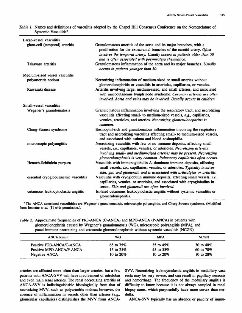
How “pauci-immune” is the gbomerulonephritis in patients
with ANCA-SVV and ANCA-GN? In a recent evaluation of
21 3 patients with glomerulonephritis and crescent formation
(excluding patients with bupus nephritis and anti-GBM dis-
ease), we concluded that, to be at least 80% predictive of
ANCA disease, “pauci-immune” should be defined as 2+ or
less staining for any immunogbobubin (on a scale of 0 to 4+)
and absence of immune complex-type electron-dense deposits
by electron microscopy (Figure 2) (7).
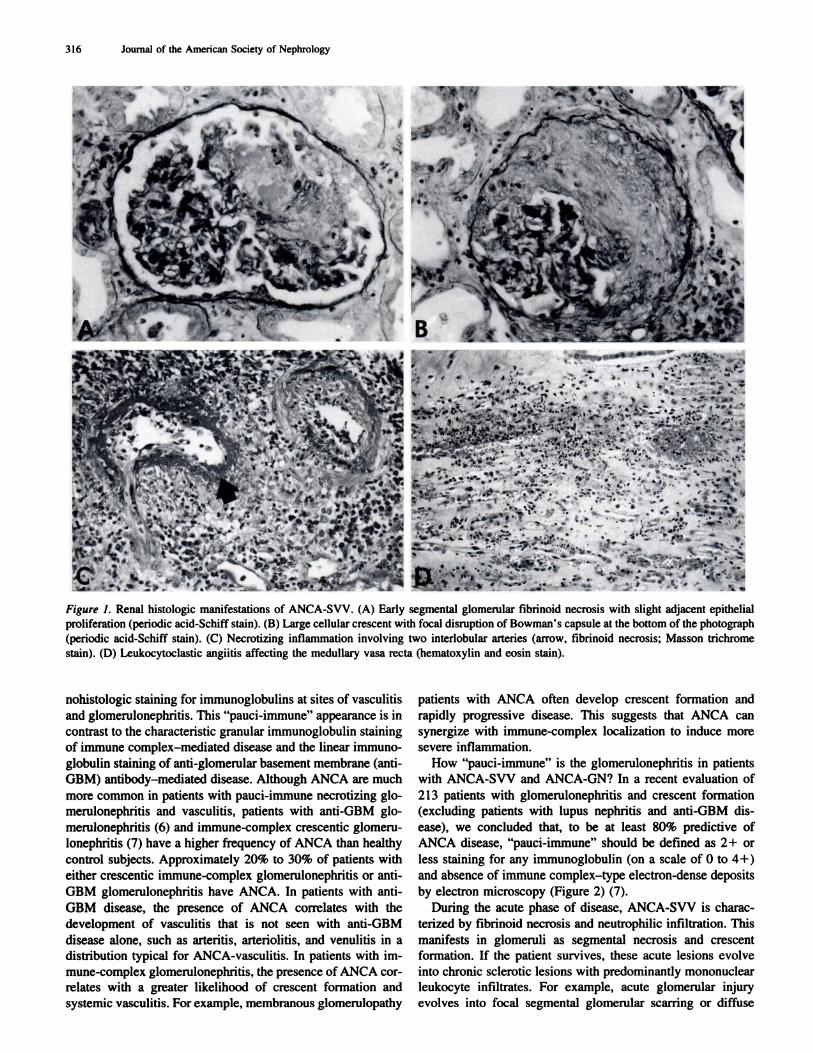
During the acute phase of disease, ANCA-SVV is charac-
terized by fibrinoid necrosis and neutrophilic infiltration. This
manifests in gbomeruli as segmental necrosis and crescent
formation. If the patient survives, these acute lesions evolve
into chronic sclerotic lesions with predominantly mononuclear
leukocyte infiltrates. For example, acute gbomerular injury
evolves into focal segmental gbomerular scarring or diffuse
#{149}IFAssay
LJELISA
liii
ANCA Small-Vessel Vasculitis 317
100
90
� 80
� 7000.60
(.)
z440
� 30
� 20
a.
0
0 0.5-1+ 1.5-2+ 2.5-3+ 3.5-4+(n=92) (n=72) (n=19) (n=17) (n=13)
Maximum intensity of glomerular staining for any immunoglobulin
Figure 2. The frequency of ANCA positivity by indirect immunoflu-orescence microscopy assay (IF assay) and enzyme immunoassay for
anti-PR3 or anti-MPO (ELISA) in patients with crescentic glomeru-
lonephntis, relative to the intensity of direct immunofluorescence
microscopy gbomerubar staining for immunoglobulin (excluding bupus
nephritis and anti-GBM nephritis patients). Note that the highest
frequency of ANCA is in the patients with the most “pauci-immune”
crescentic glomerulonephritis, but that patients with crescentic im-
mune-complex glomerulonephritis do have a higher frequency of
ANCA than healthy individuals (who have a frequency of <5%).
global gbomerular scarring, depending on the severity of the
acute injury (Figure 3).
The lungs are frequently affected by ANCA-SVV. Acute
pulmonary lesions that occur in all types of ANCA-SVV
include necrotizing inflammation of alveolar capillaries, arte-
rioles, arteries, veins, and venules (8,9). Necrotizing granulo-
matous inflammation occurs in patients with Wegener’s gran-
ulomatosis and Churg-Strauss syndrome, and may involve
vessels and airways. The acute inflammatory pulmonary injury
evolves into more chronic lesions, such as interstitial fibrosis
and bronchiobitis obliterans-organizing pneumonia. No partic-
ular pulmonary lesion is specific for either C-ANCA or
P-ANCA-positive patients; however, necrotizing granuboma-
LIGHT MICROSCOPIC MORPHOLOGY
FOCALSCLEROSING CN � CHRONIC GN
i\ INFOCAL � � NECROSIS AND � CRESCENTIC � END STAGENECROSIS FEW CRESCENTS GN KIDNEY
HEMATURIA and/or ACUTE RAPIDLY SLOWLY
PROTEINURIA NEPHRITIS PROGRESSIVE PROGRESSIVENEPHRITIS NEPHRITIS
CLINICAL PRESENTATiON
Figure 3. The relationship between the severity and stage of patho-
logic injury and the clinical manifestations of ANCA-GN. Note that
there are transitions over time between different pathologic and din-
ical phenotypes.
tous inflammation is more commonly found in C-ANCA pa-
tients.
The tissue distribution of the inflammatory lesions of
ANCA-SVV is extremely varied among patients, resulting in a
wide variety of clinical manifestations. In addition to nephritis
caused by glomerular inflammation and pulmonary hemor-
rhage caused by alveolar capillaries or granulomatous inflam-
mation, patients may have purpura caused by dermab leukocy-
toclastic angiitis, mononeuritis multiplex caused by perineural
and epineural artenolitis and arteritis, myalgias caused by
arteriolitis and arteritis in skeletal muscles, and abdominal pain
caused by venulitis, arteriolitis, and arteritis in abdominal
viscera, such as the gut, liver, pancreas, and spleen.
Clinical FeaturesANCA-SVV is more common in Caucasians than African
Americans, with a ratio of approximately 7.5: 1 . Women and
Men are equally affected, and although patients are usually 55
yr of age or older, patients of any age may develop ANCA-
Svv (10-12). The clinical renal manifestations vary according
to the severity and stage of the underlying renal injury (Figure
3). Hematuria with dysmorphic red blood cells and red blood
cell casts is a frequent clinical feature. Proteinuria tends to be
in the subnephrotic range, with a mean of 2 to 3 g/day,
although some patients have as little proteinuria as 0.5 g/day
and as much as 20 g/day. Although the presentation of ANCA
gbomerulonephritis is frequently that of rapidly progressive
glomerubonephritis, the syndrome of asymptomatic hematuria
with minimal amounts of proteinuria or acute nephritis is
common as well. Unfortunately, at their first presentation,
many patients already require dialysis because of chronic gb-
merubonephritis and severe glomerular sclerosis, and intersti-
tial fibrosis indicative of ESRD. This circumstance is often the
result of a delay in referring the patient to a nephrologist for
management.
Nephrologists are confronted with the diagnostic dilemma of
whether a renal biopsy is essential for the management of
ANCA-GN and ANCA-SVV. The answer to this question is
predicted on several variables, including the features of the
clinical syndrome, the accuracy of the ANCA serologic meth-
odobogy, and most importantly, the form of therapy that will be
used. Some investigators have concluded that PR3-ANCA and
MPO-ANCA assays can have a sensitivity for pauci-immune
necrotizing and crescentic glomerubonephritis ranging between
95% and 100%, with a specificity in excess of 99% (13). If
patients who have strong clinical evidence for ANCA-SVV are
tested with such a high-quality assay system, the positive
predictive value of the ANCA result is greater than 99% (14).
Thus, expert ANCA testing may obviate pathological confir-
mation of ANCA-SVV or ANCA-GN especially in some emer-
gency situations. For instance, in a patient who presents with
pulmonary hemorrhage who requires ventilator support, and
who has red blood cells and red blood cell casts in the urine, a
rapidly rising creatinine concentration, and palpable purpura,
renal biopsy may be unnecessary if the ANCA test is positive.
However, despite the contention by some that ANCA serobog-
ical testing has “come of age,” not all laboratories perform
antigen-specific ANCA assays, and the sensitivity and speci-
318 Journal of the American Society of Nephrology
ficity for PR3-ANCA and MPO-ANCA testing vary among
assay systems. Therefore, clinicians must have knowledge of
the capabilities of the laboratory that is performing their
ANCA testing before they can decide how much weight to give
the test result in their management decisions.
If patients with ANCA-SVV and ANCA-ON could be
treated effectively with innocuous drugs, theoretically, the
clinician could decide to treat the patient empirically without
exhaustive confirmation of the diagnosis. However, because
ANCA-SVV and ANCA-ON are typically treated with potent
anti-inflammatory and immunosuppressive drugs that predis-
pose to life-threatening infections and risk of mutagenesis, it is
our contention that a renal biopsy or other tissue confirmation
is called for whenever possible. An integration of clinical
manifestations, pathologic findings, and serologic data is re-
quired in order to provide optimum information for meaningful
patient-informed consent and for treatment decisions.
In addition to ANCA-SVV, the differential diagnosis for
patients with signs and symptoms of a systemic disease affect-
ing small vessels often includes systemic immune-complex
diseases, such as systemic bupus erythematosus, Henoch-
Sch#{246}nlein purpura, and cryogbobulinemic vasculitis, as well as
the thrombotic microangiopathies and atheroembolization.
Careful clinical, pathologic, and serologic evaluation should
distinguish among these systemic vascubopathies.
Pulmonary-Renal Vasculitic Syndrome
ANCA-SVV has a predilection for the capillary beds of the
respiratory tract (2,9, 15). At least 50% of patients with
ANCA-ON have pulmonary disease spanning the spectrum
from severe life-threatening pulmonary hemorrhage to fleeting
alveolar infiltrates. Among patients with ANCA-ON, 10%
have massive pulmonary hemorrhage, with a mortality rate of
50%. The majority of patients with ANCA-SVV present with
focal or diffuse pulmonary infiltrates with variable amounts
of hemoptysis. In other patients-especialby those with
Wegener’ 5 granubomatosis-pulmonary nodules, cavities, al-
veobar opacities, or diffuse ground-glass opacities are observed
radiographically. Because these parenchymal opacities are ill-
defined at times, routine chest x-rays miss smaller nodular or
cavitary lesions that are best seen on fine-cut computed tomog-
raphy. This form of imaging is a more sensitive approach to
unravel the pulmonary manifestations of ANCA-SVV. Fiber-
optic bronchoscopy with transbronchiab lung biopsy is a useful
procedure in patients in whom a pathological confirmation of
a SVV cannot be made by kidney or upper respiratory tract
biopsy. There may be no renal involvement at the onset of
a SVV. For example, among the first 158 patients studied at
the National Institutes of Health, only 18% of those with
Wegener’s granulomatosis presented with evidence for gbomer-
ubonephritis; however, renal involvement eventually occurred in
77% of patients, usually in the first 2 yr of illness (16).
Upper respiratory tract symptoms of ANCA-SVV include
necrotizing lesions of the nose, sinus, and ear (17). These
lesions are found not only in patients with Wegener’s granu-
lomatosis, but also in patients with microscopic polyangiitis.
Oranubomatous inflammation of the trachea, usually in the
subgbottic region, results in unilateral or bilateral edema, in-
flammation, fibrosis, and tracheal stenosis. The clinical picture
is that of stridor and dyspnea that requires persistent bong-term
immunosuppressive therapy.
The differential diagnoses of pulmonary-renal vascubitic
syndromes other than ANCA-SVV includes anti-OBM disease
(Ooodpasture’s syndrome) and some forms of immune-com-
plex diseases, such as systemic bupus erythematosus. Renal
histology is a definitive means of separating the diagnostic
possibilities. Serological studies-including tests for antinu-
clear antibodies, anti-double-stranded DNA, low serum com-
plement levels, cryogbobulins, hepatitis C antibodies, and anti-
GBM antibodies-are useful for suggesting the appropriate
diagnosis. One should bear in mind that 20% to 30% of
patients with anti-OBM disease also are ANCA-positive and
are at risk for developing any of the manifestations of ANCA-
SVV. Thus, patients with pulmonary-renal vascubitic syndrome
who have anti-OBM antibodies should also be tested for
ANCA, and vice versa.
A difficult differential diagnosis occurs in patients with
ANCA-SVV or ANCA-ON who, during treatment, develop
pulmonary infiltrates with or without hemoptysis. Recurrent
vascubitis must be differentiated from infection, especially fun-
gab or mycobacteria tuberculosis. A high ANCA titer in such a
patient is not definitive evidence for pulmonary vascubitis. A
negative ANCA result makes pulmonary vascubitis less likely.
Bronchoscopy, including bronchial alveolar lavage, may help
differentiate infection from vascubitic alveolar hemorrhage.
Similarly, infections of the upper respiratory tract can mimic
vasculitic lesions in the nose, sinus, and ear. Fiberoptic trans-
lumination of the upper airways with biopsy is useful for
identifying infections and vascular inflammation.
Renal-Dermal Vasculitic Syndrome
Renal-dermal vasculitic syndromes include Henoch-Sch#{246}n-
lein purpura, cryogbobulinemia vasculitis, systemic bupus ery-
thematosus, and ANCA-SVV (18). Common dermal vasculitic
findings in ANCA-SVV are palpable purpura (usually in the
lower extremities), petechia, ulcers, nodules, ecchymoses, and
bullae. Urticaria is a much more common manifestation of
SVV in the skin than was previously realized. The clinical
syndrome of palpable purpura, nephritis, and ANCA is nearly
diagnostic for ANCA-SVV, although tissue confirmation of
vasculitis is comforting before the institution of toxic immu-
nosuppression treatments. In a patient with renab-dermal vas-
culitic syndrome, biopsy demonstration of immunoglobulin
A-dominant immune deposits in either dermal vessels or gb-
meruli is diagnostic for Henoch-Sch#{246}nlein purpura. A renal
biopsy will include or exclude cryogbobulinemic gbomerubone-
phritis or lupus nephritis. The resolution of the differential
diagnosis in patients with renal-dermal vasculitic syndromes is
very important because the prognosis and appropriate treat-
ments are quiet different among the diagnostic possibilities,
i.e., Henoch-Sch#{246}nbein purpura, cyoglobulinemic vasculitis,
systemic bupus erythematosus, and ANCA-SVV. For example,
a 25-yr-old patient with purpura and nephritis who has He-
noch-Sch#{246}nbein purpura usually does not require aggressive
immunosuppression, whereas a patient with the same clinical
ANCA Small-Vessel Vasculitis 319
a Reprinted from Hogan et a!. (1 1) with permission.
presentation who has ANCA-SVV is at risk for life-threatening
organ injury if not promptly and appropriately treated.
Other Organ System Involvement
The most common neurological manifestation of ANCA-
SVV is peripheral neuropathy, typically manifesting as mono-
neuritis multiplex. Occasionally, patients have central nervous
system inflammation, especially granubomatosis meningial in-
flammation, which may be observed on magnetic resonance
imaging. Patients may develop seizures, which will respond to
immunosuppressive treatment.Gastrointestinal disease, found in one third of patients with
ANCA-SVV, may present with gastric or peptic ulcers de-
tected by endoscopic examination. Vasculitis of the small and
large intestine results in gastrointestinal bleeding, or-if Se-
vere-perforation of the viscus as a consequence of transmurab
infarction.
Iritis, uveitis, and episcieritis result in painful, red eyes.
These lesions are frequently present in a “subclinical” fashion
and require slit-lamp ophthalmologic evaluation for recogni-
tion.
Almost all patients have a prodrome of a “flu-bike illness”
with malaise, myalgias, and arthralgias, which is probably
mediated by the increased systemic levels of cytokines released
from the sites of inflammation. The arthralgias often migrate
from joint to joint.
OutcomeSeveral studies have examined prognostic factors in ANCA-
SVV (11,19). These studies point to the entry-level serum
creatinine value, the presence of pulmonary disease, the sever-
ity of renal disease, and even the initial white blood cell count
as important predictors of outcomes. In our own studies ( 1 1),
the most important determinant of patient survival was the
presence or absence of pulmonary hemorrhage (Table 3). The
presence of any pulmonary symptoms-that is, pulmonary
infiltrates, nodules, or cavities-did not increase the risk of
death. The risk of ESRD was largely attributable to the entry-
level serum creatinine value, even when such variables as the
presence or absence of extrarenal disease and the presence or
absence of pulmonary findings were incorporated into the
model. Crescents and necrosis found on renal biopsy did not
add to the predictive power of the model. Interstitial fibrosis on
renal biopsy was an independent risk factor in individuals with
an entry-level creatinine concentration of <3 mg/dL, whereas
in individuals with serum creatinine concentrations greater
than 4.5 mg/dL, tububointerstitiab injury was present.
TreatmentInduction Therapy
Because renal prognosis appears to be determined by early
treatment, it is reasonable to induce suppression of the vascular
inflammation promptly with aggressive therapy that usually
includes high-dose corticosteroids and cycbophosphamide. Our
approach is to give patients 7 mg/kg per day of methylpred-
nisobone for 3 days, followed by daily oral prednisone.
Whether plasmapheresis and methylprednisobone are equally
good induction therapies, or whether plasmapheresis adds ad-
ditional benefit to methylprednisobone as a means of induction
therapy is currently under study (20). Cycbophosphamide is
administered either intravenously or orally. Intravenous cycbo-
phosphamide is administered on a monthly basis beginning at
a dose of 0.5 g/m2, with adjustment of the dose upward to 1
g/m2 on the basis of 2-wk beukocyte counts ( 10, 12). Oral
cycbophosphamide should begin at a dose of 2 mg/kg per day
(2 1 ). Prednisone is administered at a dose of 1 mg/kg for the
first month, which is tapered to alternate-day therapy in the
second month, with cessation of prednisone therapy at the end
of the third to fourth months. All forms of cycbophosphamide
dosage should be titrated to the beukocyte count. Special atten-
tion to the beukocyte count must be monitored while the patient
is on tapering doses of prednisone, because cycbophosphamide
dosage may need to be adjusted downward as the prednisone
dose is decreased. This induction therapy can be initiated while
clinicians wait for renal biopsy or serologic study results.
The optimal length of cycbophosphamide therapy has not
been determined. It is clear that indefinite treatment with
cycbophosphamide is associated with prohibitable risks, includ-
ing a 15% incidence of transitional cell carcinoma in the
bladder in those patients treated with long-term oral cyclophos-
phamide (22). In patients achieving complete remission within
6 months of therapy, treatment can be stopped under the
provision that there is close patient follow-up. In those mdi-
viduals with persistently active disease at 6 months, it is
Table 3. Predictors of disease-related death in patients with ANCA-positive microscopic polyangiitis (MPA) and ANCA-
positive necrotizing and crescentic gbomerubonephritis (NCGN)�
.Predictor
Relative Risk ofDeath
95% ConfidenceInterval
Value
Pulmonary hemorrhage (present versus absent) 8.64 (3.36 to 22. 19) 0.0002
Any pulmonary symptoms (present versus absent) 2.36 (0.80 to 6.95) 0.16
Disease category (MPA versus NCGN alone) 1 .68 (0.48 to 5.82) 0.61
ANCA pattern (C-ANCA versus P-ANCA) 3.78 (1 .22 to I 1 .70) 0.031
Race (African American versus Caucasian) 2.41 (0.75 to 7.77) 0.34
Treatment (cyclophosphamide versus 0. 18 (0.05 to 0.66) 0.0 12
corticosteroids alone)
320 Journal of the American Society of Nephrology
a Reprinted from Nachman et a!. (12) with permission.
reasonable to continue cyclophosphamide therapy for a full 12
months. Another possible protocol utilizes cycbophosphamide
as the immunosuppressive agent for the first 3 months of
treatment and then azathioprine (2 mg/kg) for the duration of
the therapy (23). Most patients may stop this form of therapy
after a total of 6 to 12 months of treatment.
Intravenous and oral cyclophosphamide appear to have
equal efficacy in inducing remission. Whether oral cycbophos-
phamide treatment results in a decrease in the relapse rate when
compared with intravenous cycbophosphamide remains an is-
sue for study. Intravenous cyclophosphamide allows for a
smaller total dose of cyclophosphamide than the oral regimen.
As a consequence, there is a threefold decrease in both minor
and major infections. The most bothersome feature of cyclo-
phosphamide therapy, however, is the long-term mutagenic
risk, especially the risk of induction of transitional cell carci-
noma of the bladder (22). Gonadal toxicity in women over the
age of 35 yr results in premature menopause.
Because cycbophosphamide is toxic, the question arises
whether prednisone is an adequate treatment option; however,
the requirement of cycbophosphamide as a necessary adjunct to
prednisone has been appreciated for years (21). In our own
studies, we have determined that treatment with prednisone
alone resulted in a three-fold increased risk of relapse when
compared with treatment with cyclophosphamide (12). Thus,
patients with ANCA-SVV who are not dialysis-dependent
should be treated with combination therapy including cycbo-
phosphamide.
Alternative Treatment Strategies
DeRemee and colleagues suggested that local-regional dis-
ease of the upper respiratory tract should be treated first with
trimethoprin-sulfamethoxazole and then with corticosteroid
therapy in the event of unsuccessful antibiotic therapy (24).
The rationale for this approach is based largely on empirical
data, but is supported by the fact that chronic nasal carriage of
Staphylococcus aureus is a risk factor for relapse in Wegener’s
granubomatosis (25). In 1985, DeRemee et a!. described their
results in treating patients with Wegener’s granubomatosis
(24). One hundred new patients with Wegener’s granubomato-
sis were treated with trimethoprin-sulfamethoxazole. Eleven of
these patients improved, four of them without concurrent or
previous immunosuppressive treatment. Most recently, Rein-
hold-Keller et a!. (26) studied 19 patients with Wegener’s
granulomatosis restricted to the upper and lower airways, and
found similar results. A prospective placebo-controlled trial
with tnmethoprin-sulfamethoxazole (960 mg twice daily) was
performed in 81 patients with Wegener’s granulomatosis (27).
In 19% of these patients, therapy was stopped because of side
effects. A statistically significant reduction in the number of
relapses was observed in the groups assigned to trimethoprin-
sulfamethoxazole, although the decrease in relapse rate was
limited to relapse in the upper respiratory tract, and not to
relapse in the lower respiratory tract or the kidney.
Conflicting data are beginning to emerge, however, in that a
randomized trial comparing methotrexate and trimethoprin-
sulfamethoxazole for maintenance of remission in 65 patients
with Wegener’s granulomatosis was recently completed (25).
In this study, generalized Wegener’s granubomatosis was
brought into remission by treatment with cyciophosphamide
and prednisone. The maintenance of remission was much more
effective with methotrexate therapy than with either tri-
methoprin-sulfamethoxazole or prednisone. In fact, tri-
methoprin-sulfamethoxazole and prednisone together seemed
to increase the chance of relapse. Thus, the use of trimethoprin-
sulfamethoxazole in patients with Wegener’ s granubomatosis is
still controversial.
Methotrexate may be a useful form of therapy for SVV,
especially in individuals with normal or near-normal renal
function, and in patients with vasculitis predominantly con-
fined to the kidneys (29). Methotrexate should not be used if
the patient’s serum creatinine concentration is greater than 2
mg/dL. Pooled intravenous y-gbobulin has been tried anecdot-
ally, not only for induction therapy but also for individuals
resistant to cyclophosphamide and azathioprine therapy. The
long-term role of these agents in the treatment of ANCA-SVV
awaits further investigation.
Remission and Relapse
The terms “remission” and “relapse” for ANCA-SVV pa-
tients are defined in Table 4 (1 1). ANCA-SVV and ANCA-ON
patients treated with either intravenous or oral cycbophospha-
mide have a long-term remission rate of between 60% and
85%. Relapse occurs in up to 40% of patients within a mean of
18 months after therapy has stopped. Relapse typically occurs
in the same organ system initially affected by the disease,
Table 4. Criteria for evaluating treatment responses in
patients with ANCA-SVV and ANCA�GNa
S Remission. Stabilization or improvement of renal function
(as measured by serum creatinine concentration), resolution
of hematuria, and resolution of extrarenal manifestations of
systemic vasculitis. Persistence of proteinuria was not con-
sidered indicative of persistence of disease activity.
. Remission by therapy. The achievement of remission
while still receiving immunosuppressive medication or cor-
ticosteroids given at a dose greater than 7.5 mg per day of
prednisone or its equivalent.
S Treatment resistance. (1) Progressive decline in renal
function with the persistence of an active urine sediment or
(2) persistence of new appearance of any extrarenal mani-
festation of vasculitis despite immunosuppressive therapy.
. Relapse. Occurrence of at least one of the following: (1)
rapid rise in serum creatinine concentration, accompanied
by an active urine sediment; (2) a renal biopsy demonstrat-
ing active necrosis or crescent formation; (3) hemoptysis,
pulmonary hemorrhage, or new or expanding nodules with-
out evidence for infection; (4) active vasculitis of the respi-
ratory or gastrointestinal tracts as demonstrated by endos-
copy with biopsy; (5) iritis or uveitis; (6) new mononeuritis
multiplex; or (7) necrotizing vasculitis identified by biopsy
in any tissue.
ANCA Small-Vessel Vasculitis 321
although new organ system involvement occurs as well. Re-
lapses in the kidney are heralded by the recurrence of micro-
scopic hematuria, red blood cell casts, and worsening renal
function. Fluctuations in the amount of proteinuria are not
good indicators of active disease, and may be related to gb-
merular sclerosis. Relapse in the lungs tends to occur in the
same locus as the site of original injury. Many patients’ re-
lapses are heralded by a recurrence in a migratory polyar-
thropathy.
How to best diagnose and treat relapse is a matter of sub-
stantial investigation. Table 4 provides some clinical indicators
that relapse is occurring. There is no consensus on the value of
ANCA titers for monitoring remission. Full-blown vasculitic
relapse should be treated with a repeat course of prednisone
and cycbophosphamide. In general, these patients require main-
tenance on long-term immunosuppression with cyclophospha-
mide, azathioprine, or methotrexate. Less ominous relapse of
the upper respiratory tract may be treated with prednisone
alone or perhaps with trimethoprin-sulfamethoxazole.
Serial ANCA Testing
The value of ANCA titers for disease monitoring has been
the subject of several investigations. ANCA titers rise during
relapse in 23% to 77% of patients (30,31). ANCA titers may
increase without overt clinical relapse, and in some patients,
persistently high ANCA titers are found during clinical remis-
sion. In addition, some patients have a clinical relapse that is
not associated with any rise in ANCA titers. At the very least,
however, a rise in ANCA titer should raise the possibility of a
relapse. The disappearance of ANCA is at least suggestive of
remission. Many serologic tests, including ANCA titers, may
have greater importance in some patients than in others. For
instance, in some patients, the ANCA titers may closely follow
the clinical disease course, whereas in other patients, ANCA
titers may not correlate well at all with clinical events. Perhaps
serial monitoring of different ANCA isotypes or affinities will
prove to be a useful approach in the future. As of now, ANCA
titers should not be used as a determinant for beginning or
altering immunosuppressive therapy.
Treatment of Dialysis-Dependent Patients
At least 20% of patients with ANCA-SVV and ANCA-ON
will require dialysis at the time of diagnosis. Half of these
patients may come off dialysis within 8 to 12 wk of therapy.
Which patient will remain on long-term dialysis or will have a
dialysis-free response is not clear at the initiation of dialysis.
The dialysis-free interval may last from as little as a few weeks
to 3 to 4 yr. The degree of immunosuppression warranted
during the first 12 wk of dialysis depends on the risk-to-benefit
ratio. It is our practice to treat dialysis-dependent patients for at
beast 8 to 12 wk with pulse methylprednisolone and oral
prednisone. If patients’ renal functions improve, we continue
corticosteroid treatment and institute cyclophosphamide ther-
apy. Whether plasmapheresis is a more effective way of in-
ducing dialysis-free intervals than methylprednisobone is not
yet known.
Transplantation
ANCA-ON and ANCA-SVV rarely recur after renal trans-
plantation. Recurrence is not confined to the transplanted kid-
ney, but may also recur in nonrenal sites, such as the upper and
lower respiratory tracts. The question of whether ANCA titers
need to normalize before renal transplantation has not been
resolved. In those patients who are in clinical remission with a
negative ANCA titer, transplantation should proceed. Delay of
transplantation may be warranted in patients who have had a
persistently negative ANCA titer, but who then develop a
marked rise in titer. In patients who have persistently high
ANCA titers but no clinical evidence for SVV, it is reasonable
to proceed with renal transplantation.
References1. Jennette IC, Falk Ri, Andrassy K. Bacon PA, Churg J, Gross
WL, Hagen EC, Hoffman GS, Hunder GG, Kallenberg CG,McCluskey RT, Sinico RA, Rees Al, van Es LA, Waldherr R,
Wiik A: Nomenclature of systemic vasculitides: Proposal of an
international consensus conference. Arthritis Rheum 37: 187-
192, 1994.
2. Jennette JC, Falk Ri: The pathology of vasculitis involving the
kidney. Am J Kidney Dis 24: 130-141, 1994.
3. Falk RI, Jennette JC: Anti-neutrophil cytoplasmic autoantibodies
with specificity for myeloperoxidase in patients with systemic
vasculitis and idiopathic necrotizing and crescentic glomerulo-
nephntis. N Engl J Med 318: 1651-1657, 1988.
4. Hagen EC, Ballieux Be, van Es LA, Daha MR, van der Woude
FJ: Antineutrophil cytoplasmic autoantibodies: A review of the
antigens involved, the assays, and the clinical and possible patho-
genetic consequences. Blood 81: 1996-2002, 1993.
5. Jennette IC, Wilkman AS, Falk RI: Anti-neutrophil cytoplasmic
autoantibody-associated glomerulonephritis and vasculitis. Am J
Pathol 135: 921-930, 1989.
6. Jennette JC, Wilkman AS, Tuttle RH, Falk Ri: How pauci-immune is ANCA-associated crescentic gbomerulonephritis(CGN)? [Abstract] J Am Soc Nephrol 7: 1735, 1996.
7. Short AK, Esnault VL, Lockwood CM: Anti-neutrophil cytoplas-
mic antibodies and anti-glomerular basement membrane antibod-
ies: Two coexisting distinct autoreactivities detectable in patients
with rapidly progressive glomerulonephritis. Am J Kidney Dis
26: 439-445, 1995.
8. Fienberg R, Mark El, Goodman M, McCluskey RT, Niles IL:
Correlation of antineutrophil cytoplasmic antibodies with the
extrarenal histopathology of Wegener’s (pathergic) granulomatosis
and related forms of vasculitis. Hu,n Pathol 24: 160-168, 1993.
9. Gaudin PB, Askin FB, Falk Ri, Jennette JC: The pathologicspectrum of pulmonary lesions in patients with anti-neutrophil
cytoplasmic antibodies specific for anti-proteinase 3 and anti-
myeloperoxidase. Am J C/in Pathol 104: 7-16, 1995.
10. Falk RI, Hogan 5, Carey TS, Jennette IC: Clinical course of
anti-neutrophil cytoplasmic autoantibody-associated glomerulo-nephritis and systemic vasculitis: The Glomerular Disease Col-
laborative Network. Ann Intern Med I 13: 656-663, 1990.
1 1 . Hogan SL, Nachman PH, Wilkman AS, Jennette IC, Falk RJ, andthe Glomerular Disease Collaborative Network: Prognostic
markers in patients with ANCA-associated microscopic polyan-
giitis and glomerulonephritis. JAm Soc Nephrol 7: 23-32, 1996.
12. Nachman PH, Hogan SL, Jennette IC, Falk Ri: Treatment re-
sponse and relapse in ANCA-associated microscopic polyangiitis
and glomerulonephritis. J Am Soc Nephrol 7: 23-32, 1996.
322 Journal of the American Society of Nephrology
13. Niles IL, Pan G, Collins AB, Shannon T, Skates S, Fienberg R,
Arnaout MA, McCluskey RT : Value of antigen-specific radio-immunoassays for measuring anti-neutrophil cytoplasmic anti-
bodies (ANCA) in the differential diagnosis of rapidly progres-
sive glomerulonephritis. J Am Soc Nephrol 2: 27-36, 191.
14. Niles IL: A renal biopsy is essential for the management ofANCA-positive patients with gbomerulonephritis: The Contr
View. Sarcoidosis Vascu/itis and D�ffuse Lung Diseases 13:
232-234, 1995.
15. Bosch X, Lopez-Soto A, Mirapiex E: Antineutrophil cytoplasmic
autoantibody-associated alveolar capillaritis in patients present-
ing with pulmonary hemorrhage. Arch Patho/ Lab Med 118:517-518, 1994.
16. Hoffman GS, Kerr GS, Leavitt RY, Hallahan CW, Lebovics RS,Travis WD, Rottem M, Fauci AS: Wegener’s granulomatosis:
An analysis of 158 patients. Ann Intern Med 1 16: 488-498,
1992.
17. Andrassy K, Rasmussen N: Treatment of granulomatous disorders
of the nose and paranasal sinuses. Rhinology 27: 221-230, 1989.
18. Jennette IC, Milling DM, Falk RJ: Vasculitis affecting the skin:A review [Editorial]. Arch Dermatol 130: 899-906, 1994.
19. Savage CO, Winearls CG, Evans DI, Rees Al, Lockwood CM:Microscopic polyarteritis: Presentation, pathology and prognosis.
Q J Med 56: 467-483, 1985.
20. Pusey CD, Rees Al, Evans DI, Peters DK, Lockwood CM:
Plasma exchange in focal necrotizing gbomerubonephritis without
anti-GBM antibodies. Kidney Int 40: 757-763, 1991.2 1 . Fauci AS, Katz P, Haynes BF, Wolff SM: Cycbophosphamide
therapy of severe systemic necrotizing vasculitis. N EngI J Med
301: 235-238, 1979.
22. Tarlar-Williams C, Hijazi Y, Walther M, Linehan WM, Hallahan
CW, Lubensky I, Kerr GS, Hoffman GS, Fauci AS, Sneller MC:
Cyclophosphamide-induced cystitis and bladder cancer in pa-
tients with Wegener’s granulomatosis. Ann Intern Med 124:
477-484, 1996.
23. Gaskin 0, Savage CO, Ryan II, Jones S, Rees AJ, Lockwood
CM, Pusey CD: Anti-neutrophil cytoplasmic antibodies and dis-
ease activity during long-term follow-up of 70 patients with
systemic vasculitis. Nephro/ Dia/ Transp/ant 6: 689-694, 1991.
24. DeRemee RA, McDonald TI, Weiland LH: Wegener’s granulo-
matosis: Observations on treatment with antimicrobial agents.
Mayo C/in Proc 60: 27-32, 1985.
25. Stegemann CA, Cohen-Tervaert 1W, Sluiter WI, Manson WL, de
long PE, Kallenberg CGM: Association of chronic nasal carriage
of Staphylococcus aureus and higher relapse rates in Wegener’ s
granulomatosis. Ann Intern Med 120: 12-17, 1994.
26. Reinhold-Keller E, Dc Groot K, Rudert H, N#{246}lleB, Heller M,
Gross WL: Response to trimethoprin-sulfamethoxazole in We-
gener’s granulomatosis depends on the phase of disease. Q J Med
89: 15-23, 1996.
27. Stegman CA, Cohen Tervaert 1W, de long PE, Kallenberg CGM:
Trimethoprin-sulfamethoxazole for the prevention of relapses of
Wegener’s granubomatosis. N Engl J Med 335: 16-20, 1996.
28. De Groot K, Reinhold-Keller E, Tatsis E, Paulsen I, Heller M,
N#{246}lleB, Gross WL: Maintenance of remission therapy in 65
patients with generalized Wegener’s granubomatosis: Methotrex-
ate versus trimethoprin-sulfamethoxazole [Abstract]. Sarcoidosis
Vascu/itis and Diffuse Lung Diseases 13: 276, 1996.
29. Hoffman GS, Leavitt RY, Kerr GS, Fauci AS: The treatment of
Wegener’ S granulomatosis with glucocorticoids and methotrex-
ate.Arthritis Rheum 35: 1322-1329, 1992.
30. Cohen Tervaert 1W, Stegeman CA, Kallenberg CGM: Serial
ANCA testing is useful in monitoring disease activity of patents
with ANCA-associated vasculitides. Sarcoidosis Vascu/itis and
Diffuse Lung Disease 13: 241-245, 1996.
3 1 . DeOliveira J, Gaskin G, Dash A, Rees AJ, Pusey CD: Relation-
ship between disease activity and anti-neutrophil cytoplasmic
antibody concentration in long-term management of systemic
vascubitis. Am J Kidney Dis 25: 380-389, 1995.