dislocations of the pip joint - wordpress.com of the pip joint the pip joint may dislocate in one of...
TRANSCRIPT

DISLOCATIONS OF THE PIP JOINT
The PIP joint may dislocate in one of three directions: dorsal, lateral, and volar. These refer to theposition of the middle phalanx when joint deformation occurs.
Acute Dorsal PIP Dislocations
page 345
page 346
The mechanism of injury in dorsal dislocations is usually PIP joint hyperextension combined withsome degree of longitudinal compression, which frequently occurs in ball-handling sports if the tipof the digit is hit with the ball. In the majority of cases, dorsal dislocation produces a soft tissue orbone injury to the distal insertions of the three-dimensional ligament-box complex. The greaterthe longitudinal force, the more likely the volar lip of the middle phalanx will be sheared off orimpacted, producing a fracture-dislocation. On rare occasions, the volar plate ruptures proximallyand can become interposed, perhaps with an osteochondral fragment, between the head of theproximal phalanx and the base of the middle phalanx. This results in an irreducible dislocationand necessitates an open reduction.
13,36,55,57,82,84 The volar fracture fragment may even become
trapped within the flexor sheath and inhibit motion. 29
Dorsal displacement of the middle phalanxproduces specific lesions of the ligament system that can be classified into three major types (Fig.9-4). Each lesion represents a specific disruption of this ligament-box complex. Types I(hyperextension) and II (dorsal dislocation) rarely require surgical treatment, whereas type III(fracture-dislocation) injuries may require a more intensive, often surgical, treatment regimen.
Type I (Hyperextension)
Hyperextension injuries are characterized by partial or complete avulsion of the volar plate fromthe base of the middle phalanx, with or without a bone fragment, and minor longitudinal rents inthe collateral ligaments. In cases where the initial deformity has been severe, the middle phalanxmay actually become locked in 70 to 80 degrees of hyperextension. The articular surfaces remaincongruent, but with the middle phalanx articulating with the dorsal third of the condyle of theproximal phalanx (Fig. 9-4A). True lateral radiographs will often reveal a small, minimallydisplaced avulsion fragment from the base of the middle phalanx that should have no impact ontreatment.
Type II (Dorsal Dislocation)
With complete dorsal dislocation of the PIP joint, avulsion of the volar plate is accompanied by amajor bilateral split in the collateral ligament system. The base of the middle phalanx restsdorsally on the condyles of the proximal phalanx, usually in bayonet apposition with the shafts ofthe phalanges essentially parallel. There is no contact between the articular surfaces (see Fig. 9-4B).
Type III (Fracture-Dislocation)
Occasionally, the compressive force is of sufficient magnitude to shear off or impact the volarbase of the middle phalanx, producing a fracture-dislocation (see Fig. 9-4C).
40 Fracture-
dislocations can be subdivided into two types, stable and unstable, which have very specificimplications for reduction and treatment (Fig. 9-5).

base of the middle phalanx, producing a fracture-dislocation (see Fig. 9-4C). 40
Fracture-dislocations can be subdivided into two types, stable and unstable, which have very specificimplications for reduction and treatment (Fig. 9-5).
Stable Fracture-Dislocation.
Fracture-dislocation with a small triangular fragment representing less than 40% of the volararticular arc results in a dorsal displacement much like simple dorsal dislocations. The transversedisruption, however, occurs through the base of the phalanx rather than at the insertion of thevolar plate. The dorsal portion of the collateral ligaments remains attached to the middle phalanx,which is what renders these injuries inherently stable on reduction (see Fig. 9-5A).
Unstable Fracture-Dislocation.
Fracture or impaction of a larger segment of the volar articular surface of the middle phalanx mayalso result in dorsal dislocation. However, this represents a major loss of articular andligamentous support for the PIP joint. With disruption of greater than 40% of the volar articularsegment, the majority of the collateral ligament-volar plate complex is attached to the fragment,not the remaining intact base of the middle phalanx. Accurate closed reduction is difficult toachieve and even more difficult to maintain. Adding to the instability is the loss of the buttressingeffect of the volar margin of the middle phalanx that partially cups the proximal phalangealcondyles (see Fig. 9-5C). Treatment is described in the section on open reduction.

Add to lightbox
Figure 9-4 Pathology of dorsal dislocations of the PIP joint. A, Type I (hyperextension). The volar plate is avulsed and anincomplete longitudinal split occurs in the collateral ligaments. The articular surfaces maintain congruous contact. B, Type
II (dorsal dislocation). There is complete rupture of the volar plate and a complete split in the collateral ligaments, with themiddle phalanx resting on the dorsum of the proximal phalanx. The proximal and middle phalanges lie in almost parallelalignment. C, Type III (fracture-dislocation). The insertion of the volar plate, including a portion of the volar base of the
middle phalanx, is disrupted. The major portion of the collateral ligaments remains with the volar plate and flexor sheath.A major articular defect may be present. (From Eaton RG, Littler JW: Joint injuries and their sequelae. Clin Plast Surg
3:85-98, 1976.)

alignment. C, Type III (fracture-dislocation). The insertion of the volar plate, including a portion of the volar base of themiddle phalanx, is disrupted. The major portion of the collateral ligaments remains with the volar plate and flexor sheath.
A major articular defect may be present. (From Eaton RG, Littler JW: Joint injuries and their sequelae. Clin Plast Surg3:85-98, 1976.)
Schenck introduced a 16-component classification system in an effort to more specificallycharacterize the type III (fracture-dislocation) injuries just discussed.
98 The PIP joint is examined
on the lateral radiograph and assigned one of four fracture grades (I to IV) and one of foursubluxation grades (A to D). The injury is thereby classified into one of the 16 possiblepermutations. This was devised as an attempt to standardize published reports and facilitatecomparative analyses of different treatment methods for the specific PIP injury patterns.
Pilon Fracture.
page 346
page 347

Add to lightbox
Figure 9-5 Comparison of type III stable and unstable fracture-dislocations. A, Stable. Fracture of less than 40% of thevolar base of the middle phalanx leaves a significant portion of the collateral ligaments still attached. This portion will
guide the displaced middle phalanx to a congruous reduction. B, Normal. Collateral ligament insertion into the volar thirdof the middle phalanx and volar plate. C, Unstable. Fracture of greater than 40% of the volar base of the middle phalanxleaves little or no collateral ligament attached. Congruous reduction is unlikely without these ligaments. Frequently the
articular surface is impacted into the subcondylar bone and produces an irregular, depressed volar articular defect.
Fracture-dislocations of the PIP joint usually occur when the vector force of compression is notdirectly axial and the PIP joint is flexed to some degree. When an axial compressive force occurswith the proximal and middle phalanges being collinear, a pilon fracture may occur. (See Chapter8 on fractures of the metacarpals and phalanges.) This fracture is primarily intra-articular withoutdislocation or subluxation of the joint, but there is often widening of the base as dorsal and volarfragments are driven apart and more central cartilage surfaces are impacted into the cancellousbone of the base.
106a,123 Treatment modalities such as those used for fracture-dislocations may be
necessary.

fragments are driven apart and more central cartilage surfaces are impacted into the cancellousbone of the base.
106a,123 Treatment modalities such as those used for fracture-dislocations may be
necessary.
Depression of the concave articular surface of one or both condylar fossae of the middlephalangeal base mimics certain tibial plateau fractures. Angular deformities are commonespecially if only one condyle is involved. These fractures are subtle on plain radiographs of theinvolved digit, and the clinician should maintain a low threshold for obtaining a CT scan of the PIPjoint to better visualize the articular depression. Aggressive treatment should lessen the risk ofpersistent or worsening angular deformity, stiffness, and arthritis. Nevertheless, excellent resultsare difficult to achieve, with residual stiffness likely.
106a
Treatment of Stable PIP Joint Injuries
The PIP joint is contaminated in an open dorsal dislocation. Therefore, adequate débridementand irrigation should be performed under ideal conditions, usually in the operating room, andantibiotics given based on the condition of the wound and the nature and location of the injury.
106
Type I (hyperextension) injuries may be quite painful but are relatively benign injuries in themajority of cases.
44 The two most important aspects of treatment are patient education and the
avoidance of prolonged immobilization. Patients must be reassured that most of these injuries willreturn to normal function but that the swelling and stiffness may persist for several months, andoccasionally for more than 6 months. These injuries are stable and should be immobilized forcomfort and soft tissue recovery for no more than 1 week. It is rare to see problems arising frompremature mobilization and common to see stiffness and contracture as a result of prolongedimmobilization.
Few clinical studies have been performed on these ubiquitous injuries. Jesperson and associatesprospectively evaluated 57 PIP joint hyperextension injuries, 57% of which occurred during ball-handling sports.
48 The ring finger PIP joint was most commonly affected, and 44% had
associated avulsion fractures. The four patients with persistent pain and instability all hadhyperextension instability on the initial examination.
Type II (dorsal dislocation) injuries are usually stable to active and passive testing and requiremore protection than simple hyperextension injuries but still no more than 2 to 3 weeks ofimmobilization or controlled mobilization
101 (see Authors' Preferred Methods of Treatment).
Type III (fracture-dislocation) injuries must be carefully assessed to determine whether thedislocation is stable or unstable. Stable fracture-dislocations can generally be treatedconservatively with 3 weeks of dorsal block splinting followed by range of motion exercises.Intermittent static or dynamic splinting may be necessary to overcome the natural tendency todevelop stiffness and flexion contracture.
49,122 Most patients will require formal hand therapy to
regain full motion and function. In our experience, the patient's personality and initial response tothe injury may play more of a role than the specific injury in determining the extent to whichtherapy is needed.
Treatment of Unstable PIP Joint Injuries

Unstable type III PIP fracture-dislocations must be evaluated and treated with great attention todetail, because in the best of circumstances these injuries may still lead to complications,permanent dysfunction, and dissatisfaction. The continuing development of new treatmenttechniques for these injuries may imply that none of the current techniques produces consistentlygood to excellent results in a majority of patients for a majority of hand surgeons.
Specific Techniques
Dynamic Skeletal Traction.
page 347
page 348
Variations on this theme have been recommended for several decades.3,4,16,81,93
Both early andcurrent devices are founded on the principle of ligamen-totaxis through which the fracturefragments and the articular surface are reduced as longitudinal traction tightens the intactcomponents of the attached soft tissue envelope. Early range of motion may be instituted with thetraction maintained.
4,43,79,97,107 However, care must be taken to ensure that joint subluxation does
not occur during any part of the motion arc. A review of several recent reports showed anaverage range of motion of approximately 85 degrees at 1- to 2-year follow-up.
4,43,79,107 Pin tract
complications ranged from 0% to 74% but rarely affected final outcome. The best results wereachieved in fractures with less articular involvement and those treated acutely. Although designspecifics vary, the effective application of certain of these devices can be difficult even forexperienced hand surgeons.
4,43,79,97,107 Dynamic traction devices can be cumbersome to wear and
do not necessarily ensure optimal restoration of the articular surface, particularly when impactionof articular fracture fragments is present. Nevertheless, in certain fracture-dislocations andarticular impaction and pilon fractures a dynamic traction device may be the only option. For pilonfractures, the dynamic traction device can help to neutralize the joint reactive forces after openelevation of the depressed articular surface and bone grafting of the resultant defect. Dynamictraction devices vary in design, one type deriving the distraction force from rubber bands and theother from coiled Kirschner wires.
4,43,79,100a,100b,107 Regardless of which particular device is used,
care must be taken to prevent creating a moment arm about the middle phalanx leading to re-subluxation.
Krakauer and Stern used a hinged device to allow early active range of motion after surgicalprocedures on the PIP joint that combined distraction arthroplasty with other techniques, includingclosed reduction, open reduction and internal fixation (ORIF), and volar plate arthroplasty.
59
Satisfactory results in 20 patients led the authors to recommend the selective use of this device inthe treatment of fractures about the PIP joint.
Extension Block Splinting.
McElfresh and colleagues reported good results with active flexion using a dorsal splint to blockextension beyond the point of potential redisplacement.
75 The PIP joint is brought into extension
incrementally by reducing flexion in the splint by 10 to 15 degrees per week. However, in theirseries of 17 fingers, only four patients had a fragment size greater than 30% of the articularsurface. This suggests that the technique is most useful in fractures with small volar fragments inwhich the majority of the collateral ligament remains attached to the middle phalangeal base.
20,75
Hamer and Quinton prospectively followed 27 patients for less than 2 years, with fracture-dislocations involving an average of 50% of the articular surface treated by extension blocksplinting.
38 They reported a mean of 87 degrees of active PIP motion and good results in 70% of
patients. Green also advocated this technique. 35
Unstable fracture-dislocations tend to becomestable only in marked flexion, and several authors have recommended immobilization or fixationin up to 75 degrees of PIP flexion.
14,21,100,104,122 This, however, dramatically increases the risk for
late flexion contracture. Short, small, fingers make the fixation of the extension block splint moredifficult, and thick swollen fingers decrease the efficacy of this technique, given that re-subluxation and even re-dislocation may occur. Serial radiographs must be obtained to documentthe ongoing effectiveness of this technique in maintaining joint congruity.

splinting. 38
They reported a mean of 87 degrees of active PIP motion and good results in 70% ofpatients. Green also advocated this technique.
35 Unstable fracture-dislocations tend to become
stable only in marked flexion, and several authors have recommended immobilization or fixationin up to 75 degrees of PIP flexion.
14,21,100,104,122 This, however, dramatically increases the risk for
late flexion contracture. Short, small, fingers make the fixation of the extension block splint moredifficult, and thick swollen fingers decrease the efficacy of this technique, given that re-subluxation and even re-dislocation may occur. Serial radiographs must be obtained to documentthe ongoing effectiveness of this technique in maintaining joint congruity.
Extension Block Pinning.
With this technique, a Kirschner wire is placed into the head of the proximal phalanx at an angleto mechanically block extension of the PIP joint and prevent dorsal subluxation of the middlephalanx.
111,114 However, reports of this technique include only a small number of cases, and its
clinical efficacy must be substantiated through further study.
Trans-articular Pinning.
Simple reduction and pinning of the PIP joint with no attempt at articular reconstruction has beenadvocated in a retrospective report by Newington and associates.
83 Although preoperative data
were limited, the authors used this technique for articular involvement greater than 25% and lessthan 60%. In 11 fingers with long-term follow-up, all of the PIP joints were congruent, average PIPmotion was 85 degrees, and 3 of 10 patients had some residual pain.
Open Reduction with Internal Fixation.
This method has many advocates* and is most likely to be successful in acute cases with asingle, large fragment. Anatomic restoration of the articular surface is technically difficult evenwith a large, single fragment, especially because the remaining articular contour may bedisrupted secondary to impaction of subchondral cancellous bone. As with dynamic digitaltraction, the relatively small series and short follow-up leave unanswered the question of whetherthe articular reductions are adequate to prevent late post-traumatic arthrosis. If early activemotion is not instituted, the risk for joint contracture is greatly increased after open treatment,especially if pins are placed in positions that impale or tether the extensor mechanism.
37
Using a novel technique introduced by Hasting in 1999 (Scientific presentation, American Societyfor Surgery of the Hand, Annual Meeting, 1999) William and associates reported on 13consecutive patients treated with a hemihamate autograft. The autograft is harvested from thedorsal distal aspect of the hamate centered at the fourth and fifth carpometacarpal articulations,spanning approximately one-half of each in both the radioulnar and dorsovolar planes. The graftis then rotated 180 degrees in two planes, keyed into the prepared bed at the volar base of themiddle phalanx, and stabilized with two or three mini-fragment lag screws.
All 13 patients in this study had unstable dorsal PIP fracture-dislocations with 40% to 80% loss ofthe middle phalangeal articular base. These retrospectively studied patients achieved a mean of85 degrees (range, 65 to 100 degrees) of PIP motion at an average of 17 months. Eleven of 12patients were "very satisfied," with two of 12 cases complicated by re-subluxation and four of 12patients noting some pain at the donor site. The authors expressed cautious optimism as to thelongevity of the procedure. The authors maintained that while indications for this procedurelargely parallel those of volar plate arthroplasty, the increased risk of re-subluxation with volarplate arthroplasty in the face of more extensive articular comminution may make the hemihamategraft a better option.

longevity of the procedure. The authors maintained that while indications for this procedurelargely parallel those of volar plate arthroplasty, the increased risk of re-subluxation with volarplate arthroplasty in the face of more extensive articular comminution may make the hemihamategraft a better option.
Volar Plate Arthroplasty.
Multiple authors have reported on the technique and efficacy of using the distal aspect of thefibrocartilaginous volar plate to resurface the comminuted volar articular surface of the middlephalanx, especially when other techniques are not feasible.
8,22,26,46,47,72 The technique is
described in detail in Authors' Preferred Methods of Treatment.
*See references 34
, 46
, 47
, 64
, 66
, 70
, 74
, 78
, 103
, 117
, 121
, 123
, 125
, and 126
.
page 348
page 349
As a variation on the theme of volar plate arthroplasty, Wiley proposed débridement of thefragments and insertion of a slip of flexor superficialis tendon into the defect to reduce thedisplacement by active tendon tone.
119,120 The irregularity of the articular surface was not
specifically corrected in his series.
Authors' Preferred Methods of Treatment
Stable Injuries.
Most dorsal dislocations and fracture-dislocations of the PIP joint are amenable to nonoperativetreatment. PIP joint injuries that are stable with active motion can be treated by immobilizationwith a dorsal splint usually in 20 to 30 degrees of flexion for comfort and to rest the soft tissues.The period of immobilization ranges from as little as 3 to 5 days for mild to moderate type Ihyperextension injuries to 7 to 14 days for dislocations and stable fracture-dislocations. Durationof immobilization is individualized depending on the extent of the injury and resultant amount ofsoft tissue swelling; higher energy injuries with more swelling are rested longer. The PIP jointshould not be immobilized in more than 30 degrees of flexion or a flexion contracture is likely todevelop. After full-time splinting, the finger can be buddy-taped to an adjacent digit for furtherprotection while active use and range-of-motion exercises are begun. Stiffness and swelling maypersist for many months, and if patients are advised of this at the outset, they will better acceptthis slow but normal improvement.
Unstable Injuries.
Several satisfactory and no perfect treatment options exist for the treatment of unstable PIP jointfracture-dislocations. We are not wedded to a particular modality of treatment and allow the injurycharacteristics to guide our approach. The rare unstable pure dislocation with-out fracture isusually treated with extension block splinting and only with extension block pinning if subluxationoccurs in the splint. Timely follow-up care with radiographs to document reduction and pinposition are necessary for good outcomes. On the rare occasions that we use an extension blockpin, the digit is splinted to decrease the risk for pin migration or pin tract infection. Gentle activemotion exercises are possible with an extension block pin in place, but the pin must be placedobliquely between the central tendon and lateral bands, and patient compliance must besubstantiated.
111,114

substantiated.111,114
Operative treatment is indicated only for those unstable fracture-dislocations in which acongruous closed reduction is not possible. These are usually type III unstable fracture-dislocations that have more than 40% of the volar articular surface fractured, leaving little if anycollateral ligament attached to the middle phalanx. The goals of treatment are congruousreduction of the PIP joint, restoration of the articular surface, and active motion as soon aspossible.
For fracture-dislocations, the only treatment option we do not use is transarticular pinning withoutarticular reduction.
83 We have found extension block splinting to be quite effective for cases
where subluxation is relatively mild and articular involvement is less than 40% 20
(Fig. 9-6). Thereare two important caveats in using this technique: (1) the joint must be reduced before applyingthe splint, and (2) care must be taken to ensure that dorsal subluxation (loss of reduction) doesnot occur during the course of treatment with the splint in place. If the PIP joint injury is amenableto extension block splinting, but the digit is too short, stocky, or swollen for such treatment, thenwe will occasionally use an extension block pin.
We find dynamic skeletal traction methods useful in select cases, especially with increasedcomminution and nondisplaced fracture lines extending dorsally through the base of the middlephalanx. We have tried most of the methods noted earlier and have found the device reported bySuzuki and colleagues to be the most reproducible in our hands
43,107 (Fig. 9-7).
Add to lightbox
Add to lightbox
page 349
page 350
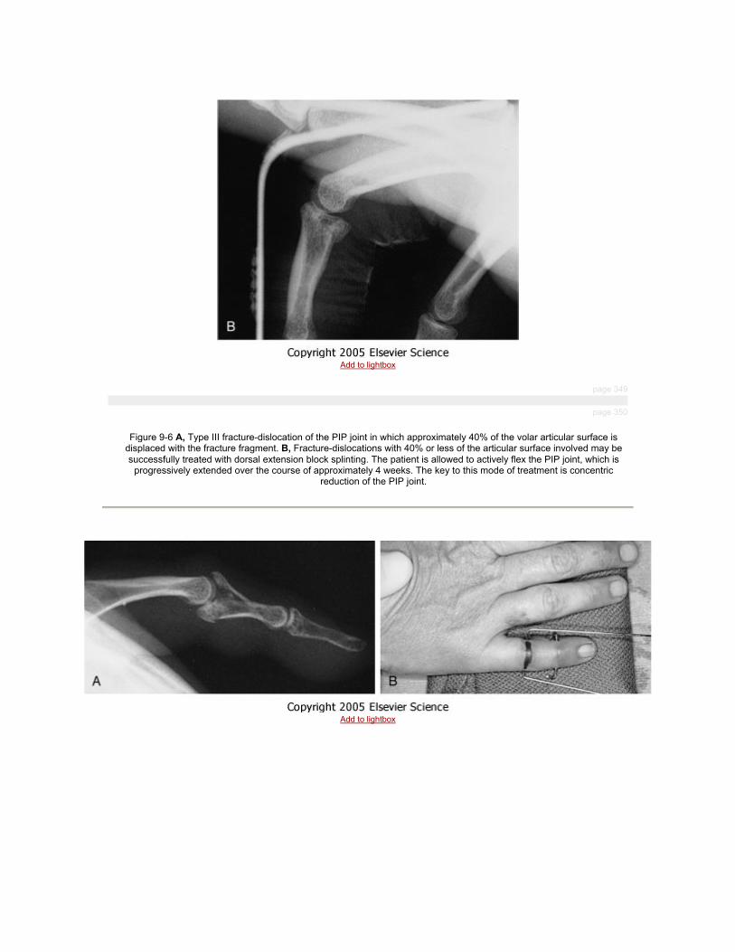
Figure 9-6 A, Type III fracture-dislocation of the PIP joint in which approximately 40% of the volar articular surface isdisplaced with the fracture fragment. B, Fracture-dislocations with 40% or less of the articular surface involved may be
successfully treated with dorsal extension block splinting. The patient is allowed to actively flex the PIP joint, which isprogressively extended over the course of approximately 4 weeks. The key to this mode of treatment is concentric
reduction of the PIP joint.
Add to lightbox

Add to lightbox
Add to lightbox
Figure 9-7 A, Lateral radiograph shows a pilon-type fracture of the middle phalangeal base. B, The wires of a dynamictraction device are placed in the proximal and middle phalanges in a manner similar to that described by Suzuki and
associates. 107
C, The two wires are connected by rubber bands. D, Lateral view of the traction device. E, Postreductionlateral radiograph shows significantly improved position of the articular fragments. (Courtesy of Benjamin Rosenstadt, MD,
New York, NY.)
ORIF is an excellent form of treatment when there is a single large volar fragment. 34
Theprocedure is performed through a volar zigzag incision, based radially in the index and longfingers and ulnarly in the ring and small fingers to reduce the potential for contact hypersensitivity.The PIP joint is entered in the interval between the flexor sheath and accessory collateralligaments on one or both sides of the joint. Careful preoperative evaluation of the lateralradiograph will have revealed any impaction of the remaining dorsal articular surface. This iselevated with a dental pick or Freer elevator, and the remaining void is filled with a smallautologous or allograft cancellous crouton.
A single, large fragment may then be reduced and held with one or two small Kirschner wires, thegoal being an anatomic, stable reduction. Mini-fragment lag screw fixation may be performed, ifpreferred over Kirschner wire fixation. In hard bone, the small screw head(s) are counter-sunk todecrease flexor sheath irritation and care is taken to avoid screw tips extending beyond the dorsalcortex and irritating the extensor mechanism. Excision of the collateral ligaments attached to thefragment greatly improves visualization and ease of manipulation, but preserving the volar plateinsertion on the fragment seems to maintain sufficient blood supply for fracture healing to occur.Protected early motion is the goal. (See Chapter 8 for more detailed description of ORIFtechniques.)

decrease flexor sheath irritation and care is taken to avoid screw tips extending beyond the dorsalcortex and irritating the extensor mechanism. Excision of the collateral ligaments attached to thefragment greatly improves visualization and ease of manipulation, but preserving the volar plateinsertion on the fragment seems to maintain sufficient blood supply for fracture healing to occur.Protected early motion is the goal. (See Chapter 8 for more detailed description of ORIFtechniques.)
The majority of unstable PIP dorsal fracture-dislocations are not amenable to ORIF, and weprefer to treat these with volar plate arthroplasty.
8,22,26,46,47,72 Although not necessarily apparent
radiographically, the volar fragment is frequently comminuted, making ORIF difficult orimpossible. Through the same exposure, the feasibility of ORIF is assessed. If ORIF isimpossible, then resurfacing of this depressed, irregular area can be achieved by advancement ofthe fibrocartilaginous volar plate.
Technique of PIP Volar Plate Arthroplasty.
The PIP joint is exposed using a radially based flap through a chevron-shaped incision with itsapex at the ulnar midaxial point. The flexor sheath is excised between the A2 and A4 pulleys, andthe flexor tendons are atraumatically retracted with a Penrose drain. The articular surfaces arevery difficult to assess with the joint reduced. Hyperextension of the joint will usually expose thedistal edge of the fracture fragment, facilitating entrance into the joint. To gain optimal exposure,the collateral ligaments that remain attached to the middle phalanx are excised except for themost volar remnants. These are preserved for later use in the procedure to suture each corner ofthe volar plate margin after advancement. Excising the collateral ligaments allows the joint to bemaximally hyper-extended, as one would open a shotgun. With both articular surfaces completelyexposed, the feasibility of reduction and fixation of the fragments can be determined.
page 350
page 351

Add to lightbox

Add to lightbox
Figure 9-8 A, Technique of volar plate arthroplasty (see the text). B, Intraoperative lateral radiograph confirming
congruous reduction of a volar plate arthroplasty. The pull-out suture marks the position of the volar plate within thearticular defect.
Congruous reduction may not be possible if the fracture fragments are markedly comminuted orimpacted. Loose bone fragments and the segment attached to the volar plate are débrided. Thedefect in the volar rim of the middle phalanx is shaped into a transverse groove perpendicular tothe long axis of the phalanx. Deeply impacted fragments may be left undisturbed to serve as abuttress for the volar plate. The interval between the volar plate and both collateral ligaments isincised, and the fibrocartilaginous plate is mobilized if necessary to allow its advancement 4 to 6mm distally into the defect in the middle phalanx. The more recent the injury, the more easily theplate will advance. In late cases, it is usually necessary to partially release the proximal checkreinligaments to gain sufficient length for advancement.
The volar plate is advanced into the middle phalangeal defect by means of a pull-out wire orsuture, which spirals along the lateral margins of the volar plate and is then passed through drillholes in the lateral margins of the middle phalangeal defect (Fig. 9-8A). The holes are made bydrilling a Keith needle from volar to dorsal, and the needles are threaded with the suture andpulled out dorsally. These holes should be as proximal as possible to draw the plate against theedge of the remaining articular cartilage. They should exit the dorsum of the middle phalanx morecentrally through the triangular ligament of the extensor mechanism to avoid binding down thelateral bands. The DIP joint should be flexed 30 degrees as the suture is passed through theextensor mechanism to avoid tethering the tendon. Traction on the sutures emerging from thedorsum of the middle phalanx facilitates reduction of the joint as the plate is advanced into thedefect.

holes in the lateral margins of the middle phalangeal defect (Fig. 9-8A). The holes are made bydrilling a Keith needle from volar to dorsal, and the needles are threaded with the suture andpulled out dorsally. These holes should be as proximal as possible to draw the plate against theedge of the remaining articular cartilage. They should exit the dorsum of the middle phalanx morecentrally through the triangular ligament of the extensor mechanism to avoid binding down thelateral bands. The DIP joint should be flexed 30 degrees as the suture is passed through theextensor mechanism to avoid tethering the tendon. Traction on the sutures emerging from thedorsum of the middle phalanx facilitates reduction of the joint as the plate is advanced into thedefect.
Lateral radiographs are obtained to confirm that a congruous reduction has been achieved (seeFig. 9-8B). Maintenance of the reduction with articular gliding through the flexion arc (as opposedto hinging on the dorsal articular surface) must be documented. This is particularly true in chronicfracture-dislocations with dorsal adhesions. If hinging is present, then additional release isnecessary, usually of the dorsal capsule, which may have become scarred and inelastic overtime. Once a congruous reduction and arc of motion is ensured, the pull-out sutures are tied overfelt and a button. A secondary suture is placed between each lateral margin of the volar plate andits adjacent remaining collateral ligament remnant. This reestablishes three-dimensional stabilityand ensures broad coverage of the condyles and phalangeal base. If there is too much laxity inthe volar plate with the joint reduced, additional tightening sutures may be placed to further tie thesides of the volar plate to the accessory collateral ligaments, although this is rarely necessary. Anoblique Kirschner wire is used to maintain the reduced joint in 20 to 30 degrees of flexion. On rareoccasions, a perceived lack of osseous support distal to the volar plate insertion may becorrected with cancellous bone graft or even the fracture fragments that are not otherwisereconstructable.
Postoperative Management
DIP joint motion is started immediately. Three weeks after surgery, the Kirschner wire is removedand active, unlimited flexion of the PIP joint is begun using a dorsal extension block splint.Unrestricted active extension is permitted at 4 weeks after surgery, and dynamic extensionsplinting is begun if full active extension is not regained by 5 weeks after surgery. Unlimitedsports activities are allowed at 8 weeks with buddy taping, and buddy taping is continued for 4 to6 months. Swelling may persist for several months, and it may take up to 6 to 8 months toachieve final range of motion.
26,46,47
Expected Outcome
page 351
page 352
CRITICAL POINTS: VOLAR PLATE ARTHROPLASTY FORDORSAL FRACTURE-DISLOCATIONS OF THE PIP JOINT
INDICATION
• Unstable dorsal PIP fracture-dislocations with more than 40%
articular surface involvement and not amenable to ORIF

PREOPERATIVE EVALUATION
• Standard radiographs (anteroposterior, lateral, oblique) of digit• Determination if remaining dorsal articular surface is anatomic or
impacted
• Age of the injury
PEARLS
• It may be used as a bailout through same approach if ORIF is notpossible.
TECHNICAL POINTS
• Use volar approach with chevron incision.• "Shotgun" joint after proper collateral ligament excision.• Excise comminuted fragments and elevate dorsal, impacted articular
surface.• Place holes for pull-out sutures as proximal as possible and avoid
lateral bands dorsally.• Establish ideal length of volar plate with joint reduced.• Reduce joint, tie sutures with DIP flexed, and document congruency
radiographically in flexion and extension.
• Pin in 20 to 30 degrees of flexion for 3 weeks only.
PITFALLS
• Leaving stable but impacted dorsal articular surface unaddressed• Impaling lateral bands with pull-out sutures• Failing to spread volar plate broadly across condyles
• Failing to identify hinged versus gliding flexion via dorsal adhesions
POSTOPERATIVE CARE
• Immobilize joint with Kirschner wire and splint for 3 weeks.• Remove pin at 3 weeks with radiographs after pin removal.• Use extension block flexion for 1 to 3 weeks and then unlimited
extension.• Use a dynamic extension splint at 5 to 6 weeks as needed.
ATHLETIC PARTICIPATION
• Contact sports including basketball can be resumed at 8 weeks withbuddy-taping.
The hand surgeon will often see PIP fracture-dislocations after patients have been initially treatedelsewhere, and the true nature of this complicated injury may not have been appreciated orconveyed to the patient. The patient must understand from the outset that while normal PIP jointfunction (as compared with the contralateral or adjacent digits) is possible, it is highly unlikely.They can be reassured that carefully planned treatment and compliance with postoperativeregimens will likely lead to long-term satisfactory to good results. Of course, overall resultsdiminish with increased time from injury to treatment, especially beyond 6 weeks.
17a,26

regimens will likely lead to long-term satisfactory to good results. Of course, overall resultsdiminish with increased time from injury to treatment, especially beyond 6 weeks.
17a,26
Eaton and Malerich reported on 24 patients who underwent volar plate arthroplasty for both acuteand chronic PIP fracture-dislocations, with a 10-year average follow-up.
26 The seven cases
performed within 6 weeks of injury attained an average of 95 degrees of motion and a 6-degreeflexion contracture. In contrast, the 17 patients with chronic (>6 weeks after injury) diseaseachieved 78 degrees of motion and 12 degrees of contracture. Only 3 patients reported any pain,and only with strenuous use. More recent reports have confirmed the reliability of this procedurein producing good results in a majority of patients.
8,22
Dionysian and Eaton have reviewed the results of 17 volar plate arthroplasties at a mean follow-up of 11.5 years and found that the long-term benefit of the procedure endured with an averagearc of PIP motion of 85 degrees and no residual pain.
19
Complications
Careful preoperative planning, intraoperative attention to detail with awareness of potentialpitfalls, and good patient compliance will minimize the risk of complications associated withtreatment of these injuries.
Redisplacement.
Failure to achieve a stable reduction, pull-out suture failure, or inadequate protection to preventextension during mobilization may result in recurrent dorsal subluxation. One cause of pull-outsuture failure is damage from the transarticular Kirschner wire fixation. This complication may beobviated by pre-positioning of the wire in the middle phalanx after Keith needle passage butbefore the sutures are pulled through. With close clinical and radiographic follow-up, suchproblems can be more expeditiously addressed and their effects minimized.
Angulation.
Asymmetrical impaction of the base of the middle phalanx or failure to create a trough for thevolar plate that is perpendicular to the long axis of the middle phalanx will occasionally result inangulation of the middle phalanx. Although postoperative angular deformity is usually mild andnot functionally significant, patient satisfaction may be compromised and secondary correctiveosteotomy at the base of the middle phalanx may become necessary.
26
Flexion Contractures.
page 352
page 353
Immobilization of the PIP joint in more than 30 degrees of flexion and failure to begin dynamicextension splinting by 5 weeks may result in lack of full extension. Recognition of this potentialand common problem should help minimize it. Even with early motion and formal hand therapy, acertain percentage of patients will develop a PIP flexion contracture.

DIP Stiffness.
Failure to flex the DIP joint approximately 30 degrees or impaling the lateral bands while passingthe pull-out suture through the extensor mechanism on the dorsum of the middle phalanx maycause limitation of DIP flexion. Modifications in the technique help to decrease the risk of DIPstiffness. These include using two separate sutures for each side of the volar plate, tying thesuture knots beneath the skin dorsally, or using small suture anchors in lieu of the pull-out suture.It is imperative to start DIP motion immediately postoperatively to regain the maximal motionpossible.
pages 345 - 353
Copyright © 2008 Elsevier Inc. All rights reserved. Read our Terms and Conditions of Use and our PrivacyPolicy.
For problems or suggestions concerning this service, please contact: [email protected]
LATERAL PIP DISLOCATIONS
The critical anatomic lesions in a lateral dislocation of the PIP joint are a rupture of one collateralligament and at least partial avulsion of the volar plate from the middle phalanx. Failure probablybegins with disruption of the origin of the collateral ligament from either the proximal phalangealhead or the middle phalangeal base,
92 proceeds through the junction of the collateral and
accessory collateral ligaments, and finally detaches the insertion of the volar plate on the middlephalanx. To assess PIP stability after spontaneous or manual reduction of the joint, the examinermust test the joint in extension to assess not only the collateral ligaments but also the secondarystabilizers (e.g., volar plate, articular contour).
76 More than 20 degrees of deformity on gentle
static lateral testing indicates complete collateral ligament disruption and injury to at least oneother secondary stabilizer.
54,77 This is a major disruption of the four-sided ligamentous box, but
with few exceptions the ligaments heal when the joint is reduced and early controlled motionbegun.
105,113 The joint should be protected with buddy-tapes to an adjacent uninjured digit. On
occasion, these injuries may benefit from a malleable splint placed along the same side as theligament tear to prevent inadvertent lateral deviation.
Repair of the ruptured collateral ligaments has been frequently reported after PIP dislocations ingeneral and lateral dislocations in particular.
5,11,35,46,47,74,91,94 Most of these series involve athletes,
and surgery can be performed in selected patients to provide more predictable stability withreduced length of disability. Kato and colleagues reported on 11 primary repairs of acute PIP jointcollateral ligament tears in athletes and manual laborers.
52 Suture anchors were utilized in the
repair, active motion was initiated at 2 to 3 weeks, and unlimited activities were allowed at 6weeks.
Nevertheless, because the usual sequela of ligament injury of the PIP joint is stiffness and notinstability, the surgical trauma of ligament repair may have an adverse effect on the ultimaterange of motion after lateral dislocations. A clearer indication for surgical repair or reconstructionis the sub-acute or chronic collateral ligament rupture with persistent PIP instability anddysfunction.

VOLAR PIP DISLOCATIONS
Volar dislocations of the PIP joint are rare injuries. The base of the middle phalanx may dislocatevolarly without rotation (volar dislocation) or may rotate on one intact collateral ligament so thatthe opposite side subluxates in a volar direction (volar rotatory subluxation). Appreciating thedifference between these two related injuries and identifying them clinically can help ensureappropriate management.
34 Volar rotatory subluxation of the PIP joint is a rare injury.
The mechanism of injury is usually a rotatory longitudinal compression force on a semiflexedmiddle phalanx that results in unilateral disruption of a collateral ligament and partial avulsion ofthe volar plate. One of the most common causes is catching the involved digit in a spin dryer thathas not stopped spinning.
33 As the middle phalanx displaces volarly, the involved condyle
ruptures through the extensor mechanism. Usually this rupture occurs between the central slipand the ipsilateral lateral band. The involved condyle may buttonhole between the central slip andthe thickened volar fibers of the lateral band. These fibers are caught behind the volar flare of thecondyle, routing the distal lateral band through the joint as it remains in continuity with the centraltendon over the middle phalanx. Because of this interposition, the usual reduction maneuver oftraction and middle phalanx extension tends to further tighten this encirclement of the condyleand block reduction.
Occasionally, when a volar dislocation occurs without a rotatory component, the central slip isruptured. If the dislocation is irreducible, there is a high likelihood of an inter-posed structure suchas the central slip, a collateral ligament, or a fracture fragment.
15,45 If the joint reduces easily, one
must still be wary of a more profound injury that may have occurred to the extensor mechanism.This must be taken into account when deciding on the position of immobilization for the PIP joint.35
Volar fracture-dislocations are also rare. Rosenstadt and coworkers reported on 13 cases treatedsurgically, of which 9 involved acute injuries.
96 Variations in the size of the dorsal fragment and
age of the injury dictated specific treatment (closed pinning versus ORIF). At an average of 4 1/2
years of follow-up, the acute injuries fared better than those that presented at 4 or more weeks.Average arc of motion was 91 degrees for the acute injuries and 70 degrees for the chronicinjuries. Five patients had an average 25-degree extensor lag at final follow-up.
Volar fracture-dislocations with a large dorsal fragment may be treated with ORIF via mini-fragment lag screw fixation. Tekkis and coworkers reported two such cases with excellent clinicaloutcomes
109 and attributed their success to rigid screw fixation and immediate active motion.
Care must be taken to avoid distal thread extension into the flexor sheath; similarly, dorsal screwhead prominence may require later screw removal.
page 355
page 356
The management of chronic volar dislocations of the PIP joint is complex and requires thesimultaneous surgical correction of an incongruous and contracted joint as well as anincompetent extensor mechanism.
87 There may be degeneration of the articular cartilage; if so,
the prospect of restoring normal joint function is greatly diminished. Therefore, it is important torecognize a volar PIP joint dislocation at the time of injury and adequately treat the disruptedextensor mechanism.
30,35,87,89 Peimer and colleagues also pointed out the necessity of repair of
the ruptured collateral ligament. 87
In this relatively large series of 15 patients with volar PIPdislocations, inspection at the time of surgery revealed disruption of the extensor mechanism, thevolar plate, and one collateral ligament in all patients. In each, the extensor mechanism andcollateral ligament were repaired, and motion with therapy was started at 4 weeks after removalof the Kirschner wire that transfixed the joint. Although all patients had painless, stable PIPmotion, no patient achieved preinjury PIP motion.

recognize a volar PIP joint dislocation at the time of injury and adequately treat the disruptedextensor mechanism.
30,35,87,89 Peimer and colleagues also pointed out the necessity of repair of
the ruptured collateral ligament. 87
In this relatively large series of 15 patients with volar PIPdislocations, inspection at the time of surgery revealed disruption of the extensor mechanism, thevolar plate, and one collateral ligament in all patients. In each, the extensor mechanism andcollateral ligament were repaired, and motion with therapy was started at 4 weeks after removalof the Kirschner wire that transfixed the joint. Although all patients had painless, stable PIPmotion, no patient achieved preinjury PIP motion.
Authors' Preferred Method of Treatment The management of volar rotatory dislocations of thePIP joint is controversial. Volar rotatory dislocation has been described as irreducible or trappedby many authors and one that necessitates open reduction.* Open reduction is advocated notonly to reduce the interposed extensor mechanism but also to repair the rent in this mechanism.Others feel compelled to repair the torn ligaments and volar plate as well.
74,102,124 We believe that
open reduction is necessary only if closed reduction fails and have a great deal of success usingthe specific reduction maneuver described next.
45-47,87
The majority of volar rotatory dislocations can be reduced without surgery by applying gentletraction while holding both the MP and PIP joints flexed.
110 This maneuver relaxes the volar-
displaced lateral band so that with a gentle rotatory motion the intra-articular portion can bedisengaged from behind the condyle and reduction accomplished. If necessary, further relaxationof the extensor mechanism can be gained by moderate wrist extension. Once the joint is reduced,active motion is tested. Because the ligament lesion is a collateral ligament disruption, when thejoint is reduced the ligaments are usually restored to their anatomic alignment. Postreductionradiographs should confirm congruous reduction. After reduction of a volar rotatory dislocation,full active extension is usually possible (under digital block) because the contralateral lateral bandand at least a portion of the central slip usually remain intact. If the patient cannot actively extendto neutral, the PIP joint should be immobilized in full extension for 6 weeks, just as one wouldtreat a closed boutonnière deformity. The only indication for open reduction, therefore, would befailure to obtain a completely congruous reduction, confirmed by radiographs, and presumablydue to the presence of ligament, capsule, or extensor mechanism trapped within the joint.
In volar dislocations without a rotatory component, the reduction is easily accomplished.However, one must assume an injury to the central slip of the extensor mechanism has occurredand thus treat the finger in the postreduction period as one would treat an acute boutonnièredeformity, in full extension for 4 to 6 weeks.
Technique of Open Reduction of Volar Rotatory PIP Dislocation
Open reduction is approached through a midaxial incision on the side of the major ligamentdisruption. The lateral band is atraumatically extricated from the joint and reduction is easilyaccomplished. With the patient under local or wrist block anesthesia it is then possible to testactive extension. If the lateral band is not severely traumatized, it may be carefully repaired.Should the band be ragged, it is best to excise it because the normal contralateral lateral band issufficient to provide intrinsic extensor power. If full extension is demonstrable on examination, thefinger should be immobilized in extension for 5 to 7 days until the wound has stabilized and thenactive range of motion begun. Dynamic extension splinting is alternated with a resting extensionsplint to protect the extensor mechanism.
*See references 15
, 17
, 33
, 46
, 47
, 50
, 51
, 56
, 76
, 80
, 85
, 90
, 99
, 102
, 118
, and 124
.

pages 355 - 356
Copyright © 2008 Elsevier Inc. All rights reserved. Read our Terms and Conditions of Use and our PrivacyPolicy.
For problems or suggestions concerning this service, please contact: [email protected]