disorders of blood circulation and of lymph. cellular adaptations ii. healing. · 2016-09-26 ·...
TRANSCRIPT

Disorders of blood circulation and of lymph.
Cellular adaptations II. Healing.
(Brown induration of lungs, chronic venostasis of liver, pulmonary artery embolism,
lung edema, hypertrophy of myocardium, metaplasia, organisation of
fibrinous perotonitis, organisation of thrombus, healing of MI,
foreign body granuloma)
General Medicine
Svetoslav Štvrtina, M.D., PhD.
Michal Palkovič, M.D., Ph.D.
Vladimír Šišovský, M.D., Ph.D. František Kiš
Department of Pathology, FMCU and FH Work place Staré mesto
Sasinkova 4, Bratislava
Prof. MUDr. Ľudovít Danihel, CSc.

Disorders of blood
circulation (B.C.)
• Systemic
• Local
Disorders can be:
+
-
Different division:

(+) Disorders
• Hyperemia
• Increased volume of blood
• Increased number of blood elements
• Increased volume of fluids and
electrolytes
• Increased permeability of capillaries
• Increased coagulability of blood
• Increased blood pressure

(-) Disorders
• Heart failure
• Decreased blood pressure
• Disorders of the amount and
composition of blood
• Disorders of circulatory system
integrity

Disorders of blood circulation
To maintain normal blood flow:
1.Normal anatomic features
2.Normal physiologic controls for blood flow
3.Normal biochemical composition of the blood
Haemodynamic disturbances:
1.Disturbances in the volume of the circulating blood
(hyperaemia, congestion, haemorrhage, shock)
2.Circulatory disturbances of obstructive nature
(thrombosis, embolism, ischaemia, infarction)

Disturbances in the volume of circulating blood
Hyperaemia (active hyperaemia) – increased
volume of blood from arterial and arteriolar
dilatation (e.g. inflammation, high grade fever)
- clinically - redness, raised temperature
Venous congestion (passive hyperaemia,
venostasis) – dilatation of veins and capillaries due to
impaired venous drainage
- clinically – bluish colour (cyanosis)
- acute / chronic
1. local – e.g. portal venous obstruction in cirrhosis of the
liver
2. systemic (general) – in left-sided heart failure
(pulmonary congestion), in right-sided heart failure
(chronic venous congestion of the liver, spleen, kidney)

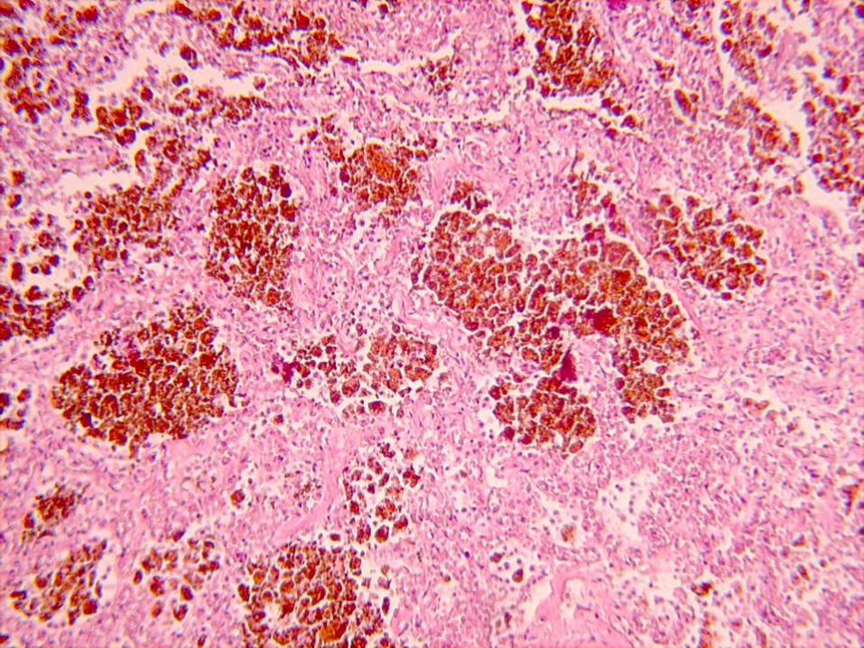
• Left heart failure
• Blood stasis in lungs with dilatated capillaries filled with Er
• Accumulation of fluid in alveoli
• Acculmulation of erythrocytes in alveoli → Phagocytosis of erythrocytes → Metabolic change of hemoglobin to hemosiderin → Creation of siderophages („heart failure cells“)
• Fibrosis of alveolar septi
• Pulmonary hypertension
Brown induration of lungs (157)


• right heart failure, occlusion of inferior vena cava, hepatic vein
• yellowbrown liver tissue with dark red foci – nutmeg liver, later opposite image
• dilatation of central veins and sinusoids → pressure atrophy or hypoxic necrosis of centrolobular hepatocytes and hypoxic steatosis of peripheral hepatocytes
• later - accumulation of
connective tissue – fibrosis
(cardiac)
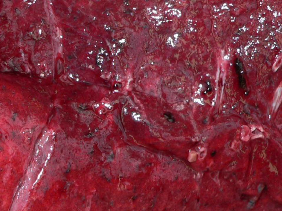
Chronic venostasis of liver (10)


Chronic venostasis of liver.


Disturbances in the volume of
circulating blood
• Collapse (orthostatic collapse) - sudden transient circulation failure
• Shock – profound failure of circulation to
maintain appropriate blood supply of vital organs
– cardiogenic
– hypovolemic
– septic (toxaemic)
– others (traumatic, neurogenic,...)

Shock kidneys (327) Macro : large, swollen kidney, congested, but the cortex
may be pale. A cross-section shows blood pooling in the
outer stripe od medulla.
Micro : acute tubular necrosis – dilatation of the proximal
tubules, focal cell necrosis, pigmented casts in the tubular
lumina, interstitial edema.


Shock adrenal gland (328)
Macro : conspicuous hemorrhage in the inner cortex.
(Waterhouse-Friderichsen syndróm)
Micro : necrosis, intracapillary haemocoagulation,
increase in ACTH secretion, widening of the space
between trabeculae (oedema) and thinning of the
trabeculae - pseudotubular degeneration


Circulatory disturbances of
obstructive nature
• trombosis
• embolism
• ischaemia
• Infarction
Ischemia
– deficient blood supply (obstruction,
compression, spasm,...)
– ischemia ... Infarction (necrosis)

Trombus, trombosis
Thrombus (aggregate of coagulated blood within vascular lumen,
adheres to EC)
- in the heart, arteries, veins, microcirculation
- Virchow trias – 1. endothelial injury
2. altered blood flow
3. hypercoagulability
Grossly:
– white thrombus (arterial)
– red thrombus (venous)
– mixed thrombus
- lysis / propagation / organisation / recanalisation
Arterial thrombi - ischaemia...necrosis
Cardiac, venous thrombi - tromboembolism
Microthrombi in microcirculation - DIC

Embolus (material carried in the bloodstream that can lead
to vessel obstruction)
a) solid (trombus, tumour cells, parasites,
bacterial clumps, foreign bodies)
b) liquid (amniotic fluid, fat, bone marrow)
c) gaseous (air)
- paradoxical embolus
- retrograde embolus
Embolus, Embolism

• Most often by blood thrombus (from deep
veins of lower extremities)
• Other possibilities: malignant cells, air, fat,
amniotic fluid, bacteria
• Results depends on size of the embolus:
- asymptomatic / minor dyspnoea →
pulmonary hypertension and RV failure
- pulmonary infarction
- cardiovascular collapse with sudden death
Pulmonary artery embolism (7)

Embolus
Lungs

Embolus
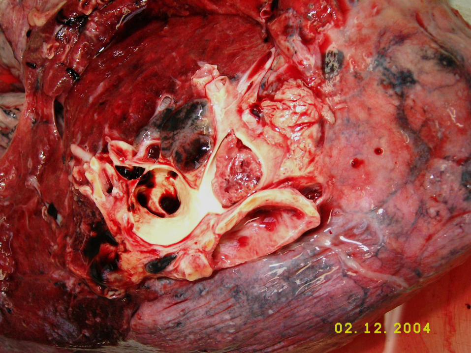

Thrombus (embolus) Lungs – venostasis and bleedig


1.Oedema
2.Dehydratation
3.Overhydratation
Disturbances of body fluids

- excess of fluid in interstitial spaces, cavities
- localised (inflammation, allergy, venous or lymphatic
obstruction)
- generalized (heart failure, renal, hepatal disease...)
• Causes
– Increase of hydrostatic pressure (art,.,venous)
– Decreased content of proteins in serum (oncotic)
– Increased permeability of capillaries
– Disorder of lymphatic drainage
– Sodium and water retention
Oedema

Arteriole Venule
Lymphatic vessel with node
Capillary
AP VP B B 4,27 kPa 1,6 kPa
3,33 kPa 3,33 kPa

Arteriole Venule
Lymphatic vessel with node
Capillary
AP VP B B
Norm (standard)
10% 100% 90%

Arteriole Venule
Lymphatic vessel with node
Capillary
AP VP B B
Venostasis
30%
X X X
X X X

Arteriole Venule
Lymphatic vessel with node
Capillary
AP VP B B
Proteins
X X X
X X X

Arteriole Venule
Lymphatic vessel with node
Capillary
AP VP B B
Inflammation
X X X
X X X

Arteriole Venule
Lymphatic vessel with node
Capillary
AP VP B B
Lymphostatic
edema
X
X
X X X
X X X

• Accumulation of liquid first in the interstitium and
later in alveoli
• Causes – left heart failure, inflammation,
disorders of blood plasma composition,
disorders of lymphatic drainage, combination
• Histology: light pink homogenous material in
alveoli – by fixation coagulated proteins of
edematous liquid
• Optically empty rounded spaces – air bubbles
Lung oedema (265)


Disorders of lymphatic drainage -
lymphoedema
• removal of axillary lymph nodes (in radical
mastectomy)
• pressure from outside (tumours)
• injury of thoracic duct
• chylothorax
• chylous ascites
• inflammation of the lymphatics (filariasis –
elephantiasis)
• occlusion of lymphatic channels by
malignant cells
• Milroy's disease -hereditary lymphoedema

Cellular adaptations
• Physiologic adaptation – to the physiologic needs
• Pathologic adaptation – to non-lethal pathologic injury
• Adaptations:
• 1. atrophy – reduction of the number and size of cells
• 2. hypertrophy – increase in size of cells
• 3. hyperplasia – increase in number of cells
• 4. metaplasia – reversible change of one type of mature cells to another type of mature cells
• 5. dysplasia – disordered cellular development

Hypertrophy
• enlargement of cell volume

Hypertrophy
• Physiologic (enlarged size of the uterus in
pregnancy, also hyperplasia)
• Pathologic:
1.hypertrophy of cardiac muscle
2.hypertrophy of smooth muscle
3.hypertrophy of skeletal muscle
4.compensatory hypertrophy

• Hypertension in PC
- acute – RV dilatation (cor pulmonale acutum)
- chronic – RV hypertrophy (cor pulm.chron.)
• Hypertension in SC
• esencial / secundary hypertension
• LV hypertrophy (cor hypertensivum)
→ cor bovinum
• Hypertrophy
– concentric – pressure overload
– eccentric – volume overload
Myocardial hypertrophy

Hypertrophy of myocardium (291)


Hyperplasia
• increas of cell number

Hyperplasia
• Physiologic:
1.hormonal (pregnant uterus, female breast during
lactation)
2.compensatory (liver after partial hepatectomy)
• Pathologic:
• endometrial hyperplasia in oestrogen excess
• intraductal epithelial hyperplasia in the breast
• skin wart from hyperplasia of epidermis (HPV)

Metaplasia
– change of one differenciated tissue to another

Metaplasia
• Epithelial:
1.squamous (bronchus, uterine cervix)
2.columnar (intestinal metaplasia in gaster, Barrett's
oesophagus)
• Mesenchymal:
1.osseous (in scar, fibrous stroma of tumour)
2.cartilaginous (in wrong healing fractures)

Metaplasia (174)


Dysplasia
= atypical hyperplasia
•most often in epithelial cells (uterine cervix,
respiratory tract) – cellular proliferation and
cytologic changes
•may progress into carcinoma in situ,
invasive cancer

Healing
• Body response to injury – to restore
normal structure and function
• Regeneration – Replacement of damaged tissue by equivalent
tissue, proliferation of parenchymal cells
• Repair – Replacement of damaged tissue by second
rate tissue, proliferation of connective tissue
elements→fibrosis, scarring
- mostly combination of both

Regeneration
– replacement of damaged tissue by equivalent tissue

Regeneration
• According to regeneration capacity
the cells are classified:
– Unstable – regularly replaced : hematopoiesis, epithelia, axon
– Stable – renewed is only damaged
tissue: glandular epithelium, proximal canaliculi,
vascular smooth muscle
– Permanent – ganglion cells, crosstriated muscle

Repair
– replacement of damaged tissue by second rate tissue (fibrous)

Repair
1. Granulation tissue formation
2. Contraction of wounds
• Wound healing
– primary (per primam intentionem)
– secondary (per secundam intentionem)
• Organisation of hematoma and
thrombus
• Healing of bone fracture
• Healing of foreign bodies
• Organisation of fibrinous exudates

Wound healing
• Combination of regeneration and repair

Healing of fractured bone

Healing of fractured bone
hematoma
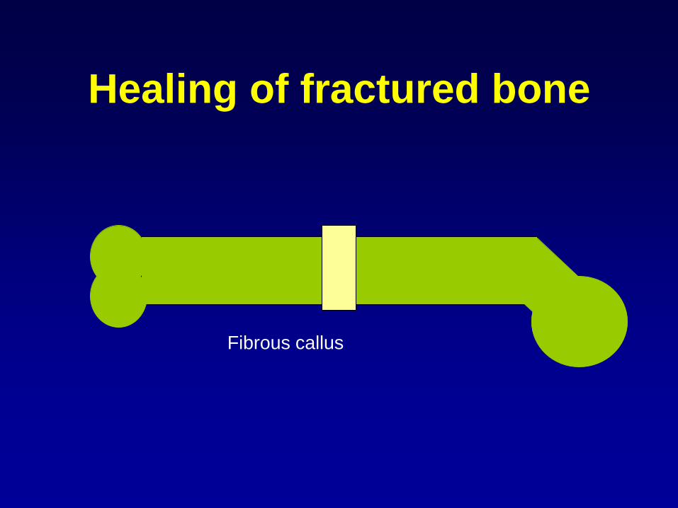
Healing of fractured bone
Fibrous callus
Healing of fractured bone
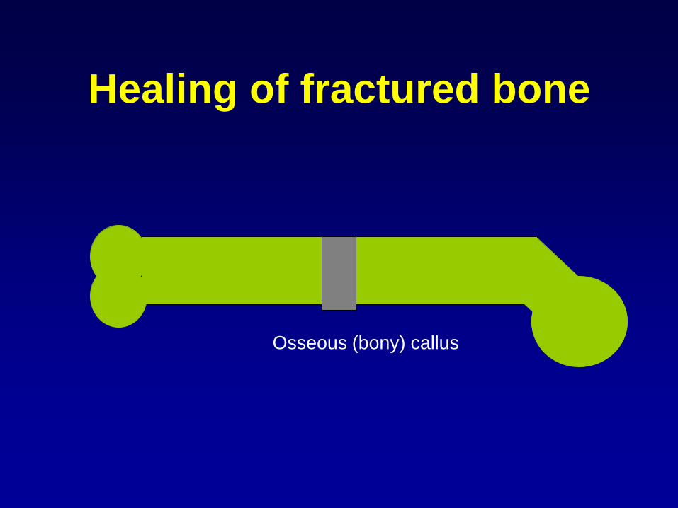
Osseous (bony) callus
Healing of fractured bone
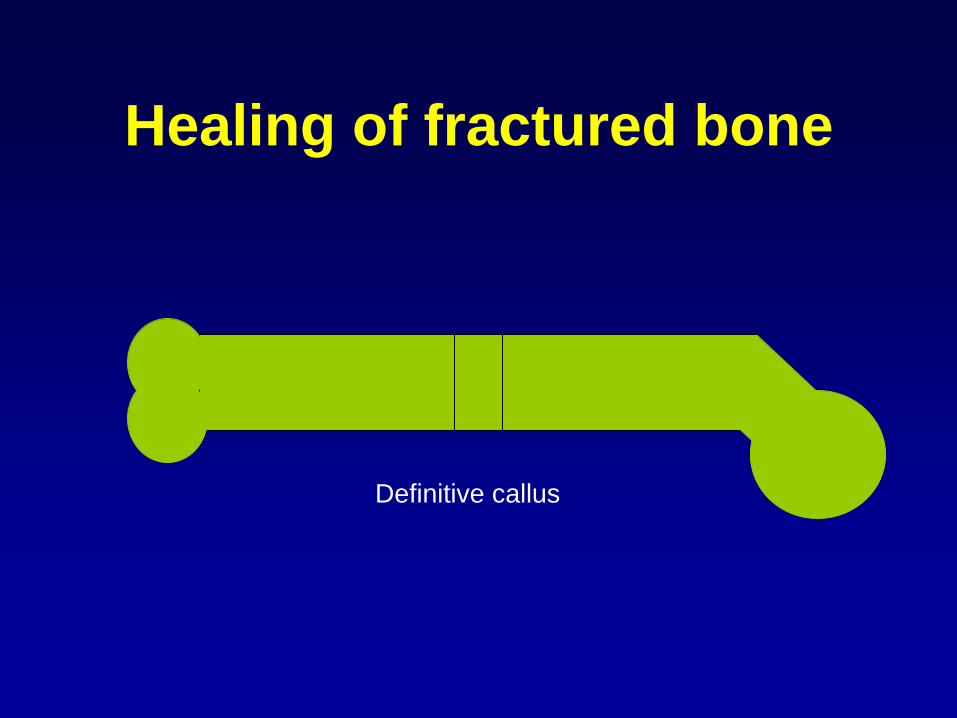
Definitive callus

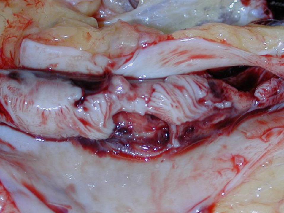
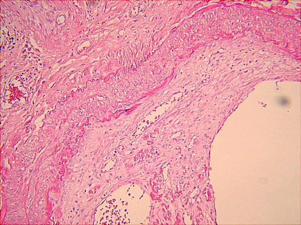
Fibrinous peritonitis in organisation(163)


Thrombus in organisation
• Thrombus is resorbed and replaced
by connective tissue
• Granulation tissue grows into the
thrombus
• Granulation tissue eliminates
thrombus and ingrowing capillaries
open their lumen - recanalisation

Organisation of thrombus

Organisation of thrombus

Organisation of thrombus

Organisation of thrombus

Organisation of thrombus

Organisation of thrombus


Mixed thrombus
Ingrowing capillaries


Healing of myocardial infarction
(293)
• granulation tissue formation
• scar maturation


Incorporation of foreign body
• Foreign body (of endogenous or
exogenous origin) – it cannot be
removed by common means such as:
– dissolving
– diluting
– phagocytosis
– elimination

Incorporation of foreign body
• Creation of granuloma around the
foreign body:
– large multinucleated cells around foreign
bodies
– connective tissue
– lymphocytes
– histiocytes
• Sometimes the granuloma grows too
large
– Schloffer’s tumor

• Scar after cholecystectomy
• Surgical stiches in 2nd picture
• Circumscribed by connective tissue
• Large cells around foreign bodies try to
phagocyte the fibres
Foreign body granuloma (19)


Large cells around
foreign bodies