dispersion staining article by walter mccrone -
TRANSCRIPT

Environmental Health PerspectivesVol. 9, pp. 57-61, 1974
Detection and Identification of Asbestosby Microscopical Dispersion Staining
by Walter C. McCrone*
Asbestos fibers as small as 1 ,m in diameter can be uniquely identified by light microscopy byemploying dispersion staining methods. The technique described herein involves suspension of fibersin liquids ofknown refractive indices and observation ofcolor display by means ofa dispersion stain-ing objective. Wavelengths or indices of refraction may be determined at right angles to and parallelto fiber axes. This method is rapid and sensitive for identification purposes.
There is a great need for a dependable, sen-sitive and rapid method for the detection andidentification of asbestos. Microscopical disper-sion staining satisfies all of these requirements.It is dependable because it is based on themeasurement of three refractive indices as wellas the dispersion of those indices. Refractive in-dices are among the most valuable identifyingcharacteristics for small particles. The methodis sensitive because the refractive indices are"read" as bright dispersion staining colorsagainst a black background. These colors can beobserved on particles well below 1 Am indiameter. It is rapid because any particle in themicroscopical preparation showing the opticalproperties of asbestos signals its presence by aunique color combination with polarized light.One particle of asbestos in a field of view con-taining many thousands of other particles willbe immediately apparent on scanning the eyeacross the field of view.
Besides optical crystallographic methods likedispersion staining, only differential thermalanalysis (DTA) and X-ray diffraction have thisability to tag a particular crystalline phase in amixture. X-ray diffraction, however, is severalorders of magnitude less sensitive than disper-sion staining and requires much more time.
*McCrone Associates, Inc., Chicago, Illinois 60616.
There are, however, compounds whose disper-sion staining colors are, at least at first glance,confused with chrysotile colors. Here, for-tunately, particle morphology is able todifferentiate between these interfering sub-stances. Quartz is one example, lizarditeanother. The latter, however, is a tabulartalclike mineral, and quartz shows conchoidalfracture into usually thin flakes. Both are easilydifferentiated from fibrous asbestos bymorphology.
Dispersion staining is, therefore, a straight-forward technique easily applied by anymicroscopist with some knowledge of opticalcrystallography. We have found it extremelyuseful for the rapid and routine examination ofany particulate samples for any of the variouskinds of asbestos (1). The necessary backgroundinformation for applying the method is given inFigure 1, which plots the matching wavelengthXo, as a function of refractive index of theCargille refractive index liquids used in thesedeterminations. Most of the asbestos mineralshave distinctive indices without overlap. Figure1 suggests, however, that amosite andcrocidolite may overlap partially. There shouldbe no confusion in this situation, however, sincey for amosite overlaps only with a forcrocidolite. The data in Figure 1 are plotted asthe averages of a considerable number of values
December 1974 57

FIGURE 1. Asbestos dispersion staining curves.
for individual mine samples previously pub-lished (1). It is interesting to look a little bitmore closely at this variation in dispersion stainingdata from mine to mine, and Table 1 lists thematching wavelength Xo for y (parallel to thefiber length) and a (perpendicular to the fiberlength) for a group of more than 30 asbestossamples from different parts of the world. Thereis some -variation from sample to sample, in-dicating composition variations. However, all ofthe data lie in the same characteristic chrysotileregion and show no overlap with any of thefibrous amphiboles.
Since dispersion staining is a relatively newtechnique requiring a certain amount of skill,not only in reading the matching wavelengthsbut also in adjusting the microscope for best dis-persion staining colors, it seems well to sum-marize a few of the common difficulties.The refractive indices given in dispersion
staining tables are not dispersion data for thatcompound. To illustrate this, we can cite thedata for the w index of quartz (Table 2).The value 1.538 for w at 486 nm is nD for
the Cargille liquid that matches quartz w at 486nm. The actual refractive index of that liquid at486 nm is 1.550, the same as quartz o). This op-eration, which simplifies the analytical pro-cedure, is used for all dispersion staining data.One can, of course, calculate the true refrac-tive indices of any substance from the disper-sion staining data. The necessary data to do thiscan be found in the table of dispersion of refrac-tive index data for the Cargille liquids.True refractive index data and dispersion
staining data are identical at 589 nm; hence,refractive index data at 589 nm for any sub-stance become dispersion staining data for thatsubstance. Chrysoberyl, for example, does notappear in the dispersion staining tables, but
Environmental Health Perspectives58

Table 1. Matching wavelength Xo in H. D. 1.550 liquid.
Sample
Quebec; Lake AsbestosQuebec; King Asbestos Corp.Quebec; Asbestos Corp.Quebec; Bell MinesQuebec; JohnsonsQuebec; Careys, BradfordQuebec; FlintkoteQuebec; NormandieOntario; ReevesOntario; MunroVermont; Hyde Park, GAFVermont; JeffreyNew Foundland; AdvocateNew FoundlandYukon; Clinton CreekBritish Columbia; CassiarCalifornia; Pacific Asbestos Corp.California; CoalingsArizonaVenezuelaRhodesiaRhodesia; ShabinaRhodesia; Havelock C and GRhodesia; Havelock HVLRhodesia; Havelock VRACyprusGreece; ZandiniYugoslaviaItalian; BalengeraRussiaAustralia; Woodsreef
Xo, nm
5I 1
510 610510 610500 610510 600500 600480 590500 610570 610480 590560 610510 620500 580510 610590 620500 580500 580480 610590 630600 620610 680
520 (460) 620 (550)480 580490 590490 590500 630600 660580 620520 590
500 (460) 600 (510)500 600610 680
Table 2. Refractive indices for quartz.
486 nm 589 nm 656 nm
True refractive indices,w 1.550 1.544 1.542Dispersion staining data 1.538 1.544 1.547
Winchell (2) gives nD = 1.746 (a), 1.748 (,B), and1.756 (-I). From these data one would mount asuspected chrysoberyl in Cargille liquid nD =1.750 and expect to see annular stop colors rang-ing from orange (,B) to greenish-yellow ( y) orgreenish-blue (a) to magenta-blue (Y) with thecentral stop. This greatly extends the usefulne§sof dispersion staining.We are often asked if the dispersion staining
objective can be supplied with a highermagnification. This gives us the opportunity to
point out that higher magnification is notdesirable. One is trying to "resolve" color of theparticles not the particles themselves. Centralstop dispersion staining is a darkfieldprocedure, hence a strong light source will showpoints of light for particles smaller than theresolving power limit of the microscope system,i.e., 1.22 A,m for NA= 0.25, 1ox objective. Thesepoints of light will be colored for particles show-ing dispersion in that liquid. The lower limit ofdetection is a function of light intensity and con-trast.There are a number of points of technique
which greatly improve the sensitivity of the dis-persion staining procedure. The particles mustbe well separated in the mounting liquid sincenonstained particles close to or overlappingstained asbestos can mask their presence. Thedispersion staining colors for chrysotile and thefiber amphiboles are more brilliant if one usesthe high dispersion Cargille set of refractive in-dex liquids. In spite of the above injunction con-cerning high magnification, it is sometimesuseful to use a 20-25X ocular with the 1OX dis-persion staining objective. The optics for the dis-persion staining objective, the axis of stage rota-tion, the substage apertures and lenses must bewell aligned on the same optical axis. It is a goodidea to take special pains to align the opticalsystem and to maintain that microscope for dis-persion staining examination only. The problemof glare from other particles in the field of viewis solved to a great extent by having a centeredand nearly closed field diaphragm in the opticalsystem. This concentrates attention on particlesin the center of the field and eliminates wellover 90% of the glare which makes it difficult tosee very fine asbestos fibers. It is desirable to beable to change the orientation of any particleswhich appear at first sight to give the distinctivedispersion staining colors characteristic ofchrysotile (or other fibrous amphiboles). Rollingthe particle by sliding the cover slip with aviscous liquid prep is the ideal way of doing thisand helps greatly in differentiating quartz,paper fibers and mineral wool from chrysotile.The slides and cover slips used for dispersionstaining preparations should be unusually cleansince any optical discontinuities on any surfaceof the preparation cause glare and interferewith visibility of the dispersion staining colors.
December 1974 59
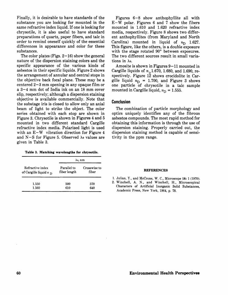
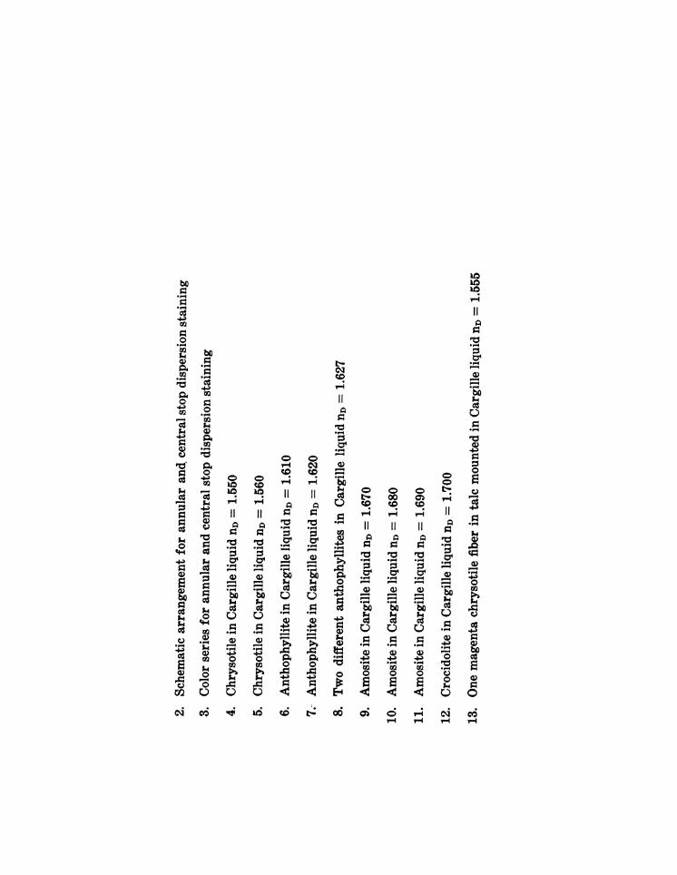
Finally, it is desirable to have standards of thesubstance you are looking for mounted in thesame refractive index liquid. If one is looking forchrysotile, it is also useful to have standardpreparations of quartz, paper fibers, and talc inorder to remind oneself quickly of the essentialdifferences in appearance and color for thesesubstances.The color plates (Figs. 2-16) show the general
nature of the dispersion staining colors and thespecific appearance of the various kinds ofasbestos in their specific liquids. Figure 2 showsthe arrangement of annular and central stops inthe objective back focal plane. These may be acentered 2-3 mm opening in any opaque film ora 3-4 mm dot of India ink on an 18 mm coverslip, respectively; although a dispersion stainingobjective is available commercially. Note thatthe substage iris is closed to allow only an axialbeam of light to strike the object. The colorseries obtained with each stop are shown inFigure 3. Chrysotile is shown in Figures 4 and 5mounted in two different standard Cargillerefractive index media. Polarized light is usedwith an E-W vibration direction for Figure 4and N-S for Figure 5. Observed Xo values aregiven in Table 3.
Table 3. Matching wavelengths for chrysotile.
Xo, nm
Refractive index Parallel to Crosswise toof Cargille liquid n D fiber length fiber
1.550 500 5701.560 610 640
Figures 6-8 show anthophyllite all withE-W polar. Figures 6 and 7 show the fibersmounted in 1.610 and 1.620 refractive indexmedia, respectively. Figure 8 shows two differ-ent anthophyllites (from Maryland and NorthCarolina) mounted in liquid of nD 1.627.This figure, like the others, is a double exposurewith the stage rotated 900 between exposures.The two different sources result in small varia-tions in Xo.Amosite is shown in Figures 9-11 mounted in
Cargille liquids of nD1.670, 1.680, and 1.690, re-spectively. Figure 12 shows crocidolite in Car-gille liquid nD = 1.700, and Figure 3 showsone particle of chrysotile in a talc samplemounted in Cargille liquid, nD = 1.555.
ConclusionThe combination of particle morphology and
optics uniquely identifies any of the fibrousasbestos compounds. The most rapid method forobtaining this information is through the use ofdispersion staining. Properly carried out, thedispersion staining method is capable of sensi-tivity in the ppm range.
REFERENCES
1. Julian, Y., and McCrone, W. C., Microscope 18: 1 (1970).2. Winchell, A. N., and Winchell, H., Microscopical
Characters of Artificial Inorganic Solid Substances,Academic Press, New York, 1964, p. 78.
60 Environmental Health Perspectives

YANNULAR
STOP
)
CE NEr RAL I
STOP
x
PREPARATION
CONDENSERSUBSTAGE IRIS
r,162 656T1 C
X
400 420 440 460 500 540 580 640 700
mIII Xo (m,s.)
IIr
11
I
co~~~~~~~~~~~~~~u
0~~~~~~~~~~~~~~~1r-4 .
~~~~~~~~~~~~~~~~~~~~~~~-4o
0~~~~~~~~~~~~~~~~~~0
* - *~~C\ -
4-A 0
11
co .o r.)C)~~~~~~~~~~~~~~a
0 ~~~~~~0
co xo w' r .4 1
Cd co t.. ooH>4 a) -11 1 C ° c
aO D4 1- .4 a)
E , 3 0c II - T-4 e- - 0C
=a Z r II IIo"-4 '~~~~~~~ ~~~ -r.~~~ -"4 - --~~~~~~~~~P4aa)-~~~~~~ ~~~ bo bo Pr0' 0' 0' a
ce-4b~o boO 0 +0 - -co 4 p.. -4 .- $
k. Ca Caco bO bo bVyco 4
p..o00 p.4~~~~~~~~~~~~~c Cacp.4%.4 a a)+rCU a)w w)- p4~6
o~~~~~~~~.~~~~~~~o.
ro U
0120 00 46 01E- 41414100d- r-I c-i c.4