disruption of the ciliary gtpase arl13b suppresses sonic ... · medulloblastoma is the most common...
TRANSCRIPT

Disruption of the ciliary GTPase Arl13b suppressesSonic hedgehog overactivation and inhibitsmedulloblastoma formationSarah N. Baya,b,1, Alyssa B. Longa, and Tamara Casparya,2
aDepartment of Human Genetics, Emory University, Atlanta, GA 30322; and bGenetics and Molecular Biology Program, Emory University, Atlanta, GA 30322
Edited by Kathryn V. Anderson, Sloan Kettering Institute, New York, NY, and approved January 2, 2018 (received for review April 26, 2017)
Medulloblastoma (MB) is the most common malignant pediatricbrain tumor, and overactivation of the Sonic Hedgehog (Shh)signaling pathway, which requires the primary cilium, causes 30%of MBs. Current treatments have known negative side effects orresistance mechanisms, so new treatments are necessary. Shhsignaling mutations, like those that remove Patched1 (Ptch1) oractivate Smoothened (Smo), cause tumors dependent on the pres-ence of cilia. Genetic ablation of cilia prevents these tumors byremoving Gli activator, but cilia are a poor therapeutic target sincethey support many biological processes. A more appropriate strategywould be to identify a protein that functionally disentangles Gliactivation and ciliogenesis. Our mechanistic understanding of theciliary GTPase Arl13b predicts that it could be such a target. Arl13bmutants retain short cilia, and loss of Arl13b results in ligand-independent, constitutive, low-level pathway activation but preventsmaximal signaling without disrupting Gli repressor. Here, we showthat deletion of Arl13b reduced Shh signaling levels in the presenceof oncogenic SmoA1, suggesting Arl13b acts downstream of knowntumor resistance mechanisms. Knockdown of ARL13B in human MBcell lines and in primary mouse MB cell culture decreased prolifera-tion. Importantly, loss of Arl13b in a Ptch1-deleted mouse model ofMB inhibited tumor formation. Postnatal depletion of Arl13b doesnot lead to any overt phenotypes in the epidermis, liver, or cerebel-lum. Thus, our in vivo and in vitro studies demonstrate that disrup-tion of Arl13b inhibits cilia-dependent oncogenic Shh overactivation.
Arl13b | cilia | Shh signaling | medulloblastoma
Medulloblastoma (MB) is the most common pediatric tumorof the central nervous system (1). Patients are treated with
surgery, radiation, and chemotherapy, curing 60% of MBs, butthese treatments are often associated with significant negative sideeffects (1). The identification of four genetic subgroups of MBprovided a rationale for the study of molecular-based therapies inan effort to improve disease survival and reduce treatment-relatedside effects (1). Overactivated Sonic hedgehog (Shh) signalingcauses about 30% of MBs (2) and also causes basal cell carcinoma(BCC)—the most common type of cancer in North America.Much research focuses on molecularly modulating the Shh path-way in these Shh-driven malignancies (3).Vertebrate Shh signaling requires the primary cilium (4). When
the Shh ligand is absent, the 12-transmembrane receptor Patched(Ptch1) is enriched in cilia and inhibits activation of the G protein-coupled receptor Smoothened (Smo) (5). Without activation ofSmo, full-length Gli proteins (GliFL) are cleaved into their re-pressor form (GliR) and suppress target gene transcription (6).When the Shh ligand is present, it binds Ptch1, and the ligand–receptor complex exits the cilium. Smo becomes enriched in ciliaand is subsequently activated, leading to the production of Gliactivator (GliA) and transcription of target genes; these includepathway members Ptch1 and Gli1 (7). The output of Shh signaltransduction is mediated by the GliA/GliR ratio, which directseach cell’s Shh-dependent transcriptional profile (8).
As the obligate transducer of the pathway, Smo’s activity is key(3). SMOA1 (also known as SmoM2) is a point mutation identi-fied in a BCC patient that results in a Trp → Leu conversion,causing constitutive activation of Smo; it is often used in researchto model oncogenic Smo (9). In contrast, bioavailable derivativesof the Smo antagonist cyclopamine are FDA-approved to treatShh-derived BCCs, but some tumors develop conformationalresistance to these drugs through secondary Smo mutations—indicating that molecular treatment is a viable strategy yet to befully realized (10). Pharmacological manipulation of Smo allowsfor testing hypotheses related to Smo’s localization and its acti-vation state. The antagonist SANT-1 prevents ciliary accumula-tion and activation of Smo, whereas cyclopamine traps Smo in thecilium while preventing its activation and downstream pathwayactivation (Fig. S1) (11, 12). The agonist SAG drives downstreampathway response by directly activating Smo in contrast to Shhligand, which activates Smo through removal of inhibitory Ptch1.During normal cerebellar development, Shh acts as a mitogen,
stimulating proliferation of cerebellar granule neuron precursors(CGNPs) (13). CGNPs are specified beginning at embryonic day13.5 (E13.5) in mouse and express the transcription factor Math1(14, 15). After specification, CGNPs undergo intense Shh-driven,cilia-dependent proliferation before becoming postmitotic andmigrating inward to form the internal granular layer of the maturecerebellum (16, 17). Mutations resulting in overactivation and/orderegulation of Shh signaling in these cells, including loss of Ptch1and the point mutation SmoA1, can lead to MB formation (18).
Significance
Medulloblastoma is the most common malignant pediatricbrain tumor. Current therapies are associated with negativeside effects, and one-fourth of patients are treatment-resistantor develop tumor progression. Since 30% of medulloblastomasexhibit activation of the Sonic hedgehog (Shh) pathway, muchresearch centers on identifying molecular targets that are ableto reduce the high levels of Shh pathway activity that causetumors. As cilia are required for Shh signaling, we provideevidence that inactivation of a ciliary protein called Arl13b re-duces Shh-dependent transcription and proliferation, inhibitingtumor formation in a mouse model of medulloblastoma. Arl13bdisruption moderately affects cilia, indicating that Arl13b is apotential candidate for therapeutic drug development.
Author contributions: S.N.B. and T.C. designed research; S.N.B. and A.B.L. performed re-search; S.N.B. analyzed data; and S.N.B. and T.C. wrote the paper.
The authors declare no conflict of interest.
This article is a PNAS Direct Submission.
Published under the PNAS license.1Present address: Genetics Society of America, Bethesda, MD 20814.2To whom correspondence should be addressed. Email: [email protected].
This article contains supporting information online at www.pnas.org/lookup/suppl/doi:10.1073/pnas.1706977115/-/DCSupplemental.
1570–1575 | PNAS | February 13, 2018 | vol. 115 | no. 7 www.pnas.org/cgi/doi/10.1073/pnas.1706977115
Dow
nloa
ded
by g
uest
on
Aug
ust 7
, 202
0
Cilia play a complex role in Shh-mediated carcinogenesis.Mutations that mimic constitutively activated Gli, such as thetruncated form of human GLI2 called GLI2ΔN, cause cilia-independent tumors; here, the presence of a cilium protectsagainst tumor formation (19, 20). However, mutations like lossof Ptch1 or activation of Smo cause cilia-dependent tumors asthe cilium must transduce the activated signal to produce highGliA/GliR ratios (19, 20). Without the cilium, stable, full-lengthGli is produced but is neither activated nor cleaved, so suchtumors are suppressed. Loss of cilia in extant tumors fromPtch1+/− mice leads to growth arrest and tumor regression (21).The cilium’s utility as a therapeutic target remains limited. The
postnatal requirement of cilia is well established in multiple or-gan systems. In mouse models, postnatal genetic ablation of ciliaresults in phenotypes including respiratory difficulties, mistimedgrowth plate proliferation, ovarian malfunction/infertility, andneurologic/memory problems (22–25). A more effective strat-egy would be identifying cilia proteins whose loss could loweroncogenic pathway output without complete loss of cilia orsignaling (21).We identified the ciliary GTPase ADP ribosylation factor-like
13b (Arl13b) and characterized its function through studies ofnull and conditional mouse alleles (26–28). Loss of Arl13b re-sults in ultrastructural defects in the cilium and affects ciliarytrafficking without resulting in the absence of cilia or Shh sig-naling (26, 27). Loss of Arl13b results in low-level, ligand-independent, constitutive activation of Shh signaling in the de-veloping neural tube (26) and controls the ligand-gated enrich-ment of Smo in cilia (27). Analysis of cell-fate specification in theneural tubes of Arl13b Gli2 and Arl13b Gli3 double mutantsshowed that Arl13b regulates GliA without affecting GliR (26).Thus, in contrast to mutants that ablate cilia and lose both GliAand GliR production, loss of Arl13b functionally disentangles therelationship among GliA, GliR, and cilia. This mechanistic un-derstanding of Arl13b in Shh signaling led us to study it in anactivated-Shh tumor capacity.Here, we demonstrate that oncogenic Shh overactivation is
inhibited by Arl13b disruption in vitro and in vivo. We show thatArl13b functions downstream of activated Smo and demonstratethat knockdown of ARL13B in human MB tumor cells reducesSHH-signaling levels and proliferation. We find that knockdownof Arl13b in primary mouse tumor culture also reduces pro-liferation and that deletion of Arl13b inhibits MB formation in anestablished Ptch1-deleted MB mouse model. We demonstratepostnatal depletion of Arl13b does not lead to any overt phe-notypes in the epidermis, liver, or cerebellum. Taken together,our data indicate that oncogenic Shh signaling can be reduced bydisrupting the cilia gene Arl13b.
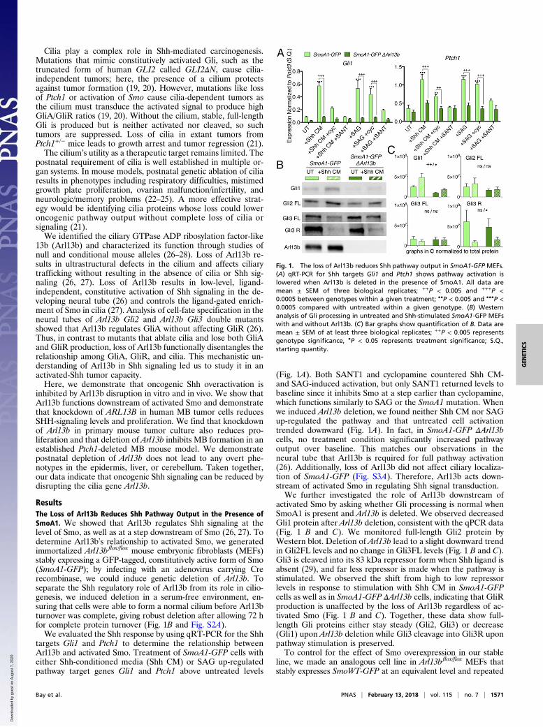
ResultsThe Loss of Arl13b Reduces Shh Pathway Output in the Presence ofSmoA1. We showed that Arl13b regulates Shh signaling at thelevel of Smo, as well as at a step downstream of Smo (26, 27). Todetermine Arl13b’s relationship to activated Smo, we generatedimmortalized Arl13bflox/flox mouse embryonic fibroblasts (MEFs)stably expressing a GFP-tagged, constitutively active form of Smo(SmoA1-GFP); by infecting with an adenovirus carrying Crerecombinase, we could induce genetic deletion of Arl13b. Toseparate the Shh regulatory role of Arl13b from its role in cilio-genesis, we induced deletion in a serum-free environment, en-suring that cells were able to form a normal cilium before Arl13bturnover was complete, giving robust deletion after allowing 72 hfor complete protein turnover (Fig. 1B and Fig. S2A).We evaluated the Shh response by using qRT-PCR for the Shh
targets Gli1 and Ptch1 to determine the relationship betweenArl13b and activated Smo. Treatment of SmoA1-GFP cells witheither Shh-conditioned media (Shh CM) or SAG up-regulatedpathway target genes Gli1 and Ptch1 above untreated levels
(Fig. 1A). Both SANT1 and cyclopamine countered Shh CM-and SAG-induced activation, but only SANT1 returned levels tobaseline since it inhibits Smo at a step earlier than cyclopamine,which functions similarly to SAG or the SmoA1 mutation. Whenwe induced Arl13b deletion, we found neither Shh CM nor SAGup-regulated the pathway and that untreated cell activationtrended downward (Fig. 1A). In fact, in SmoA1-GFP ΔArl13bcells, no treatment condition significantly increased pathwayoutput over baseline. This matches our observations in theneural tube that Arl13b is required for full pathway activation(26). Additionally, loss of Arl13b did not affect ciliary localiza-tion of SmoA1-GFP (Fig. S3A). Therefore, Arl13b acts down-stream of activated Smo in regulating Shh signal transduction.We further investigated the role of Arl13b downstream of
activated Smo by asking whether Gli processing is normal whenSmoA1 is present and Arl13b is deleted. We observed decreasedGli1 protein after Arl13b deletion, consistent with the qPCR data(Fig. 1 B and C). We monitored full-length Gli2 protein byWestern blot. Deletion of Arl13b lead to a slight downward trendin Gli2FL levels and no change in Gli3FL levels (Fig. 1 B and C).Gli3 is cleaved into its 83 kDa repressor form when Shh ligand isabsent (29), and far less repressor is made when the pathway isstimulated. We observed the shift from high to low repressorlevels in response to stimulation with Shh CM in SmoA1-GFPcells as well as in SmoA1-GFP ΔArl13b cells, indicating that GliRproduction is unaffected by the loss of Arl13b regardless of ac-tivated Smo (Fig. 1 B and C). Together, these data show full-length Gli proteins either stay steady (Gli2, Gli3) or decrease(Gli1) upon Arl13b deletion while Gli3 cleavage into Gli3R uponpathway stimulation is preserved.To control for the effect of Smo overexpression in our stable
line, we made an analogous cell line in Arl13bflox/flox MEFs thatstably expresses SmoWT-GFP at an equivalent level and repeated
Fig. 1. The loss of Arl13b reduces Shh pathway output in SmoA1-GFP MEFs.(A) qRT-PCR for Shh targets Gli1 and Ptch1 shows pathway activation islowered when Arl13b is deleted in the presence of SmoA1. All data aremean ± SEM of three biological replicates; ++P < 0.005 and +++P <0.0005 between genotypes within a given treatment; ••P < 0.005 and •••P <0.0005 compared with untreated within a given genotype. (B) Westernanalysis of Gli processing in untreated and Shh-stimulated SmoA1-GFP MEFswith and without Arl13b. (C) Bar graphs show quantification of B. Data aremean ± SEM of at least three biological replicates; ++P < 0.005 representsgenotype significance, •P < 0.05 represents treatment significance; S.Q.,starting quantity.
Bay et al. PNAS | February 13, 2018 | vol. 115 | no. 7 | 1571
GEN
ETICS
Dow
nloa
ded
by g
uest
on
Aug
ust 7
, 202
0

all analyses in that line (Figs. S2 B and C, S3B, and S4). UntreatedSmoWT-GFP cells showed no active signaling, indicating thatstable overexpression of tagged Smo did not alter normal Smofunction. Consistent with our analysis of the SmoA1-GFP ΔArl13bMEFs, we found (i) lower Gli1 and Ptch1 transcription, (ii) lowerGli1 levels, (iii) normal Gli2FL and Gli3FL levels, (iv) normalGli3R processing, and (v) stereotypic Smo localization patterns inSmoWT-GFP ΔArl13b MEFs, confirming that deletion of Arl13blowers the Shh transcriptional response to Shh CM/SAG regard-less of Smo activation.
ARL13B Knockdown Reduces SHH Signaling and Proliferation in HumanMB Cell Lines. To test whether manipulating ARL13B has similareffects in human cells as in mouse cells, we knocked downARL13B in two human MB cell lines: DAOY and D556 (30, 31).The DAOY cell line was isolated from a desmoplastic MB—ahistological variant of MB associated with activation of SHHsignaling. D556 cells are MYCC-amplified, derived from an an-aplastic MB, and often used in culture experiments as a “non-SHH” tumor cell line. After confirming that DAOY cells wereShh-sensitive while D556 were not (Fig. S5A), we hypothesizedthat disruption of ARL13B would affect DAOY but not D556cells. We infected cells with a lentiviral shRNA targetingARL13B or a scrambled control and assayed SHH target genetranscription in response to ligand by qRT-PCR (Fig. 2 A and Band Fig. S5B). DAOY cells up-regulate GLI1 and PTCH1 inresponse to Shh CM, while D556 cells do not. Knockdown ofARL13B reduces the stimulated SHH response in DAOY cellsand, surprisingly, in D556 cells regardless of pathway stimulation.
To test whether the knockdown of ARL13B affected pro-liferation in these cells, we examined BrdU incorporation. ARL13Bknockdown resulted in a significant decrease in proliferationin both DAOY and D556 cells (Fig. 2 C and D). Together,these data show that loss of ARL13B causes down-regulationof SHH signaling and reduces proliferation in human MBcell lines.
Arl13b Knockdown Reduces Proliferation of Mouse MB Cells.We nextturned to an ex vivo system to investigate whether Arl13bfunctions in tumor cell maintenance by knocking down Arl13b vialentiviral shRNA in primary mouse MB cells and assaying forproliferation. We used a well-established MB model expressingSmoA1 under the control of the neuroD2 promoter, known asnD2::SmoA1 (32). We infected cells with lentiviruses carryingone of two shRNAs against Arl13b (designated 442 or 968) or ascrambled control and compared tumors left unstimulated ortreated with ShhN. We monitored proliferation via BrdU in-corporation. Modest knockdown of Arl13b in these cells reducedproliferation with or without ShhN stimulation (Fig. 3 A and Band Fig. S6).Since constitutive activation of Smo is only one tumorigenic
mechanism, we repeated this experiment in a Ptch1-deleted mousemodel of MB (33, 34). We usedMath1-CreER to induce Ptch1flox/flox
recombination and deletion specifically in CGNPs through ta-moxifen treatment at E14.5 (referred to as Ptch1ΔMath1-Cre–E14.5
animals). We labeled cells in S phase through BrdU incorpora-tion and discovered few stained cells, indicating the proliferationrate was too low for us to measure differences between genotypesand conditions. To circumvent this, we monitored cells in anyactive cell cycle phase using Ki67. Knockdown of Arl13b resultedin significantly less Ki67 staining than the scrambled control re-gardless of treatment with ShhN (Fig. 3 C and D). Taken together,these data show that loss of Arl13b can reduce proliferation inprimary culture of cilia-dependent tumors.
Loss of Arl13b in the Developing Cerebellum Inhibits MB Formation.Ptch1ΔMath1-Cre–E14.5 animals developed tumors quickly and ro-bustly, so we used this model to test whether the loss of Arl13bcould prevent MB formation in vivo. We compared a controltumor model in which we deleted Ptch1 to an experimentalmodel in which we concurrently deleted both Ptch1 and Arl13b.We used Math1-CreER to induce deletion via tamoxifen treat-ment at E14.5. We followed control Ptch1ΔMath1-Cre–E14.5 mice(n = 23) and experimental Arl13b Ptch1ΔMath1-Cre–E14.5 mice (n =23), monitoring them for head doming, ataxia, and weight loss assymptoms of MB formation. Only 9% (2/23) of Ptch1-deletedmice survived to the study endpoint of 150 d, with a mediansurvival of 99 d (Fig. 4 A and B). In contrast, 78% (18/23) ofPtch1 Arl13b-deleted animals survived to the endpoint with noanimal dying before 124 d (Fig. 4 A and E). Twenty-two percentof experimental animals developed tumors (Fig. 4F), but thesetumors formed later than in the control mice. In contrast tocontrol animals, these tumors did not wholly disrupt cerebel-lar structure, leaving some intact internal granule layer (IGL) visible(Fig. 4 B and F).The fact that the tumors in the experimental animals did not
affect the entire cerebellum raised the possibility that they de-rived from a subpopulation of Ptch1-deleted cells that did notalso delete Arl13b. To investigate, we looked for the presence ofArl13b-positive cilia within experimental tumors. We found that,like control tumors, the late-forming tumors in the experimentalanimals were Arl13b-positive (Fig. 4 C and G), indicating thatCre-mediated recombination had not occurred in those cellsor that Arl13b protein had not turned over. In contrast, theremaining IGL of tumor-positive experimental animals displayedfew Arl13b-positive cilia—similar to the IGL of surviving experi-mental animals (Fig. 4 E, H, and I). Previous reports documented
Fig. 2. Knockdown of ARL13B reduces Shh signaling and proliferation in hu-manMB cell lines. (A and B) qRT-PCR shows expression of SHH targets GLI1 andPTCH1 is reduced when ARL13B is knocked down in both DAOY and D556 celllines. qRT-PCR for ARL13B demonstrates robustness of KD. All data are mean ±SEM of three biological replicates; ++P < 0.005 and +++P < 0.0005 betweengenotypes within a given treatment; •P < 0.05 and ••P < 0.005 compared withuntreated within a given genotype. (C and D) Knockdown of ARL13B reducesproliferation in DAOY and D556 human MB cell lines, shown by percentageof BrdU+ cells (red). Bar graphs show mean ± SEM of ≥2 biological replicates;+++P < 0.0001; Student’s t test; S.Q., starting quantity. (Magnification, 40×.)
1572 | www.pnas.org/cgi/doi/10.1073/pnas.1706977115 Bay et al.
Dow
nloa
ded
by g
uest
on
Aug
ust 7
, 202
0

that distinct floxed alleles recombine discordantly (35, 36), so weinterpret these tumors as deriving from a subpopulation of cells inwhich Ptch1 deleted more efficiently than Arl13b. These data in-dicate that the loss of Arl13b disrupts Ptch1-deleted MB forma-tion and is likely to function cell autonomously.
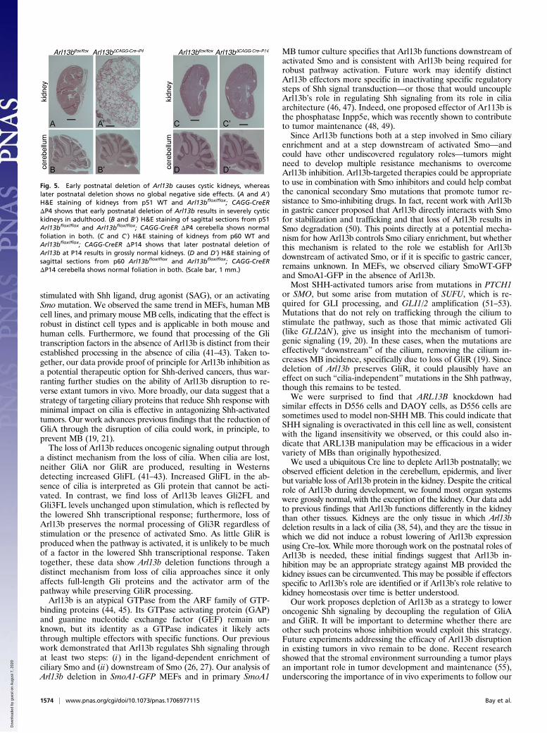
Postnatal Function of Arl13b. Embryonic loss of Arl13b is lethal,and tissue-specific embryonic deletion of Arl13b in the devel-oping kidney leads to cystic kidneys and death (26, 37, 38). Totest whether Arl13b continues to be required postnatally, weinduced Arl13b depletion by administering tamoxifen toArl13bflox/flox and Arl13bflox/flox; CAGG-CreER mice at postnataldays 4, 6, and 8 (P4, P6, and P8) and followed these mice into
adulthood. Deleted mice, noted as Arl13bΔCAGG-Cre–P4, werenoticeably smaller than their Cre-negative littermates, and 73%(8/11) died with cystic kidneys between P27 and P51. We ex-amined kidneys of surviving animals at P51 and found over-grown, cystic kidneys in depleted animals but not Cre-negativelittermates (Fig. 5 A and A′). Since Arl13b also functions incerebellar development, we examined cerebellar morphology viaH&E staining and found size and foliation were comparablebetween the two genotypes (Fig. 5 B and B′).Previous work defined a critical window for kidney development
ending around P14 (39, 40), so we next treated Arl13bflox/flox andArl13bflox/flox; CAGG-CreER mice with tamoxifen at P14, P16, andP18 and followed them until P60. In contrast to the earlier timepoint, these animals (n = 11), noted as Arl13bΔCAGG-Cre–P14, hadsimilar body sizes to Cre-negative littermates (n = 7) (Fig. S7A).Cre-positive animals displayed no obvious ataxia or motor controlissues, and females were capable of reproduction. At P60, weperformed a gross necropsy and found no visible defects in theheart, lungs, liver, reproductive organs, or brain. We further ex-amined kidney and cerebellar morphology via H&E staining (Fig.5 C, C′, D, and D′). The CAGG-CreER allele is expressed ubiq-uitously, and we observed substantial yet variable deletion upontamoxifen induction. We found low-level Arl13b protein in thecerebellum, as expected, but residual Arl13b protein expressionin the kidneys of Arl13bΔCAGG-Cre–P14 mice (Fig. S7 B and C).We observed widely variable Arl13b loss in kidney across theArl13bΔCAGG-Cre–P14 cohort—indicating either inefficient Cre–loxrecombination or protein turnover in the kidney. We observed afew small cysts largely localized to the renal cortex in theArl13bΔCAGG-Cre–P14 animals (Fig. 5C′); this agrees with obser-vations that postnatal loss of cilia genes can result in slow-growing renal cysts and reinforces the importance of Arl13b’srole in ciliogenesis in the kidney (38, 40). We found re-combination of the floxed allele in ear punch and liver DNA,indicating that, like cerebellum, Arl13b is depleted in these tis-sues (Fig. S7D). Depleted animals had normal cerebellar mor-phology, as in the Arl13bΔCAGG-Cre–P4 animals (Fig. 5 D and D′).Taken together, these data argue that Arl13b depletion afterP14 in mouse does not lead to any overt phenotypes in liver, skin,or cerebellar tissue.
DiscussionOur data show that deletion of Arl13b reduced Shh signaling ac-tivity and inhibited tumor formation in vivo. Arl13b deletion di-minished the Shh transcriptional response when the pathway was
Fig. 4. Deletion of Arl13b inhibits tumor formationin a mouse model of MB. Control: Ptch1ΔMath1-Cre–E14.5
Experimental: Arl13b Ptch1ΔMath1-Cre–E14.5. (A) Graphshows significant difference in survival curves be-tween control and experimental mice. ***P < 0.0001;log-rank test. (B) H&E staining of a representativecontrol brain with MB. (C) Image at 100× of tumorfrom B shows Arl13b-positive cilia in white. (D) H&Estaining of a surviving experimental brain with notumor. (E) Image at 100× of IGL from D shows fewArl13b-postive cilia in white. (F) H&E staining of a tu-mor-positive experimental brain shows normal cere-bellar structure in addition to tumor. G–I show thedifference in Arl13b staining (white) between tumortissue (G at 100×) and normal IGL (I at 100×). H showsthe boundary between tumor and IGL at 60×. Solidline: identifiable cerebellar foliation; dotted line: tu-mor. (Scale bars, 1 cm.)
Fig. 3. Knockdown ofArl13b in primary mouse MB cells reduces proliferation.(A) BrdU staining (red) in primary MB culture isolated from nD2::SmoA1/nD2::SmoA1 mice infected with scramble or shArl13b 442 and left untreated ortreated with recombinant ShhN. (B) Bar graph shows quantification of exper-iments represented in A: BrdU-positive cells as a percentage of total cellnumber; data are mean ± SEM of three biological replicates. ++P < 0.01 sig-nificance due to genotype, no significance due to treatment. (C) Ki67 staining(green) in primary MB culture isolated from Ptch1ΔMath1-Cre–E14.5 mice infectedwith scramble or shArl13b 442 and left untreated or treated with recombinantShhN. (D) Bar graph shows quantification of experiments represented in C:Ki67-positive cells as a percentage of total cell number; data are mean ± SEMof three biological replicates. +++P < 0.001 significance due to genotype, nosignificance due to treatment. (Magnification, 40×.)
Bay et al. PNAS | February 13, 2018 | vol. 115 | no. 7 | 1573
GEN
ETICS
Dow
nloa
ded
by g
uest
on
Aug
ust 7
, 202
0
stimulated with Shh ligand, drug agonist (SAG), or an activatingSmo mutation. We observed the same trend in MEFs, human MBcell lines, and primary mouse MB cells, indicating that the effect isrobust in distinct cell types and is applicable in both mouse andhuman cells. Furthermore, we found that processing of the Glitranscription factors in the absence of Arl13b is distinct from theirestablished processing in the absence of cilia (41–43). Taken to-gether, our data provide proof of principle for Arl13b inhibition asa potential therapeutic option for Shh-derived cancers, thus war-ranting further studies on the ability of Arl13b disruption to re-verse extant tumors in vivo. More broadly, our data suggest that astrategy of targeting ciliary proteins that reduce Shh response withminimal impact on cilia is effective in antagonizing Shh-activatedtumors. Our work advances previous findings that the reduction ofGliA through the disruption of cilia could work, in principle, toprevent MB (19, 21).The loss of Arl13b reduces oncogenic signaling output through
a distinct mechanism from the loss of cilia. When cilia are lost,neither GliA nor GliR are produced, resulting in Westernsdetecting increased GliFL (41–43). Increased GliFL in the ab-sence of cilia is interpreted as Gli protein that cannot be acti-vated. In contrast, we find loss of Arl13b leaves Gli2FL andGli3FL levels unchanged upon stimulation, which is reflected bythe lowered Shh transcriptional response; furthermore, loss ofArl13b preserves the normal processing of Gli3R regardless ofstimulation or the presence of activated Smo. As little GliR isproduced when the pathway is activated, it is unlikely to be muchof a factor in the lowered Shh transcriptional response. Takentogether, these data show Arl13b deletion functions through adistinct mechanism from loss of cilia approaches since it onlyaffects full-length Gli proteins and the activator arm of thepathway while preserving GliR processing.Arl13b is an atypical GTPase from the ARF family of GTP-
binding proteins (44, 45). Its GTPase activating protein (GAP)and guanine nucleotide exchange factor (GEF) remain un-known, but its identity as a GTPase indicates it likely actsthrough multiple effectors with specific functions. Our previouswork demonstrated that Arl13b regulates Shh signaling throughat least two steps: (i) in the ligand-dependent enrichment ofciliary Smo and (ii) downstream of Smo (26, 27). Our analysis ofArl13b deletion in SmoA1-GFP MEFs and in primary SmoA1
MB tumor culture specifies that Arl13b functions downstream ofactivated Smo and is consistent with Arl13b being required forrobust pathway activation. Future work may identify distinctArl13b effectors more specific in inactivating specific regulatorysteps of Shh signal transduction—or those that would uncoupleArl13b’s role in regulating Shh signaling from its role in ciliaarchitecture (46, 47). Indeed, one proposed effector of Ar113b isthe phosphatase Inpp5e, which was recently shown to contributeto tumor maintenance (48, 49).Since Arl13b functions both at a step involved in Smo ciliary
enrichment and at a step downstream of activated Smo—andcould have other undiscovered regulatory roles—tumors mightneed to develop multiple resistance mechanisms to overcomeArl13b inhibition. Arl13b-targeted therapies could be appropriateto use in combination with Smo inhibitors and could help combatthe canonical secondary Smo mutations that promote tumor re-sistance to Smo-inhibiting drugs. In fact, recent work with Arl13bin gastric cancer proposed that Arl13b directly interacts with Smofor stabilization and trafficking and that loss of Arl13b results inSmo degradation (50). This points directly at a potential mecha-nism for how Arl13b controls Smo ciliary enrichment, but whetherthis mechanism is related to the role we establish for Arl13bdownstream of activated Smo, or if it is specific to gastric cancer,remains unknown. In MEFs, we observed ciliary SmoWT-GFPand SmoA1-GFP in the absence of Arl13b.Most SHH-activated tumors arise from mutations in PTCH1
or SMO, but some arise from mutation of SUFU, which is re-quired for GLI processing, and GLI1/2 amplification (51–53).Mutations that do not rely on trafficking through the cilium tostimulate the pathway, such as those that mimic activated Gli(like GLI2ΔN), give us insight into the mechanism of tumori-genic signaling (19, 20). In these cases, when the mutations areeffectively “downstream” of the cilium, removing the cilium in-creases MB incidence, specifically due to loss of GliR (19). Sincedeletion of Arl13b preserves GliR, it could plausibly have aneffect on such “cilia-independent” mutations in the Shh pathway,though this remains to be tested.We were surprised to find that ARL13B knockdown had
similar effects in D556 cells and DAOY cells, as D556 cells aresometimes used to model non-SHHMB. This could indicate thatSHH signaling is overactivated in this cell line as well, consistentwith the ligand insensitivity we observed, or this could also in-dicate that ARL13B manipulation may be efficacious in a widervariety of MBs than originally hypothesized.We used a ubiquitous Cre line to deplete Arl13b postnatally; we
observed efficient deletion in the cerebellum, epidermis, and liverbut variable loss of Arl13b protein in the kidney. Despite the criticalrole of Arl13b during development, we found most organ systemswere grossly normal, with the exception of the kidney. Our data addto previous findings that Arl13b functions differently in the kidneythan other tissues. Kidneys are the only tissue in which Arl13bdeletion results in a lack of cilia (38, 54), and they are the tissue inwhich we did not induce a robust lowering of Arl13b expressionusing Cre–lox. While more thorough work on the postnatal roles ofArl13b is needed, these initial findings suggest that Arl13b in-hibition may be an appropriate strategy against MB provided thekidney issues can be circumvented. This may be possible if effectorsspecific to Arl13b’s role are identified or if Arl13b’s role relative tokidney homeostasis over time is better understood.Our work proposes depletion of Arl13b as a strategy to lower
oncogenic Shh signaling by decoupling the regulation of GliAand GliR. It will be important to determine whether there areother such proteins whose inhibition would exploit this strategy.Future experiments addressing the efficacy of Arl13b disruptionin existing tumors in vivo remain to be done. Recent researchshowed that the stromal environment surrounding a tumor playsan important role in tumor development and maintenance (55),underscoring the importance of in vivo experiments to follow our
Fig. 5. Early postnatal deletion of Arl13b causes cystic kidneys, whereaslater postnatal deletion shows no global negative side effects. (A and A′)H&E staining of kidneys from p51 WT and Arl13bflox/flox; CAGG-CreERΔP4 shows that early postnatal deletion of Arl13b results in severely cystickidneys in adulthood. (B and B′) H&E staining of sagittal sections from p51Arl13bflox/flox and Arl13bflox/flox; CAGG-CreER ΔP4 cerebella shows normalfoliation in both. (C and C′) H&E staining of kidneys from p60 WT andArl13bflox/flox; CAGG-CreER ΔP14 shows that later postnatal deletion ofArl13b at P14 results in grossly normal kidneys. (D and D′) H&E staining ofsagittal sections from p60 Arl13bflox/flox and Arl13bflox/flox; CAGG-CreERΔP14 cerebella shows normal foliation in both. (Scale bar, 1 mm.)
1574 | www.pnas.org/cgi/doi/10.1073/pnas.1706977115 Bay et al.
Dow
nloa
ded
by g
uest
on
Aug
ust 7
, 202
0

work. Our data that targeting Arl13b reduces tumorigenic Shhsignaling lays the foundation for future work into drug devel-opment and investigating the role of Arl13b in other Shh-derivedtumors. BCC and MB share common tumorigenic mechanisms,so it is likely that Arl13b inhibition would similarly impact BCCand should be investigated.
Materials and MethodsAnimal workwas carriedoutunder InstitutionalAnimal CareandUseCommittee-approved protocols at Emory University, and all cell culture work was done underan Emory Environmental Health and Safety-approved biosafety protocol. Lenti-virus was produced using the Sigma MISSION Lentiviral Packaging Mix (SHP001)and Promega FuGENE 6 (E2691) according to the manufacturer’s instruc-tions. Coverslips were mounted in ProLong Gold antifade reagent (P36934;
ThermoFisher Scientific) and imaged using anOlympus Fluoview FV1000 confocalmicroscope and Olympus Fluoview v4.2 or a Leica CTR6000 microscope withSimplePCI. All statistical analysis was done using GraphPad Prism 7 software.
ACKNOWLEDGMENTS. For mice and reagents at Emory, we thank R. CraigCastellino, Anna Kenney, Andrew Kowalczyk, Tobey McDonald, and Tracy-AnnRead. We thank Ching-Fang Chang and Samantha Brugmann (Cinncinnati Child-ren’s) for Gli Western blot positive controls and advice, the T.C. laboratory andR. Craig Castellino for discussion and manuscript comments, and Deborah A.Cook for editing. This workwas supported by a research project grant from theChildren’s Brain Tumor Foundation, CURE Childhood Cancer Grant60608002002, and NIH Grants R01GM110663 and R01NS090029. S.N.B. wassupported by NIH training Grants T32MH087977 and T32GM008490. Thisresearch project was supported in part by the Emory University IntegratedCellular Imaging Microscopy Core of the Emory Neuroscience National Instituteof Neurological Disorders and Stroke (NINDS) Core Facilities Grant P30NS055077.
1. Northcott PA, Korshunov A, Pfister SM, Taylor MD (2012) The clinical implications ofmedulloblastoma subgroups. Nat Rev Neurol 8:340–351.
2. Louis DN, et al. (2016) The 2016 World Health Organization classification of tumors ofthe central nervous system: A summary. Acta Neuropathol 131:803–820.
3. Ruat M, Hoch L, Faure H, Rognan D (2014) Targeting of smoothened for therapeuticgain. Trends Pharmacol Sci 35:237–246.
4. Huangfu D, et al. (2003) Hedgehog signalling in the mouse requires intraflagellartransport proteins. Nature 426:83–87.
5. Rohatgi R, Milenkovic L, Scott MP (2007) Patched1 regulates hedgehog signaling atthe primary cilium. Science 317:372–376.
6. Hui CC, Angers S (2011) Gli proteins in development and disease. Annu Rev Cell DevBiol 27:513–537.
7. Briscoe J, Thérond PP (2013) The mechanisms of hedgehog signalling and its roles indevelopment and disease. Nat Rev Mol Cell Biol 14:416–429.
8. Dessaud E, McMahon AP, Briscoe J (2008) Pattern formation in the vertebrate neuraltube: A sonic hedgehog morphogen-regulated transcriptional network. Development135:2489–2503.
9. Xie J, et al. (1998) Activating smoothened mutations in sporadic basal-cell carcinoma.Nature 391:90–92.
10. Von Hoff DD, et al. (2009) Inhibition of the hedgehog pathway in advanced basal-cellcarcinoma. N Engl J Med 361:1164–1172.
11. Chen JK, Taipale J, Young KE, Maiti T, Beachy PA (2002) Small molecule modulation ofsmoothened activity. Proc Natl Acad Sci USA 99:14071–14076.
12. Rohatgi R, Milenkovic L, Corcoran RB, Scott MP (2009) Hedgehog signal transductionby smoothened: Pharmacologic evidence for a 2-step activation process. Proc NatlAcad Sci USA 106:3196–3201.
13. Wang VY, Zoghbi HY (2001) Genetic regulation of cerebellar development. Nat RevNeurosci 2:484–491.
14. Machold R, Fishell G (2005) Math1 is expressed in temporally discrete pools of cere-bellar rhombic-lip neural progenitors. Neuron 48:17–24.
15. Ben-Arie N, et al. (1997) Math1 is essential for genesis of cerebellar granule neurons.Nature 390:169–172.
16. Spassky N, et al. (2008) Primary cilia are required for cerebellar development and Shh-dependent expansion of progenitor pool. Dev Biol 317:246–259.
17. Chizhikov VV, et al. (2007) Cilia proteins control cerebellar morphogenesis by pro-moting expansion of the granule progenitor pool. J Neurosci 27:9780–9789.
18. Zurawel RH, et al. (2000) Analysis of PTCH/SMO/SHH pathway genes in medulloblas-toma. Genes Chromosomes Cancer 27:44–51.
19. Han Y-G, et al. (2009) Dual and opposing roles of primary cilia in medulloblastomadevelopment. Nat Med 15:1062–1065.
20. Wong SY, et al. (2009) Primary cilia can both mediate and suppress hedgehogpathway-dependent tumorigenesis. Nat Med 15:1055–1061.
21. Barakat MT, Humke EW, Scott MP (2013) Kif3a is necessary for initiation and main-tenance of medulloblastoma. Carcinogenesis 34:1382–1392.
22. Johnson ET, et al. (2008) Role for primary cilia in the regulation of mouse ovarianfunction. Dev Dyn 237:2053–2060.
23. Song B, Haycraft CJ, Seo HS, Yoder BK, Serra R (2007) Development of the post-natalgrowth plate requires intraflagellar transport proteins. Dev Biol 305:202–216.
24. Gilley SK, et al. (2014) Deletion of airway cilia results in noninflammatory bronchi-ectasis and hyperreactive airways. Am J Physiol Lung Cell Mol Physiol 306:L162–L169.
25. Amador-Arjona A, et al. (2011) Primary cilia regulate proliferation of amplifyingprogenitors in adult hippocampus: Implications for learning and memory. J Neurosci31:9933–9944.
26. Caspary T, Larkins CE, Anderson KV (2007) The graded response to sonic hedgehogdepends on cilia architecture. Dev Cell 12:767–778.
27. Larkins CE, Aviles GDG, East MP, Kahn RA, Caspary T (2011) Arl13b regulates ciliogenesisand the dynamic localization of Shh signaling proteins. Mol Biol Cell 22:4694–4703.
28. Su C-Y, Bay SN, Mariani LE, Hillman MJ, Caspary T (2012) Temporal deletion of Arl13breveals that a mispatterned neural tube corrects cell fate over time. Development139:4062–4071.
29. Wang B, Fallon JF, Beachy PA (2000) Hedgehog-regulated processing of Gli3 produces ananterior/posterior repressor gradient in the developing vertebrate limb. Cell 100:423–434.
30. Jacobsen PF, Jenkyn DJ, Papadimitriou JM (1985) Establishment of a human medul-loblastoma cell line and its heterotransplantation into nude mice. J Neuropathol ExpNeurol 44:472–485.
31. Aldosari N, et al. (2002) Comprehensive molecular cytogenetic investigation ofchromosomal abnormalities in human medulloblastoma cell lines and xenograft.Neuro-oncol 4:75–85.
32. Hatton BA, et al. (2008) The Smo/Smo model: Hedgehog-induced medulloblastomawith 90% incidence and leptomeningeal spread. Cancer Res 68:1768–1776.
33. Yang Z-J, et al. (2008) Medulloblastoma can be initiated by deletion of patched inlineage-restricted progenitors or stem cells. Cancer Cell 14:135–145.
34. Goodrich LV, Milenkovi�c L, Higgins KM, Scott MP (1997) Altered neural cell fates andmedulloblastoma in mouse patched mutants. Science 277:1109–1113.
35. Liu J, et al. (2013) Non-parallel recombination limits Cre-LoxP-based reporters asprecise indicators of conditional genetic manipulation. Genesis 51:436–442.
36. Schmidt-Supprian M, Rajewsky K (2007) Vagaries of conditional gene targeting. NatImmunol 8:665–668.
37. Li Y, et al. (2016) Deletion of ADP ribosylation factor-like GTPase 13B leads to kidneycysts. J Am Soc Nephrol 27:3628–3638.
38. Seixas C, et al. (2015) Arl13b and the exocyst interact synergistically in ciliogenesis.Mol Biol Cell 27:308–320.
39. Piontek K, Menezes LF, Garcia-Gonzalez MA, Huso DL, Germino GG (2007) A criticaldevelopmental switch defines the kinetics of kidney cyst formation after loss of Pkd1.Nat Med 13:1490–1495.
40. Davenport JR, et al. (2007) Disruption of intraflagellar transport in adult mice leads toobesity and slow-onset cystic kidney disease. Curr Biol 17:1586–1594.
41. Chang CF, Chang YT, Millington G, Brugmann SA (2016) Craniofacial ciliopathies re-veal specific requirements for GLI proteins during development of the facial midline.PLoS Genet 12:e1006351.
42. Huangfu D, Anderson KV (2005) Cilia and hedgehog responsiveness in the mouse.Proc Natl Acad Sci USA 102:11325–11330.
43. Liu A, Wang B, Niswander LA (2005) Mouse intraflagellar transport proteins regulateboth the activator and repressor functions of Gli transcription factors. Development132:3103–3111.
44. Kahn RA, et al. (2006) Nomenclature for the human Arf family of GTP-binding pro-teins: ARF, ARL, and SAR proteins. J Cell Biol 172:645–650.
45. Hori Y, Kobayashi T, Kikko Y, Kontani K, Katada T (2008) Domain architecture of theatypical Arf-family GTPase Arl13b involved in cilia formation. Biochem Biophys ResCommun 373:119–124.
46. Kuai J, Kahn RA (2000) Residues forming a hydrophobic pocket in ARF3 are deter-minants of GDP dissociation and effector interactions. FEBS Lett 487:252–256.
47. Joneson T, White MA, Wigler MH, Bar-Sagi D (1996) Stimulation of membrane ruf-fling and MAP kinase activation by distinct effectors of RAS. Science 271:810–812.
48. Conduit SE, et al. (2017) A compartmentalized phosphoinositide signaling axis at ciliais regulated by INPP5E to maintain cilia and promote sonic hedgehog medulloblas-toma. Oncogene 36:5969–5984.
49. Humbert MC, et al. (2012) ARL13B, PDE6D, and CEP164 form a functional network forINPP5E ciliary targeting. Proc Natl Acad Sci USA 109:19691–19696.
50. Shao J, et al. (2017) Arl13b promotes gastric tumorigenesis by regulating Smo traf-ficking and activation of the hedgehog signaling pathway. Cancer Res 77:4000–4013.
51. Northcott PA, et al. (2009) Multiple recurrent genetic events converge on control ofhistone lysine methylation in medulloblastoma. Nat Genet 41:465–472.
52. Taylor MD, et al. (2002) Mutations in SUFU predispose to medulloblastoma. Nat Genet31:306–310.
53. Brugières L, et al. (2012) High frequency of germline SUFU mutations in children withdesmoplastic/nodular medulloblastoma younger than 3 years of age. J Clin Oncol 30:2087–2093.
54. Duldulao NA, Lee S, Sun Z (2009) Cilia localization is essential for in vivo functions ofthe Joubert syndrome protein Arl13b/scorpion. Development 136:4033–4042.
55. Bassett EA, et al. (2016) Norrin/Frizzled4 signalling in the preneoplastic niche blocksmedulloblastoma initiation. Elife 5:1–27.
56. Caspary T, Marazziti D, Berbari NF (2016) Methods for visualization of neuronal cilia.Cilia: Methods and Protocols, eds Satir P, Christensen ST (Springer New York, NewYork), pp 203–214.
57. Mariani LE, et al. (2016) Arl13b regulates Shh signaling from both inside and outsidethe cilium. Mol Biol Cell mbc.E16-03-0189.
58. Nachtergaele S, et al. (2013) Structure and function of the smoothened extracellulardomain in vertebrate hedgehog signaling. Elife 2:e01340.
59. Carpenter AE, et al. (2006) CellProfiler: Image analysis software for identifying andquantifying cell phenotypes. Genome Biol 7:R100.
Bay et al. PNAS | February 13, 2018 | vol. 115 | no. 7 | 1575
GEN
ETICS
Dow
nloa
ded
by g
uest
on
Aug
ust 7
, 202
0