disruption of the ste22 gene encoding a glycosyltransferase and its function in biosynthesis of...
TRANSCRIPT

Disruption of the ste22 Gene Encoding a Glycosyltransferase and ItsFunction in Biosynthesis of Ebosin in Streptomyces sp. 139
Tianyu Zhang,1 Lingyan Wang,1 Guiyun Xu,2 Yang Chen,2 Yang Zhang,1 Yuan Li1
1Institute of Medicinal Biotechnology, Chinese Academy of Medical Sciences, Peking Union Medical College, 100050, Beijing, China2Institute of Chemistry, Chinese Academy of Sciences, 100080, Beijing, China
Received: 19 April 2005 / Accepted: 20 September 2005
Abstract. Streptomyces sp.139 produces an exopolysaccharide (EPS) designated Ebosin with remark-able anti-rheumatic arthritis activity in vivo. The ste (Streptomyces eps) gene cluster required for Ebosinbiosynthesis has been identified. According to similarities with other proteins in the database, ste22shows high homology with glycosyltransferases originated from different microorganisms. In this study,the ste22 gene was disrupted by double crossover via homologous recombination. The EPS produced bythe mutant strain Streptomyces sp.139 (ste22)) has a different monosaccharide composition profile incomparison with that of Ebosin. This derivative of Ebosin retained the original antagonistic activity ofIL-1R in vitro but lost the bioactivities of anti-inflammation and pain relief in vivo.
Microbial EPSs are long-chain, high-molecular-masspolymers secreted into the environment by a largevariety of different bacteria. These polymers are be-lieved to play a protective role in the native state, andcan be dissolved or dispersed in water to give interestingthickening, gelling, and emulsifying effects, propertiesthat are indispensable tools in the food and many otherindustries [9]. Many EPSs secreted by bacteria haveregular structures composed of complex chemicalrepeating units. These repeating units may contain anumber of different sugars linked in a variety of ways.The structure can be branched or multi-branched, andmay be decorated with non-carbohydrate substitutes. Itis widely known that the functional properties of an EPSare determined by its primary structure. To assess thecontribution of each sugar unit or linkage to the poly-saccharide functionality, it is desirable to engineer novelpolymers that are variants of the common structure [2].
The polysaccharide biosynthesis gene clusters ofsome gram-positive microorganisms including Staphy-lococcus aureus and Streptococcus pneumoniae haverecently been characterized [5, 6]. The general organi-zation of these clusters seems to be conserved: a central
region with similarity to glycosyltransferase genes isflanked by two regions exhibiting similarity to genesinvolved in polymerization and export, and a putativeregulatory region can be found at the beginning of eachcluster. The sequential transfer of sugar residues onto alipophilic carrier by specific glycosyltransferasesassembles the repeating unit. Of the enzymes requiredfor the biosynthesis of EPS, researchers are especiallyinterested in glycosyltransferases, because their sugarspecificities determine the nature of the polysaccharide.
Streptomyces, a Gram-positive bacterium, is wellknown as an important industrial microorganism for itsproduction of natural derived antibiotics. However, EPSproduction in Streptomyces has only been reported veryrecently despite the fast progress at a molecular level inother bacterial species (such as lactic acid bacteria) [11].In our laboratory, S. sp.139 has been identified to pro-duce a new EPS designated Ebosin with anti-rheumaticarthritis activity in vivo and antagonist activity forinterleukin I receptor (IL-1R) in vitro. Ebosin is an EPSwith a repeating unit consisting of galactose, mannose,glucose, arabinose, fucose, xylose, and rhamnose [3].The biosynthesis gene cluster of Ebosin has been iden-tified and characterized recently [10]. It consists of 22ORFs named ste1 through ste22 located on a 31.3-kbchromosomal region of S.sp.139. According to similar-Correspondence to: Yuan Li; email: [email protected]
CURRENT MICROBIOLOGY Vol. 52 (2006), pp. 55–59DOI: 10.1007/s00284-005-0096-9 Current
MicrobiologyAn International Journal
ª Springer Science+Business Media, Inc. 2005

ities with other proteins in the database, Ste22 showshigh homology with glycosyltransferases. In this study,the ste22 gene was disrupted by double crossover viahomologous recombination for functional study of theEbosin biosynthesis. As a result, the monosaccharidecomposition changed significantly in EPS (EPS1) pro-duced by the mutant S.sp.139 (ste22)) compared withEbosin. At functional level, EPS1 retained the in vitroantagonistic activity of IL-1R but lost the in vivoactivities in anti-inflammation and pain relief.
Materials and Methods
Bacterial strains, plasmids and media. E. coli DH5a, E. coliET12567, pKC1139, and pUC19 were all maintained in our laboratory.E. coli strains were grown, transformed, and screened as described bySambrook et al. [8]. Streptomyces sp.139 (China Central MicrobiologyCulture Collection Center, No. 0405) was grown at 28�C, either in TSBmedium supplemented with 5 mM MgCl2 and 0.5% glycine, or infermentation medium (1% glucose, 2% starch, 2% soybean extract,0.2% tryptone, 0.2% beef extract, 0.4% yeast extract, 0.05% K2HPO4,0.3% CaCO3, pH 7.3). Culture of Streptomyces and preparation andtransformation of protoplasts were described previously [4].
General DNA manipulation. Isolation of E. coli plasmid DNA andstandard recombinant DNA techniques were performed as described bySambook et al. [8]. According to the methods described by Kieser et al.[4], Streptomyces genomic DNA and plasmid DNA were isolated.Southern hybridization analyses were performed following theinstruction of the manufacturer (Amersham Life Science).
Gene disruption. The plasmid pKC22 used for the disruption of ste22was constructed as follows: On the one hand, primers were preparedfrom the sequence of the ste22 gene and its flanking sequence: P1, 5¢-ATAAGCTTCTGCCTCGGCGTCGTCCTG-3¢ (HindIII) and P2, 3¢-GCGATGCGGTTCGTCCTGGGAGATCTCA-5¢ (XbaI, restrictionsites are underlined) for fragment F1 (a �1.1-kb fragment upstreamof ste22 gene); P3, 5¢-CGTCTAGACTGCTACCGGCACCTCGCC-3¢(XbaI) and P4, 3¢-TCCACTTCGTCC GCACTCGCCTTAAGCA-5¢(EcoRI) for fragment F2 (a �1.9-kb fragment downstream of ste22).F1 and F2 were amplified by PCR from S. sp.139 genomic DNA. ThePCR primers were designed to contain HindIII-XbaI sites and XbaI-EcoRI sites, respectively, to facilitate cloning into plasmid pUC19digested by the same appropriate endonucleases for sequencing and thefollowing cloning into EcoRI-HindIII digested pKC1139 by three-fragment ligation to produce pKCste22LR. A �1.2-kb DNA fragmentcontaining the Kmr gene cassette from pFD666IC has an XbaI site ateach end to facilitate cloning into XbaI digested pKCste22LR to createpKC22.
pKC22, passed through E. coli ET12567 [7], was transformedinto the protoplasts of S. sp.139. Plasmid pKC22 bears a temperature-sensitive Streptomyces replication origin [1] that is unable to replicateat temperature above 34�C. The spores of transformants containingpKCm22 were harvested and spread on R2 plates containing Km. Aftergrowing for 4 days at 37�C, the grown colonies were replicated on R2plates containing Am and Km, respectively. The ste22 disruptants wereselected by both the Am sensitivity (Ams) and Km resistance (Kmr).
Isolation and purification of EPSs. The filtrate obtained by filtrationwas applied to a Diaion HP-20 column. The eluate was subjected to001 · 7 styrene cation exchange resin. After 60% ethanol being added
to the eluate to precipitate EPS twice, the resultant product (pellet) wasdissolved in a minimum amount of water and loaded on a DEAE-Dextran A-25 column. The column was eluted with water, 0.2 mol l)1
NH4Cl and 0.5 mol l)1 NH4Cl, and then fractions were monitored byELISA (see below). The active fractions were dialyzed and freeze-dried.
Sugar analyses of EPSs. The composition analysis was performed asdescribed elsewhere [12]. GPC (Gel Permeation Chromatography) wasemployed to determine Mr of EPSs. The sample of EPS (25 lL) wasinjected to a G4000 PWxL column using 0.7% Na2SO4 as mobile phaseat a flow rate of 0.5 ml min)1 at 35�C (Water 201, Detector: RI R401).
ELISA for EPSs. IL-1ra was coated onto a 96-well immunoplate(Nunc) at 4�C overnight (100 ng well)1). To each well, 250 lL of 3%BSA in PBS buffer (KH2PO4 0.024%, Na2HPO4 0.363%, KCl 0.02%,and NaCl 0.8%, pH 7.4) was added and the plate was kept at 4�C for 8h, followed by washing 5 times with PBS buffer and being dried.Fermentation filtrates or pure EPSs were then diluted in PBScontaining 0.1% BSA and 50 lL added to each well, as well as50 lL of IL-1R (1:1000). The plate was incubated at 4�C overnight.After binding and washing the plate 5 times with PBST (PBS + 0.1%Tween20), 100 lL of diluted solution (1:1000) of goat anti-human IL-1R monoclonal antibody (R&D systems) was transferred into each welland the plate was allowed to stand at 4�C for 1h. The plate was washedagain as above before the reaction with the 100 lL second antibody ofdiluted solution (1:5000) rabbit monoclonal anti-goat IgG conjugatedwith SA-HRP (streptavidin-horseradish peroxidase) (Promega) at 4�Cfor 1 h. After the final wash as above, 100 lL of TMB (3,3¢, 5,5¢-tetramethyl benzidine dihydrochloride) solution was added to eachwell and reaction took place at room temperature for 1 h (solutionshould turn blue) before being stopped by adding 100 lL of 2 mol l)1
HCl. The absorbance at 450 nm was recorded. Antagonist rate= (IL-1ROD450-EPS OD450)/ IL-1R OD450 · 100%.
Results
Sequence analysis of the deduced product ofste22. The DNA sequence of ste22 reported in thisstudy has been deposited in GenBank under accessionnumber AY131229. Database searches revealed that thededuced protein of ste22 shows 84% identity and 87%similarity over a 311-aa region to SCF62.24 ofStreptomyces coelicolor A3 (2) (GenBank accessionnumber AL121855). To SMb21066 of Sinorhizobiummeliloti (GenBank accession number AL603646), theidentity is 31% and similarity 46% over a 277-aa region.It shows 31% identity and 46% similarity over a 227-aato lgtD of Rickettsia conorti (GenBank accessionnumber AE008622). They are all glycosyltransferases.According to these results, Ste22 is most homologous tothe family II glycosyltransferases that transfer sugarfrom UDP-glucose, UDP-N-acetyl-galactoamine, andGDP-mannose to polysaccharides of cellulose and sialicacid.
Gene replacement of ste22. More than 15 colonieswith a selective marker KmrAms were obtained byreplacing ste22 with Km r (Fig. 1). Two of them selected
56 CURRENT MICROBIOLOGY Vol. 52 (2006)

randomly were proved by PCR using Primers P5 and P6.Primers P5 (5¢-GCGAGCTCGGTGGCTGCATCTGCG-3¢) and P6 (3¢-GCTTCGGCACGACCTGTTCGAAGC-5¢) were designed from ste22 flanking sequences at bothends. As expected, in the wild type, a single 1.1-kb bandwas recognized and in contrast, ste22 mutant wasrecognized as a single 1.7-kb band instead of the 1.1-kb band (Fig. 2a). One of the two strains was furtherconfirmed to be ste22 mutant by Southern hybridizationanalysis. Genomic DNA was isolated and digested withKpnI. Southern hybridization was performed with a 0.8-kb XbaI-KpnI cut F1 DNA fragment as probe. Theresults showed that in colonies with KmrAms, thekanamycin resistance cassette had replaced ste22 gene,since hybridization signal appeared with the expected1.75-kb (wild-type) and 2.35-kb (ste22 mutant strain)genomic DNA fragments (Fig. 2b). The positive strainwas designated as S. sp. 139 (ste22)).
Sugar analysis of EPSs. The results showed that theEPS1 analyzed with GC-MS (Gas Chromatography-Mass Spectrometry) was still composed of rhamnose,xylose, glucose, mannose, arabinose, fucose, andgalactose (Table 1). In comparison to Ebosin,however, the monosaccharide composition profile ofEPS1 has changed, with mannose becoming thequantitatively predominant component increasing from7.2% to 49.9%, while arabinose diminished remarkablyfrom 31.4% to 14.2%. Rhamnose, fucose, xylose,galactose, and glucose in the EPS1 also all reducedtheir proportions compared with Ebosin.
Fig. 1. Replacement of ste22 via double crossover. Restriction mapsof the wild type Streptomyces sp.139 and the mutant Streptomycessp.139 (ste22)) show the predicted fragment sizes upon KpnI digestion.The broad arrows show the regions and orientations of correspondinggenes.
Fig. 2. Verification of replacement of ste22 via double crossover byPCR (a) and Southern hybridization (b). (A) PCR was carried out withprimers P5 and P6. 1, HindIII-digested kDNA; 2, PCR product withStreptomyces sp.139 genomic DNA (wild-type strain) as template; 3–4,PCR products with Streptomyces sp.139 (ste22)) (ste22 disruptants)genomic DNA as templates. (B) Genomic DNA was digested withKpnI and probed with the 0.8-kb XbaI-KpnI cut fragment F1 (Fig. 1). 1,EcoRI/HindIII-digested kDNA; 2, Genomic DNA from Streptomycessp.139 genomic DNA (wild-type strain); 3, Genomic DNA fromStreptomyces sp.139 (ste22)) (ste22 disruptant).
Table 1. Monosaccharide Composition of the EPSs Produced by S. sp.139 (Ebosin) and Its ste22 Disruption Mutant (EPS1)
Monosaccharide Ebosin EPS1
Galactose 4l.6 30.1Arabinose 31.4 14.2Mannose 7.2 50.0Fucose 7.2 2.0Xylose 5.2 1.7Glucose 4.1 1.4Rhamnose 3.4 0.8
Data of GC-MS are expressed as relative percentage, compared tostandard peaks. Values are mean of three determinations.
T. Zhang et al.: Disruption of ste22 57
With GPC analysis, Mr of the EPS1 was determinedto be 82.9661 · 104, which is similar to that of Ebosin(80.3953 · 104). This indicated that the ste22 genedisruption did not significantly alter the size of the EPSin spite of the dramatic change in the monosaccharidecomposition profile.
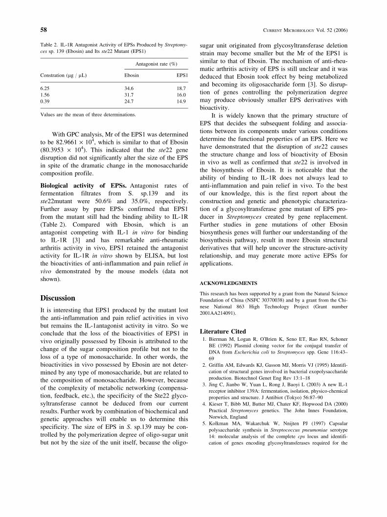
Biological activity of EPSs. Antagonist rates offermentation filtrates from S. sp.139 and itsste22mutant were 50.6% and 35.0%, respectively.Further assay by pure EPSs confirmed that EPS1from the mutant still had the binding ability to IL-1R(Table 2). Compared with Ebosin, which is anantagonist competing with IL-1 in vitro for bindingto IL-1R [3] and has remarkable anti-rheumaticarthritis activity in vivo, EPS1 retained the antagonistactivity for IL-1R in vitro shown by ELISA, but lostthe bioactivities of anti-inflammation and pain relief invivo demonstrated by the mouse models (data notshown).
Discussion
It is interesting that EPS1 produced by the mutant lostthe anti-inflammation and pain relief activities in vivobut remains the IL-1antagonist activity in vitro. So weconclude that the loss of the bioactivities of EPS1 invivo originally possessed by Ebosin is attributed to thechange of the sugar composition profile but not to theloss of a type of monosaccharide. In other words, thebioactivities in vivo possessed by Ebosin are not deter-mined by any type of monosaccharide, but are related tothe composition of monosaccharide. However, becauseof the complexity of metabolic networking (compensa-tion, feedback, etc.), the specificity of the Ste22 glyco-syltransferase cannot be deduced from our currentresults. Further work by combination of biochemical andgenetic approaches will enable us to determine thisspecificity. The size of EPS in S. sp.139 may be con-trolled by the polymerization degree of oligo-sugar unitbut not by the size of the unit itself, because the oligo-
sugar unit originated from glycosyltransferase deletionstrain may become smaller but the Mr of the EPS1 issimilar to that of Ebosin. The mechanism of anti-rheu-matic arthritis activity of EPS is still unclear and it wasdeduced that Ebosin took effect by being metabolizedand becoming its oligosaccharide form [3]. So disrup-tion of genes controlling the polymerization degreemay produce obviously smaller EPS derivatives withbioactivity.
It is widely known that the primary structure ofEPS that decides the subsequent folding and associa-tions between its components under various conditionsdetermine the functional properties of an EPS. Here wehave demonstrated that the disruption of ste22 causesthe structure change and loss of bioactivity of Ebosinin vivo as well as confirmed that ste22 is involved inthe biosynthesis of Ebosin. It is noticeable that theability of binding to IL-1R does not always lead toanti-inflammation and pain relief in vivo. To the bestof our knowledge, this is the first report about theconstruction and genetic and phenotypic characteriza-tion of a glycosyltransferase gene mutant of EPS pro-ducer in Streptomyces created by gene replacement.Further studies in gene mutations of other Ebosinbiosynthesis genes will further our understanding of thebiosynthesis pathway, result in more Ebosin structuralderivatives that will help uncover the structure-activityrelationship, and may generate more active EPSs forapplications.
ACKNOWLEDGMENTS
This research has been supported by a grant from the Natural ScienceFoundation of China (NSFC 30370038) and by a grant from the Chi-nese National 863 High Technology Project (Grant number2001AA214091).
Literature Cited1. Bierman M, Logan R, O�Brien K, Seno ET, Rao RN, Schoner
BE (1992) Plasmid cloning vector for the conjugal transfer ofDNA from Escherichia coli to Streptomyces spp. Gene 116:43–69
2. Griffin AM, Edwards KJ, Gasson MJ, Morris VJ (1995) Identifi-cation of structural genes involved in bacterial exopolysaccharideproduction. Biotechnol Genet Eng Rev 13:1–18
3. Jing C, Jianbo W, Yuan L, Rong J, Baoyi L (2003) A new IL-1receptor inhibitor 139A: fermentation, isolation, physico-chemicalproperties and structure. J Antibiot (Tokyo) 56:87–90
4. Kieser T, Bibb MJ, Butter MJ, Chater KF, Hopwood DA (2000)Practical Streptomyces genetics. The John Innes Foundation,Norwich, England
5. Kolkman MA, Wakarchuk W, Nnijten PJ (1997) Capsularpolysaccharide synthesis in Streptococcus pneumoniae serotype14: molecular analysis of the complete cps locus and identifi-cation of genes encoding glycosyltransferases required for the
Table 2. IL-1R Antagonist Activity of EPSs Produced by Streptomy-ces sp. 139 (Ebosin) and Its ste22 Mutant (EPS1)
Antagonist rate (%)
Constration (lg / lL) Ebosin EPS1
6.25 34.6 18.71.56 31.7 16.00.39 24.7 14.9
Values are the mean of three determinations.
58 CURRENT MICROBIOLOGY Vol. 52 (2006)

biosynthesis of the tetrasaccharide subunit. Mol Microbiol26:197–208
6. Liu D, Cole RA, Reeves PR (1996) An O-antigen processingfunction for Wzx (RfbX): a promising candidate for O-unit flip-pase. J Bacteriol 178:931–935
7. MacNeil DJ, Gewain KM, Ruby CL, Dzency G, Gibbons PH,MacNeil T (1992) Analysis of Streptomyces avermitilis gene re-quired for avermectin biosynthesis utilizing a novel integrationvector. Gene 111:61–68
8. Sambrook J, Fritisch EF, Maniatis T (1989) Molecular cloning: Alaboratory manual, 2 nd ed. Cold Spring Harbor, NY: Cold SpringHarbor Laboratory Press
9. Sutherland IW (1998) Novel and established applications ofmicrobial polysaccharides. Trends Biotechnol 16:41–46
10. Wang L, Li S, Li Y (2003) Identification and characterization of anew exopolysaccharide biosynthesis gene cluster from Strepto-myces. FEMS Microbiol Lett 220:21–27
11. Welman AD, Maddox IS (2003) Exopolysaccharides from lacticacid bacteria: perspectives and challenges. Trends Biotechnol12:269–274
12. Xu G, Chang W, Fei LH (1998) Composition analysis of carbo-hydrate released from bovine sumaxillarymucin by capillary gaschromatography. Chinese J Anal Chem 26:922–926
T. Zhang et al.: Disruption of ste22 59