dissecting functional cooperation among protein subunits in
TRANSCRIPT
Dissecting functional cooperation amongprotein subunits in archaeal RNase P, acatalytic ribonucleoprotein complexWen-Yi Chen1,2,3, Dileep K. Pulukkunat1,3,4, I-Ming Cho1,3,5, Hsin-Yue Tsai1,2,3 and
Venkat Gopalan1,2,3,4,5,*
1Department of Biochemistry, 2Molecular Cellular Developmental Biology Program, 3Center for RNA Biology,The Ohio State University, 4Ohio State Biochemistry Program and 5Department of Molecular Genetics,Columbus, OH 43210, USA
Received June 8, 2010; Revised July 10, 2010; Accepted July 14, 2010
ABSTRACT
RNase P catalyzes the Mg2+-dependent50-maturation of precursor tRNAs. Biochemicalstudies on the bacterial holoenzyme, composed ofone catalytic RNase P RNA (RPR) and one RNase Pprotein (RPP), have helped understand the pleio-tropic roles (including substrate/Mg2+ binding) bywhich a protein could facilitate RNA catalysis. As amodel for uncovering the functional coordinationamong multiple proteins that aid an RNA catalyst,we use archaeal RNase P, which comprises onecatalytic RPR and at least four RPPs. Exploitingour previous finding that these archaeal RPPsfunction as two binary RPP complexes(POP5�RPP30 and RPP21�RPP29), we prepared re-combinant RPP pairs from three archaea and estab-lished interchangeability of subunits throughhomologous/heterologous assemblies. Our findingthat archaeal POP5�RPP30 reconstituted with bac-terial and organellar RPRs suggests functionaloverlap of this binary complex with the bacterialRPP and highlights their shared recognition of aphylogenetically-conserved RPR catalytic core,whose minimal attributes we further definedthrough deletion mutagenesis. Moreover, single-turnover kinetic studies revealed that whilePOP5�RPP30 is solely responsible for enhancingthe RPR’s rate of precursor tRNA cleavage(by 60-fold), RPP21�RPP29 contributes to increasedsubstrate affinity (by 16-fold). Collectively,
these studies provide new perspectives on the func-tioning and evolution of an ancient, catalyticribonucleoprotein.
INTRODUCTION
RNase P, a ribonucleoprotein (RNP), is theendoribonuclease that catalyzes the removal of 50-leadersin precursor tRNAs (pre-tRNAs) in all three domains oflife (1–4). The bacterial variant is composed of 1 catalyticRNase P RNA (RPR) and 1 RNase P protein (RPP)cofactor. Eukaryal (nuclear) RNase P from yeast andhuman contain 1 RPR with 9 and 10 RPPs, respectively.Intermediate in complexity, the archaeal holoenzyme isassociated with 1 RPR and at least 4 RPPs (POP5,RPP30, RPP21 and RPP29), which are homologous toeukaryal RPPs.
Several years after the remarkable finding that the bac-terial RPR is a true RNA enzyme in the presence of Mg2+
and monovalent ions (5), archaeal and eukaryal RPRswere also shown to be catalytically active in vitro (6,7).This common attribute of evolutionarily divergent RPRswas anticipated from their shared ancestry, attested bysequence and structural similarity of their putative cata-lytic core (8–15). However, dramatic variations (106-fold)in catalytic potential indicate that not all RPRs are equal:activity of RPRs from bacteria> archaea> eukarya (5–7).Although RPRs display activity in vitro without RPPs,they are dependent on their cognate protein cofactorsfor cellular function. Interestingly, there is an inverse re-lationship between RPR activity and RNP composition—the protein:RNA mass ratio is 70% in eukaryal and 50%
*To whom correspondence should be addressed. Tel: +1 614 292 1332; Fax: +1 614 292 6773; Email: [email protected]
The authors wish it to be known that, in their opinion, the first two authors should be regarded as joint First Authors.
Present addresses:Dileep K. Pulukkunat, Department of Chemistry, Columbia University, New York, NY, USA.Hsin-Yue Tsai, Program in Molecular Medicine, University of Massachusetts Medical School, Worcester, MA, USA.
8316–8327 Nucleic Acids Research, 2010, Vol. 38, No. 22 Published online 12 August 2010doi:10.1093/nar/gkq668
� The Author(s) 2010. Published by Oxford University Press.This is an Open Access article distributed under the terms of the Creative Commons Attribution Non-Commercial License (http://creativecommons.org/licenses/by-nc/2.5), which permits unrestricted non-commercial use, distribution, and reproduction in any medium, provided the original work is properly cited.
Downloaded from https://academic.oup.com/nar/article-abstract/38/22/8316/1040188by gueston 19 March 2018

in archaeal RNase P compared to 10% in their bacterialcounterpart. Thus, elucidating the intimate cooperationbetween the RNA and protein subunits of RNase Pvariants with differing RNP make-up offers a paradigmto understand how structural and functional attributes ofRNAs might have been reassigned to protein cofactorsduring the evolutionary transition from an RNA toRNP world (1–3,15). Toward this goal, we have focusedour efforts on the simpler and biochemically tractablearchaeal version, especially as an experimental surrogatefor the eukaryal relative which is yet to be reconstitutedin vitro. Rapid advances in functional reconstitution(16–19) and structural studies (16,20–29) have validatedthis choice.
We have shown earlier that robust RNase P activitycould be obtained from assembling recombinantsubunits of Pyrococcus furiosus (Pfu; type A), and thatthe four RPPs functioned as two binary complexes(POP5�RPP30 and RPP21�RPP29) (18). Since the struc-tural diversity of RNase P is exemplified even withinarchaea where the type A and M RPRs resemble thebacterial and eukaryal relatives, respectively (Figure 1)(13,15), we reasoned that heterologous reconstitutionof types A and M archaeal RNase P variants will facilitatedelineation of their conserved and divergent features,and thereby help to understand the dynamic co-evolutionof the RNA and protein subunits in RNase P fromall domains of life. Indeed, heterologous reconstitutionsof archaeal RPPs with bacterial and organellar RPRshave helped define a minimal RNP catalytic core that isobscured by natural variations in the subunit make-up ofRNase P. Our single-turnover studies also provide keyinsights into the division of labor among archaeal RPPs,and permit formulation of a simple kinetic framework tohighlight this functional cooperation. These findings,together with the structures of the binary RPPs(25,28,29), should aid efforts to establish structure–function correlations in archaeal RNase P.
MATERIALS AND METHODS
Cloning, overexpression and purification of variousRPRs and RPPs
Complete details are provided in the Supplementary Data.
RNase P assays
All assays were performed with Escherichia coli (Eco)pre-tRNATyr as the substrate, a trace amount of whichwas labeled with [a-32P] GTP. Although we providebelow the details for the individual assemblies for differenthomologous and heterologous reconstitutions, we list heresome common features. All reconstitutions and assayswere performed in a thermal cycler. RPRs (unless other-wise stated) were folded as follows: incubation at 50�C for50min in water followed by 37�C for 30min in 50mMTris–HCl (pH 7.5), 800mM NH4OAc and 10mMMgCl2. The optimal RPR:RPP ratios for each combin-ation and the substrate concentrations were empiricallydetermined. Optimal RNP assembly entailed successiveincubations at 37 and 55�C for 5–10min each. Assayswere always initiated by adding pre-tRNATyr, which hadbeen pre-incubated at 55�C for 2min. Aliquots wereremoved at defined time intervals and added to an equalvolume of stop solution [10M urea, 5mM EDTA, 0.05%(w/v) bromophenol blue, 0.05% (w/v) xylene cyanol, 10%(v/v) phenol] to terminate the reaction. For short incuba-tions (e.g. 5 s), reactions were first terminated byimmersing the reaction tubes in liquid nitrogen beforeadding stop solution. The reaction contents wereseparated using denaturing PAGE [8% (w/v) polyacryl-amide, 7M urea].
Multiple-turnover assays with Methanocaldococcusjannaschii, Methanothermobacter thermautotrophicusand Pfu RNase P
To facilitate qualitative comparisons with multiple-turnover assays reported for Pfu RNase P (18), we
Figure 1. Secondary structure representations of (A) Eco, (B) Mth, (C) Mja and (D) Hsa RPRs (12,15). Letters indicate universally conservednucleotides (12). Paired helices (e.g. P1, P2) are numbered consecutively from 50 to 30 and according to the Eco RPR nomenclature (12). Alternativesecondary structure representations (69) are provided in Supplementary Figure S1.
Nucleic Acids Research, 2010, Vol. 38, No. 22 8317
Downloaded from https://academic.oup.com/nar/article-abstract/38/22/8316/1040188by gueston 19 March 2018
assayed Methanocaldococcus jannaschii (Mja) andMethanothermobacter thermautotrophicus (Mth) RNase Punder conditions identical to those used for the Pfu coun-terpart. Partially reconstituted RNase P holoenzymes wereassayed in 50mM Tris–HCl (pH 7.5), 100mM NH4OAcand 120mM MgCl2 with 10 nM RPR+100 nMPOP5�RPP30 or 250 nM RPR+625nM RPP21�RPP29.For the holoenzyme with four RPPs, 10 nM foldedRPR was incubated with 100 nM of all 4 RPPs in50mM Tris–HCl (pH 7.5), 800mM NH4OAc and30mM MgCl2. In all cases, the resulting RNPs wereincubated with 500 nM pre-tRNATyr for 30min at 55�C.Reconstitutions of Mja RPR �S and RPR �S Min
were performed by combining 500 nM of each RNAwith 1 mM of all four Mja RPPs in assay buffer [50mMTris–HCl (pH 7.5), 400mM NH4OAc and 30mMMg(OAc)2]. For reconstitutions with binary RPPs,Mg(OAc)2 was increased to 120mM. The resultingRNPs were incubated with 1 mM pre-tRNATyr for15min at 55�C.In Figures 3 and 4, the archaeal RPRs were assayed
under conditions as those used for the respectiveRPR+4 RPPs.
Multiple-turnover assays with heterologously-reconstitutedRNase P holoenzymes
A total of 50 nM archaeal type A RPR and 250 nM typeM RPPs (or type M RPR and type A RPPs) werereconstituted in 50mM Tris–HCl (pH 7.5), 800mMNH4OAc and 30mM Mg(OAc)2. The resulting RNPswere incubated with 500 nM pre-tRNATyr for 15minat 55�C.A total of 50 nM of folded Eco (30), Bacillus subtilis
(Bsu) (31) or Reclinomonas americana (Ram) mitochon-drial (mt) (32) RPR was reconstituted with archaealRPPs (500 nM) in 50mM Tris–HCl (pH 7.5), 800mMNH4OAc and 30mM Mg(OAc)2. The resulting RNPswere incubated with 200 nM pre-tRNATyr at 55�C for5min (Eco and Bsu RPRs) or 15min (Ram mt RPR).
Single-turnover kinetic studies with Mth RNase P
For these experiments, 50mM 2-(N-morpholino)etha-nesulfonic acid (MES)–HCl, pH 5.8 (unless otherwiseindicated) was used instead of Tris–HCl in RPR foldingand assays. The folded RPR was assayed either alone orwith a 2-fold molar excess of RPPs using the optimalconcentrations of MgCl2 and NH4OAc for each RNP(Table 1). In the case of RNPs, the amount of RPR inthe assay was used as the concentration of enzyme basedon the assumption that all of the RPR is assembledinto RNPs under the conditions employed. To determinethe maximal kobs (max. kobs) under single-turnover condi-tions at 55�C, we incubated �2 nM pre-tRNATyr witha range of enzyme concentrations: RPR, 0.3–20 mM;RPR+RPP21�RPP29, 0.5–3 mM; RPR+POP5�RPP30,1–10mM; and RPR+4 RPPs, 0.3–3 mM.Because the Mth RPR+4 RPPs reaction is too rapid
at pH 5.8 (t1/2� 8 s) when [E]> 1mM, we could obtainreliable data only by decreasing the assay pH to 5.4.To establish the dependence of the rates of product
formation catalyzed by Mth RPR+4 RPPs on assaypH, the assays were performed in 20mM instead of30mM Mg2+ and in the pH range 5.4–6.15(Supplementary Figure S6). [In an earlier publication(19), the rates reported for a self-cleaving Mja (type M)RPR were at pH 5.4 and not pH 5.1 as was mistakenlynoted].
Data analysis
After denaturing PAGE, the reaction products werevisualized by phosphorimaging on the Typhoon(GE Healthcare). The resulting bands were quantitatedby ImageQuant (GE Healthcare) to assess the extent ofsubstrate cleaved. To obtain the rate of product formation(kobs) in single-turnover reactions, the percentage productformed at time t (Pt) was fit to Pt=P1(1� e�kt) usingKaleidagraph software (Synergy). For optimal curve fits,the amplitudes were defined based on experimentallyobserved values (Supplementary Figure S5). The individ-ual curve-fit errors for kobs did not exceed 8%. For allkinetic studies, at least three replicates were performedto obtain the mean and standard deviation values.
The plot of kobs versus [E0] displayed hyperbolicdependence on the Mth RPR in the absence andpresence of its RPPs (Figure 7). Kaleidagraph was usedto fit these data to
kobs ¼max: kobs� ½E0�
KMðSTOÞ+½E0�
to derive values for max. kobs and KM(STO).
RESULTS
While type A archaeal RPRs process pre-tRNAs in vitro inthe absence of their cognate protein cofactors, type Mcounterparts display such an activity only when the sub-strate is provided in cis, perhaps reflecting their inability tobind substrates (6,19). Compared to bacterial andarchaeal type A relatives, type M RPRs exhibit twostriking structural changes (Figure 1) (13). First, they aremissing P8, which is part of the P7-9 cruciform in bacterialRPRs that is involved in T-loop recognition and binding(33–36). Second, type M RPRs lack P6, P16, P17 and theloop L15 that connects P15 and P16; L15 interacts withthe 30-RCCA of the pre-tRNA substrate (37,38). If type MRPPs have evolved to compensate for these structuralalterations in their cognate RPRs, then the effects ofRPPs on RPR catalysis will be distinctive in type A andM archaeal RNase P. As a first step to elucidate suchco-evolutionary trends, we sought to reconstitute RNaseP from Mja (type M), and compare it with those from Pfuand Mth (type A).
Purification of archaeal RPPs as binary complexes andtheir functional validation
To expediently assemble different archaeal RNase Pholoenyzmes in vitro, we explored a strategy thatentailed (i) purifying the four RPPs as two binary RPPcomplexes, after their co-overexpression in Eco, and
8318 Nucleic Acids Research, 2010, Vol. 38, No. 22
Downloaded from https://academic.oup.com/nar/article-abstract/38/22/8316/1040188by gueston 19 March 2018
(ii) reconstituting these recombinant RPP pairs with theircognate (or another heterologous) RPR prepared byin vitro transcription. The rationale for this approachwas based on several observations. First, biochemicaland genetic studies had already established pair-wise inter-actions between POP5�RPP30 and RPP21�RPP29(18,19,39,40). Second, NMR studies revealed significantchemical-shift perturbations in the HSQC spectrum ofeach RPP upon addition of its binary complex partner,indicating strong macromolecular interactions within thepairs even in the absence of the RPR (26,27,29). Also, thecrystal structure of Pyrococcus horikoshii (Pho) POP5 wassolved as a heterodimer with RPP30 (25). Third, there is agrowing realization that multi-component proteincomplexes are better reconstituted in vivo than in vitro,presumably due to better protein folding (41,42). Theinteracting pair of proteins from such complexes couldbe expressed from either a bicistronic construct in onevector or from two compatible plasmids each of whichharbors one ORF. Similar to the findings which wedescribe below, two yeast RPPs (Pop6 and Pop7) wereisolated as a recombinant heterodimer and their crystalstructure determined in complex with an RNA structuraldomain derived from the RNase MRP RNA (43,44).
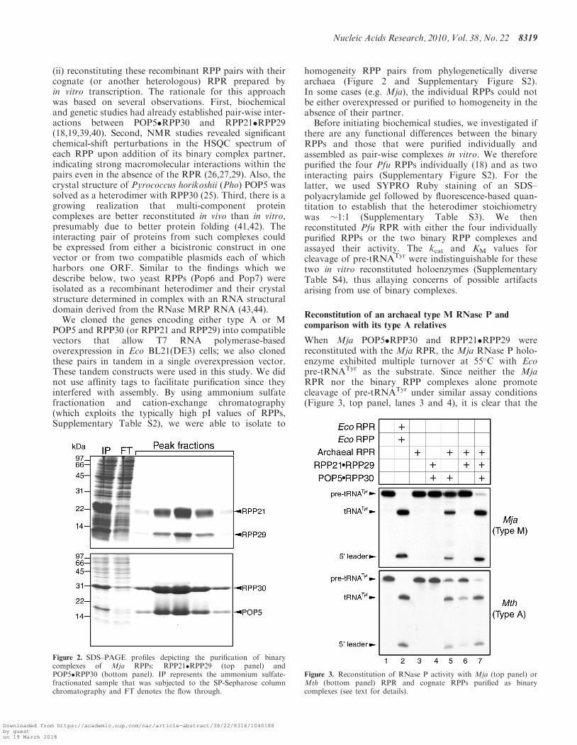
We cloned the genes encoding either type A or MPOP5 and RPP30 (or RPP21 and RPP29) into compatiblevectors that allow T7 RNA polymerase-basedoverexpression in Eco BL21(DE3) cells; we also clonedthese pairs in tandem in a single overexpression vector.These tandem constructs were used in this study. We didnot use affinity tags to facilitate purification since theyinterfered with assembly. By using ammonium sulfatefractionation and cation-exchange chromatography(which exploits the typically high pI values of RPPs,Supplementary Table S2), we were able to isolate to
homogeneity RPP pairs from phylogenetically diversearchaea (Figure 2 and Supplementary Figure S2).In some cases (e.g. Mja), the individual RPPs could notbe either overexpressed or purified to homogeneity in theabsence of their partner.Before initiating biochemical studies, we investigated if
there are any functional differences between the binaryRPPs and those that were purified individually andassembled as pair-wise complexes in vitro. We thereforepurified the four Pfu RPPs individually (18) and as twointeracting pairs (Supplementary Figure S2). For thelatter, we used SYPRO Ruby staining of an SDS–polyacrylamide gel followed by fluorescence-based quan-titation to establish that the heterodimer stoichiometrywas �1:1 (Supplementary Table S3). We thenreconstituted Pfu RPR with either the four individuallypurified RPPs or the two binary RPP complexes andassayed their activity. The kcat and KM values forcleavage of pre-tRNATyr were indistinguishable for thesetwo in vitro reconstituted holoenzymes (SupplementaryTable S4), thus allaying concerns of possible artifactsarising from use of binary complexes.
Reconstitution of an archaeal type M RNase P andcomparison with its type A relatives
When Mja POP5�RPP30 and RPP21�RPP29 werereconstituted with the Mja RPR, the Mja RNase P holo-enzyme exhibited multiple turnover at 55�C with Ecopre-tRNATyr as the substrate. Since neither the MjaRPR nor the binary RPP complexes alone promotecleavage of pre-tRNATyr under similar assay conditions(Figure 3, top panel, lanes 3 and 4), it is clear that the
Figure 3. Reconstitution of RNase P activity with Mja (top panel) orMth (bottom panel) RPR and cognate RPPs purified as binarycomplexes (see text for details).
Figure 2. SDS–PAGE profiles depicting the purification of binarycomplexes of Mja RPPs: RPP21�RPP29 (top panel) andPOP5�RPP30 (bottom panel). IP represents the ammonium sulfate-fractionated sample that was subjected to the SP-Sepharose columnchromatography and FT denotes the flow through.
Nucleic Acids Research, 2010, Vol. 38, No. 22 8319
Downloaded from https://academic.oup.com/nar/article-abstract/38/22/8316/1040188by gueston 19 March 2018
RNP complex is the functional unit under theseconditions.Although Pfu (type A) RPR is capable of multiple
turnover in the absence of RPPs, the presence of eitherRPP21�RPP29 or POP5�RPP30 increases activity par-ticularly at lower substrate and Mg2+ concentrations,demonstrating that each partial holoenyzme constitutesa minimal functional complex (18). Compared to thereaction with RPR+4 RPPs, these partial holoenzymesdisplayed 600- or 100-fold lower kcat/KM values forcleavage of pre-tRNATyr (18). To test whether thesefindings are qualitatively applicable for a type M RNaseP, we assayed the partial RNP complexes assembledfrom Mja RPR and either Mja RPP21�RPP29 orPOP5�RPP30. Under multiple-turnover conditions,activity was observed with Mja RPR+POP5�RPP30 butnot RPP21�RPP29 (Figure 3, top panel, lanes 5 versus 6),a notable difference with Pfu RNase P. However, in bothtypes A and M, the RPR+4 RPPs has a 30mM Mg2+
requirement compared to 120mM Mg2+ needed by theRNPs assembled with only one of the binary RPPs.The observed differences in behavior of the partially
reconstituted Mja (type M) and Pfu (type A) RNase Pcomplexes could be attributed either to fundamental dif-ferences in the structure and functioning of these twoclasses of archaeal RNase P or to the remote possibilitythat Pfu RNase P might somehow differ from other typeA relatives. To rule out the latter, we purified RPPs fromMth, another member of the type A family and repeatedthe partial reconstitution studies. The results obtainedwith Mth RNase P (Figure 3, bottom panel) parallelthose we reported for Pfu RNase P (18), confirmingsimilar patterns for the two type A variants and highlight-ing the distinctive reconstitution results obtained withtypes A and M.
Reconstitution of Mja RPR deletion derivativeswith Mja RPPs
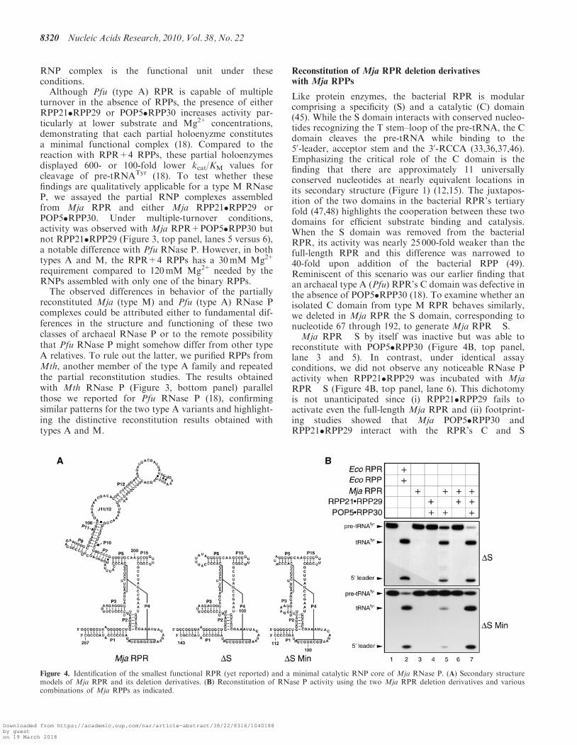
Like protein enzymes, the bacterial RPR is modularcomprising a specificity (S) and a catalytic (C) domain(45). While the S domain interacts with conserved nucleo-tides recognizing the T stem–loop of the pre-tRNA, the Cdomain cleaves the pre-tRNA while binding to the50-leader, acceptor stem and the 30-RCCA (33,36,37,46).Emphasizing the critical role of the C domain is thefinding that there are approximately 11 universallyconserved nucleotides at nearly equivalent locations inits secondary structure (Figure 1) (12,15). The juxtapos-ition of the two domains in the bacterial RPR’s tertiaryfold (47,48) highlights the cooperation between these twodomains for efficient substrate binding and catalysis.When the S domain was removed from the bacterialRPR, its activity was nearly 25 000-fold weaker than thefull-length RPR and this difference was narrowed to40-fold upon addition of the bacterial RPP (49).Reminiscent of this scenario was our earlier finding thatan archaeal type A (Pfu) RPR’s C domain was defective inthe absence of POP5�RPP30 (18). To examine whether anisolated C domain from type M RPR behaves similarly,we deleted in Mja RPR the S domain, corresponding tonucleotide 67 through 192, to generate Mja RPR �S.
Mja RPR �S by itself was inactive but was able toreconstitute with POP5�RPP30 (Figure 4B, top panel,lane 3 and 5). In contrast, under identical assayconditions, we did not observe any noticeable RNase Pactivity when RPP21�RPP29 was incubated with MjaRPR �S (Figure 4B, top panel, lane 6). This dichotomyis not unanticipated since (i) RPP21�RPP29 fails toactivate even the full-length Mja RPR and (ii) footprint-ing studies showed that Mja POP5�RPP30 andRPP21�RPP29 interact with the RPR’s C and S
Figure 4. Identification of the smallest functional RPR (yet reported) and a minimal catalytic RNP core of Mja RNase P. (A) Secondary structuremodels of Mja RPR and its deletion derivatives. (B) Reconstitution of RNase P activity using the two Mja RPR deletion derivatives and variouscombinations of Mja RPPs as indicated.
8320 Nucleic Acids Research, 2010, Vol. 38, No. 22
Downloaded from https://academic.oup.com/nar/article-abstract/38/22/8316/1040188by gueston 19 March 2018

domains, respectively (29). However, we were surprisedthat RPP21�RPP29 was able to stimulate the activity ofMja RPR �S+POP5�RPP30 (Figure 4B, top panel, lane7), likely reflecting indirect effects arising from inter-actions between the two RPP pairs. Such an effect wasnot observed when we tested Pfu RPR �S with the twocognate binary RPP pairs (18).
Relative to a type A RPR, the C domain of a type MRPR is smaller due to the lack of certain structuralelements (e.g. P6, P16 and P17) (13). To identify thesmallest RPR variant that might yet generate a functionalRNP, we trimmed the P1 and P3 helices and the length ofthe loop that caps P5 in Mja RPR �S. The resultingminimal Mja RPR �S Min (112 nt; Figure 4A) reconsti-tutes with POP5�RPP30 and is further activated uponaddition of RPP21�RPP29, similar in behavior to MjaRPR �S (Figure 4B, lanes 5 and 7).
Heterologous reconstitutions within and across domainshighlights a universal catalytic core
The results with Mja RPR �S Min revealed that theuniversally conserved catalytic core embedded in aminimal structural fold suffices for functional reconstitu-tion with Mja RPPs. To test the premise that a commonfunctional core might exist in all archaeal RNase P holo-enzymes, we mixed type A RPRs with type M RPPsand vice versa. Indeed, heterologous assemblies did yieldfunctional holoenzymes (Supplementary Figure S3).However, type A and M RPRs functioned more effectivelywith their cognate RPPs; such type-preferentialreconstitution patterns might reflect the co-evolution ofRPRs with their respective RPPs. These data neverthelessled us to examine if the bacterial and organellar RPR,which possess this conserved core, could also reconstitutewith archaeal RPPs (minimally with POP5�RPP30).
Under optimal conditions for reconstitution, both Eco(bacterial type A) and Bsu (bacterial type B) RPRsreconstituted with both type A (Pfu) and M (Mja)archaeal RPPs to generate functional RNase P holoen-zymes [Figure 5, (50) and data not shown]. To examine
which subset of RPPs suffices to promote bacterial RPRcatalysis, we tested the Eco RPR with Pfu POP5+RPP30or RPP21+RPP29 at 55�C and pH 7.5. We observed thatwhile POP5+RPP30 could enhance the activity of EcoRPR, RPP21+RPP29 has no effect (Figure 5, lanes 8and 9). Both type A and B RPRs were stimulatedroughly 10- to 15-fold, respectively, by PfuPOP5�RPP30 [(50) and data not shown]. Given the re-markable tertiary structure similarity between POP5 andthe bacterial RPP (26), we further examined if POP5 alonecould stimulate the bacterial RPR’s pre-tRNA cleavageactivity; however, this is not the case (Figure 5, lane 4).
Figure 6. Heterologous reconstitution of an RNase P holoenzymeusing Ram mt RPR and archaeal (Mja) RPPs. (A) Secondary structurerepresentation of Ram mt RPR. (B) Reconstitution of RNase P usingRam mt RPR and Mja RPPs (in the combinations indicated).
Figure 5. Heterologous reconstitution of an RNase P holoenzymeusing Eco RPR and Pfu RPPs (in the combinations indicated).
Nucleic Acids Research, 2010, Vol. 38, No. 22 8321
Downloaded from https://academic.oup.com/nar/article-abstract/38/22/8316/1040188by gueston 19 March 2018
Taken together, these results suggest that POP5�RPP30 isfunctionally equivalent to the bacterial RPP.We then assessed whether Mja RPPs could support
organellar RPR catalysis. We focused on themitochondrially (mt)-encoded RPR of Ram, a jakobid fla-gellate. Ram mt RPR represents a highly reduced versionof the bacterial RPR, lacking P6, P16, P17 and L15(Figure 6A) and was found to be catalytically inactiveunder various conditions tested in vitro (32).Interestingly, Mja POP5�RPP30 (and notRPP21�RPP29) can heterologously reconstitute withRam mt RPR (Figure 6B, lanes 6 and 7); this
activity is enhanced upon addition of RPP21�RPP29(Figure 6B, lane 8), in a manner reminiscent of the recon-stitutions of Mja RPR �S or �S Min with Mja RPPs(Figure 4).
Single-turnover kinetic studies to elucidate the role ofRPPs in aiding the cleavage step
From the various homologous and heterologous reconsti-tutions that we performed, it was clear that the two RPPpairs have different functional effects. Therefore, wesought to dissect these differences further using detailed
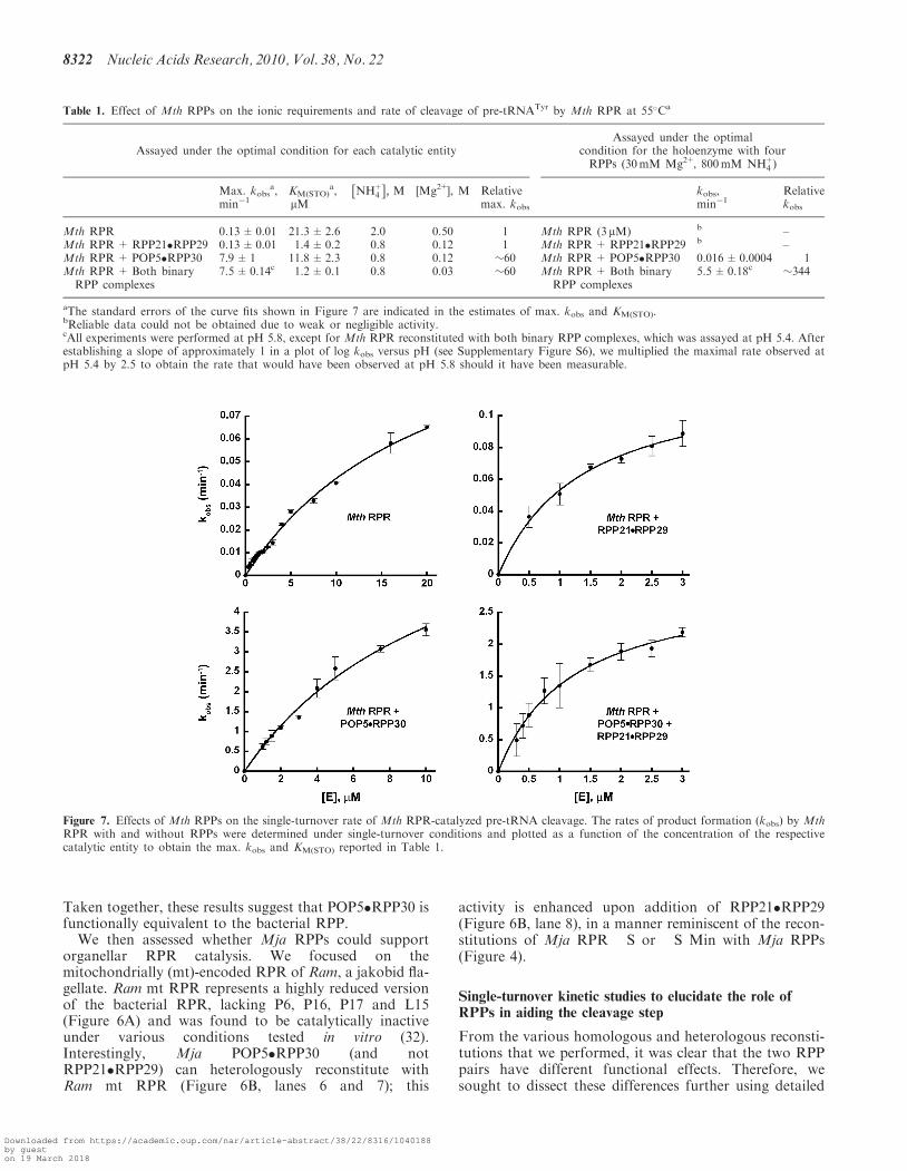
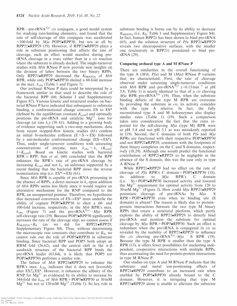
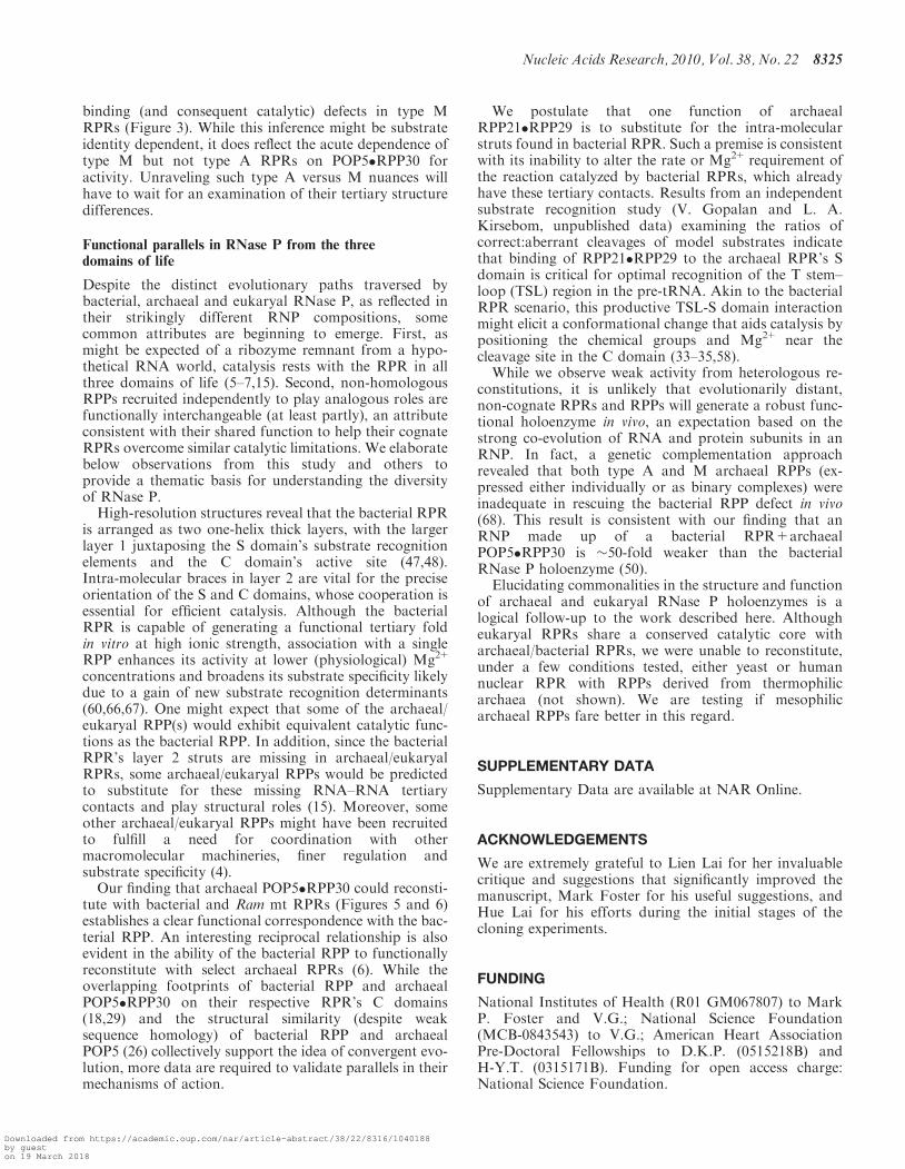
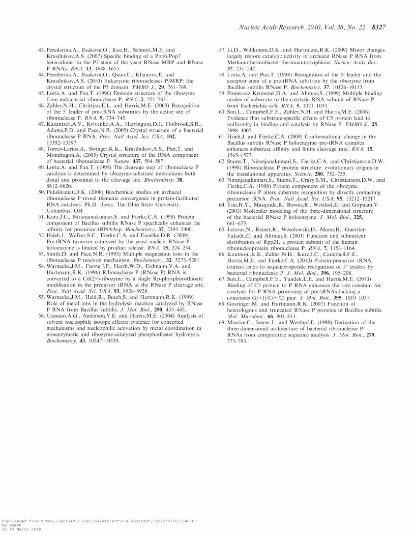
Figure 7. Effects of Mth RPPs on the single-turnover rate of Mth RPR-catalyzed pre-tRNA cleavage. The rates of product formation (kobs) by MthRPR with and without RPPs were determined under single-turnover conditions and plotted as a function of the concentration of the respectivecatalytic entity to obtain the max. kobs and KM(STO) reported in Table 1.
Table 1. Effect of Mth RPPs on the ionic requirements and rate of cleavage of pre-tRNATyr by Mth RPR at 55�Ca
Assayed under the optimal condition for each catalytic entityAssayed under the optimal
condition for the holoenzyme with fourRPPs (30mM Mg2+, 800mM NH+
4 Þ
Max. kobsa,
min�1KM(STO)
a,mM
NH+4
� �, M [Mg2+], M Relative kobs,
min�1Relative
max. kobs kobs
Mth RPR 0.13±0.01 21.3±2.6 2.0 0.50 1 Mth RPR (3mM) b –Mth RPR+RPP21�RPP29 0.13±0.01 1.4±0.2 0.8 0.12 1 Mth RPR+RPP21�RPP29 b –Mth RPR+ POP5�RPP30 7.9±1 11.8±2.3 0.8 0.12 �60 Mth RPR+ POP5�RPP30 0.016±0.0004 1Mth RPR+ Both binaryRPP complexes
7.5±0.14c 1.2±0.1 0.8 0.03 �60 Mth RPR+ Both binaryRPP complexes
5.5±0.18c �344
aThe standard errors of the curve fits shown in Figure 7 are indicated in the estimates of max. kobs and KM(STO).bReliable data could not be obtained due to weak or negligible activity.cAll experiments were performed at pH 5.8, except for Mth RPR reconstituted with both binary RPP complexes, which was assayed at pH 5.4. Afterestablishing a slope of approximately 1 in a plot of log kobs versus pH (see Supplementary Figure S6), we multiplied the maximal rate observed atpH 5.4 by 2.5 to obtain the rate that would have been observed at pH 5.8 should it have been measurable.
8322 Nucleic Acids Research, 2010, Vol. 38, No. 22
Downloaded from https://academic.oup.com/nar/article-abstract/38/22/8316/1040188by gueston 19 March 2018

rate measurements. Although we previously showed thatonly the addition of Pfu POP5�RPP30 to Pfu RPR in-creases its kcat (18), it remains unclear whether productrelease is rate limiting under multiple-turnover conditions[as demonstrated for bacterial and yeast RNase P (51,52)].Hence, we undertook single-turnover assays to measurethe RPR’s rate of pre-tRNA cleavage either alone orwhen aided by each archaeal RPP pair.
To facilitate manual single-turnover measurements,we slowed down the reaction rate by decreasing theassay pH from the typical 7.5 to 5.8, since the hydroxidenucleophile, which attacks the scissile phosphodiesterlinkage in the pre-tRNA, is believed to result fromdeprotonation of a hydrated Mg2+ ion in the RPR’sactive site (53–56). To shorten incubation times, weused Mth in lieu of Pfu RNase P because the Mth RPRis more active in vitro (6).
An excess of enzyme over substrate ([E]=0.3–20 mM,[S] �2 nM) was used in our single-turnover studies withMth RNase P. We found that a single-exponentialfunction describes the rate of product formation by theMth RPR±RPPs (Supplementary Figure S5). We usemax. kobs to indicate rates determined at saturating con-centrations of the RPR±RPPs in the presence of optimallevels of NH+
4 and Mg2+, which were established byscreening different assay conditions. The term KM(STO) isused to refer to the KM calculated under single-turnoverconditions.
To justify using KM(STO) [(k–1+k2)/k1] as a measure ofKS (k–1/k1), we performed a pulse-chase experiment to in-vestigate if the ES complex dissociates faster than sub-strate cleavage (i.e. k�1 >> k2; Scheme I) under thesingle-turnover conditions used. Through large dilutionof a pre-formed Mth RNase P–pre-tRNATyr complex,we dissociated the substrate from the enzyme andexpected a plateau in product formation post-dilution ifk–1 >> k2 (due to the pre-tRNA’s inability to rebind forcleavage). Indeed, when the reactions catalyzed by MthRPR±RPP21�RPP29 were diluted a few minutes aftermixing with pre-tRNATyr, product formation did notincrease post dilution (Supplementary Figure S4).
At pH 5.8 and 55�C, both Mth POP5�RPP30 andRPP21�RPP29 decreased the concentration of NH+
4 andMg2+ required for RPR-mediated pre-tRNATyr process-ing; the Mg2+ requirement decreased from 500 to120mM with either binary complex and to 30mM withboth (Table 1). The max. kobs for processing ofpre-tRNATyr by Mth RPR increased 60-fold uponaddition of POP5�RPP30 with no further changeupon inclusion of RPP21�RPP29 (Table 1; Figure 7).Also, RPP21�RPP29 alone was unable to increase theRPR’s max. kobs. However, RPP21�RPP29 was able toreduce KM(STO) from 21.3 to 1.4 mM (to the same extentas four RPPs; Table 1). These results demonstrate the
importance of POP5�RPP30 in cleavage andRPP21�RPP29 in substrate binding (however, see‘Discussion’ section for additional comments on the roleof POP5�RPP30).The above studies were performed at the optimal Mg2+
concentration for each of the RNP complexes assembledwith Mth RPR. We inquired if the results would be dif-ferent if the Mth RPR was assayed with and without eachbinary complex at 30mM Mg2+and 800mM NH+
4 , a con-dition optimal for the holoenzyme assembled with all fourRPPs but not for the partial RNPs. There is no detectableactivity in the RPR-alone reaction and weak (not reliablyquantifiable) activity with RPR+RPP21�RPP29. Incontrast, the RPR+POP5�RPP30 exhibits a kobs of0.016min�1, which increased to 5.5min�1 (344-fold)upon addition of RPP21�RPPP29, suggesting that thelatter significantly facilitates catalysis at lower Mg2+
concentrations.Some rate comparisons are instructive. First, although
Li et al. (57) did not report a max. kobs, they documentedpre-tRNAGly cleavage by 10 mM Mth RPR at pH 6 with arate of 0.034min�1, which is similar to the 0.04min�1
that we observed for cleavage of pre-tRNATyr by 10 mMMth RPR at pH 5.8; this coincidence is reassuringgiven the different substrates and assay conditions usedin these two studies. Second, the max. kobs forpre-tRNATyr cleavage by Mth RPR is 0.13min�1 atpH 5.8 and 55�C (Table 1) compared to the bacterialRPR’s max. kobs of �5min�1 at pH 6 and 37�C (57).These data, together with earlier studies on chimericMth-Eco RPRs (57), reaffirm the idea that the bacterialRPR structural elements which are missing in the type Aarchaeal RPR contribute to both substrate binding andcleavage rate.
DISCUSSION
We have shown that purification of RPPs as binarycomplexes allows expedient assembly of RNase P holoen-zymes from different archaea. In addition to halving theprotein preparation effort, co-overexpression was a neces-sary measure to obtain recombinant RPPs (e.g. MjaRPP30) that do not express by themselves. These facileassemblies represent an important first step to investigatethe stoichiometry and oligomeric state of individualsubunits, and to initiate high-resolution structuralstudies. The latter objective might also benefit fromuse of pared-down RPRs such as Mja RPR �S Min(112 nt), the smallest known RPR that is functionalin the presence of POP5�RPP30. Importantly, these bio-chemical reconstitutions help uncover the mechan-istic basis of protein-aided RNA catalysis in archaealRNase P.
Delineating the roles of archaeal RPPs in aiding RPRcatalysis
Hint of the role of POP5�RPP30 in promoting pre-tRNAcleavage first came from multiple-turnover studieson Pfu RNase P where it was shown to increase theRPR’s kcat by 25-fold (18). We then used an MjaScheme I.
Nucleic Acids Research, 2010, Vol. 38, No. 22 8323
Downloaded from https://academic.oup.com/nar/article-abstract/38/22/8316/1040188by gueston 19 March 2018
RPR—pre-tRNATyr cis conjugate, a good model systemfor studying rate-limiting chemistry, and found that therate of self-cleavage of this conjugate was accelerated�100-fold by Mja POP5�RPP30, but not at all byRPP21�RPP29 (19). However, if RPP21�RPP29 plays arole in substrate positioning that affects the rate ofcleavage, such an effect would manifest during pre-tRNA cleavage in a trans rather than in a cis reactionwhere the substrate is already docked. The single-turnoverstudies with Mth RNase P now provide new insights onthe division of labor between the two binary RPPs.Only RPP21�RPP29 decreased the KM(STO) of MthRPR, while only POP5�RPP30 elicited a 60-fold increasein the max. kobs (Table 1 and Figure 7).Our archaeal RNase P data could be interpreted by a
framework similar to that used to describe the role ofthe bacterial RPP (60; Scheme I and SupplementaryFigure S7). Various kinetic and structural studies on bac-terial RNase P have indicated that subsequent to substratebinding, a conformational change converts ES to ES*(defined by the equilibrium constant Kconf) and optimallypositions the pre-tRNA and catalytic Mg2+ ions forcleavage (at rate kc) (58–61). Adding to a growing bodyof evidence supporting such a two-step mechanism, datafrom recent stopped-flow kinetic studies (61) confirman initial bi-molecular collision (E+S!ES) followedby a uni-molecular conformational change (ES!ES*).Thus, under single-turnover conditions with saturatingconcentrations of enzyme, max. kobs= kc (Kconf/1+Kconf). Based on kinetic studies with bacterialRPR±RPP, Sun et al. (60) concluded that the RPPenhances the RPR’s rate of pre-tRNA cleavage byincreasing Kconf and not kc, an inference supported by arecent finding that the bacterial RPP slows the reverseisomerization step (i.e. ES*!ES) (61).Since Mth RPR is capable of pre-tRNA processing in
the absence of RPPs, a direct increase in kc upon additionof Mth RPPs seems less likely since it would require analternative mechanism for the RNP compared to theRPR, an unsupported premise. Therefore, we hypothesizethat increased conversion of ES!ES* must underlie theability of cognate POP5�RPP30 to elicit a 60- and100-fold increase, respectively, in the Mth RPR’s max.kobs (Figure 7) and the pre-tRNATyr—Mja RPRself-cleavage rate (19). Because POP5�RPP30 significantlyincreases the rate of the cleavage step, we cannot assess ifKM(STO)�KS (as we did for RPP21�RPP29;Supplementary Figure S4). Thus, without determiningthe microscopic rate constants that contribute to KS, wecannot rule out the role of POP5�RPP30 in substratebinding. Since bacterial RPP and POP5 both adopt anRRM fold (26,62), and the central cleft in the a–bsandwich structure of the bacterial RPP binds thepre-tRNA leader (63,64), it is likely that POP5 (orPOP5�RPP30) performs a similar role.The failure of Mth RPP21�RPP29 to enhance the
RPR’s max. kobs indicates that it does not favorablyalter ES !ES*. However, it enhances the affinity of theRNP for Mg2+ as evidenced by its ability to increase by344-fold the kobs of Mth RPR+POP5�RPP30 at 30mMMg2+ but not at 120mM Mg2+ (Table 1). Its key role in
substrate binding is borne out by its ability to decreaseKM(STO) (i.e. KS; Table 1 and Supplementary Figure S4).In fact, human RPP21 has been shown to bind pre-tRNA(65), and the solution structure of Pfu RPP21�RPP29reveals two electropositive surfaces, with the smallerone (exclusively in RPP21) postulated to bind pre-tRNA (29).
Comparing archaeal type A and M RNase P
There are similarities in the overall functioning ofthe type A (Mth, Pfu) and M (Mja) RNase P variantsthat we characterized. First, the rate of cleavageobserved under saturating single-turnover conditionswith Mth RPR and pre-tRNATyr (�0.13min�1 at pH5.8; Table 1) is nearly identical to that of a cis cleavingMja RPR–pre-tRNATyr (19). Thus, when the substratebinding defects of the type M RPR are overcomeby providing the substrate in cis, its activity coincideswith the type A relative. In fact, the fullyreconstituted type A and M holoenzymes also displaysimilar rates (Table 1) (19). Such a comparisontakes into consideration the fact that the rates re-ported for the self-cleaving Mja (type M) RPR wereat pH 5.4 and not pH 5.1 as was mistakenly reportedin (19). Second, the C domains of both Pfu and MjaRPRs are functional with their respective POP5�RPP30and not RPP21�RPP29, consistent with the footprints ofthese binary complexes on the C and S domains, respect-ively (18,29). Although one would predict the functionalcontribution of RPP21�RPP29 to be negligible in theabsence of the S domain, this was the case only in typeA RNase P.
While RPP21�RPP29 had no effect on the rate ofcleavage of Pfu RPR’s C domain+POP5�RPP30 (18),its addition to Mja RPR’s C domain(i.e. �S)+POP5�RPP30 increased activity and loweredthe Mg2+ requirement for optimal activity from 120 to30mM Mg2+ (Figure 3). How could Mja RPP21�RPP29potentiate cleavage of pre-tRNAs by Mja �SRPR+POP5�RPP30 even when its binding site (Sdomain) is absent? The reason is likely due to protein–protein interactions between the two type M binaryRPPs that create a structural platform, which partlyexploits the ability of RPP21�RPP29 to directly bindpre-tRNA and position the substrate for optimalcleavage by Mja RPR+POP5�RPP30. Such an effect isredundant when the pre-tRNA is conjugated in cis asrevealed by the inability of RPP21�RPP29 to influencethe cis cleaving pre-tRNATyr–Mja �S RPR (19).Because the type M RPR is smaller than the type ARPR (13), it offers fewer possibilities for nucleating inde-pendent, cooperative interactions with multiple RPPs,thus accentuating the need for protein-protein interactionsin type M RNase P.
Our studies on type A and M RNase P indicate that thesubstrate- and metal ion-positioning effects ofRPP21�RPP29 contribute to an increased rate whenenabled by POP5�RPP30 already bound to the Cdomain. However, it is intriguing that type MRPP21�RPP29 alone is unable to alleviate the substrate
8324 Nucleic Acids Research, 2010, Vol. 38, No. 22
Downloaded from https://academic.oup.com/nar/article-abstract/38/22/8316/1040188by gueston 19 March 2018
binding (and consequent catalytic) defects in type MRPRs (Figure 3). While this inference might be substrateidentity dependent, it does reflect the acute dependence oftype M but not type A RPRs on POP5�RPP30 foractivity. Unraveling such type A versus M nuances willhave to wait for an examination of their tertiary structuredifferences.
Functional parallels in RNase P from the threedomains of life
Despite the distinct evolutionary paths traversed bybacterial, archaeal and eukaryal RNase P, as reflected intheir strikingly different RNP compositions, somecommon attributes are beginning to emerge. First, asmight be expected of a ribozyme remnant from a hypo-thetical RNA world, catalysis rests with the RPR in allthree domains of life (5–7,15). Second, non-homologousRPPs recruited independently to play analogous roles arefunctionally interchangeable (at least partly), an attributeconsistent with their shared function to help their cognateRPRs overcome similar catalytic limitations. We elaboratebelow observations from this study and others toprovide a thematic basis for understanding the diversityof RNase P.
High-resolution structures reveal that the bacterial RPRis arranged as two one-helix thick layers, with the largerlayer 1 juxtaposing the S domain’s substrate recognitionelements and the C domain’s active site (47,48).Intra-molecular braces in layer 2 are vital for the preciseorientation of the S and C domains, whose cooperation isessential for efficient catalysis. Although the bacterialRPR is capable of generating a functional tertiary foldin vitro at high ionic strength, association with a singleRPP enhances its activity at lower (physiological) Mg2+
concentrations and broadens its substrate specificity likelydue to a gain of new substrate recognition determinants(60,66,67). One might expect that some of the archaeal/eukaryal RPP(s) would exhibit equivalent catalytic func-tions as the bacterial RPP. In addition, since the bacterialRPR’s layer 2 struts are missing in archaeal/eukaryalRPRs, some archaeal/eukaryal RPPs would be predictedto substitute for these missing RNA–RNA tertiarycontacts and play structural roles (15). Moreover, someother archaeal/eukaryal RPPs might have been recruitedto fulfill a need for coordination with othermacromolecular machineries, finer regulation andsubstrate specificity (4).
Our finding that archaeal POP5�RPP30 could reconsti-tute with bacterial and Ram mt RPRs (Figures 5 and 6)establishes a clear functional correspondence with the bac-terial RPP. An interesting reciprocal relationship is alsoevident in the ability of the bacterial RPP to functionallyreconstitute with select archaeal RPRs (6). While theoverlapping footprints of bacterial RPP and archaealPOP5�RPP30 on their respective RPR’s C domains(18,29) and the structural similarity (despite weaksequence homology) of bacterial RPP and archaealPOP5 (26) collectively support the idea of convergent evo-lution, more data are required to validate parallels in theirmechanisms of action.
We postulate that one function of archaealRPP21�RPP29 is to substitute for the intra-molecularstruts found in bacterial RPR. Such a premise is consistentwith its inability to alter the rate or Mg2+ requirement ofthe reaction catalyzed by bacterial RPRs, which alreadyhave these tertiary contacts. Results from an independentsubstrate recognition study (V. Gopalan and L. A.Kirsebom, unpublished data) examining the ratios ofcorrect:aberrant cleavages of model substrates indicatethat binding of RPP21�RPP29 to the archaeal RPR’s Sdomain is critical for optimal recognition of the T stem–loop (TSL) region in the pre-tRNA. Akin to the bacterialRPR scenario, this productive TSL-S domain interactionmight elicit a conformational change that aids catalysis bypositioning the chemical groups and Mg2+ near thecleavage site in the C domain (33–35,58).While we observe weak activity from heterologous re-
constitutions, it is unlikely that evolutionarily distant,non-cognate RPRs and RPPs will generate a robust func-tional holoenzyme in vivo, an expectation based on thestrong co-evolution of RNA and protein subunits in anRNP. In fact, a genetic complementation approachrevealed that both type A and M archaeal RPPs (ex-pressed either individually or as binary complexes) wereinadequate in rescuing the bacterial RPP defect in vivo(68). This result is consistent with our finding that anRNP made up of a bacterial RPR+archaealPOP5�RPP30 is �50-fold weaker than the bacterialRNase P holoenzyme (50).Elucidating commonalities in the structure and function
of archaeal and eukaryal RNase P holoenzymes is alogical follow-up to the work described here. Althougheukaryal RPRs share a conserved catalytic core witharchaeal/bacterial RPRs, we were unable to reconstitute,under a few conditions tested, either yeast or humannuclear RPR with RPPs derived from thermophilicarchaea (not shown). We are testing if mesophilicarchaeal RPPs fare better in this regard.
SUPPLEMENTARY DATA
Supplementary Data are available at NAR Online.
ACKNOWLEDGEMENTS
We are extremely grateful to Lien Lai for her invaluablecritique and suggestions that significantly improved themanuscript, Mark Foster for his useful suggestions, andHue Lai for his efforts during the initial stages of thecloning experiments.
FUNDING
National Institutes of Health (R01 GM067807) to MarkP. Foster and V.G.; National Science Foundation(MCB-0843543) to V.G.; American Heart AssociationPre-Doctoral Fellowships to D.K.P. (0515218B) andH-Y.T. (0315171B). Funding for open access charge:National Science Foundation.
Nucleic Acids Research, 2010, Vol. 38, No. 22 8325
Downloaded from https://academic.oup.com/nar/article-abstract/38/22/8316/1040188by gueston 19 March 2018

Conflict of interest statement. None declared.
REFERENCES
1. Liu,F. and Altman,S. (2010) Ribonuclease P. Protein ReviewsSeries. New York, Springer-Verlag.
2. Evans,D., Marquez,S.M. and Pace,N.R. (2006) RNase P:interface of the RNA and protein worlds. Trends Biochem. Sci.,31, 333–341.
3. Walker,S.C. and Engelke,D.R. (2006) Ribonuclease P: theevolution of an ancient RNA enzyme. Crit. Rev. Biochem. Mol.Biol., 41, 77–102.
4. Lai,L.B., Vioque,A., Kirsebom,L.A. and Gopalan,V. (2010)Unexpected diversity of RNase P, an ancient tRNA processingenzyme: challenges and prospects. FEBS Lett., 584, 287–296.
5. Guerrier-Takada,C., Gardiner,K., Marsh,T., Pace,N. andAltman,S. (1983) The RNA moiety of ribonuclease P is thecatalytic subunit of the enzyme. Cell, 35, 849–857.
6. Pannucci,J.A., Haas,E.S., Hall,T.A., Harris,J.K. and Brown,J.W.(1999) RNase P RNAs from some Archaea are catalyticallyactive. Proc. Natl Acad. Sci. USA, 96, 7803–7808.
7. Kikovska,E., Svard,S.G. and Kirsebom,L.A. (2007) EukaryoticRNase P RNA mediates cleavage in the absence of protein. Proc.Natl Acad. Sci. USA, 104, 2062–2067.
8. Haas,E.S., Banta,A.B., Harris,J.K., Pace,N.R. and Brown,J.W.(1996) Structure and evolution of ribonuclease P RNA inGram-positive bacteria. Nucleic Acids Res., 24, 4775–4782.
9. Haas,E.S., Armbruster,D.W., Vucson,B.M., Daniels,C.J. andBrown,J.W. (1996) Comparative analysis of ribonuclease P RNAstructure in Archaea. Nucleic Acids Res., 24, 1252–1259.
10. Brown,J.W., Nolan,J.M., Haas,E.S., Rubio,M.A., Major,F. andPace,N.R. (1996) Comparative analysis of ribonuclease P RNAusing gene sequences from natural microbial populations revealstertiary structural elements. Proc. Natl Acad. Sci. USA, 93,3001–3006.
11. Siegel,R.W., Banta,A.B., Haas,E.S., Brown,J.W. and Pace,N.R.(1996) Mycoplasma fermentans simplifies our view of the catalyticcore of ribonuclease P RNA. RNA, 2, 452–462.
12. Brown,J.W. (1999) The ribonuclease P database. Nucleic AcidsRes., 27, 314.
13. Harris,J.K., Haas,E.S., Williams,D., Frank,D.N. and Brown,J.W.(2001) New insight into RNase P RNA structure fromcomparative analysis of the archaeal RNA. RNA, 7, 220–232.
14. Marquez,S.M., Harris,J.K., Kelley,S.T., Brown,J.W.,Dawson,S.C., Roberts,E.C. and Pace,N.R. (2005) Structuralimplications of novel diversity in eucaryal RNase P RNA. RNA,11, 739–751.
15. Gopalan,V. (2007) Uniformity amid diversity in RNase P.Proc. Natl Acad. Sci. USA, 104, 2031–2032.
16. Boomershine,W.P., McElroy,C.A., Tsai,H.Y., Wilson,R.C.,Gopalan,V. and Foster,M.P. (2003) Structure of Mth11/MthRpp29, an essential protein subunit of archaeal and eukaryoticRNase P. Proc. Natl Acad. Sci. USA, 100, 15398–15403.
17. Kouzuma,Y., Mizoguchi,M., Takagi,H., Fukuhara,H.,Tsukamoto,M., Numata,T. and Kimura,M. (2003) Reconstitutionof archaeal ribonuclease P from RNA and four proteincomponents. Biochem. Biophys. Res. Commun., 306, 666–673.
18. Tsai,H.Y., Pulukkunat,D.K., Woznick,W.K. and Gopalan,V.(2006) Functional reconstitution and characterization ofPyrococcus furiosus RNase P. Proc. Natl Acad. Sci. USA, 103,16147–16152.
19. Pulukkunat,D.K. and Gopalan,V. (2008) Studies onMethanocaldococcus jannaschii RNase P reveal insights into theroles of RNA and protein cofactors in RNase P catalysis.Nucleic Acids Res., 36, 4172–4180.
20. Sidote,D.J. and Hoffman,D.W. (2003) NMR structure of anarchaeal homologue of ribonuclease P protein Rpp29.Biochemistry, 42, 13541–13550.
21. Numata,T., Ishimatsu,I., Kakuta,Y., Tanaka,I. and Kimura,M.(2004) Crystal structure of archaeal ribonuclease P proteinPh1771p from Pyrococcus horikoshii OT3: an archaeal homologof eukaryotic ribonuclease P protein Rpp29. RNA, 10, 1423–1432.
22. Sidote,D.J., Heideker,J. and Hoffman,D.W. (2004) Crystalstructure of archaeal ribonuclease P protein aRpp29 fromArchaeoglobus fulgidus. Biochemistry, 43, 14128–14138.
23. Takagi,H., Watanabe,M., Kakuta,Y., Kamachi,R., Numata,T.,Tanaka,I. and Kimura,M. (2004) Crystal structure of theribonuclease P protein Ph1877p from hyperthermophilic archaeonPyrococcus horikoshii OT3. Biochem. Biophys. Res. Commun.,319, 787–794.
24. Kakuta,Y., Ishimatsu,I., Numata,T., Kimura,K., Yao,M.,Tanaka,I. and Kimura,M. (2005) Crystal structure of aribonuclease P protein Ph1601p from Pyrococcus horikoshii OT3:an archaeal homologue of human nuclear ribonuclease P proteinRpp21. Biochemistry, 44, 12086–12093.
25. Kawano,S., Nakashima,T., Kakuta,Y., Tanaka,I. and Kimura,M.(2006) Crystal structure of protein Ph1481p in complex withprotein Ph1877p of archaeal RNase P from Pyrococcus horikoshiiOT3: implication of dimer formation of the holoenzyme.J. Mol. Biol., 357, 583–591.
26. Wilson,R.C., Bohlen,C.J., Foster,M.P. and Bell,C.E. (2006)Structure of Pfu Pop5, an archaeal RNase P protein.Proc. Natl Acad. Sci. USA, 103, 873–878.
27. Amero,C.D., Boomershine,W.P., Xu,Y. and Foster,M. (2008)Solution structure of Pyrococcus furiosus RPP21, a component ofthe archaeal RNase P holoenzyme, and interactions with itsRPP29 protein partner. Biochemistry, 47, 11704–11710.
28. Honda,T., Kakuta,Y., Kimura,K., Saho,J. and Kimura,M. (2008)Structure of an archaeal homolog of the human protein complexRpp21-Rpp29 that is a key core component for the assembly ofactive ribonuclease P. J. Mol. Biol., 384, 652–662.
29. Xu,Y., Amero,C.D., Pulukkunat,D.K., Gopalan,V. andFoster,M.P. (2009) Solution structure of an archaeal RNase Pbinary protein complex: formation of the 30-kDa complexbetween Pyrococcus furiosus RPP21 and RPP29 is accompaniedby coupled protein folding and highlights critical features forprotein-protein and protein-RNA interactions. J. Mol. Biol., 393,1043–1055.
30. Vioque,A., Arnez,J. and Altman,S. (1988) Protein-RNAinteractions in the RNase P holoenzyme from Escherichia coli.J. Mol. Biol., 202, 835–848.
31. Waugh,D.S. and Pace,N.R. (1993) Gap-scan deletion analysis ofBacillus subtilis RNase P RNA. FASEB J., 7, 188–195.
32. Seif,E., Cadieux,A. and Lang,B.F. (2006) Hybrid E. coli–Mitochondrial ribonuclease P RNAs are catalytically active.RNA, 12, 1661–1670.
33. Pan,T., Loria,A. and Zhong,K. (1995) Probing of tertiaryinteractions in RNA: 2’-hydroxyl-base contacts between theRNase P RNA and pre-tRNA. Proc. Natl Acad. Sci. USA, 92,12510–12514.
34. Loria,A. and Pan,T. (1997) Recognition of the T stem-loop of apre-tRNA substrate by the ribozyme from Bacillus subtilisribonuclease P. Biochemistry, 36, 6317–6325.
35. Brannvall,M., Kikovska,E., Wu,S. and Kirsebom,L.A. (2007)Evidence for Induced Fit in Bacterial RNase P RNA-mediatedCleavage. J. Mol. Biol., 372, 1149–1164.
36. Kirsebom,L.A. and Trobro,S. (2009) RNase P RNA-mediatedcleavage. IUBMB Life, 61, 189–200.
37. Kirsebom,L.A. and Svard,S.G. (1994) Base pairing betweenEscherichia coli RNase P RNA and its substrate. EMBO J., 13,4870–4876.
38. Oh,B.K. and Pace,N.R. (1994) Interaction of the 3’-end of tRNAwith ribonuclease P RNA. Nucleic Acids Res., 22, 4087–4094.
39. Hall,T.A. and Brown,J.W. (2004) Interactions between RNase Pprotein subunits in archaea. Archaea, 1, 247–254.
40. Kifusa,M., Fukuhara,H., Hayashi,T. and Kimura,M. (2005)Protein-protein interactions in the subunits of ribonuclease P inthe hyperthermophilic archaeon Pyrococcus horikoshii OT3.Biosci. Biotechnol. Biochem., 69, 1209–1212.
41. Tan,S. (2001) A modular polycistronic expression system foroverexpressing protein complexes in Escherichia coli. ProteinExpr. Purif., 21, 224–234.
42. Finkelstein,J., Antony,E., Hingorani,M.M. and O’Donnell,M.(2003) Overproduction and analysis of eukaryotic multiproteincomplexes in Escherichia coli using a dual-vector strategy.Anal. Biochem., 319, 78–87.
8326 Nucleic Acids Research, 2010, Vol. 38, No. 22
Downloaded from https://academic.oup.com/nar/article-abstract/38/22/8316/1040188by gueston 19 March 2018
43. Perederina,A., Esakova,O., Koc,H., Schmitt,M.E. andKrasilnikov,A.S. (2007) Specific binding of a Pop6/Pop7heterodimer to the P3 stem of the yeast RNase MRP and RNaseP RNAs. RNA, 13, 1648–1655.
44. Perederina,A., Esakova,O., Quan,C., Khanova,E. andKrasilnikov,A.S. (2010) Eukaryotic ribonucleases P/MRP: thecrystal structure of the P3 domain. EMBO J., 29, 761–769.
45. Loria,A. and Pan,T. (1996) Domain structure of the ribozymefrom eubacterial ribonuclease P. RNA, 2, 551–563.
46. Zahler,N.H., Christian,E.L. and Harris,M.E. (2003) Recognitionof the 50 leader of pre-tRNA substrates by the active site ofribonuclease P. RNA, 9, 734–745.
47. Kazantsev,A.V., Krivenko,A.A., Harrington,D.J., Holbrook,S.R.,Adams,P.D. and Pace,N.R. (2005) Crystal structure of a bacterialribonuclease P RNA. Proc. Natl Acad. Sci. USA, 102,13392–13397.
48. Torres-Larios,A., Swinger,K.K., Krasilnikov,A.S., Pan,T. andMondragon,A. (2005) Crystal structure of the RNA componentof bacterial ribonuclease P. Nature, 437, 584–587.
49. Loria,A. and Pan,T. (1999) The cleavage step of ribonuclease Pcatalysis is determined by ribozyme-substrate interactions bothdistal and proximal to the cleavage site. Biochemistry, 38,8612–8620.
50. Pulukkunat,D.K. (2008) Biochemical studies on archaealribonuclease P reveal thematic convergence in protein-facilitatedRNA catalysis. Ph.D. thesis. The Ohio State University,Columbus, OH.
51. Kurz,J.C., Niranjanakumari,S. and Fierke,C.A. (1998) Proteincomponent of Bacillus subtilis RNase P specifically enhances theaffinity for precursor-tRNAAsp. Biochemistry, 37, 2393–2400.
52. Hsieh,J., Walker,S.C., Fierke,C.A. and Engelke,D.R. (2009)Pre-tRNA turnover catalyzed by the yeast nuclear RNase Pholoenzyme is limited by product release. RNA, 15, 224–234.
53. Smith,D. and Pace,N.R. (1993) Multiple magnesium ions in theribonuclease P reaction mechanism. Biochemistry, 32, 5273–5281.
54. Warnecke,J.M., Furste,J.P., Hardt,W.D., Erdmann,V.A. andHartmann,R.K. (1996) Ribonuclease P (RNase P) RNA isconverted to a Cd(2+)-ribozyme by a single Rp-phosphorothioatemodification in the precursor tRNA at the RNase P cleavage site.Proc. Natl Acad. Sci. USA, 93, 8924–8928.
55. Warnecke,J.M., Held,R., Busch,S. and Hartmann,R.K. (1999)Role of metal ions in the hydrolysis reaction catalyzed by RNaseP RNA from Bacillus subtilis. J. Mol. Biol., 290, 433–445.
56. Cassano,A.G., Anderson,V.E. and Harris,M.E. (2004) Analysis ofsolvent nucleophile isotope effects: evidence for concertedmechanisms and nucleophilic activation by metal coordination innonenzymatic and ribozyme-catalyzed phosphodiester hydrolysis.Biochemistry, 43, 10547–10559.
57. Li,D., Willkomm,D.K. and Hartmann,R.K. (2009) Minor changeslargely restore catalytic activity of archaeal RNase P RNA fromMethanothermobacter thermoautotrophicus. Nucleic Acids Res.,37, 231–242.
58. Loria,A. and Pan,T. (1998) Recognition of the 5’ leader and theacceptor stem of a pre-tRNA substrate by the ribozyme fromBacillus subtilis RNase P. Biochemistry, 37, 10126–10133.
59. Pomeranz Krummel,D.A. and Altman,S. (1999) Multiple bindingmodes of substrate to the catalytic RNA subunit of RNase Pfrom Escherichia coli. RNA, 5, 1021–1033.
60. Sun,L., Campbell,F.E., Zahler,N.H. and Harris,M.E. (2006)Evidence that substrate-specific effects of C5 protein lead touniformity in binding and catalysis by RNase P. EMBO J., 25,3998–4007.
61. Hsieh,J. and Fierke,C.A. (2009) Conformational change in theBacillus subtilis RNase P holoenzyme–pre-tRNA complexenhances substrate affinity and limits cleavage rate. RNA, 15,1565–1577.
62. Stams,T., Niranjanakumari,S., Fierke,C.A. and Christianson,D.W.(1998) Ribonuclease P protein structure: evolutionary origins inthe translational apparatus. Science, 280, 752–755.
63. Niranjanakumari,S., Stams,T., Crary,S.M., Christianson,D.W. andFierke,C.A. (1998) Protein component of the ribozymeribonuclease P alters substrate recognition by directly contactingprecursor tRNA. Proc. Natl Acad. Sci. USA, 95, 15212–15217.
64. Tsai,H.Y., Masquida,B., Biswas,R., Westhof,E. and Gopalan,V.(2003) Molecular modeling of the three-dimensional structureof the bacterial RNase P holoenzyme. J. Mol. Biol., 325,661–675.
65. Jarrous,N., Reiner,R., Wesolowski,D., Mann,H., Guerrier-Takada,C. and Altman,S. (2001) Function and subnucleardistribution of Rpp21, a protein subunit of the humanribonucleoprotein ribonuclease P. RNA, 7, 1153–1164.
66. Koutmou,K.S., Zahler,N.H., Kurz,J.C., Campbell,F.E.,Harris,M.E. and Fierke,C.A. (2010) Protein-precursor tRNAcontact leads to sequence-specific recognition of 5’ leaders bybacterial ribonuclease P. J. Mol. Biol., 396, 195–208.
67. Sun,L., Campbell,F.E., Yandek,L.E. and Harris,M.E. (2010)Binding of C5 protein to P RNA enhances the rate constant forcatalysis for P RNA processing of pre-tRNAs lacking aconsensus G(+1)/C(+72) pair. J. Mol. Biol., 395, 1019–1037.
68. Gosringer,M. and Hartmann,R.K. (2007) Function ofheterologous and truncated RNase P proteins in Bacillus subtilis.Mol. Microbiol., 66, 801–813.
69. Massire,C., Jaeger,L. and Westhof,E. (1998) Derivation of thethree-dimensional architecture of bacterial ribonuclease PRNAs from comparative sequence analysis. J. Mol. Biol., 279,773–793.
Nucleic Acids Research, 2010, Vol. 38, No. 22 8327
Downloaded from https://academic.oup.com/nar/article-abstract/38/22/8316/1040188by gueston 19 March 2018