dissecting the mechanism underlying epigenetic …
TRANSCRIPT

The Pennsylvania State University
The Graduate School
Graduate Program in Plant Physiology
DISSECTING THE MECHANISM UNDERLYING
EPIGENETIC ACTIVATION
OF THE MAIZE SPM TRANSPOSON
BY THE ELEMENT-ENCODED TNPA PROTEIN
A Thesis in
Plant Physiology
by
Hongchang Cui
2003 Hongchang Cui
Submitted in Partial Fulfillment
of the Requirements
for the Degree of
Doctor of Philosophy
August 2003

The thesis of Hongchang Cui has been reviewed and approved by the following:
Nina V. FedoroffWillaman Professor of Life SciencesEvan Pugh ProfessorThesis Co-AdvisorChair of Committee
Jerry L.WorkmanAssociate Investigator, Howard Hughes Medical InstitutePaul Berg Professor of Biochemistry and Molecular BiologyThesis Co-Advisor
Hong MaProfessor of Biology
Teh-hui KaoProfessor of Biochemistry and Molecular BiologyHead of the Graduate Program in Plant Physiology
* Signatures are on file in the Graduate School.
iii
ABSTRACT
The activity of the maize Spm transposon is epigenetically regulated. The promoter
and its downstream GC-rich sequence are extensively methylated when the transposon is
inactive and unmethylated when the element is active. A methylated, inactive Spm
transposon can be activated by an active element, and the activation is accompanied by
loss of DNA methylation in both the promoter and the GC-rich sequence. Previous
studies have identified TnpA, one of the Spm-encoded transposase proteins, as the trans-
acting factor that mediates the epigenetic activation of an inactive Spm. However, how
TnpA promotes DNA demethylation has yet to be determined.
To facilitate elucidating the underlying mechanism, I have developed a novel assay
system that permits demethylation of the Spm sequence to be controlled by inducing
expression of TnpA in transgenic tobacco cells. Using this inducible DNA demethylation
system, I show that TnpA-mediated DNA demethylation occurs at a rate much faster than
that attributable to interference with methylation maintenance during DNA replication.
This observation strongly suggests an active process.
In further studies, I show that TnpA is a weak transcriptional activator and that
deletions that disrupt its ability to activate transcription also eliminate its ability to
promote DNA demethylation. Since the fusion protein between the truncated protein and
the viral VP16 activation domain has stronger DNA demethylation activity as well as
transcriptional activity, it is thus concluded that TnpA’s transcriptional activity plays an
important role in promoting DNA demethylation.
iv
Although TnpA-mediated demethylation of the Spm sequence is rapid over the
whole process, initial DNA demethylation is very slow. Moreover, DNA demethylation
has been observed only in callus and suspension cells, but not in leaves that have ceased
to divide. Using inhibitors of DNA replication and cell division, I show that DNA
replication is required as well for TnpA-mediated Spm demethylation. By using a gel
mobility shift assay I show that the binding affinity of TnpA to fully methylated DNA
fragments derived from Spm termini is much lower than its affinity for the same
fragments when hemi-methylated or unmethylated. Based on these observations, a two-
step DNA demethylation mechanism is suggested where TnpA binds to the post-
replicative, hemi-methylated Spm sequence and promotes demethylation either by
creating an appropriate demethylation substrate or by itself participating in or recruiting a
demethylase complex.
In animals cells several enzymatic activities have been identified that appear to be
capable of converting 5-methylated cytosine (5mC) into normal cytosine through distinct
mechanisms. Although there are cytological observations that suggest the presence of
DNA demethylase in plant cells, no biochemical evidence has ever been obtained. In the
present study, I have developed an in vitro DNA demethylation assay for which a
hemimethylated DNA fragment derived from Spm promoter and GC-rich sequence is
used as the substrate. A DNA demethylase activity is detected in nuclear extract that is
prepared from suspension cultured tobacco cells.
To investigate whether TnpA is itself a sequence-specific DNA demethylase or it
recruits a DNA demethylase activity to the Spm sequence, I have purified native TnpA
protein and its truncated derivatives from tobacco suspension cells by

v
coimmunoprecipitation. No DNA demethylase activity is detected in the TnpA-
containing immunoprecipitate, however, indicating the lack of a specific physical
interaction between TnpA and DNA demethylase. These results clearly rule out the
possibility that TnpA is a sequence-specific DNA demethylase.
To further explore the connection between transcriptional activation and DNA
demethylation, I have generated transgenic tobacco lines with the Spm promoter and GC-
rich sequence under the control of a glucocorticoid-inducible promoter. DNA
demethylation is observed in lines that show inducible transcription through the Spm
sequence. Spm demethylation is rapid, again suggesting an active DNA demethylation
process. Taken together, these observations strongly suggest that TnpA promotes Spm
demethylation by facilitating the access of DNA demethylase activities to methylated
DNA as a consequence of transcriptional activation.
Since several other trans-acting factors that are known to promote DNA
demethylation are also transcription factors, the present study has likely revealed a
common mechanism for epigenetic regulation. The theoretical and practical implications
of the present findings are discussed. Lastly, several future directions and approaches are
briefly described.

vi
TABLE OF CONTENTS
LIST OF TABLES …………………………………………………………………. ix
LIST OF FIGURES ………………………………………………………………... x
ACKNOWLEDGEMENTS ………………………………………………………... xii
Chapter 1 INTRODUCTION
DNA Methylation is Essential for Growth and Development …….……………. . 1
Biochemistry of DNA Methylation …….……………………………………….. 3
DNA Methylation Represses Transcription …………………………………….... 6
Interplay between DNA Methylation, Chromatin Remodeling, Histone
Modifications and Gene Silencing ……………………………………..…… 9
DNA Methylation is Reversible ………………………………………..………… 13
The Maize Spm Transposon is Epigenetically Regulated …..……………………. 18
References ………………………………………………………………………... 22
Chapter 2 TNPA PROMOTES DEMETHYLATION OF THE SPM
SEQUENCE THROUGH AN ACTIVE PROCESS
Introduction …………………………………………………… …….………….. 33
Materials and Methods …….…………………………………………………….. 35
Results …………………………..……………………………………..…………. 39
Discussion …………………………………..…………………………..………... 52
References ………………………………………………………………………... 55
Chapter 3 TNPA IS A SPM-SPECIFIC TRANSCRIPTIONAL ACTIVATOR
Introduction …………………………………………………… …….………….. 57
Materials and Methods …….……………………………………………………. 58
Results …………………………..……………………………………..…………. 61
Discussion …………………………………..…………………………..……….. 65
References ………………………………………………………………………... 69

vii
Chapter 4 THE TRANSCRIPTIONAL ACTIVITY OF TNPA IS ESSENTIAL
FOR ITS ABILITY TO PROMTE SPM DEMETHYLATION
Introduction …………………………………………………… …….………….. 71
Materials and Methods …….……………………………………………………. 73
Results …………………………..……………………………………..…………. 75
Discussion …………………………………..…………………………..………... 84
References ………………………………………………………………………... 88
Chapter 5 DNA REPLICATION IS REQUIRED FOR TNPA-
MEDIATED SPM DEMETHYLATION
Introduction …………………………………………………… …….………….. 91
Materials and Methods …….……………………………………………………. 92
Results …………………………..……………………………………..…………. 94
Discussion …………………………………..…………………………..……….. 98
References ……………………………………………………………………….. 101
Chapter 6 DNA DEMETHYLASE ACTIVITY IS PRESENT IN
DIVIDING TOBACCO CELLS
Introduction …………………………………………………… …….………….. 102
Materials and Methods …….……………………………………………………. 104
Results …………………………..……………………………………..………… 106
Discussion …………………………………..…………………………..……….. 111
References ……………………………………………………………………….. 114
Chapter 7 DNA DEMETHYLASE ACTIVITY IS RECRUITED TO THE SPM
SEQUENCE AS A RESULT OF TRANSCRIPTIONAL ACTIVATION
Introduction …………………………………………………… …….………….. 116
Materials and Methods …….……………………………………………………. 117
Results …………………………..……………………………………..………… 120
Discussion …………………………………..…………………………..……….. 131
References ……………………………………………………………………….. 139

viii
Chapter 8 CONCLUSION
Mechanism of TnpA-Mediated DNA Demethylation …………….…………….. 142
Significance of the Present Findings .…….………..……………………………. 145
Future Directions .…………………………………..…………………………... 148
References .……………………………………………………………………… 152

ix
LIST OF TABLES
Table 1 Expected and observed extent of EcoO109I cleavage of hemimethylated
DNA with different number of methylated cytosine residues (5mC) ………. 51

x
LIST OF FIGURES
Figure 1. T-DNA region of the binary vector used for inducible expression of
FLAG-TnpA, deletion derivatives, and fusion proteins …………… … …. 39
Figure 2. Inducible TnpA-mediated Spm demethylation ………….……………….. 42
Figure 3. Rapid DNA demethylation after TnpA expression ……….……………….. 45
Figure 4. Sensitivity to restriction enzymes of hemimethylated DNA
containing the Spm promoter and DCR .……………….………………… . 47
Figure 5. A diagram depicting the method for preparation of hemimethylated DNA
using biotin/streptavidin interaction …..……….……………….…………. 49
Figure 6. TnpA is a transcriptional activator in yeast ….…………………………….. 61
Figure 7. TnpA is a weak transcriptional activator in tobacco cells. ……………….... 63
Figure 8. Demethylation of the Spm sequence by TnpA deletion derivatives
and the TnpA-VP16 fusion protein ………..….…………………………… 76
Figure 9. Truncated TnpA proteins do not activate transcription in yeast cells .……. 78
Figure 10. Truncated TnpA proteins do not activate transcription in
tobacco cells. ……………………………………………………………… 80
Figure 11. Gel mobility shift assay showing binding of TnpA540 and TnpA420
to DNA derived from the Spm promoter sequence. ……………...………. 82
Figure 12. A model depicting the role of TnpA in Spm demethylation ………….…... 87
Figure 13. Inhibitors of DNA synthesis and cell division interfere with
TnpA-mediated Spm demethylation ..…………….……...….…………….. 95

xi
Figure 14. Electrophoretic mobility shift assay of TnpA binding to DNA
fragments with various number of TnpA binding sites ……..……….…… 97
Figure 15. A modified model illustrating the mechanism underlying
TnpA-mediated Spm demethylation ……………………………………. .. 99
Figure 16. Optimization of conditions for an in vitro DNA demethylation
assay…………………………………………………………….. 107
Figure 17. DNase activity in tobacco nuclear extracts and inhibition under the
conditions for the in vitro DNA demethylation assay ….………….………109
Figure 18. A representative in vitro DNA demethylation assay, showing the presence
of DNA demethylase activity in tobacco nuclear extracts ………………... 110
Figure 19. Immunoprecipitation of FLAG-tagged TnpA protein and derivatives
from tobacco suspension cultured cells………………..….………………. 121
Figure 20. TnpA does not associate specifically with DNA demethylase activity …... 123
Figure 21. TnpA protein expressed in plant cells binds well to the Spm promoter
sequence………………………………………………………………….. 125
Figure 22. DNA demethylation assay using hemimethylated DNA containing
DCR only …………………………………………………………………. 126
Figure 23. Detection of abasic sites in DNA using endonuclease IV . ………………. 127
Figure 24. TnpA does not co-fractionate with DNA demethylase activity. …….…… 128
Figure 25. Spm demethylation after induction of transcription ..…………………….. 130
Figure 26. Transgenes in close proximity are co-regulated……………...……….…... 137

xii
ACKNOWLEDGEMENTS
This work has been conducted under the supervision of Dr. Nina Fedoroff. DNA
methylation and the underlying epigenetic mechanisms have been an area of long-lasting
interest in animal studies, cancer research particularly, and such research with plants has
become intense in recent years. At the time I embarked on this project, however,
understanding in this area, especially about DNA demethylation, was still very
rudimentary. Not surprisingly, I have encountered many challenges, both technical and
conceptual, in the early stage of my studies. One of the challenges has been to develop a
new assay system for dissecting the mechanism by which TnpA promotes DNA
demethylation. Although some of my ideas worked well, yielding interesting results, for a
long time I had difficulty working out the key assay system. Eventually I was successful
with the assay, which has allowed me to address many important questions. However,
this would not have been possible without the invaluable advice and much-needed
encouragement as well as continuing support from Dr. Fedoroff. As my mentor, Dr.
Fedoroff has also helped me profoundly in my developing critical thinking skills as well
as understanding of the Western culture. There is no doubt that the training in her lab has
set a new stage for me and this experience will continue to benefit my future academic
career. I am deeply indebted to her for giving me the opportunity to study and work in her
laboratory.
I also would like to express my gratitude to Dr. Teh-hui Kao, Dr. Hong Ma and Dr.
Jerry Workman for their service on my committee and invaluable suggestions. I am
particularly thankful to Dr. Jerry Workman for his willingness to serve as my co-advisor.

xiii
My special thanks go to my wife, Yueling Hao, for her understanding and
unconditional support of my studies. In order for me to focus on research, she has given
up her job to stay at home taking care of our children and me. In the course of my studies,
my second child was born and this has further increased her workload. Since she is not
eligible for work in this country, she has to manage to make the both ends meet for an
already tight budget. She surely has sacrificed a lot for me and certainly deserves to share
all the credits I have earned so far.

1
Chapter 1
INTRODUCTION
DNA methylation is an almost universal phenomenon in living organisms ranging
from bacteria to the human being (Palmer and Marinus, 1994; Yoder and Bestor, 1996).
In prokaryotic cells, methylation occurs on both adenosine and cytosine, giving rise to 6’-
N-methyl adenosine and 5-methylcytosine (5mC) respectively, and is functionally
important in such processes as methylation-directed DNA repair (Modrich, 1989), control
of DNA replication initiation (Abeles et al., 1993), as well as protection of the host cell
against foreign nucleic acid invasion (Palmer and Marinus, 1994). By contrast, DNA
methylation in eukaryotes is normally found as 5mC only. Whether 5mC maintains these
prototypic functions is still unclear, but there are indications that it may play a role in
controlling initiation of DNA replication (Rein et al., 1997). Extensive studies over the
past decades, however, have shown that DNA methylation is an important epigenetic
modification for normal growth and development in eukaryotes.
DNA Methylation is Essential for Growth and Development
In mammals, DNA methylation is linked to a variety of phenomena such as parental
imprinting (Constancia et al., 1998; Reik and Walter, 1998), X-inactivation (Allaman-
Pillet et al., 1998), and allelic exclusion of gene expression (Mostoslavsky et al., 1998).

2
Since 5mC is normally associated with virus sequences, transposons and retrotransposons
as well as other repetitive sequences (Kochanek et al., 1995; ten Lohuis et al., 1995;
Bingham, 1997; Yoder et al., 1997; Fedoroff, 2000), DNA methylation has been regarded
as a safeguarding mechanism against genome instability. DNA methylation has also been
shown to repress homologous recombination (Maloisel and Rossignol, 1998).
The importance of DNA methylation to eukaryotic organisms has been revealed by
studies of animal and plant mutants that have a reduced level of DNA methylation. In the
mouse, a deficiency in DNA methylation results in embryonic death early in development
(Li et al., 1992; Okano et al., 1999). A variety of diseases have been associated with a
genome-wide decrease in DNA methylation (Hendrich, 2000), among them Rett
syndrome (Van den Veyver and Zoghbi, 2000) and immunodeficiency centromeric
instability and facial anomalies (ICF) (Hansen et al., 1999). Although the survival of
Arabidopsis ddm1 mutant is not affected, its growth and development are seriously
impaired (Vongs et al., 1993; Kakutani et al., 1996). Deficiency in genome-wide DNA
methylation causes abnormal flower development (Jacobsen and Meyerowitz, 1997;
Jacobsen et al., 2000; Soppe et al., 2000) and loss of imprinting (Baroux et al., 2002;
Grini et al., 2002). Moreover, many tissue- and cell-specific genes have specific patterns
of DNA methylation, and maintenance of this methylation pattern seems essential for
normal cell growth and differentiation (Rossi et al., 1997; Leegwater et al., 1998; Samac
et al., 1998). Programmed changes in the DNA methylation pattern of flower specific
genes has been reported during the process of vernalization, a treatment that promotes
flowering (Sheldon et al., 1999). In human cancer cells, genes that are essential for cell

3
proliferation are often found to be associated with aberrant DNA methylation (Schmutte
and Jones, 1998).
Biochemistry of DNA Methylation
Maintenance of DNA Methylation
DNA methylation is heritable, that is, it can be passed through DNA replication and
to succeeding generations. DNA methylation is maintained by maintenance DNA
methyltransferase (Bestor and Verdine, 1994; Adams, 1995). In animals, two
maintenance DNA methyltransferase genes have been identified, DNMT1 and DNMT2,
but only DNMT1 has been shown to have DNA methyltransferase activity (Hsieh, 1999a;
Robert et al., 2003). It has been reported recently that not only the C-terminal catalytic
domain, but also the N-terminal regulatory domain, is essential for DNA methylation
(Araujo et al., 2001). The lack of the N-terminal domain in DNMT2 is the likely reason
why DNMT2 does not have methyltransferase activity (Okano et al., 1998), although it
contains all the catalytic domains (Yoder and Bestor, 1998). In contrast, plants have two
functional DNA methyltransferase genes. In addition to MET1, which is the counterpart
of DNMT1 in animals (Jeddeloh et al., 1998), a new methyltransferase,
CHROMOMETHYLASE 3, appears to be a plant specific DNA methyltransferase.
Unlike DNMT1 or MET1, which maintains methylation in CpG dinucleotides (Finnegan
and Kovac, 2000), CHROMOMETHYLASE 3 (CMT3) is responsible for the
4
maintenance of CpXpG methylation, which is a major DNA modification in plants but
essentially absent in animals (Jeddeloh and Richards, 1996; Lindroth et al., 2001).
Consistent with a role of DNMT1 in maintenance of DNA methylation, in vitro
studies have shown that DNMT1 recognizes and preferentially methylates
hemimethylated duplex DNA. Moreover, maintenance of DNA methylation appears to be
coupled with DNA replication, as DNMT1 colocalizes at the replication loci with PCNA,
a DNA polymerase accessory factor (Leonhardt et al., 1992; Chuang et al., 1997; Liu et
al., 1998). Physical interaction between DNMT1 and PCNA has also been reported
(Araujo et al., 2001).
De novo DNA Methylation
Unmethylated DNA sequences, either exogenous or endogenous, can also be
methylated, which is defined as de novo DNA methylation. Since DNMT1 also shows de
novo methyltransferase activity by an in vitro assay and for a long time has remained the
only known DNA methyltransferase, it has been regarded as the de novo methylation
methyltransferase as well. However, in vivo studies have clearly demonstrated that
DNMT1 functions only in maintenance of DNA methylation (Hsieh, 1999a). In the
meantime, other proteins with de novo DNA methyltransferase activity have been
identified.
In most organisms, de novo DNA methyltransferases are distinct from the
maintenance DNA methyltransferases. The fungus Neurospora is an exception, however,
where the only DNA methyltransferase, DIM-2, is responsible for both the maintenance
and de novo DNA methylation (Kouzminova and Selker, 2001). In mammals, two de

5
novo DNA methyltransferases, DNMT3a and DNMT3b, are well characterized. Both
proteins show de novo methyltransferase activity by both in vitro (Okano et al., 1999) and
in vivo assays (Hsieh, 1999a) and use unmethylated DNA as the preferred substrate
(Yokochi and Robertson, 2002). DNMT3b has a weaker methyltransferase activity and
different cellular localization than DNMT3a (Bachman et al., 2001). DNMT3a is
primarily involved in CpG methylation, but also catalyzes cytosine methylation in an
asymmetric (CpN) context (Ramsahoye et al., 2000).
Recent studies show that DNMT3a and 3b may also play an important role in
maintenance of DNA methylation in cooperation with DNMT1. DNMT3a and DNMT3b
could form a complex and colocalize with DNMT1 in the nucleus (Kim et al., 2002).
Since hemimethylated DNA of a repetitive sequence can be converted to fully methylated
DNA by overexpression of DNMT3a and DNMT3b, but not DNMT1, it is suggested that
maintenance of DNA methylation in some sequences could be partly achieved through de
novo methylation (Liang et al., 2002). This accounts for the observation that substantial
level of DNA methylation is still detected in human cells lacking the DNMT1 gene (Rhee
et al., 2000).
A mammalian de novo methyltransferase-like gene, DNMT3L, has been identified
as well (Aapola et al., 2000). Unlike DNMT3a and DNMT3b, DNMT3L lacks DNA
methyltransferase catalytic domains (Bourc'his et al., 2001). Nevertheless, it still
stimulates de novo methylation in maternal imprinting (Bourc'his et al., 2001).
Subsequent studies have revealed that DNA methylation due to DNMT3L is achieved by
recruitment of the DNMT3a protein (Chedin et al., 2002; Hata et al., 2002).

6
Two homologs of the animal de novo DNA methyltransferase genes, DRM1 and
DRM2, have been identified in Arabidopsis (Cao et al., 2000), and there is genetic
evidence that they are indeed required for de novo DNA methylation (Cao and Jacobsen,
2002a). All the conserved methyltransferase motifs are present in these proteins but in a
noncanical order, which gives their names (DRM stands for Domain Rearranged
Methyltransferase) (Cao et al., 2000). Unlike DNMT3a and DNMT3b, DRM1 and DRM2
appear to be redundant genes because only double mutants show decrease in DNA
methylation (Cao and Jacobsen, 2002a). Moreover, DRM1 and DRM2 also show
overlapping function with CMT3 in maintaining CpNpG methylation (Cao and Jacobsen,
2002b). These findings indicate that plants have evolved some unique feature in
controlling DNA methylation.
DNA Methylation Represses Transcription
In eukaryotes, a major function of DNA methylation is to represse transcription. In
some cases, DNA methylation may inhibit transcription directly by reducing the binding
affinity between transcription factors and their target promoter sequences (Bednarik et
al., 1991; Inamdar et al., 1991; Kochanek et al., 1995). In others, DNA methylation does
not apparently affect DNA binding by transcription factors, but transcription is still
efficiently repressed (Holler et al., 1988; Silke et al., 1995; Rossi et al., 1997). Obviously,
in the latter situation DNA methylation alone is insufficient to bring about transcription
inhibition, suggesting the involvement of additional mechanisms. In agreement with this
view, a family of methylated-DNA binding proteins (MBDs) has been identified (Meehan

7
et al., 1989; Lewis et al., 1992) (Hendrich and Bird, 1998), and most of them can target to
methylated DNA for transcription repression (Fujita et al., 1999; Ng et al., 1999; Ng et
al., 2000; Hendrich et al., 2001).
Methylated DNA Binding Proteins
The essential role of MBDs for transcription repression by DNA methylation has
been elegantly demonstrated in studies of MeCP2, which, due to its abundance in the
nucleus, has received the earliest attention. MeCP2 contains two domains that are
required for transcription repression: one is a methylated DNA binding domain and the
other a repressor domain (Nan et al., 1997). In Drosophila melanogaster, which lacks
substantial level of DNA methylation (Lyko et al., 2000), transcription activity from the
leukosialin promoter does not differ substantially whether the promoter is unmethylated
or methylated. Strikingly, however, transcription from the methylated promoter is
significantly inhibited when the human MeCP2 gene is expressed in Drosophila (Kudo,
1998).
DNA Methylation Functions through Chromatin Remodeling
Despite a general correlation between DNA methylation and transcription
repression, numerous examples exist where heavily methylated DNA is still actively
transcribed (Garrick et al., 1996; Walsh and Bestor, 1999). These observations support
the view that DNA methylation alone is not sufficient for gene silencing. They further
suggest that the presence of MBDs does not necessarily leads to transcriptional
repression. More recent studies have revealed that transcription repression by DNA
8
methylation interfaces with another gene regulatory mechanism, i.e., chromatin
remodeling and chromatin protein modification, histones in particular (Geiman and
Robertson, 2002; Robertson, 2002).
A functional link between DNA methylation and chromatin remodeling is
suggested by an early study on transcription regulation of the HSV tk gene by DNA
methylation (Kass et al., 1997). Kass et al found that methylated templates are initially
assembled into active transcription complexes and transcription inactivation occurred
only when chromatin structure was reconstituted (Kass et al., 1997). In agreement with a
key role of chromatin remodeling in transcriptional regulation, heavily methylated and
transcriptionally inactive genes are generally associated with heterochromatin, a
facultatively condensed form of chromatin (Geiman and Robertson, 2002; Robertson,
2002) and transcriptional activation results in chromatin decondensation (Tumbar et al.,
1999; Muller et al., 2001; Nye et al., 2002).
Further studies have shown that all known transcription repressor-like methyl-DNA
binding proteins, including MBD1, MBD2, MBD3 as well as MeCP2, are components of
larger repressor complexes that contain histone deacetylase activity (Yoder and Bestor,
1996; Razin, 1998; Ballestar and Wolffe, 2001). Hyperacetylation of histones, on the 9th
lysine of histone H3 particularly, is a hallmark of transcriptionally active genes. These
observations suggest that histone modification may determine the function of DNA
methylation. Evidence that histone deacetylase is indeed essential for MBD-mediated
gene silencing is derived from the observation that repression of transcription by MBD1
and MBD2 is relieved by trichostatin A, a specific histone deacetylase inhibitor (Ng et
al., 2000; Magdinier and Wolffe, 2001). Moreover, disruption of the histone deacetylase

9
activity in the MeCP1 complex abolishes its ability to represses transcription from
methylated DNA (Feng and Zhang, 2001). Interestingly, many transcriptional factors
such as GCN5 and the viral VP16 protein have been shown to contain histone
acetyltransferase activity or interact with other proteins with such an activity (Tumbar et
al., 1999; Dyda et al., 2000; Marmorstein, 2001). Notably, yeast and Drosophila, which
lack substantial levels of DNA methylation, still show a full spectrum of epigenetic
phenomena (Paro et al., 1998; Lyko et al., 2000). Although some MBD-like proteins have
been identified in Drosophila, they appear to repress transcription independently of DNA
methylation (Roder et al., 2000; Ballestar et al., 2001). DNA methylation has thus come
to be regarded as an additional layer of regulation enhancing gene silencing and
maintaining low level of background transcription.
Interplay between DNA Methylation, Chromatin Remodeling,
Histone Modifications and Gene Silencing
More recent studies have revealed intimate links between DNA methylation,
chromatin remodeling, histone modifications, and gene silencing. On the one hand,
chromatin remodeling and histone modifications are essential not only for the function
but also for the maintenance or occurrence of DNA methylation. On the other hand, DNA
methylation is able to alter the structure of chromatin and the status of histone
modifications by recruiting chromatin-remodeling proteins. By such a positive feedback

10
mechanism, transcriptional repression by DNA methylation is enhanced, ensuring robust
control of gene regulation.
Dependence of DNA Methylation on Chromatin Remodeling and Histone Modifications
An important role of chromatin proteins in maintaining DNA methylation is
uncovered by the finding in plants that mutations in DDM1, a ATP-dependent SWI/SNF-
like chromatin remodeling gene, cause a genome-wide decrease in DNA methylation
(Jeddeloh et al., 1999). As the ddm1 mutation does not affect the expression or the
activity of DNA methyltransferase (Bartee and Bender, 2001), it is likely that the DDM1
protein itself is required for the maintenance of DNA methylation. A similar phenotype
has been observed in mammals that lack the related chromatin remodeling proteins LSH
in the mouse (Dennis et al., 2001) and ATRX in the human (Gibbons et al., 2000a;
Bourc'his and Bestor, 2002).
How chromatin-remodeling proteins affect the status of DNA methylation is still
unclear, but there is convincing data that histone methylation, particularly at the 9th
lysine of histone H3 (H3mK9), and heterochromatin formation are key determinants of
DNA methylation. In the fungus Neurospora, a mutation in DIM-5, the sole histone
methyltransferase gene, leads to a complete loss of DNA methylation (Tamaru and
Selker, 2001; Selker et al., 2002). Mutations in KRYPTONITE, a histone
methyltransferase gene in Arabidopsis, cause loss of DNA methylation as well, albeit
primarily in the CpNpG context (Jackson et al., 2002). Moreover, the ddm1 and kryp
mutants have a reduced level of histone methylation in regions where the level of DNA
methylation decreases (Gendrel et al., 2002). The latter study also showed that
11
heterochromatin protein HP1, but not methylated H3, interacts with CMT3, the CpNpG
DNA methyltransferase (Jackson et al., 2002). The role of HP1 in heterochromatin
formation has been demonstrated in Drosophila (Li et al., 2003). These studies have
therefore revealed an important role of heterochromatin in DNA methylation. Indeed,
both the ddm1 and atrx mutants show defects in chromatin condensation (Gibbons et al.,
2000; Probst et al., 2003).
In addition to methylation, histones have other modifications such as acetylation,
phosphorylation and ubiquitinization. Each seems to play a different role in gene
regulation (Richards and Elgin, 2002). Since acetylation and methylation cannot occur
simultaneously at the same position (K9), acetylated histones must be deacetylated
before being methylated. In the ddm1 and kryp mutants, a decrease in the extent of DNA
methylation is associated not only with the loss of histone methylation, but also with
histone hyperacetylation (Gendrel et al., 2002; Johnson et al., 2002). Histone
deacetylation thus could play a critical role in the occurrence of DNA methylation.
Transcriptional Gene Silencing Leads to DNA Methylation
Heterochromatin, histone hypoacetylation and methylation at K9 are generally
associated with transcriptionally inactive sequences such as the centromeres, transposons
and other repetitive sequences (Avramova, 2002; Richards and Elgin, 2002). Since this is
also true for such organisms as yeast and Drosophila, which are either deficient in DNA
methylation or lack substantial levels of DNA methylation (Schotta et al., 2002), DNA
methylation could be secondary to transcriptional inactivation. Supporting this view,
silencers derived from Drosophila not only repress transcription but also become

12
methylated in transgenic mice (Brenton et al., 1999). Conversely, mammalian sequences
that remain methylated in their host behave as silencers in Drosophila without
involvement of DNA methylation (Drewell et al., 2000). Importantly, accumulating
evidence indicates that DNA methyltransferases do not bind DNA themselves but are
recruited to target DNA sequences through sequence-specific DNA binding
transcriptional repressors (Fuks et al., 2001; Kishimoto et al., 2001; Zilberman et al.,
2003). This is also true for DNMT3L, a DNA methyltransferase homolog (Aapola et al.,
2000) that has no DNA methyltransferase activity (Aapola et al., 2002). Recently
DNMT3L has been shown to be a transcriptional repressor that interacts with histone
deacetylase and directs DNA methylation through recruitment of de novo DNA
methyltransferases DNMT3A and DNMT3B (Deplus et al., 2002). Although there is
disagreement over whether DNA methylation determines the status of histone
methylation and hypoacetylation (Soppe et al., 2002), most studies strongly suggest that
transcriptional gene silencing determines or precedes the occurrence of DNA
methylation.
Feedback of DNA Methylation on Gene Silencing, Histone Modifications and Chromatin
Remoldelling
Once established, DNA methylation not only maintains gene silencing, but also
affects transcription and DNA methylation in flanking sequences by altering the status of
histone modification and chromatin structure. As discussed previously, there exist a
number of MBDs that are associated with histone deacetylase. All DNA
methyltransferases are associated with histone deacetylases (Burgers et al., 2002).

13
Hypoacetylated histones arising from the binding of MBDs and DNA methyltransferases
would become potential sites for histone methyltransferase, which in turn modifies the
histones and eventually effects DNA methylation. By such a positive feedback
mechanism, DNA methylation and heterochromatin can be spread into flanking
sequences, and in the case of X-inactivation it even spreads to the whole chromosome
(Brockdorff, 2002).
Spreading of DNA methylation is not unlimited, however, due to the counteraction
by transcriptional activators. Many transcriptional co-activators such GCN5 and
P300/CBP have histone acetyltransferase activity and they can prevent the formation of
heterochromatin by maintaining histones in a hyperacetylated state (Frit et al., 2002).
Transcriptional activators can also resolve compact heterochromatin by recruiting
chromatin-remodeling factors like SWI/SNF proteins (Varga-Weisz, 2001; Li et al.,
2002). The boundary of heterochromatin thus seems to be determined by the relative
strength of the two opposing forces: histone hyperacetylation resulting from
transcriptional activation and histone hypoacetylation and methylation as a consequence
of repressor binding (Dillon and Festenstein, 2002). Since different genes need to be
transcribed at different stages of development, the boundary is not static, but dynamic.
DNA Methylation is Reversible
DNA methylation is both heritable and reversible. Methylated DNA can become
demethylated in one of two ways, designated “passive” and “active” (Hsieh, 2000; Kress
et al., 2001; Reik et al., 2001). Passive DNA demethylation occurs by the replication of

14
methylated DNA without remethylation. This gradually dilutes the concentration of
methyl groups and methylated DNA strands in a cell population. Passive DNA
demethylation is a predictable function of cell division time, since fully methylated DNA
becomes hemimethylated after one cycle of replication and only half of the daughter
molecules are unmethylated after two replication cycles. Active DNA demethylation
occurs rapidly and can, in some cases, be independent of DNA replication. In mammals,
genome-wide DNA demethylation occurs in germ cells, as well as in early embryonic
development (Dean et al., 2001; Reik et al., 2001). Loss of DNA methylation in the
paternal genome after fertilization is rapid (within 4 hrs) and independent of DNA
replication (Mayer et al., 2000; Oswald et al., 2000; Santos et al., 2002). However, the
heavily methylated maternal genome is not demethylated as rapidly as the paternal
genome (Oswald et al., 2000; Dean et al., 2001). Little is known about methylation and
demethylation during plant development, but it appears that DNA is actively
demethylated during pollen development (Oakeley et al., 1997).
DNA demethylation also occurs during later stages of plant and animal
development, but in a sequence-specific manner. Genome-wide DNA demethylation in
early animal embryonic development is followed by de novo methylation (Reik et al.,
2001). The new methylation pattern is stably propagated during development, except for
the selective demethylation of genes in specific tissues or at particular developmental
stages (Migeon et al., 1991; Brunk et al., 1996; Kirillov et al., 1996; Grange et al., 2001).
Tissue-specific gene demethylation has been observed in response to hormone treatment
(Wilks et al., 1984; Grange et al., 2001; Thomassin et al., 2001). Gene-specific DNA
demethylation has also been detected during vernalization in certain flower-specific

15
genes (Sheldon et al., 1999; Soppe et al., 2000). The most extensively studied
demethylation mechanisms in plants are those associated with transposable elements and
there is evidence for both global demethylation of plant transposons and sequence-
specific demethylation (Fedoroff et al., 1995; Jeddeloh et al., 1999; Hirochika et al.,
2000; Bartee and Bender, 2001).
Biochemistry of DNA Demethylation.
While substantial progress has been made in identifying and analyzing the multiple
DNA methyltransferases of both higher plants and animals, relatively little is known
about the biochemistry of DNA demethylation. Several DNA demethylation mechanisms
have been identified in animal cells. The nucleotide excision repair (NER) pathway
entails nicking of the DNA duplex at 5-methylcytosine (5mC) residues and removal of
the nucleotide (Jost, 1993; Vairapandi and Duker, 1993). The base excision repair (BER)
pathway commences with the cleavage of the glycosidic bond and removal of the 5-
methylcytosine by a DNA glycosylase (Jost and Jost, 1995; Jost et al., 1995; Vairapandi
and Duker, 1996). The resulting gap from the NER pathway or the abasic site from BER
is subsequently replaced by cytosine as a result of DNA repair activities (Jost and Jost,
1995; Jost et al., 1995; Vairapandi and Duker, 1996; Scharer and Jiricny, 2001). Two
distinct 5-methylcytosine DNA glycosylase activities have been reported, one is specific
for fully methylated DNA (Vairapandi and Duker, 1996) and the other has a preference
for hemimethylated DNA as the substrate (Jost and Jost, 1995; Jost et al., 1995). Both 5-
methylcytosine DNA glycosylase enzymes appear to require an RNA moiety for activity
(Fremont et al., 1997; Jost et al., 1997; Vairapandi et al., 2000).

16
DNA demethylation can also be achieved by deamination of 5mC to thymine (T),
followed by repair of the G/T mismatch by the G/T mismatch DNA glycosylase activity
of methyl-binding protein 4 (MBD4) and DNA repair enzymes that remove and repair the
abasic residue with cytosine (Bellacosa et al., 1999; Hendrich et al., 1999; Petronzelli et
al., 2000).
DNA demethylation by direct removal of the methyl group from 5-mC is
controversial. Although it has been claimed that the methyl DNA-binding protein 2
(MBD2) has such a ‘bona fide’ DNA demethylase activity (Bhattacharya et al., 1999),
efforts in several laboratories have failed to replicate these findings (Ng et al., 1999;
Wade et al., 1999). On the contrary, evidence that MBD2 is a transcriptional repressor
has steadily accumulated (Ng et al., 1999; Boeke et al., 2000; Tatematsu et al., 2000;
Magdinier and Wolffe, 2001; Yu et al., 2001). Moreover, active DNA demethylation was
not affected even in cells that are deficient in the MDB2 protein, suggesting that MBD2
does not have detectable DNA demethylase activity (Santos et al., 2002). Direct
enzymatic demethylation has long been considered unlikely because it is highly
unfavorable energetically, but it is not impossible (Cedar and Verdine, 1999). Hence the
question of whether DNA is demethylated by direct removal of methyl groups remains
open.
5-methylcytosine could be demethylated as well by an oxidative demethylation
mechanism. The bacterial AlkB protein, a 2-oxoglutarate-dependent and iron-dependent
oxygenase, recently has been shown to correct DNA with alkylated adenine and cytosine
(Falnes et al., 2002; Trewick et al., 2002). AlkB can use both double- and single-

17
stranded DNA as substrate, but has a preference for single-stranded DNA. Homologs of
the bacterial AlkB gene have been identified in human and other mammals.
Although the animal enzymes described above have been implicated in DNA
demethylation, whether they participate in regulating DNA methylation in vivo still
remains mysterious. Much less has been learned about the biochemistry of DNA
demethylation in plants. Very recently, two Arabidopsis DNA glycosylase genes, ROS1
and DEMETER, have been shown to encode DNA glycosylase. Whereas ROS1 was
shown to be essential for preventing DNA methylation and maintaining active
transcription of a stress-inducible gene in the presence of homologous transgenes,
DEMETER does not appear to function in regulating DNA methylation at all (Fremont et
al., 1997).
Sequence-Specific DNA Demethylation.
Several trans-acting factors have been shown to promote sequence-specific DNA
demethylation, among them the ubiquitous transcription factor Sp1 (Silke et al., 1995), as
well as NF-kappa B (Kirillov et al., 1996), the EBNA-1 protein (Hsieh, 1999b) and the
glucocorticoid receptor (Grange et al., 2001; Thomassin et al., 2001). There is evidence
that these proteins promote active DNA demethylation, rather than just interfering with
maintenance DNA methylation, another mechanism by which DNA can be demethylated
(Wilks et al., 1984; Matsuo et al., 1998; Hsieh, 1999b; Grange et al., 2001; Lin and
Hsieh, 2001; Thomassin et al., 2001). The target specificity of DNA demethylation by
transcription factors is likely to be determined by the DNA binding domain, but
transcription activation may also play a role (Matsuo et al., 1998). Consistent with the

18
view that transcription activation is important for DNA demethylation, a recent study
indicates that histone acetylation, which generally accompanies transcription, might mark
the site for DNA demethylase action (Cervoni and Szyf, 2001). Studies on the NF-Kappa
B factor indicate that additional proteins may be required as well (Matsuo et al., 1998).
Interestingly, 5-methylcytosine DNA glycosylase, which can catalyze DNA
demethylation through the DNA repair pathway, has been shown to interact with the
retinoid or the estradiol receptors, both of which are known to promote sequence-specific
DNA demethylation (Zhu et al., 2001; Jost et al., 2002). However, whether this is the
case for other trans-acting factors still remains to be determined.
The Maize Spm Transposon is Epigenetically Regulated
Some of the earliest evidence for epigenetic regulation of gene expression came
from McClintock’s elegant genetic studies on the maize Suppressor-mutator (Spm)
transposable element. McClintock reported that an active Spm could undergo what she
termed a “change of phase”, by which she meant a heritable, but reversible, genetic
inactivation (McClintock, 1958, 1963). Peterson, who identified the element
independently but named it as “Enhancer” (En), made similar observations (Peterson,
1966). In further studies, McClintock identified epigenetic variants of the Spm transposon
exhibiting different developmental patterns of activation and inactivation (McClintock,
1957-58, 1961, 1968, 1971). Importantly, she deduced that an active Spm element could
transiently reactivate an inactive one when they were brought together by a genetic cross

19
(McClintock, 1958, 1959, 1971), suggesting that Spm encodes a trans-acting factor that
can reactivate a genetically silent transposon.
More recent experiments showed that the genetic activity of Spm elements is
correlated with the extent of DNA methylation of the transposon’s 5’ end comprising its
promoter and the adjacent GC-rich sequence, termed the downstream control region
(DCR) (Banks et al., 1988). Both are unmethylated in an active Spm element and become
progressively methylated as the heritability of the inactive state increases (Banks and
Fedoroff, 1989).
A moderately methylated inactive Spm transposon is readily reactivated by the
genetic introduction of an active element (Fedoroff, 1989). Transient reactivation of an
inactive element requires the simultaneous presence of an active element. A heavily
methylated element, termed a “cryptic” Spm, is extremely resistant to either spontaneous
or Spm-mediated reactivation. However, exposure to an active Spm element gradually
promotes the heritable reactivation of both inactive and cryptic Spm transposons.
Heritable activation of the latter is a slow process that occurs over several plant
generations and requires continuous exposure to the active element (Fedoroff, 1989).
Activation of a genetically inactive transposon is correlated with the loss of DNA
methylation in the promoter and the DCR sequences in maize (Banks et al., 1988). Spm
undergoes genetic inactivation and methylation of the 5’ terminus in transgenic tobacco,
as it does in maize (Masson and Fedoroff, 1989). Further studies have identified TnpA,
one of the two Spm-encoded proteins required for transposition, as the protein that
promotes demethylation of the transposon’s 5’ terminal sequences (Schläppi et al., 1993;
Schläppi et al., 1994).

20
TnpA is a sequence-specific DNA-binding protein. It recognizes slight variations
of a 12-bp sequence (CCGACACTCTTA) present in 9 copies at the 5’ or promoter end
of the element and in 15 copies at the 3’ end of the element (Masson et al., 1987; Gierl et
al., 1988). The DNA binding domain is located between amino acids 120 and 420 at the
N-terminus of the protein (Gierl et al., 1988; Trentmann et al., 1993). At its C-terminus is
a leucine-zipper domain that is involved in protein dimerization (Trentmann et al., 1993).
As homodimers TnpA can thus bind two cognate binding sites on one or two DNA
fragments. Accordingly, TnpA could bring together the two ends of the Spm sequence,
forming the transposition complex. Notably, the TnpA protein easily aggregates and can
bind its cognate DNA only under reduced condition (Gierl et al., 1988; Trentmann et al.,
1993).
The TnpA-binding motif is present in both direct and inverted orientations at both
ends of the Spm transposon. The orientation and the number of binding site appear to
have an effect on TnpA binding. It has been reported that TnpA forms a more stable
complex with DNA fragments with two binding sites in a tail-to-tail orientation than in
other orientations (Trentmann et al., 1993). Cooperative TnpA binding between TnpA
binding sites is suggested by the observation that DNA fragments with an increasing
number of binding site form complexes with TnpA at decreasing protein concentrations
(Raina et al., 1998). These observations could explain why truncation in both ends of the
Spm element delays the timing and frequency of transposition (Masson et al., 1987; Raina
et al., 1998).
The Spm promoter sequence is co-extensive with the TnpA-binding region at the 5’
end of the element and lacks a conventional TATA sequence (Raina et al., 1993).

21
Truncation from the 5’ end gradually reduces but never eliminates its promoter activity
(Raina et al., 1993). Inactivation of the promoter entails methylation of both the promoter
and the GC-rich DCR, which encodes the untranslated leader sequence of the single Spm
transcription unit. TnpA-mediated promoter demethylation encompasses both the
promoter and DCR sequence (Banks et al., 1988), although there is no TnpA-binding
sites in the DCR (Raina et al., 1998). There is evidence that both the N-terminal DNA
binding domain and C-terminal protein dimerization domain are essential for promoting
demethylation of the Spm sequence (Schläppi et al., 1994; Raina et al., 1998). These
observations imply that demethylation does not occur by a simple competition between
TnpA and DNA methyltransferases for binding to the TnpA promoter (Schläppi et al.,
1994; Raina et al., 1998).

22
References
Aapola, U., Kawasaki, K., Scott, H.S., Ollila, J., Vihinen, M., Heino, M., Shintani,A., Minoshima, S., Krohn, K., Antonarakis, S.E., Shimizu, N., Kudoh, J., andPeterson, P. (2000). Isolation and initial characterization of a novel zinc finger gene,DNMT3L, on 21q22.3, related to the cytosine-5-methyltransferase 3 gene family.Genomics 65, 293-298.
Aapola, U., Liiv, I., and Peterson, P. (2002). Imprinting regulator DNMT3L is atranscriptional repressor associated with histone deacetylase activity. Nucleic AcidsRes. 30, 3602-3608.
Aapola, U., Kawasaki, K., Scott, H.S., Ollila, J., Vihinen, M., Heino, M., Shintani,A., Minoshima, S., Krohn, K., Antonarakis, S.E., Shimizu, N., Kudoh, J., andPeterson, P. (2000). Isolation and initial characterization of a novel zinc finger gene,DNMT3L, on 21q22.3, related to the cytosine-5-methyltransferase 3 gene family.Genomics 65, 293-298.
Abeles, A., Brendler, T., and Austin, S. (1993). Evidence of two levels of control of P1oriR and host oriC replication origins by DNA adenine methylation. J. Bacteriol. 175,7801-7807.
Adams, R.L. (1995). Eukaryotic DNA methyltransferases--structure and function.Bioessays 17, 139-145.
Allaman-Pillet, N., Djemai, A., Bonny, C., and Schorderet, D.F. (1998). Methylationstatus of CpG sites and methyl-CpG binding proteins are involved in the promoterregulation of the mouse Xist gene. Gene Expr. 7, 61-73.
Araujo, F.D., Croteau, S., Slack, A.D., Milutinovic, S., Bigey, P., Price, G.B., Zannis-Hajopoulos, M., and Szyf, M. (2001). The DNMT1 target recognition domainresides in the N terminus. J. Biol. Chem. 276, 6930-6936.
Avramova, Z.V. (2002). Heterochromatin in animals and plants. Similarities anddifferences. Plant Physiol. 129, 40-49.
Bachman, K.E., Rountree, M.R., and Baylin, S.B. (2001). Dnmt3a and Dnmt3b aretranscriptional repressors that exhibit unique localization properties toheterochromatin. J. Biol. Chem. 276, 32282-32287.
Ballestar, E., and Wolffe, A.P. (2001). Methyl-CpG-binding proteins. Targeting specificgene repression. Eur. J. Biochem. 268, 1-6.
Ballestar, E., Pile, L.A., Wassarman, D.A., Wolffe, A.P., and Wade, P.A. (2001). ADrosophila MBD family member is a transcriptional corepressor associated withspecific genes. Eur. J. Biochem. 268, 5397-5406.
Banks, J.A., and Fedoroff, N. (1989). Patterns of developmental and heritable change inmethylation of the Suppressor-mutator transposable element. Dev. Genet. 10, 425-437.
Banks, J.A., Masson, P., and Fedoroff, N. (1988). Molecular mechanisms in thedevelopmental regulation of the maize Suppressor-mutator transposable element.Genes Dev. 2, 1364-1380.
Baroux, C., Spillane, C., and Grossniklaus, U. (2002). Genomic imprinting during seeddevelopment. Adv. Genet. 46, 165-214.

23
Bartee, L., and Bender, J. (2001). Two Arabidopsis methylation-deficiency mutationsconfer only partial effects on a methylated endogenous gene family. Nucleic AcidsRes. 29, 2127-2134.
Bednarik, D.P., Duckett, C., Kim, S.U., Perez, V.L., Griffis, K., Guenthner, P.C.,and Folks, T.M. (1991). DNA CpG methylation inhibits binding of NF-kappa Bproteins to the HIV- 1 long terminal repeat cognate DNA motifs. New Biol. 3, 969-976.
Bellacosa, A., Cicchillitti, L., Schepis, F., Riccio, A., Yeung, A.T., Matsumoto, Y.,Golemis, E.A., Genuardi, M., and Neri, G. (1999). MED1, a novel human methyl-CpG-binding endonuclease, interacts with DNA mismatch repair protein MLH1.Proc. Natl. Acad. Sci. USA 96, 3969-3974.
Bestor, T.H., and Verdine, G.L. (1994). DNA methyltransferases. Curr. Opin. Cell.Biol. 6, 380-389.
Bhattacharya, S.K., Ramchandani, S., Cervoni, N., and Szyf, M. (1999). Amammalian protein with specific demethylase activity for mCpG DNA. Nature 397,579-583.
Bingham, P.M. (1997). Cosuppression comes to the animals. Cell 90, 385-387.Boeke, J., Ammerpohl, O., Kegel, S., Moehren, U., and Renkawitz, R. (2000). The
minimal repression domain of MBD2b overlaps with the methyl-CpG-bindingdomain and binds directly to Sin3A. J. Biol. Chem. 275, 34963-34967.
Bourc'his, D., Xu, G.L., Lin, C.S., Bollman, B., and Bestor, T.H. (2001). Dnmt3L andthe establishment of maternal genomic imprints. Science 294, 2536-2539.
Bourc'his, D., and Bestor, T.H. (2002). Helicase homologues maintain cytosinemethylation in plants and mammals. Bioessays 24, 297-299.
Brenton, J.D., Drewell, R.A., Viville, S., Hilton, K.J., Barton, S.C., Ainscough, J.F.,and Surani, M.A. (1999). A silencer element identified in Drosophila is required forimprinting of H19 reporter transgenes in mice. Proc. Natl. Acad. Sci. USA 96, 9242-9247.
Brockdorff, N. (2002). X-chromosome inactivation: closing in on proteins that bind XistRNA. Trends Genet. 18, 352-358.
Brunk, B.P., Goldhamer, D.J., and Emerson, C.P., Jr. (1996). Regulateddemethylation of the myoD distal enhancer during skeletal myogenesis. Dev. Biol.177, 490-503.
Burgers, W.A., Fuks, F., and Kouzarides, T. (2002). DNA methyltransferases getconnected to chromatin. Trends Genet. 18, 275-277.
Cao, X., Springer, N.M., Muszynski, M.G., Phillips, R.L., Kaeppler, S., andJacobsen, S.E. (2000). Conserved plant genes with similarity to mammalian de novoDNA methyltransferases. Proc. Natl. Acad. Sci. USA 97, 4979-4984.
Cao, X., and Jacobsen, S.E. (2002a). Role of the arabidopsis DRM methyltransferasesin de novo DNA methylation and gene silencing. Curr. Biol. 12, 1138-1144.
Cao, X., and Jacobsen, S.E. (2002b). Locus-specific control of asymmetric and CpNpGmethylation by the DRM and CMT3 methyltransferase genes. Proc. Natl. Acad. Sci.USA 99 Suppl 4, 16491-16498.
Cedar, H., and Verdine, G.L. (1999). Gene expression. The amazing demethylase.Nature 397, 568-569.

24
Cervoni, N., and Szyf, M. (2001). Demethylase activity is directed by histoneacetylation. J. Biol. Chem. 276, 40778-40787.
Chedin, F., Lieber, M.R., and Hsieh, C.L. (2002). The DNA methyltransferase-likeprotein DNMT3L stimulates de novo methylation by Dnmt3a. Proc. Natl. Acad. Sci.USA 99, 16916-16921.
Chuang, L.S., Ian, H.I., Koh, T.W., Ng, H.H., Xu, G., and Li, B.F. (1997). HumanDNA-(cytosine-5) methyltransferase-PCNA complex as a target for p21WAF1.Science 277, 1996-2000.
Constancia, M., Pickard, B., Kelsey, G., and Reik, W. (1998). Imprinting mechanisms.Genome Res. 8, 881-900.
Dean, W., Santos, F., Stojkovic, M., Zakhartchenko, V., Walter, J., Wolf, E., andReik, W. (2001). Conservation of methylation reprogramming in mammaliandevelopment: aberrant reprogramming in cloned embryos. Proc. Natl. Acad. Sci.USA 98, 13734-13738.
Dennis, K., Fan, T., Geiman, T., Yan, Q., and Muegge, K. (2001). Lsh, a member ofthe SNF2 family, is required for genome-wide methylation. Genes Dev. 15, 2940-2944.
Deplus, R., Brenner, C., Burgers, W.A., Putmans, P., Kouzarides, T., de Launoit, Y.,and Fuks, F. (2002). Dnmt3L is a transcriptional repressor that recruits histonedeacetylase. Nucleic Acids Res. 30, 3831-3838.
Dillon, N., and Festenstein, R. (2002). Unravelling heterochromatin: competitionbetween positive and negative factors regulates accessibility. Trends Genet. 18, 252-258.
Drewell, R.A., Brenton, J.D., Ainscough, J.F., Barton, S.C., Hilton, K.J., Arney,K.L., Dandolo, L., and Surani, M.A. (2000). Deletion of a silencer element disruptsH19 imprinting independently of a DNA methylation epigenetic switch. Development127, 3419-3428.
Dyda, F., Klein, D.C., and Hickman, A.B. (2000). GCN5-related N-acetyltransferases:a structural overview. Annu. Rev. Biophys. Biomol. Struct. 29, 81-103.
Falnes, P.O., Johansen, R.F., and Seeberg, E. (2002). AlkB-mediated oxidativedemethylation reverses DNA damage in Escherichia coli. Nature 419, 178-182.
Fedoroff, N. (1989). The heritable activation of cryptic Suppressor-mutator elements byan active element. Genetics 121, 591-608.
Fedoroff, N., Schläppi, M., and Raina, R. (1995). Epigenetic regulation of the maizeSpm transposon. Bioessays 17, 291-297.
Fedoroff, N. (2000). Transposons and genome evolution in plants. Proc. Natl. Acad. Sci.USA 97, 7002-7007.
Feng, Q., and Zhang, Y. (2001). The MeCP1 complex represses transcription throughpreferential binding, remodeling, and deacetylating methylated nucleosomes. GenesDev. 15, 827-832.
Finnegan, E.J., and Kovac, K.A. (2000). Plant DNA methyltransferases. Plant Mol.Biol. 43, 189-201.
Fremont, M., Siegmann, M., Gaulis, S., Matthies, R., Hess, D., and Jost, J.P. (1997).Demethylation of DNA by purified chick embryo 5-methylcytosine-DNA glycosylaserequires both protein and RNA. Nucleic Acids Res. 25, 2375-2380.

25
Frit, P., Kwon, K., Coin, F., Auriol, J., Dubaele, S., Salles, B., and Egly, J.M. (2002).Transcriptional activators stimulate DNA repair. Mol. Cell 10, 1391-1401.
Fujita, N., Takebayashi, S., Okumura, K., Kudo, S., Chiba, T., Saya, H., and Nakao,M. (1999). Methylation-mediated transcriptional silencing in euchromatin by methyl-CpG binding protein MBD1 isoforms [In Process Citation]. Mol. Cell. Biol. 19, 6415-6426.
Fuks, F., Burgers, W.A., Godin, N., Kasai, M., and Kouzarides, T. (2001). Dnmt3abinds deacetylases and is recruited by a sequence-specific repressor to silencetranscription. EMBO J. 20, 2536-2544.
Garrick, D., Sutherland, H., Robertson, G., and Whitelaw, E. (1996). Variegatedexpression of a globin transgene correlates with chromatin accessibility but notmethylation status. Nucleic Acids Res. 24, 4902-4909.
Geiman, T.M., and Robertson, K.D. (2002). Chromatin remodeling, histonemodifications, and DNA methylation-how does it all fit together? J. Cell Biochem.87, 117-125.
Gendrel, A.V., Lippman, Z., Yordan, C., Colot, V., and Martienssen, R.A. (2002).Dependence of heterochromatic histone H3 methylation patterns on the Arabidopsisgene DDM1. Science 297, 1871-1873.
Gibbons, R.J., McDowell, T.L., Raman, S., O'Rourke, D.M., Garrick, D., Ayyub, H.,and Higgs, D.R. (2000). Mutations in ATRX, encoding a SWI/SNF-like protein,cause diverse changes in the pattern of DNA methylation. Nat. Genet. 24, 368-371.
Gierl, A., Lutticke, S., and Saedler, H. (1988). TnpA product encoded by thetransposable element En-1 of Zea mays is a DNA binding protein. EMBO J. 7, 4045-4053.
Grange, T., Cappabianca, L., Flavin, M., Sassi, H., and Thomassin, H. (2001). Invivo analysis of the model tyrosine aminotransferase gene reveals multiple sequentialsteps in glucocorticoid receptor action. Oncogene 20, 3028-3038.
Grini, P.E., Jurgens, G., and Hulskamp, M. (2002). Embryo and EndospermDevelopment Is Disrupted in the Female Gametophytic capulet Mutants ofArabidopsis. Genetics 162, 1911-1925.
Hansen, R.S., Wijmenga, C., Luo, P., Stanek, A.M., Canfield, T.K., Weemaes, C.M.,and Gartler, S.M. (1999). The DNMT3B DNA methyltransferase gene is mutated inthe ICF immunodeficiency syndrome. Proc. Natl. Acad. Sci. USA 96, 14412-14417.
Hata, K., Okano, M., Lei, H., and Li, E. (2002). Dnmt3L cooperates with the Dnmt3family of de novo DNA methyltransferases to establish maternal imprints in mice.Development 129, 1983-1993.
Hendrich, B., and Bird, A. (1998). Identification and characterization of a family ofmammalian methyl-CpG binding proteins. Mol. Cell. Biol. 18, 6538-6547.
Hendrich, B., Hardeland, U., Ng, H.H., Jiricny, J., and Bird, A. (1999). The thymineglycosylase MBD4 can bind to the product of deamination at methylated CpG sites.Nature 401, 301-304.
Hendrich, B. (2000). Methylation moves into medicine. Curr. Biol. 10, R60-63.Hendrich, B., Guy, J., Ramsahoye, B., Wilson, V.A., and Bird, A. (2001). Closely
related proteins MBD2 and MBD3 play distinctive but interacting roles in mousedevelopment. Genes Dev. 15, 710-723.

26
Hirochika, H., Okamoto, H., and Kakutani, T. (2000). Silencing of retrotransposons inarabidopsis and reactivation by the ddm1 mutation. Plant Cell 12, 357-369.
Holler, M., Westin, G., Jiricny, J., and Schaffner, W. (1988). Sp1 transcription factorbinds DNA and activates transcription even when the binding site is CpG methylated.Genes Dev. 2, 1127-1135.
Hsieh, C.L. (1999a). In Vivo Activity of Murine De Novo Methyltransferases, Dnmt3aand Dnmt3b. Mol. Cell. Biol. 19, 8211-8218.
Hsieh, C.L. (1999b). Evidence that protein binding specifies sites of DNAdemethylation. Mol. Cell. Biol. 19, 46-56.
Hsieh, C.L. (2000). Dynamics of DNA methylation pattern. Curr. Opin. Genet. Dev. 10,224-228.
Inamdar, N.M., Ehrlich, K.C., and Ehrlich, M. (1991). CpG methylation inhibitsbinding of several sequence-specific DNA- binding proteins from pea, wheat,soybean and cauliflower. Plant Mol. Biol. 17, 111-123.
Jackson, J.P., Lindroth, A.M., Cao, X., and Jacobsen, S.E. (2002). Control of CpNpGDNA methylation by the KRYPTONITE histone H3 methyltransferase. Nature 416,556-560.
Jacobsen, S.E., and Meyerowitz, E.M. (1997). Hypermethylated SUPERMANepigenetic alleles in arabidopsis. Science 277, 1100-1103.
Jacobsen, S.E., Sakai, H., Finnegan, E.J., Cao, X., and Meyerowitz, E.M. (2000).Ectopic hypermethylation of flower-specific genes in Arabidopsis. Curr. Biol. 10,179-186.
Jeddeloh, J.A., and Richards, E.J. (1996). mCCG methylation in angiosperms. Plant J.9, 579-586.
Jeddeloh, J.A., Bender, J., and Richards, E.J. (1998). The DNA methylation locusDDM1 is required for maintenance of gene silencing in Arabidopsis. Genes Dev. 12,1714-1725.
Jeddeloh, J.A., Stokes, T.L., and Richards, E.J. (1999). Maintenance of genomicmethylation requires a SWI2/SNF2-like protein. Nat. Genet. 22, 94-97.
Johnson, L., Cao, X., and Jacobsen, S. (2002). Interplay between two epigenetic marks.DNA methylation and histone H3 lysine 9 methylation. Curr. Biol. 12, 1360-1367.
Jost, J.P. (1993). Nuclear extracts of chicken embryos promote an active demethylationof DNA by excision repair of 5-methyldeoxycytidine. Proc Natl Acad Sci U S A 90,4684-4688.
Jost, J.P., and Jost, Y.C. (1995). Mechanism of active DNA demethylation duringembryonic development and cellular differentiation in vertebrates. Gene 157, 265-266.
Jost, J.P., Siegmann, M., Sun, L., and Leung, R. (1995). Mechanisms of DNAdemethylation in chicken embryos. Purification and properties of a 5-methylcytosine-DNA glycosylase. J. Biol. Chem. 270, 9734-9739.
Jost, J.P., Fremont, M., Siegmann, M., and Hofsteenge, J. (1997). The RNA moiety ofchick embryo 5-methylcytosine- DNA glycosylase targets DNA demethylation.Nucleic Acids Res. 25, 4545-4550.
Jost, J.P., Thiry, S., and Siegmann, M. (2002). Estradiol receptor potentiates, in vitro,the activity of 5-methylcytosine DNA glycosylase. FEBS Lett. 527, 63-66.
27
Kakutani, T., Jeddeloh, J.A., Flowers, S.K., Munakata, K., and Richards, E.J.(1996). Developmental abnormalities and epimutations associated with DNAhypomethylation mutations. Proc. Natl. Acad. Sci. USA 93, 12406-12411.
Kass, S.U., Landsberger, N., and Wolffe, A.P. (1997). DNA methylation directs a time-dependent repression of transcription initiation. Curr. Biol. 7, 157-165.
Kim, G.D., Ni, J., Kelesoglu, N., Roberts, R.J., and Pradhan, S. (2002). Co-operationand communication between the human maintenance and de novo DNA (cytosine-5)methyltransferases. EMBO J. 21, 4183-4195.
Kirillov, A., Kistler, B., Mostoslavsky, R., Cedar, H., Wirth, T., and Bergman, Y.(1996). A role for nuclear NF-kappaB in B-cell-specific demethylation of the Igkappalocus. Nat. Genet. 13, 435-441.
Kishimoto, N., Sakai, H., Jackson, J., Jacobsen, S.E., Meyerowitz, E.M., Dennis,E.S., and Finnegan, E.J. (2001). Site specificity of the Arabidopsis METI DNAmethyltransferase demonstrated through hypermethylation of the superman locus.Plant Mol. Biol. 46, 171-183.
Kochanek, S., Renz, D., and Doerfler, W. (1995). Transcriptional silencing of humanAlu sequences and inhibition of protein binding in the box B regulatory elements by5'-CG-3' methylation. FEBS Lett. 360, 115-120.
Kouzminova, E., and Selker, E.U. (2001). dim-2 encodes a DNA methyltransferaseresponsible for all known cytosine methylation in Neurospora. EMBO J. 20, 4309-4323.
Kress, C., Thomassin, H., and Grange, T. (2001). Local DNA demethylation invertebrates: how could it be performed and targeted? FEBS Lett. 494, 135-140.
Kudo, S. (1998). Methyl-CpG-binding protein MeCP2 represses Sp1-activatedtranscription of the human leukosialin gene when the promoter is methylated. Mol.Cell. Biol. 18, 5492-5499.
Leegwater, P.A., De Abreu, R.A., and Albertioni, F. (1998). Analysis of DNAmethylation of the 5' region of the deoxycytidine kinase gene in CCRF-CEM-sensitive and cladribine (CdA)- and 2-chloro-2'- arabino-fluoro-2'-deoxyadenosine(CAFdA)-resistant cells. Cancer Lett. 130, 169-173.
Leonhardt, H., Page, A.W., Weier, H.U., and Bestor, T.H. (1992). A targetingsequence directs DNA methyltransferase to sites of DNA replication in mammaliannuclei. Cell 71, 865-873.
Lewis, J.D., Meehan, R.R., Henzel, W.J., Maurer-Fogy, I., Jeppesen, P., Klein, F.,and Bird, A. (1992). Purification, sequence, and cellular localization of a novelchromosomal protein that binds to methylated DNA. Cell 69, 905-914.
Li, E., Bestor, T.H., and Jaenisch, R. (1992). Targeted mutation of the DNAmethyltransferase gene results in embryonic lethality. Cell 69, 915-926.
Li, G., Hall, T.C., and Holmes-Davis, R. (2002). Plant chromatin: development andgene control. Bioessays 24, 234-243.
Li, Y., Danzer, J.R., Alvarez, P., Belmont, A.S., and Wallrath, L.L. (2003). Effects oftethering HP1 to euchromatic regions of the Drosophila genome. Development 130,1817-1824.
Liang, G., Chan, M.F., Tomigahara, Y., Tsai, Y.C., Gonzales, F.A., Li, E., Laird,P.W., and Jones, P.A. (2002). Cooperativity between DNA methyltransferases in themaintenance methylation of repetitive elements. Mol. Cell. Biol. 22, 480-491.

28
Lin, I.G., and Hsieh, C.L. (2001). Chromosomal DNA demethylation specified byprotein binding. EMBO Rep. 2, 108-112.
Lindroth, A.M., Cao, X., Jackson, J.P., Zilberman, D., McCallum, C.M., Henikoff,S., and Jacobsen, S.E. (2001). Requirement of CHROMOMETHYLASE3 formaintenance of CpXpG methylation. Science 292, 2077-2080.
Liu, Y., Oakeley, E.J., Sun, L., and Jost, J.P. (1998). Multiple domains are involved inthe targeting of the mouse DNA methyltransferase to the DNA replication foci.Nucleic Acids Res. 26, 1038-1045.
Lyko, F., Ramsahoye, B.H., and Jaenisch, R. (2000). DNA methylation in Drosophilamelanogaster. Nature 408, 538-540.
Magdinier, F., and Wolffe, A.P. (2001). Selective association of the methyl-CpGbinding protein MBD2 with the silent p14/p16 locus in human neoplasia. Proc. Natl.Acad. Sci. USA 98, 4990-4995.
Maloisel, L., and Rossignol, J.L. (1998). Suppression of crossing-over by DNAmethylation in Ascobolus. Genes Dev. 12, 1381-1389.
Marmorstein, R. (2001). Structure and function of histone acetyltransferases. Cell Mol.Life Sci. 58, 693-703.
Masson, P., Surosky, R., Kingsbury, J.A., and Fedoroff, N.V. (1987). Genetic andmolecular analysis of the Spm-dependent a-m2 alleles of the maize a locus. Genetics117, 117-137.
Masson, P., and Fedoroff, N.V. (1989). Mobility of the maize suppressor-mutatorelement in transgenic tobacco cells. Proc. Natl. Acad. Sci. USA 86, 2219-2223.
Matsuo, K., Silke, J., Georgiev, O., Marti, P., Giovannini, N., and Rungger, D.(1998). An embryonic demethylation mechanism involving binding of transcriptionfactors to replicating DNA. EMBO J. 17, 1446-1453.
Mayer, W., Niveleau, A., Walter, J., Fundele, R., and Haaf, T. (2000). Demethylationof the zygotic paternal genome. Nature 403, 501-502.
McClintock, B. (1957). Genetic and cytological studies of maize. Carnegie Inst. Wash.Yr. Bk. 56, 393-401.
McClintock, B. (1958). The suppressor-mutator system of control of gene action inmaize. Carnegie Inst. Wash. Yr. Bk. 57, 415-429.
McClintock, B. (1959). Genetic and cytological studies of maize. Carnegie Inst. Wash.Yr. Bk. 58, 452-456.
McClintock, B. (1961). Further studies of the suppressor-mutator system of control ofgene action in maize. Carnegie Inst. Wash. Yr. Bk. 60, 469-476.
McClintock, B. (1963). Further studies of gene-control systems in maize. Carnegie Inst.Wash. Yr. Bk. 62, 486-493.
McClintock, B. (1968). The states of a gene locus in maize. Carnegie Inst. Wash. Yr. Bk.66, 20-28.
McClintock, B. (1971). The contribution of one component of a control system toversatility of gene expression. Carnegie Inst. Wash. Yr. Bk. 70, 5-17.
Meehan, R.R., Lewis, J.D., McKay, S., Kleiner, E.L., and Bird, A.P. (1989).Identification of a mammalian protein that binds specifically to DNA containingmethylated CpGs. Cell 58, 499-507.

29
Migeon, B.R., Holland, M.M., Driscoll, D.J., and Robinson, J.C. (1991). Programmeddemethylation in CpG islands during human fetal development. Somat. Cell. Mol.Genet. 17, 159-168.
Modrich, P. (1989). Methyl-directed DNA mismatch correction. J. Biol. Chem. 264,6597-6600.
Mostoslavsky, R., Singh, N., Kirillov, A., Pelanda, R., Cedar, H., Chess, A., andBergman, Y. (1998). Kappa chain monoallelic demethylation and the establishmentof allelic exclusion. Genes Dev. 12, 1801-1811.
Muller, W.G., Walker, D., Hager, G.L., and McNally, J.G. (2001). Large-scalechromatin decondensation and recondensation regulated by transcription from anatural promoter. J. Cell Biol. 154, 33-48.
Nan, X., Campoy, F.J., and Bird, A. (1997). MeCP2 is a transcriptional repressor withabundant binding sites in genomic chromatin. Cell 88, 471-481.
Ng, H.H., Zhang, Y., Hendrich, B., Johnson, C.A., Turner, B.M., Erdjument-Bromage, H., Tempst, P., Reinberg, D., and Bird, A. (1999). MBD2 is atranscriptional repressor belonging to the MeCP1 histone deacetylase complex. Nat.Genet. 23, 58-61.
Ng, H.H., Jeppesen, P., and Bird, A. (2000). Active repression of methylated genes bythe chromosomal protein MBD1. Mol. Cell. Biol. 20, 1394-1406.
Nye, A.C., Rajendran, R.R., Stenoien, D.L., Mancini, M.A., Katzenellenbogen, B.S.,and Belmont, A.S. (2002). Alteration of large-scale chromatin structure by estrogenreceptor. Mol. Cell. Biol. 22, 3437-3449.
Oakeley, E.J., Podesta, A., and Jost, J.P. (1997). Developmental changes in DNAmethylation of the two tobacco pollen nuclei during maturation. Proc. Natl. Acad.Sci. USA 94, 11721-11725.
Okano, M., Xie, S., and Li, E. (1998). Dnmt2 is not required for de novo andmaintenance methylation of viral DNA in embryonic stem cells. Nucleic Acids Res.26, 2536-2540.
Okano, M., Bell, D.W., Haber, D.A., and Li, E. (1999). DNA methyltransferasesDnmt3a and Dnmt3b are essential for de novo methylation and mammaliandevelopment. Cell 99, 247-257.
Oswald, J., Engemann, S., Lane, N., Mayer, W., Olek, A., Fundele, R., Dean, W.,Reik, W., and Walter, J. (2000). Active demethylation of the paternal genome in themouse zygote. Curr. Biol. 10, 475-478.
Palmer, B.R., and Marinus, M.G. (1994). The dam and dcm strains of Escherichia coli--a review. Gene 143, 1-12.
Paro, R., Strutt, H., and Cavalli, G. (1998). Heritable chromatin states induced by thePolycomb and trithorax group genes. Novartis Found. Symp. 214, 51-61; discussion61-56, 104-113.
Peterson, P.A. (1966). Phase variation of regulatory elements in maize. Genetics 54,249-266.
Petronzelli, F., Riccio, A., Markham, G.D., Seeholzer, S.H., Stoerker, J., Genuardi,M., Yeung, A.T., Matsumoto, Y., and Bellacosa, A. (2000). Biphasic kinetics of thehuman DNA repair protein MED1 (MBD4), a mismatch-specific DNA N-glycosylase. J. Biol. Chem. 275, 32422-32429.

30
Probst, A.V., Fransz, P.F., Paszkowski, J., and Scheid, O.M. (2003). Two means oftranscriptional reactivation within heterochromatin. Plant J. 33, 743-749.
Raina, R., Cook, D., and Fedoroff, N. (1993). Maize Spm transposable element has anenhancer-insensitive promoter. Proc. Natl. Acad. Sci. USA 90, 6355-6359.
Raina, R., Schläppi, M., Karunanandaa, B., Elhofy, A., and Fedoroff, N. (1998).Concerted formation of macromolecular Suppressor-mutator transposition complexes.Proc. Natl. Acad. Sci. USA 95, 8526-8531.
Ramsahoye, B.H., Biniszkiewicz, D., Lyko, F., Clark, V., Bird, A.P., and Jaenisch, R.(2000). Non-CpG methylation is prevalent in embryonic stem cells and may bemediated by DNA methyltransferase 3a. Proc. Natl. Acad. Sci. USA 97, 5237-5242.
Razin, A. (1998). CpG methylation, chromatin structure and gene silencing-a three-wayconnection [In Process Citation]. EMBO J. 17, 4905-4908.
Reik, W., and Walter, J. (1998). Imprinting mechanisms in mammals. Curr. Opin.Genet. Dev. 8, 154-164.
Reik, W., Dean, W., and Walter, J. (2001). Epigenetic reprogramming in mammaliandevelopment. Science 293, 1089-1093.
Rein, T., Zorbas, H., and DePamphilis, M.L. (1997). Active mammalian replicationorigins are associated with a high-density cluster of mCpG dinucleotides. Mol. Cell.Biol. 17, 416-426.
Rhee, I., Jair, K.W., Yen, R.W., Lengauer, C., Herman, J.G., Kinzler, K.W.,Vogelstein, B., Baylin, S.B., and Schuebel, K.E. (2000). CpG methylation ismaintained in human cancer cells lacking DNMT1. Nature 404, 1003-1007.
Richards, E.J., and Elgin, S.C. (2002). Epigenetic codes for heterochromatin formationand silencing: rounding up the usual suspects. Cell 108, 489-500.
Robert, M.F., Morin, S., Beaulieu, N., Gauthier, F., Chute, I.C., Barsalou, A., andMacLeod, A.R. (2003). DNMT1 is required to maintain CpG methylation andaberrant gene silencing in human cancer cells. Nat. Genet. 33, 61-65.
Robertson, K.D. (2002). DNA methylation and chromatin - unraveling the tangled web.Oncogene 21, 5361-5379.
Roder, K., Hung, M.S., Lee, T.L., Lin, T.Y., Xiao, H., Isobe, K.I., Juang, J.L., andShen, C.J. (2000). Transcriptional repression by Drosophila methyl-CpG-bindingproteins. Mol. Cell. Biol. 20, 7401-7409.
Rossi, V., Motto, M., and Pellegrini, L. (1997). Analysis of the methylation pattern ofthe maize opaque-2 (O2) promoter and in vitro binding studies indicate that the O2 B-Zip protein and other endosperm factors can bind to methylated target sequences. J.Biol. Chem. 272, 13758-13765.
Samac, S., Rice, J.C., and Ehrlich, M. (1998). Analysis of methylation in the 5' regionof the human alpha- galactosidase A gene containing a binding site for methylatedDNA- binding protein/RFX1-4. Biol. Chem. 379, 541-544.
Santos, F., Hendrich, B., Reik, W., and Dean, W. (2002). Dynamic reprogramming ofDNA methylation in the early mouse embryo. Dev. Biol. 241, 172-182.
Scharer, O.D., and Jiricny, J. (2001). Recent progress in the biology, chemistry andstructural biology of DNA glycosylases. Bioessays 23, 270-281.
Schläppi, M., Smith, D., and Fedoroff, N. (1993). TnpA trans-activates methylatedmaize Suppressor-mutator transposable elements in transgenic tobacco. Genetics 133,1009-1021.
31
Schläppi, M., Raina, R., and Fedoroff, N. (1994). Epigenetic regulation of the maizeSpm transposable element: novel activation of a methylated promoter by TnpA. Cell77, 427-437.
Schmutte, C., and Jones, P.A. (1998). Involvement of DNA methylation in humancarcinogenesis. Biol. Chem. 379, 377-388.
Schotta, G., Ebert, A., Krauss, V., Fischer, A., Hoffmann, J., Rea, S., Jenuwein, T.,Dorn, R., and Reuter, G. (2002). Central role of Drosophila SU(VAR)3-9 in histoneH3-K9 methylation and heterochromatic gene silencing. EMBO J. 21, 1121-1131.
Selker, E.U., Freitag, M., Kothe, G.O., Margolin, B.S., Rountree, M.R., Allis, C.D.,and Tamaru, H. (2002). Induction and maintenance of nonsymmetrical DNAmethylation in Neurospora. Proc. Natl. Acad. Sci. USA 99 Suppl 4, 16485-16490.
Sheldon, C.C., Burn, J.E., Perez, P.P., Metzger, J., Edwards, J.A., Peacock, W.J.,and Dennis, E.S. (1999). The FLF MADS box gene: a repressor of flowering inArabidopsis regulated by vernalization and methylation. Plant Cell 11, 445-458.
Silke, J., Rother, K.I., Georgiev, O., Schaffner, W., and Matsuo, K. (1995). Complexdemethylation patterns at Sp1 binding sites in F9 embryonal carcinoma cells. FEBSLett. 370, 170-174.
Soppe, W.J., Jacobsen, S.E., Alonso-Blanco, C., Jackson, J.P., Kakutani, T.,Koornneef, M., and Peeters, A.J. (2000). The late flowering phenotype of fwamutants is caused by gain-of-function epigenetic alleles of a homeodomain gene.Mol. Cell 6, 791-802.
Soppe, W.J., Jasencakova, Z., Houben, A., Kakutani, T., Meister, A., Huang, M.S.,Jacobsen, S.E., Schubert, I., and Fransz, P.F. (2002). DNA methylation controlshistone H3 lysine 9 methylation and heterochromatin assembly in Arabidopsis. EmboJ 21, 6549-6559.
Tamaru, H., and Selker, E.U. (2001). A histone H3 methyltransferase controls DNAmethylation in Neurospora crassa. Nature 414, 277-283.
Tatematsu, K.I., Yamazaki, T., and Ishikawa, F. (2000). MBD2-MBD3 complexbinds to hemi-methylated DNA and forms a complex containing DNMT1 at thereplication foci in late S phase. Genes Cells 5, 677-688.
ten Lohuis, M., Muller, A., Heidmann, I., Niedenhof, I., and Meyer, P. (1995). Arepetitive DNA fragment carrying a hot spot for de novo DNA methylation enhancesexpression variegation in tobacco and petunia. Plant J. 8, 919-932.
Thomassin, H., Flavin, M., Espinas, M.L., and Grange, T. (2001). Glucocorticoid-induced DNA demethylation and gene memory during development. EMBO J. 20,1974-1983.
Trentmann, S.M., Saedler, H., and Gierl, A. (1993). The transposable elementEn/Spm-encoded TNPA protein contains a DNA binding and a dimerization domain.Mol. Gen. Genet. 238, 201-208.
Trewick, S.C., Henshaw, T.F., Hausinger, R.P., Lindahl, T., and Sedgwick, B.(2002). Oxidative demethylation by Escherichia coli AlkB directly reverts DNA basedamage. Nature 419, 174-178.
Tumbar, T., Sudlow, G., and Belmont, A.S. (1999). Large-scale chromatin unfoldingand remodeling induced by VP16 acidic activation domain. J. Cell Biol. 145, 1341-1354.

32
Vairapandi, M., and Duker, N.J. (1993). Enzymic removal of 5-methylcytosine fromDNA by a human DNA-glycosylase. Nucleic Acids Res. 21, 5323-5327.
Vairapandi, M., and Duker, N.J. (1996). Partial purification and characterization ofhuman 5-methylcytosine-DNA glycosylase. Oncogene 13, 933-938.
Vairapandi, M., Liebermann, D.A., Hoffman, B., and Duker, N.J. (2000). HumanDNA-demethylating activity: A glycosylase associated with RNA and PCNA. J. CellBiochem. 79, 249-260.
Van den Veyver, I.B., and Zoghbi, H.Y. (2000). Methyl-CpG-binding protein 2mutations in Rett syndrome. Curr. Opin. Genet. Dev. 10, 275-279.
Varga-Weisz, P. (2001). ATP-dependent chromatin remodeling factors: nucleosomeshufflers with many missions. Oncogene 20, 3076-3085.
Vongs, A., Kakutani, T., Martienssen, R.A., and Richards, E.J. (1993). Arabidopsisthaliana DNA methylation mutants. Science 260, 1926-1928.
Wade, P.A., Gegonne, A., Jones, P.L., Ballestar, E., Aubry, F., and Wolffe, A.P.(1999). Mi-2 complex couples DNA methylation to chromatin remodelling andhistone deacetylation. Nat. Genet. 23, 62-66.
Walsh, C.P., and Bestor, T.H. (1999). Cytosine methylation and mammaliandevelopment. Genes Dev. 13, 26-34.
Wilks, A., Seldran, M., and Jost, J.P. (1984). An estrogen-dependent demethylation atthe 5' end of the chicken vitellogenin gene is independent of DNA synthesis. NucleicAcids Res. 12, 1163-1177.
Yoder, J.A., and Bestor, T.H. (1996). Genetic analysis of genomic methylation patternsin plants and mammals. Biol. Chem. 377, 605-610.
Yoder, J.A., Walsh, C.P., and Bestor, T.H. (1997). Cytosine methylation and theecology of intragenomic parasites. Trends Genet. 13, 335-340.
Yoder, J.A., and Bestor, T.H. (1998). A candidate mammalian DNA methyltransferaserelated to pmt1p of fission yeast. Hum. Mol. Genet. 7, 279-284.
Yokochi, T., and Robertson, K.D. (2002). Preferential methylation of unmethylatedDNA by Mammalian de novo DNA methyltransferase Dnmt3a. J. Biol. Chem. 277,11735-11745.
Yu, F., Zingler, N., Schumann, G., and Stratling, W.H. (2001). Methyl-CpG-bindingprotein 2 represses LINE-1 expression and retrotransposition but not Alutranscription. Nucleic Acids Res. 29, 4493-4501.
Zhu, B., Benjamin, D., Zheng, Y., Angliker, H., Thiry, S., Siegmann, M., and Jost,J.P. (2001). Overexpression of 5-methylcytosine DNA glycosylase in humanembryonic kidney cells EcR293 demethylates the promoter of a hormone-regulatedreporter gene. Proc. Natl. Acad. Sci. USA 98, 5031-5036.
Zilberman, D., Cao, X., and Jacobsen, S.E. (2003). ARGONAUTE4 control of locus-specific siRNA accumulation and DNA and histone methylation. Science 299, 716-719.

33
Chapter 2
TNPA PROMOTES RAPID DEMETHYLATION OF THE
SPM SEQUENCE
Introduction
Previous studies have identified TnpA, one of the transposase proteins encoded by
the Spm transposon, as the trans-acting factor that mediates both genetic activation and
demethylation of the Spm sequence (Banks et al., 1988; Banks and Fedoroff, 1989;
Fedoroff, 1989; Schläppi et al., 1993; Schläppi et al., 1994). However, the mechanism by
which TnpA promotes DNA demethylation is unknown.
One possible mechanism by which TnpA promotes demethylation of the Spm
sequence is blockage of DNA remethylation, generally termed passive DNA
demethylation. Multiple copies of the consensus TnpA binding site have been identified
in the Spm promoter sequence (Raina et al., 1993), and there is evidence that the DNA
binding domain is indispensable for TnpA’s ability to promote Spm demethylation
(Schläppi et al., 1994). Conceivably, during DNA replication TnpA could interfere with
the binding of maintenance DNA methyltransferases to the strand that is newly
synthesized and thus unmethylated, causing loss of DNA methylation.
Active DNA demethylation, or enzymatic removal of methylated cytosine, has been
reported in plants as well as in animals (Aoyama and Chua, 1997; Oakeley et al., 1997;

34
Hsieh, 1999; Oswald et al., 2000; Dean et al., 2001; Reik et al., 2001), although it does
not seem to occur in some organisms, such as Xenopus laevis (Stancheva et al., 2002).
Thus, it is possible that an active process could be involved in TnpA-mediated DNA
demethylation. Since a passive mechanism is solely dependent on the number of cell
cycles, whereas an active process is not, these two possible mechanisms can be
distinguished by examining the rate of DNA demethylation relative to the DNA
replication rate. In order to study the mechanism underlying TnpA-mediated DNA
demethylation, I first sought to develop a novel assay system that would meet the
following criteria. First, the assay should be compatible with ongoing DNA replication,
as DNA demethylation may be achieved through a passive mechanism. Second, TnpA
expression should be controllable so that initiation of DNA demethylation can be
accurately timed. Finally, the Spm sequence should remain methylated before interaction
with TnpA. To this end, I used an inducible promoter to control the expression of the
TnpA protein in existing transgenic tobacco plants that carry a methylated Spm sequence.
I also prepared hemimethylated DNA containing the Spm sequence and examined its
sensitivity to methylation-sensitive enzymes SalI and Eco109I, which were used for
assaying DNA methylation in the Spm sequence. The results show that TnpA-mediated
DNA demethylation is rapid, suggesting the involvement of an active DNA
demethylation mechanism.
35
Materials and Methods
Plant Materials and Treatments
Nicotiana tabacum L. cv Petite Havana line SR1 plants were used in this study.
Transgenic plants were obtained by the Agrobacterium leaf-disc transformation method.
Where indicated, callus cultures were established from leaves of month-old F1 plants
derived from crosses of Spm-LUC plants by plants carrying the dexamethasone-inducible
construct for FLAG-TnpA or FLAG-TnpA420. Callus was maintained on agar medium
containing 1X Murashige minimal organics medium (MS) (Life Technologies), 0.8%
agar, 3% sucrose, 1 mg/L α-naphthaleneacetic acid (NAA) (Life Technologies) and 0.1
mg/L 6-benzylaminopurine (BA) (Sigma-Aldrich). Expression of TnpA and TnpA420 was
induced in callus by incubating the callus overnight in liquid growth medium containing
10 µM dexamethasone, then transferring it to an agar plate containing 1 mg/L NAA and
0.1 mg/L BA. For continuous TnpA induction in callus, 1 ml liquid growth medium
containing 10 µM dexamethasone was added to the agar plate every 2 days. A similar
regime was followed for other chemical treatments.
The cell doubling time was determined by weighing callus, transferring it onto fresh
callus maintenance medium, and then reweighing it daily.
Plasmid Constructs
To clone the FLAG-TnpA cDNA, a 750-bp fragment containing the FLAG epitope
sequence at the 5’ end was PCR amplified from pRR483 (Raina et al., 1998) using primer
pairs FLAG-1 (5’ATCTTATGGACTACAAGGACGAC3’) and FLAG-2

36
(5’ATACATCATACCCTTTACAGC3’). The Vent DNA polymerase (New England
Biolabs) was used for the PCR reaction so that the amplified product was blunt-ended
and ready for subsequent cloning. After restriction with SplI, the PCR products were
resolved on a low melting-point agarose gel and the longer fragment (626 bp) was
recovered and purified. This DNA fragment and a SplI-SpeI fragment from pRR483 were
then cloned by a tripartite ligation into pBluescript II KS (+) (Stratagene) that was cut
with EcoRV and SpeI. Clones with the full-length FLAG-TnpA cDNA insert (pFLAG-
TnpA) were identified by PCR using the primer pair FLAG-1 and FLAG-2 and were
further confirmed by sequencing.
pFLAG-TnpA420 were cloned by ligating the XhoI to SplI fragment from pFLAG-
TnpA and the SplI to SalI fragment from pMS198 (Schläppi et al., 1994) (the SalI site
was filled in using the DNA polymerase Klenow fragment) together into pBluescript II
KS (+) that was already cut with XhoI and EcoRV. pFLAG-TnpA420 encodes the N-
terminal 420 amino acids of TnpA, as a result of a nonsense mutation introduced by site-
directed mutagenesis at the 3’ end of the cDNA for TnpA (Schläppi et al., 1996).
For inducible protein expression, the XhoI to SpeI fragment from pFLAG-TnpA
and pFLAG-TnpA420 were cloned into pTA7002 (Aoyama and Chua, 1997), giving rise to
pTA-FLAG-TnpA and pTA-FLAG-TnpA420, respectively. For constitutive protein
expression, the cDNAs for FLAG-TnpA was first cloned as an XhoI to XbaI fragment
from pFLAG-TnpA into pAVA120 (von Arnim et al., 1998), yielding pAVA-FLAG-
TnpA. The expression cassette was then subcloned as a HindIII fragment into pCGN1549
(McBride and Summerfelt, 1990), giving rise to pCGN-35S-FLAG-TnpA.

37
Spm Methylation Analysis
Genomic DNA was prepared using a CTAB protocol as described
(http://www.arabidopsis.org/info/Protocols_Mundy2.html). Six µg of genomic DNA
were digested in a 40 µl reaction for 16 hrs with 20 units each of EcoRV and the
methylation-sensitive enzymes EcoO109I or SalI (New England BioLabs). The digest
was resolved on a 1.25% Seakem LE agarose gel (BMA) and transferred to Hybond+
(Amersham) nylon membrane. A BamHI-EcoRV fragment from pDC107, part of the
luciferase gene, was used as the probe for subsequent Southern analysis. The signal was
quantified using a Phosphorimager, and the extent of DNA methylation is represented as
the percentage of the total signal in the methylated band.
Preparation and Restriction Analysis of Hemimethylated DNA
A 647-bp DNA fragment containing the Spm promoter and DCR, amplified by PCR
from pDC105 (Raina et al., 1993) using primers KS and SK, was used for determining
the sensitivity of hemimethylated DNA to EcoO109I and SalI. 0.2 µg DNA was digested
for 1 hr in a 20 µl reaction with 5 units of either enzyme and the digest was resolved on a
1.8% MetaPhor agarose gel (BMA). The amount of DNA cleaved was quantified using
the AlphaImager 2200 gel documentation and analysis system (Alpha Innotech Corp.).
Methylated DNA was prepared either by using SssI methylase (New England
BioLabs) or by incorporating 5-methyl-dCTP (Roche Applied Science) during PCR
amplification. Hemimethylated DNA was prepared either by annealing complementary
unmethylated and methylated single-stranded (ss) DNA or by incorporating 5-methyl-
dCTP into the complementary strand of an unmethylated ssDNA template. Less than
38
fully methylated DNA was prepared by synthesizing DNA using a mixture of 5-methyl-
dCTP and dCTP. The composition of DNA was calculated based on the random
probability of dCTP and 5-methyl-dCTP incorporation from a substrate mixture. For
example, at a 5-methyl-dCTP:dCTP ratio of 2:1, the probability of 5-methyl-dCTP
incorporation at each cytosine residue is 2/3; thus, for the EcoO109I cleavage site in the
Spm-Luc transgene, GGGTCCC, hemimethylated DNA with all cytosines methylated
would comprise 2/3 x 2/3 x 2/3 =8/27 of the total DNA, whereas those with 2 or 1
cytosines methylated would be 2/3 x 2/3 x 1/3 x 3 =12/27 and 2/3 x 1/3 x 1/3 x 3 =6/27
respectively. Likewise, unmethylated DNA would be 1/3 x 1/3 x 1/3 =1/27.
A biotin-streptavidin technique was used to prepare ssDNA. Briefly, dsDNA was
PCR-amplified using one primer with 5’ biotin modification and the other without
modification. After the dsDNA was bound to streptavidin-paramagnetic beads and
washed thoroughly following the vendor’s protocol (CPG Inc.), the unmodified ssDNA
was released by incubation in 0.1N NaOH for 8 min. The eluate was neutralized by
adding 0.1 volume 1N HCl and 1M Tris-HCl, pH8.0, and the ssDNA was further desalted
using the Qiagen PCR purification kit.
To examine enzyme sensitivity under the digestion conditions used for analysis of
genomic DNA, approximately 1 ng 32P-labeled hemimethylated DNA was added to 6 µg
tobacco genomic DNA and digested with 20 units of SalI in a 40 µl reaction for 16 hours.
1/5 of the digest was resolved on a 2% agarose gel. After drying, the gel was exposed to
an X-ray film for visualization. DNA was labeled by preincubation with Klenow and
dTTP at 30° C for 15 min, followed by addition of dATP, dGTP and 32P-dCTP (10 µCi in

39
a 20 µl reaction) and an additional 15-min incubation. All DNA preparations were
purified using Qiagen columns.
Results
Inducible TnpA-Mediated Demethylation of the Spm Sequence
To study the mechanism of TnpA-mediated Spm demethylation, the early events in
TnpA-DNA interactions need to be examined. The TnpA cDNA was therefore expressed
from a glucocorticoid-inducible promoter (Aoyama and Chua, 1997) in transgenic
L BR B
Dex
Cytoplasm Nucleus
GVG E9 Nos P Hyg Nos GR P 3AFLAG TnpA35S
Gal4 BD VP16 AD Gal4 BD VP16 AD GR
GR
Dex
HS protein
Figure 1. T-DNA region of the binary vector used for inducible expression of FLAG-
TnpA, deletion derivatives, and fusion proteins. FLAG, the FLAG polypeptide sequence;
Dex, dexamethasone, GVG, a hybrid transcription factor consisting of the Gal4 binding
domain (Gal4 BD), the viral VP16 activation domain (VP16 AD) and the glucocorticoid
receptor domain (GR). GVG is constitutively expressed under the CaMV 35S promoter
(35S), however, in the absence of glucocorticoid ligands it is bound by a heat shock protein
(HS protein) and remains inactive in the cytoplasm . Upon binding of dexamethasone (Dex),
GVG is activated and transported into the nucleus, where it promotes transcription from the
GVG responsive promoter (GR P). Nos P, the nopaline synthase promoter; hyg, the
bacterial HPTII gene, conferring hygromycin resistance; Nos, the nopaline synthase poly
(A) addition sequence; E9 and 3A, the poly (A) addition sequences of pea ribulose
bisphosphate carboxylase small subunits rcbS-E9 and rcbS-3A, respectively.
40
tobacco lines containing a methylated Spm sequence in a luciferase reporter construct
designated Spm-LUC (Schläppi et al., 1994) (Fig. 1 and 2A). To facilitate protein
monitoring, a FLAG-epitope tag was fused in-frame to TnpA’s N-terminus based on the
previous observation that an N-terminal 120-amino acid deletion did not affect its ability
to mediate Spm demethylation (Schläppi et al., 1994). To determine whether the FLAG-
tagged TnpA protein promotes demethylation, a plant expressing the fusion protein from
a Cauliflower Mosaic Virus (CaMV) 35S promoter was crossed to a plant containing a
heavily methylated Spm-LUC transgene and was monitored for the methylation of the
Spm sequence. The Spm sequence was demethylated in all 6 progeny plants examined
(not shown), indicating that the FLAG-TnpA retains its demethylation activity.
The inducible TnpA construct, designated pTA-FLAG-TnpA, was transformed into
an Spm-LUC transgenic tobacco line, SR1-83, in which the Spm sequence showed about
50% methylation. The Spm sequence was demethylated in the resulting transgenic
calli and methylation was restored to a very low level even in the second generation of
regenerated plants. I therefore screened many independent lines of transgenic plants that
contain only the Spm-LUC construct and selected a line in which the Spm sequence was
the most heavily methylated (SR1-1, about 90% and 70% methylation at the sites
monitored in the promoter and DCR, respectively), and introduced the dexamethasone-
inducible TnpA construct by a genetic cross with a transgenic pTA-FLAG-TnpA plant.
The Spm promoter was methylated in leaves of the F1 plants, but methylation was
unaffected by dexamethasone.
Because cell division has ceased in leaves and continuing cell division may be
necessary for demethylation, callus cultures were established from leaves of 1 month-old

41
F1 progeny plants carrying both pTA-FLAG-TnpA and Spm-LUC constructs. These also
maintained a high level of Spm methylation in the absence of dexamethasone, but
exhibited dexamethasone-inducible demethylation. Figure 2A shows a diagram of the
Spm promoter region in the Spm-LUC reporter gene construct that was tested for
methylation and the location of the methylation-sensitive restriction endonuclease sites
used to monitor its methylation status, as well as a representation of the expected band
sizes for the methylated and unmethylated fragments. The two monitored sites, EcoO109I
and Sal1, are respectively located in the Spm promoter and the DCR. Both sites in the
Spm sequence became fully sensitive to restriction in some calli but not in others after 3
weeks of induction with dexamethasone. Fig. 2B shows a Southern blot of genomic DNA
digested with EcoRV and either EcoO109I or SalI. In Fig. 2C the data obtained from
replicates of assays on the same calli are expressed as the percentage of the total signal in
the band corresponding to the methylated fragment (% methylation). The extent of Spm
methylation observed in the absence of dexamethasone shows some variation among
independently derived callus lines (Fig. 2C). This variation may be attributable to small
differences in the background expression levels of the TnpA transgene, since the
prolonged maintenance of callus lines containing the gene results in the gradual
disappearance of methylation. Sibling lines lacking the TnpA gene maintain Spm
methylation levels. Nonetheless, as evident in Fig. 2C, replicate measurements made on
an individual callus line at a similar growth stage, both with and without dexamethasone
induction, are highly reproducible.

42
A E+O E+S
2.0 (m)
kb
1.86 (m)
1.72 (u)
EEO O* S*
probe
LuciferaseDCRSpm P Spm 3’
B
18S rRNA
FLAG-TnpA
→
→
Callus 1 Callus 2 Callus 3
Ctl Ind Ctl Ind Ctl Ind
D
B
Ctl Ind
Callus 1 Callus 2 Callus 3
Ctl Ind Ctl Ind
O S O S O S O S O S O S
2.0 kb 1.86 kb
1.72 kb
Eco0109I, Spm promoter Sal1, DCR
C
0
2 0
4 0
6 0
8 0
100
Ctl Ind Ctl Ind Ctl Ind
Callus 1
DN
A m
ethy
latio
n (%
)
Callus 2 Callus 3
Luc5’ Luc3’←→
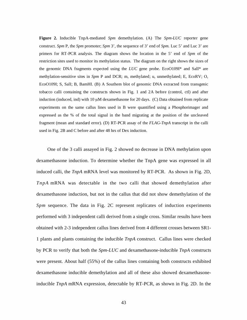
43
One of the 3 calli assayed in Fig. 2 showed no decrease in DNA methylation upon
dexamethasone induction. To determine whether the TnpA gene was expressed in all
induced calli, the TnpA mRNA level was monitored by RT-PCR. As shown in Fig. 2D,
TnpA mRNA was detectable in the two calli that showed demethylation after
dexamethasone induction, but not in the callus that did not show demethylation of the
Spm sequence. The data in Fig. 2C represent replicates of induction experiments
performed with 3 independent calli derived from a single cross. Similar results have been
obtained with 2-3 independent callus lines derived from 4 different crosses between SR1-
1 plants and plants containing the inducible TnpA construct. Callus lines were checked
by PCR to verify that both the Spm-LUC and dexamethasone-inducible TnpA constructs
were present. About half (55%) of the callus lines containing both constructs exhibited
dexamethasone inducible demethylation and all of these also showed dexamethasone-
inducible TnpA mRNA expression, detectable by RT-PCR, as shown in Fig. 2D. In the
Figure 2. Inducible TnpA-mediated Spm demethylation. (A) The Spm-LUC reporter gene
construct. Spm P, the Spm promoter; Spm 3’, the sequence of 3’ end of Spm. Luc 5’ and Luc 3’ are
primers for RT-PCR analysis. The diagram shows the location in the 5’ end of Spm of the
restriction sites used to monitor its methylation status. The diagram on the right shows the sizes of
the genomic DNA fragments expected using the LUC gene probe. EcoO109I* and SalI* are
methylation-sensitive sites in Spm P and DCR; m, methylated; u, unmethylated; E, EcoRV; O,
EcoO109I; S, SalI; B, BamHI. (B) A Southern blot of genomic DNA extracted from transgenic
tobacco calli containing the constructs shown in Fig. 1 and 2A before (control, ctl) and after
induction (induced, ind) with 10 µM dexamethasone for 20 days. (C) Data obtained from replicate
experiments on the same callus lines used in B were quantified using a Phosphorimager and
expressed as the % of the total signal in the band migrating at the position of the uncleaved
fragment (mean and standard error). (D) RT-PCR assay of the FLAG-TnpA transcript in the calli
used in Fig. 2B and C before and after 48 hrs of Dex induction.
44
remaining lines, dexamethasone was unable to induce either demethylation or TnpA
mRNA expression. Because Spm methylation remained unchanged after inducible
expression of the FLAG-TnpA420 protein (Figure 8, Chapter 4), the observed DNA
demethylation in the TnpA-inducible lines cannot be attributed to the inducible system. It
follows that demethylation of the Spm sequence can be controlled by inducing expression
of TnpA.
TnpA Promotes Rapid Spm Demethylation
The complete loss of Spm methylation observed after 20 days of dexamethasone
induction in the foregoing experiments suggests that TnpA-mediated DNA demethylation
is an active process. If demethylation occurs by interference with remethylation of newly
replicated DNA, the extent of demethylation should depend on the number of cell
divisions and some methylation should still be detectable at 20 days, the precise amount
depending on the exact doubling time. The extent of Spm DNA demethylation was
therefore determined after treatment of calli with 5-aza-cytosine (5-aza-C) or 5-aza-
deoxycytosine (5-aza-dC), both of which are incorporated into DNA in place of cytosine,
but cannot be methylated (Bender et al., 1999). Little effect on Spm methylation was
observed after 10 days of treatment with either compound at concentrations of either 10
or 50 µM. Even after 20 days of treatment, methylation was reduced by less than 50% at
the higher concentration of 5-aza-dC (Fig. 3A). By contrast, no methylation was detected
in calli treated with dexamethasone, either in the presence or absence of 5-aza-dC. Thus
TnpA-mediated demethylation is either more rapid or more efficient than the
demethylation resulting from the incorporation of 5-aza-dC.

45
To determine how rapidly the Spm sequence was demethylated after induction of
TnpA expression relative to the cell doubling time, the callus growth curve was first
determined under our experimental conditions (Fig. 3B). There is an initial lag phase,
BTobacco Callus Growth Curve
0
100
200
300
400
500
0 2 4 6 8 1 0 1 2
Days after transfer
wei
ght i
ncre
ase(
%)
↓
Spm promoter DCR
0
20
40
60
80
100
Ctl 10 µM 50 µM Dex +
Aza-dCDex
Aza-dC
DN
A m
ethy
latio
n (%
)
A
C
0
20
40
60
80
100
Ctl Ind Ctl Ind
36 h 6 daysD
NA
met
hyla
tion
(%)
Spm promoter DCR
Figure 3. Rapid DNA demethylation after TnpA expression. (A) Spm promoter and DCR
methylation in genomic DNA from transgenic tobacco calli after treatment for 20 days with
5-aza-deoxycytosine (aza-dC), 10 µM dexamethasone (Dex) or both. (B) The growth of
tobacco callus after transfer to fresh medium. Each time point represents the mean (and
standard error) of fresh weight measurements of 6 calli. The arrow indicates the initial
doubling time. (C) Spm promoter and DCR methylation after 36 hrs and after 6 days of TnpA
induction on 10 µM Dex. The values are the means and standard errors of measurements
from two replicate experiments performed with each callus line at a similar growth stage.

46
giving an initial doubling time of 6-7 days, followed by an exponential growth phase with
a doubling time of about 2 days. The methylation status of the Spm sequence was
examined under precisely these growth conditions 1.5 and 6 days after transfer to
dexamethasone-containing medium. As shown in Figure 3C, the Spm DNA was almost
completely demethylated after 6 days of dexamethasone treatment and significant loss of
methylation could already be detected by 36 hrs. If DNA demethylation is a consequence
of interference with remethylation of newly replicated DNA, the Spm sequence should be
hemimethylated after one cycle of DNA replication. If the hemimethylated DNA is still
resistant to restriction by methylation-sensitive enzymes SalI and EcoO109I, no change
in methylation is expected after a single doubling time, unless the DNA is actively
demethylated.
Restriction Analysis of Hemimethylated DNA
Although it has been reported that the fully hemimethylated SalI site with both of
its C residues methylated is resistant to cleavage, there is no information about either the
sensitivity of the fully hemimethylated EcoO109I site or on either site when less than
fully methylated (Nelson et al., 1993). A DNA fragment containing the Spm promoter
and DCR was therefore methylated using the bacterial methyltransferase SssI, which only
methylates the cytosine (C) residue in CpG dinucleotides. The sequence of the EcoO109I
cleavage site in the Spm promoter is GGGTCCC with the last C followed by a G, placing
it in the CG dinucleotide context. The SalI site sequence is GTCGAC. Thus, SssI
methylates only one of the 3 C residues in the EcoO109I site and one of the two C
residues in the SalI site.

47
As shown in Figure 4A, double-strand DNA methylated by SssI is completely
resistant to SalI, but is still sensitive to EcoO109I. Because genomic Spm DNA is largely
resistant to EcoO109I in the present experiments, it is likely that the EcoO109I site
contains more than 1 methylated C residue (5mC), although it is possible that resistance
is conferred by methylation of an internal C, but not the site’s terminal C. To methylate
all of the C residues in one strand, hemimethylated DNA was prepared by incorporating
B Unmeth
- O S
Hemi-L
- O Sbp
350
160
500
650
A
bp
350
160
Unmeth
- O S
Meth
- O S
500650
600
200
bp
C
dsD
NA
ss-S
K
ss-K
S
Anne
alD
Unmeth
(-) +
Hemi-L
(-) +
Hemi-U
(-) + (-) +
Meth
600
200
bp
E
Unmeth
(-) + (-) + (-) + (-) +
NaOH NH4oAcDe/re-nature
FUnmeth Hemi-L Hemi-U
- Sbp
350
300
- S - S
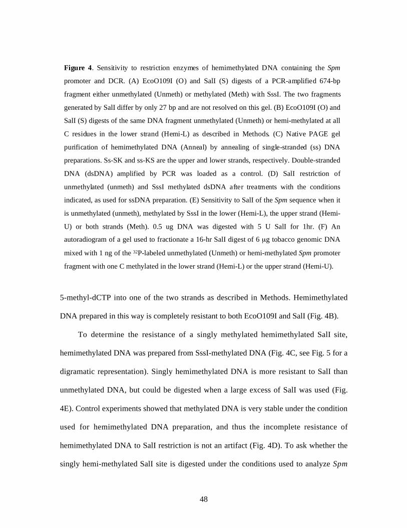
48
5-methyl-dCTP into one of the two strands as described in Methods. Hemimethylated
DNA prepared in this way is completely resistant to both EcoO109I and SalI (Fig. 4B).
To determine the resistance of a singly methylated hemimethylated SalI site,
hemimethylated DNA was prepared from SssI-methylated DNA (Fig. 4C, see Fig. 5 for a
digramatic representation). Singly hemimethylated DNA is more resistant to SalI than
unmethylated DNA, but could be digested when a large excess of SalI was used (Fig.
4E). Control experiments showed that methylated DNA is very stable under the condition
used for hemimethylated DNA preparation, and thus the incomplete resistance of
hemimethylated DNA to SalI restriction is not an artifact (Fig. 4D). To ask whether the
singly hemi-methylated SalI site is digested under the conditions used to analyze Spm
Figure 4. Sensitivity to restriction enzymes of hemimethylated DNA containing the Spm
promoter and DCR. (A) EcoO109I (O) and SalI (S) digests of a PCR-amplified 674-bp
fragment either unmethylated (Unmeth) or methylated (Meth) with SssI. The two fragments
generated by SalI differ by only 27 bp and are not resolved on this gel. (B) EcoO109I (O) and
SalI (S) digests of the same DNA fragment unmethylated (Unmeth) or hemi-methylated at all
C residues in the lower strand (Hemi-L) as described in Methods. (C) Native PAGE gel
purification of hemimethylated DNA (Anneal) by annealing of single-stranded (ss) DNA
preparations. Ss-SK and ss-KS are the upper and lower strands, respectively. Double-stranded
DNA (dsDNA) amplified by PCR was loaded as a control. (D) SalI restriction of
unmethylated (unmeth) and SssI methylated dsDNA after treatments with the conditions
indicated, as used for ssDNA preparation. (E) Sensitivity to SalI of the Spm sequence when it
is unmethylated (unmeth), methylated by SssI in the lower (Hemi-L), the upper strand (Hemi-
U) or both strands (Meth). 0.5 ug DNA was digested with 5 U SalI for 1hr. (F) An
autoradiogram of a gel used to fractionate a 16-hr SalI digest of 6 µg tobacco genomic DNA
mixed with 1 ng of the 32P-labeled unmethylated (Unmeth) or hemi-methylated Spm promoter
fragment with one C methylated in the lower strand (Hemi-L) or the upper strand (Hemi-U).

49
sequence methylation in genomic DNA, 32P-labeled singly hemimethylated Spm DNA
was prepared using SssI methylation and a trace amount of it was added to a genomic
DNA digest (1 ng labeled DNA/6 mg genomic DNA). As shown in Fig. 3F, the singly
hemimethylated DNA was almost completely resistant to SalI digestion, irrespective of
the strand into which the methyl group was introduced (hemi-L or -U).
Figure 5. A diagram depicting the method for preparation of hemimethylated
DNA using biotin/streptavidin interaction. See methods for details.
3) Binding to Streptavidin beads
4) Denature in 0.1M NaOH
5) Collect methylated ssDNA
2) In vitro DNA methylation CH3
CH3
CH3
CH3
CH3
CH3
CH3
CH3
CH3 CH3
+
CH3 CH3
1) PCR amplification of dsDNA
6) Hemimethylated DNA preparation
CH3 CH3
CH3 CH3
Spm P DCR
5’ Biotin KS
SK
Ecoo109I SalI
+ unmethylated ssDNAs
Biotin modification Streptavidin beads
CH3 5 methylcytosine Primers

50
To examine the sensitivity of hemimethylated DNA with fewer 5mC residues per
site to EcoO109I, hemimethylated DNA was prepared using a mixture of 5-methyl-dCTP
and dCTP at ratios of 3:1 and 2:1. From the ratio of methylated to unmethylated dCTP,
the expected frequencies of tri-methylated, di-methylated, monomethylated and
unmethylated EcoO109I sites can be calculated (see Methods). Based on this calculation,
the extent of digestion can in turn be predicted if just unmethylated and monomethylated
sites are cut (Table 1, 15.6% at 3:1 and 25.9% at 2:1) and if dimethylated sites are cut, as
well (Table 1, 57.8% at 3:1 and 70.4% at 2:1). The observed extent of EcoO109I
cleavage, also given in Table 1, is very close to that predicted if only the unmethylated
and monomethylated sites can be cleaved in hemimethylated DNA (Table 1, 15.3% and
22.4%). It is therefore concluded that hemimethylated DNA with any 2 of its 3 C’s
methylated at the EcoO109I site is resistant to digestion. Since genomic Spm DNA is
highly resistant to EcoO109I and is therefore likely to have at least two methylated C
residues in its EcoO109I site, it follows that the newly replicated, hemimethylated site
will also be resistant to cleavage by the enzyme.
The foregoing results clearly show that the hemimethylated EcoO109I and SalI sites
are resistant to enzymatic digestion when methylated at all internal C residues. They
further provide evidence that under the conditions employed in the present experiments,
both sites are resistant even when less than fully hemimethylated. The initial doubling
time for the callus cultures under the assay conditions was about 6 days, with subsequent
doubling times of about 2 days. If Spm DNA is demethylated by TnpA’s interference
with remethylation of hemimethylated DNA, it should be fully resistant to restriction by
SalI and EcoO109I after one doubling time (day 6), 50% resistant after 2 (day 8) and

51
25% resistant after 3 doubling times (day 10). Since methylation was reduced by about
40% after 36 hrs, much less than the initial doubling time, and virtually gone by 6 days,
we conclude that TnpA-mediated DNA demethylation is an active process.
Table I. Expected and observed extent of EcoO109I cleavage of hemimethylated
DNA with different number of methylated cytosine residues (5mC) at the
recognition site. The hemimethylated DNA was generated by copying
unmethylated single-stranded DNA using Tag DNA polymerase and a mixture of
dCTP and 5-methyl-dCTP.
EcoO109I site (GGGTCCCa)
Ratiob
5m-dCTP:dCTPExpectedDNA compositionc
5mC (1) +5mC (0)
5mC (2) +5mC (1) +5mC (0)
DNAcleaved
3:15mC (3): 42.2%5mC (2): 42.2%5mC (1): 14.0%5mC (0): 1.6%
15.6% 57.8% 15.3%
2:15mC (3): 29.6%5mC (2): 44.5%5mC (1): 22.2%5mC (0): 3.7%
25.9% 70.4% 22.4%
a EcoO109I recognition sequence in the Spm promoter.b The ratio of 5-methyl-dCTP to dCTP used in preparing hemimethylated DNA.c 5mC(N), the calculated percentage of DNA with the indicated number (N) of
5mC residues in the EcoO109I site (see Methods for calculation).

52
Discussion
An Inducible DNA Demethylation Assay
Most studies on DNA demethylation rely on the use of in vitro premethylated DNA,
which is transfected either transiently or stably into cells. There are some caveats with
this approach, however. First of all, naked DNA introduced by this method readily gets
demethylated even in the absence of the trans-acting demethylation-promoting factors,
presumably by general DNA demethylase activities in the cell (Qu and Ehrlich, 1999;
Cervoni and Szyf, 2001). Because known DNA demethylase activities do not show
sequence specificity (Jost et al., 1995; Neddermann et al., 1996; Bhattacharya et al.,
1999; Vairapandi et al., 2000; Jost et al., 2001), the observed demethylation may not at
all reflect the activity of the trans-acting factor under investigation. In the case of
transient transfection, a large amount of DNA is generally used but only a small fraction
can get into the cells and even less DNA is demethylated, making it difficult to detect the
product of DNA demethylation.
Preliminary studies in the laboratory indicate that in tobacco cells there is a DNA
demethylase activity that does not have sequence specificity (Chen and Fedoroff,
unpublished observation). To overcome the problems associated with transient
transfection, I have thus developed an in vivo assay system for dissecting the mechanism
of TnpA-mediated DNA demethylation.
By controlling DNA demethylation through inducible TnpA expression, we are able
to characterize the initial stages of DNA demethylation through several cell division
cycles in the native chromatin context. The importance of chromatin structure in

53
controlling DNA methylation has been revealed in recent studies (see Introduction). This
has not been possible previously because methylation was studied either in transformed
cells or in regenerated plants many cell generations after the introduction of a TnpA gene
or Spm transposon by transformation. The ability to induce DNA demethylation will
facilitate further molecular analysis of TnpA-mediated DNA demethylation. To our
knowledge, this is the first inducible DNA demethylation system ever reported. This
approach should be useful as well for studies of other trans-acting factors that promote
sequence-specific DNA demethylation.
Active DNA demethylation in plants
Although there are both cytological and biochemical evidence that active DNA
demethylation occurs in animals cells, very little is known about plants in this regard. The
first evidence that DNA is actively demethylated in plants is derived from the observation
that DNA methylation is dramatically reduced in the generative but not the vegetative
nucleus of germinating pollen in the absence of DNA replication (Oakeley et al., 1997).
The present study has provided further support for the existence of an active DNA
demethylation mechanism in plants and represents the first example of sequence-specific
DNA demethylation mediated by trans-acting factors. More recently, the ROS protein in
Arabidospsis has been shown to be able to nick methylated DNA, implicating its role in
DNA demethylation through a DNA repair pathway (Gong et al., 2002).

54
Sensitivity of Hemimethylated DNA
DNA methylation-sensitive restriction enzymes are often used for studies of DNA
methylation and demethylation. However, artifacts may result if this method is not used
properly, because the extent of digestion of some restriction sites is dependent on not
only the extent of methylation (the number of 5mC and whether one or both strands are
methylated) but also the amount of enzyme and DNA used in the restriction reaction, as
manifested by this and other studies (Nelson et al., 1993). In the present study, I have
found that although the SalI recognition site is completely resistant to SalI restriction
when methylated at both strands, it is only partially resistant to this enzyme when only
one strand is methylated with a single 5mC. While hemimethylated DNA with one
cytosine residue methylated is still restrictable, hemimethylated DNA with both cytosine
residues methylated becomes completely resistant to SalI restriction. This study also
showed that DNA hemimethylated at the SalI site with one 5mC is protected from SalI
restriction by an excess of unrelated DNA that also contains SalI restriction sites.
Similarly, the EcoO109I site is sensitive to restriction when only one cytosine is
methylated but becomes completely resistant when more than two cytosine residues
methylated. Since not all restriction sites have been characterized regarding sensitivity to
restriction when they are variously methylated, caution must be exercised when
methylation-sensitive enzymes are used for studies of DNA methylation and
demethylation. The methods for preparation of hemimethylated DNA developed in this
study would be helpful in characterizing the sensitivity of other restriction sites.

55
References
Aoyama, T., and Chua, N.H. (1997). A glucocorticoid-mediated transcriptionalinduction system in transgenic plants. Plant J. 11, 605-612.
Banks, J.A., Masson, P., and Fedoroff, N. (1988). Molecular mechanisms in thedevelopmental regulation of the maize Suppressor-mutator transposable element.Genes Dev. 2, 1364-1380.
Banks, J.A., and Fedoroff, N. (1989). Patterns of developmental and heritable change inmethylation of the Suppressor-mutator transposable element. Dev. Genet. 10, 425-437.
Bender, C.M., Gonzalgo, M.L., Gonzales, F.A., Nguyen, C.T., Robertson, K.D., andJones, P.A. (1999). Roles of cell division and gene transcription in the methylation ofCpG islands. Mol. Cell. Biol. 19, 6690-6698.
Bhattacharya, S.K., Ramchandani, S., Cervoni, N., and Szyf, M. (1999). Amammalian protein with specific demethylase activity for mCpG DNA [seecomments]. Nature 397, 579-583.
Cervoni, N., and Szyf, M. (2001). Demethylase activity is directed by histoneacetylation. J. Biol. Chem. 276, 40778-40787.
Dean, W., Santos, F., Stojkovic, M., Zakhartchenko, V., Walter, J., Wolf, E., andReik, W. (2001). Conservation of methylation reprogramming in mammaliandevelopment: aberrant reprogramming in cloned embryos. Proc. Natl. Acad. Sci.USA 98, 13734-13738.
Fedoroff, N. (1989). The heritable activation of cryptic Suppressor-mutator elements byan active element. Genetics 121, 591-608.
Gong, Z., Morales-Ruiz, T., Ariza, R.R., Roldan-Arjona, T., David, L., and Zhu,J.K. (2002). ROS1, a repressor of transcriptional gene silencing in Arabidopsis,encodes a DNA glycosylase/lyase. Cell 111, 803-814.
Hsieh, C.L. (1999). Evidence that protein binding specifies sites of DNA demethylation.Mol. Cell. Biol. 19, 46-56.
Jost, J.P., Siegmann, M., Sun, L., and Leung, R. (1995). Mechanisms of DNAdemethylation in chicken embryos. Purification and properties of a 5-methylcytosine-DNA glycosylase. J. Biol. Chem. 270, 9734-9739.
Jost, J.P., Oakeley, E.J., Zhu, B., Benjamin, D., Thiry, S., Siegmann, M., and Jost,Y.C. (2001). 5-Methylcytosine DNA glycosylase participates in the genome-wideloss of DNA methylation occurring during mouse myoblast differentiation. NucleicAcids Res. 29, 4452-4461.
McBride, K.E., and Summerfelt, K.R. (1990). Improved binary vectors forAgrobacterium-mediated plant transformation. Plant Mol. Biol. 14, 269-276.
Neddermann, P., Gallinari, P., Lettieri, T., Schmid, D., Truong, O., Hsuan, J.J.,Wiebauer, K., and Jiricny, J. (1996). Cloning and expression of human G/Tmismatch-specific thymine-DNA glycosylase. J. Biol. Chem. 271, 12767-12774.
Nelson, P.S., Papas, T.S., and Schweinfest, C.W. (1993). Restriction endonucleasecleavage of 5-methyl-deoxycytosine hemimethylated DNA at high enzyme-to-substrate ratios. Nucleic Acids Res. 21, 681-686.

56
Oakeley, E.J., Podesta, A., and Jost, J.P. (1997). Developmental changes in DNAmethylation of the two tobacco pollen nuclei during maturation. Proc. Natl. Acad.Sci. USA 94, 11721-11725.
Oswald, J., Engemann, S., Lane, N., Mayer, W., Olek, A., Fundele, R., Dean, W.,Reik, W., and Walter, J. (2000). Active demethylation of the paternal genome in themouse zygote. Curr. Biol. 10, 475-478.
Qu, G., and Ehrlich, M. (1999). Demethylation and expression of methylated plasmidDNA stably transfected into HeLa cells [In Process Citation]. Nucleic Acids Res. 27,2332-2338.
Raina, R., Cook, D., and Fedoroff, N. (1993). Maize Spm transposable element has anenhancer-insensitive promoter. Proc. Natl. Acad. Sci. USA 90, 6355-6359.
Raina, R., Schläppi, M., Karunanandaa, B., Elhofy, A., and Fedoroff, N. (1998).Concerted formation of macromolecular Suppressor-mutator transposition complexes.Proc. Natl. Acad. Sci. USA 95, 8526-8531.
Reik, W., Dean, W., and Walter, J. (2001). Epigenetic reprogramming in mammaliandevelopment. Science 293, 1089-1093.
Schläppi, M., Smith, D., and Fedoroff, N. (1993). TnpA trans-activates methylatedmaize Suppressor-mutator transposable elements in transgenic tobacco. Genetics 133,1009-1021.
Schläppi, M., Raina, R., and Fedoroff, N. (1994). Epigenetic regulation of the maizeSpm transposable element: novel activation of a methylated promoter by TnpA. Cell77, 427-437.
Schläppi, M., Raina, R., and Fedoroff, N. (1996). A highly sensitive plant hybridprotein assay system based on the Spm promoter and TnpA protein for detection andanalysis of transcription activation domains. Plant Mol. Biol. 32, 717-725.
Stancheva, I., El-Maarri, O., Walter, J., Niveleau, A., and Meehan, R.R. (2002).DNA methylation at promoter regions regulates the timing of gene activation inXenopus laevis embryos. Dev. Biol. 243, 155-165.
Vairapandi, M., Liebermann, D.A., Hoffman, B., and Duker, N.J. (2000). HumanDNA-demethylating activity: A glycosylase associated with RNA and PCNA. J. Cell.Biochem. 79, 249-260.
von Arnim, A.G., Deng, X.W., and Stacey, M.G. (1998). Cloning vectors for theexpression of green fluorescent protein fusion proteins in transgenic plants. Gene 221,35-43.
57
Chapter 3
TNPA IS A SPM-SPECIFIC TRANSCRIPTIONAL ACTIVATOR
Introduction
A prominent feature of TnpA-mediated DNA demethylation is that it is not
confined to the Spm promoter, which contains multiple TnpA binding sites (Raina et al.,
1998), but also occurs in the downstream GC-rich sequence termed the DCR, which
encodes the first untranslated exon of the full-length Spm transcript (Masson et al., 1991).
Since there are no TnpA binding sites in the DCR (Raina et al., 1998), it is unlikely that
DNA demethylation of the Spm sequence, particularly the DCR, is conferred by TnpA
alone. The extended region of DNA demethylation suggests that other trans-acting factors
might be involved as well. Interestingly, a leucine-zipper domain has been identified at
the C-terminus of the TnpA protein (Trentmann et al., 1993), it is therefore possible that
TnpA recruits other proteins that participate in the demethylation of the Spm sequence .
To investigate this possibility, I first attempted to screen for plant proteins that
interact with TnpA using the yeast two-hybrid interaction trap system (Fields and Song,
1989). Surprisingly, TnpA strongly activated transcription as indicated by two reporter
genes in the yeast cells. That TnpA is an Spm-specific transcriptional activator is
confirmed by further studies using a transgenic line of tobacco that shows inducible
TnpA expression and contains an unmethylated Spm sequence. Since the Spm sequence

58
remains unmethylated during the assay, transcriptional activation cannot be attributed to
DNA demethylation. The present study resolves a long-standing paradoxical observation
that TnpA acts as both a transcriptional activator and repressor depending on the
methylation status of the Spm promoter.
Materials and Methods
Plant materials and treatments
Calli for expression of FLAG-TnpA540-VP16 were generated and maintained in
similar ways for the FLAG-TnpA expressing lines as described in Chapter 2. Suspension
cultures were derived from transgenic calli and were maintained at 24°C with shaking
(120 rpm) in MS medium supplemented with 3% sucrose and 0.2 mg/L 2,4-
dichlorophenoxyacetic acid (2,4-D) (Life Technologies). Expression of the FLAG-tagged
proteins was induced in suspension cultures by adding 10 µM dexamethasone (Sigma-
Aldrich) to the growth medium. Induction of protein expression in calli was performed in
the same was as described in Chapter 2.
Plasmid Constructs
All FLAG-tagged clones were derivatives of pFLAG-TnpA. pFLAG-TnpA540-Vp16
was obtained by ligating the XhoI-SplI fragment of pFLAG-TnpA and the SplI-NcoI
fragment (the NcoI site was filled in using the Klenow fragment) from pMS249
(Schläppi et al., 1996) together into the pBluescript II KS (+) vector cut with XhoI and

59
EcoRV. pTA-FLAG-TnpA540-Vp16 was cloned by ligating a ClaI to SpeI fragment from
pFLAG-TnpA540-VP16 and pTA7002 that was cut with XhoI and SpeI and the XhoI site
was partially filled in to the adenine residue.
The construct for expression of the fusion protein between the Gal4 DNA-binding
domain and TnpA in yeast cells was cloned as follows. The yeast expression vector pAS2
was first cut with NcoI, filled in with Klenow and then cut with SalI. pRR466 (Raina et
al., 1993) was cut with BamHI, filled in with Klenow and then cut with SalI to release the
cDNAs for TnpA, which were then cloned into pAS2, giving rise to pAS2-TnpA.
Transcription Assays
Plasmid pAS2-TnpA as well as the vector pAS2 was transformed into yeast strain
Y190. Yeast transformation and the subsequent transcription assay were conducted
according to the protocol described for the HybriZAP two-hybrid cDNA Gigapack
cloning kit (Stratagene).
RT-PCR was used to assay transcriptional activation of the Spm-LUC gene by
TnpA in plant cells. Total RNA was prepared using either the Qiagen RNeasy Plant Mini
kit or the GibcoBRL TRIzol reagent. To remove residual DNA, RNA was treated with
DNase I for 5 min as recommended by the vendor (Life Technologies). For reverse
transcription, 1 µl of 100 ng/µl total RNA was mixed with 3 µl of 100 ng/µl random
primer and 8 µl RNase-free water, then denatured at 70°C for 10 min. The solution was
chilled on ice and collected by centrifugation. A premixed solution of the following
reagents was then added: 4 µl of 5x first strand buffer (Life Technologies), 2 µl of 0.1 M
DTT, 1 µl 10 µM dNTPs and 2 units RNase block inhibitor. The solution was incubated
at 25° C for 10 min, and then at 42° C for 2 min, followed by the addition of 1 µl

60
Superscript RT (Life Technologies) and incubation at 42° C for 50 min. The reaction was
terminated by heating at 70° C for 15 min and 1-2 µl was used in a 20 µl PCR reaction.
Simultaneous amplification of the transcripts for the FLAG-TnpA gene and the luciferase
(LUC) reporter gene was achieved by including both primer sets in the same reaction. For
semi-quantitative analysis, the PCR reaction was stopped after 15 cycles, fractionated on
an agarose gel and then transferred to Hybond+ nitrocellulose membrane (Amersham).
The amplification products for the TnpA and luciferase genes were detected by Southern
blot analysis. To ensure that equal amounts of total RNA had been used in the reaction,
the 18S rRNA was also amplified by RT-PCR for 10 cycles using the primer pair 18S-5’
(5’TACCGTCCTAGTCTCAACCA3’) and
18S-3’ (5’AACATCTAAGGGCATCACAG3’).
Western Blot Assay
The FLAG-tagged TnpA expressed in transgenic tobacco plants were detected in
transgenic plants extracts using the polyclonal anti-FLAG antibody (Sigma-Aldrich).
Total plant protein was extracted by grinding plant tissue in 2 volumes of TBS buffer (50
mM Tris-HCl, pH 7.8, 150 mM NaCl) supplemented with 1 mM PMSF and 1x protease
inhibitor cocktail for plants (Sigma-Aldrich). The extract was centrifuged at 14,000 rpm
at 4° C for 10 min, the supernatant was mixed with 6x SDS loading buffer (Scopes,
1998), boiled for 5 min, and 30 µl was loaded on an 8% polyacrylamide gel for
fractionation. Proteins expressed in both yeast and plant cells were visualized using the
WesternBreezeTM chemifluorescence immunodetection system (Invitrogen).

61
Results
TnpA Activates Transcription in Yeast Cells
To identify proteins that could interact with TnpA, I first used the yeast two-hybrid
system. However, attempts to use TnpA and TnpA fragments as bait (Fig. 6A) revealed
that a Gal4 fusion with the full-length TnpA protein strongly activates transcription of
both the histidine synthase and β-galactosidase reporter genes (Fig. 6B).
pAS2-TnpA Gal4 BD TnpA
UAS TATA Histidine synthaseReporter genes
UAS TATA �-galactosidase
A
Figure 6. TnpA is a transcriptional activator in yeast. (A) Construct used for expression of
the fusion protein between the Gal4 binding domain (BD) and TnpA as well as the reporter
genes for transcriptional activity assay in yeast cells. UAS, Gal4 binding sequence.
(B) Growth of transformed yeast cells on histidine (-) medium (left) and filter paper lift
assay for β-galactosidase activity (right). Cells expressing the Gal4 BD was used a negative
control. Snf1/Snf4 cells express the Gal4 BD-SNF1 and SNF4-Gal4 activation domain
fusion proteins and serve as a weak positive control. The middle panel is a diagram showing
the position of yeast cells transformed with the constructs as indicated.
B
Gal4 BD (-)
Snf1/Snf4 (+)
Gal4 BD-TnpA
Gal4 BD-TnpA540
Gal4 BD-TnpA420

62
TnpA is a Weak Transcriptional Activator in Plant Cells
To assess the transcriptional activity of TnpA on an unmethylated Spm promoter, I
used suspension cultures derived from tobacco callus lines containing a dexamethasone-
inducible TnpA gene and an Spm-LUC gene in which the Spm promoter was not
methylated. As a positive control, suspension cultures were also generated from calli that
express the FLAG-TnpA540-VP16 fusion protein under the control of the dexamethasone-
inducible promoter (Fig 7A). After addition of dexamethasone into the cell culture, the
FLAG-TnpA transcript peaked at 24-48 hrs (Fig. 7B-1). Expression of the tagged proteins
peaked at about 72 hrs (Figs. 7C). Induction of TnpA enhanced transcription of the
luciferase reporter gene, albeit weakly, and maximal induction was observed when the
TnpA protein, rather than its transcript, reached maximum (Fig. 7B-1). Activation of
transcription from the Spm promoter by TnpA was reproducible and was observed when
independent lines were examined (data not shown). Notably, there is some background
level of transcription even in the absence of TnpA induction, which is likely due to leaky
expression from the inducible promoter or the Spm promoter. Nevertheless, repression of
transcription was not observed, even at elevated concentrations of dexamethasone (data
not shown). Because the strong TnpA induction could artifactually enhance the LUC
mRNA signal, the blot was stripped and reprobed with just a LUC probe. The result
confirmed that the LUC transcript level indeed increased (Fig. 7B-2). By contrast,
although induction of the FLAG-TnpA540-VP16 gene was very slight, it substantially
enhanced transcription of the LUC gene from the Spm promoter (Fig. 7, B-1 and B-2).
Taken together, these results showed that TnpA can stimulate transcription from the

63
unmethylated Spm promoter and is therefore a transcriptional activator, albeit a much
weaker one than the FLAG-TnpA540-VP16 fusion protein.
FLAG-TnpA-VP16
FLAG tag DNA binding domain
Leucine zipper domain Q Glutamine-rich motif
VP16 AD674
Q 5401
A
FLAG-TnpA
EkMTQMEEsmAQYKQQVQQqMQQMQNQ�WmLHQ
a.a.453-483
Q 540 6211
420
→ FLAG-TnpAprotein
Transcript
B
18S rRNA
LUC
TnpA →
→
→
Ctl Ind
FLAG-TnpA
0 0.5 1 8 24 72 h 1 wk
FLAG-TnpA-Vp16
LUC
TnpA →
→
Reprobed
1)
2)
3)
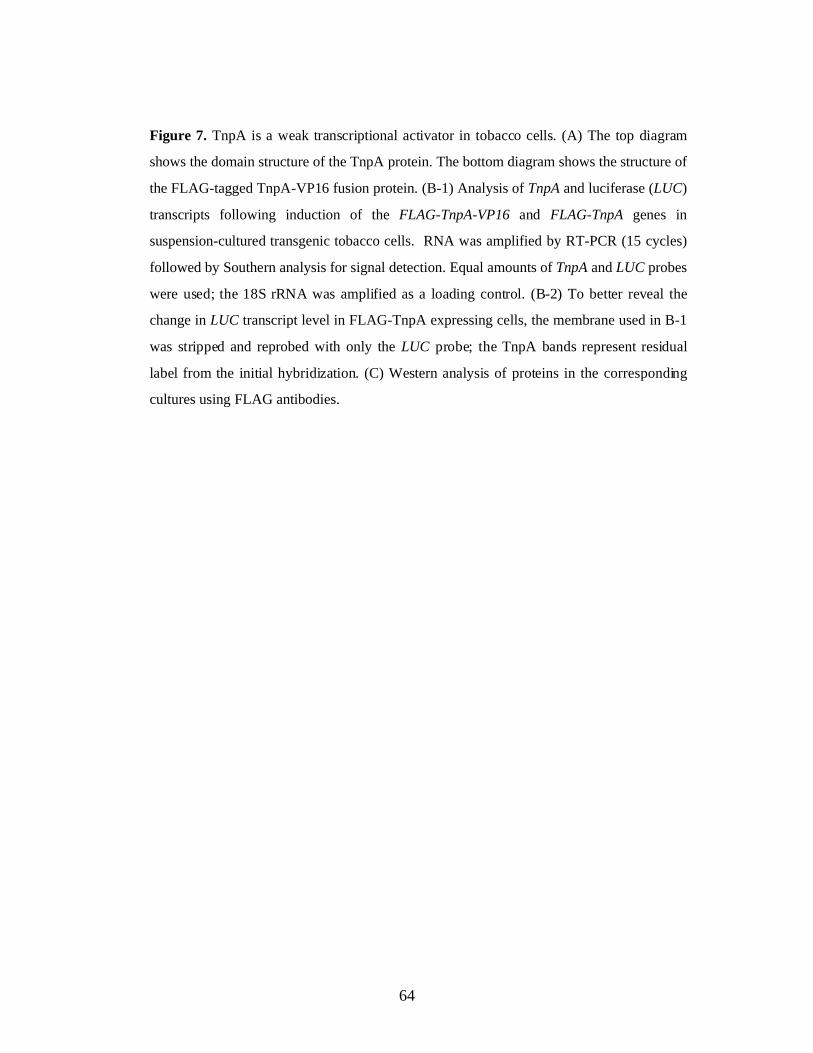
64
Figure 7. TnpA is a weak transcriptional activator in tobacco cells. (A) The top diagram
shows the domain structure of the TnpA protein. The bottom diagram shows the structure of
the FLAG-tagged TnpA-VP16 fusion protein. (B-1) Analysis of TnpA and luciferase (LUC)
transcripts following induction of the FLAG-TnpA-VP16 and FLAG-TnpA genes in
suspension-cultured transgenic tobacco cells. RNA was amplified by RT-PCR (15 cycles)
followed by Southern analysis for signal detection. Equal amounts of TnpA and LUC probes
were used; the 18S rRNA was amplified as a loading control. (B-2) To better reveal the
change in LUC transcript level in FLAG-TnpA expressing cells, the membrane used in B-1
was stripped and reprobed with only the LUC probe; the TnpA bands represent residual
label from the initial hybridization. (C) Western analysis of proteins in the corresponding
cultures using FLAG antibodies.

65
Discussion
TnpA has been shown to activate transcription from the Spm promoter when
methylated, but represses transcription from an unmethylated Spm promoter .
Paradoxically, however, these studies also showed that the Spm promoter has stronger
transcription activity when it becomes demethylated due to the presence of TnpA
(Schläppi et al., 1994). The present study shows that TnpA is a weak transcriptional
activator of the unmethylated Spm promoter in plant cells, although its transcription
activation domain functions as a strong transcriptional activator when fused to a
heterologous DNA binding domain in yeast. Since the Spm sequence is unmethylated
even before TnpA expression, the observed transcriptional activation is unlikely a
consequence of DNA demethylation. DOPA, the TnpA homolog encoded by the Spm-
like maize Doppia transposon, recently has been reported to be a transcriptional activator
(Bercury et al., 2001). Interestingly, analysis of the TnpA sequence using a motif-
recognition program (http://www.motif.genome.ad.jp/) revealed the presence of a
glutamine-rich motif, a common transcription activation domain motif, within the C-
terminal leucine-zipper domain (Fig. 7A). However, further studies are needed to
determine whether it is responsible for the observed transcriptional activation. In yeast
cells glutamine-rich domain activates transcription weakly.
The paradoxical observation about TnpA in the previous studies could arise from
the intrinsic drawbacks of the transcription assays used in that experiment. In the case of
the transient assay, where TnpA expression construct was introduced into plant cells by
the biolistic delivery method, transcription repression could occur as a consequence of
66
‘self-interference’ or ‘coactivator dilution-out’ due to protein over-expression (Kannan
and Tainsky, 1999; Nucifora et al., 2001). Similar situation could have occurred in the
studies using stable transgenic plants, where TnpA was expressed under strong promoters
(Schläppi et al., 1994). Another possible cause for the observed repression by TnpA of
transcription from an unmethylated Spm promoter is the high background activity of the
reporter gene in the line used for the transcription assay. Conceivably, a protein with an
intermediate transcription activity can act as an activator on a promoter that is located in
a transcriptionally inactive locus or as a repressor when the reporter gene is in an actively
transcribing region. In this sense, transcription activation or repression by a trans-acting
factor is only relative to the background level of the reporter gene. Accordingly,
transcription repression by TnpA in one line does not necessarily mean that TnpA also
represses transcription in other lines. To further investigate this possibility, it would be
interesting to examine the transcription activity of TnpA in a line that shows high
luciferase activity using the TnpA inducible strategy. This experiment is worth doing
since other proteins with dual properties have been reported elsewhere (Ohlsson et al.,
2001).
In the present study transcription assay has been conducted in the same line of
transgenic plants that has an unmethylated Spm sequence, the results therefore should
better reflect TnpA’s intrinsic activity. Although transcription activation from the Spm
promoter by TnpA is rather weak, it is nevertheless reproducible and consistent when
independent transgenic lines are examined. Since the transcription activity before and
after induction is measured in the same transgenic line, variability due to sample
heterogeneity is minimized, thus permitting reliable detection of even small changes in

67
transcriptional activity. Variability has been further reduced by using suspension cultures,
which has a more homogeneous cell constitution.
The resolution of the apparent paradox that TnpA functions as both an activator and
repressor may also lie in the protein concentration-dependent competition between
TnpA’s functions in transcription and transposition. TnpA is a bi-functional protein, and
the Spm sequence comprising its promoter likewise has two functions. TnpA is required
both for transcription from the transposon’s promoter and for transposition of Spm
(Masson et al., 1991; Schläppi et al., 1993). There are multiple TnpA binding sites at both
transposon ends (Masson et al., 1987). Those at the 5’ end are required for promoter
function and the TnpA binding sites at both ends are involved in transposition (Masson et
al., 1987; Raina et al., 1993).
Previous experiments to assess the ability of TnpA to activate transcription from an
unmethylated Spm promoter were carried out in transient transfection assays at high DNA
concentrations (Schläppi et al., 1996). The experiments described here were carried out
by inducing expression of a TnpA gene in vivo and measuring transcription from an
integrated chromosomal copy of the Spm-LUC reporter gene. Hence the concentrations
of both the target promoter and the transcription factor are likely to have been much
lower in these experiments than in the previous transient expression assays (Schläppi et
al., 1996). Because the Spm promoter region is co-extensive with the TnpA-binding
region at the 5’ terminus (Raina et al., 1993), the likeliest explanation is that TnpA-
mediated association of Spm termini competes with other types of protein-protein
interactions involving TnpA, including transcriptional activation. Evidence that it is
TnpA and not the inherent weakness of the Spm promoter that limits transcriptional

68
activity is provided by the observation that the TnpA540-VP16 fusion protein is a strong
transcriptional activator of the Spm promoter (Schläppi et al., 1996). The observation that
TnpA is a much weaker transcriptional activator of the Spm promoter than might be
anticipated from the ability of the Gal4-TnpA fusion protein to activate transcription in
yeast is consistent with the interpretation that TnpA is a transcriptional activator at low
concentrations, but becomes a repressor by homodimerization as its concentration
increases. However, the possibility can not be ruled out that TnpA’s transcriptional
activity is modulated by co-repressors present in plant cells, but not in yeast. In maize,
host factors that bind to the subterminal sequences of Ac/Ds and the Mutator element and
regulate their transpositional activity have been identified (Becker and Kunze, 1996;
Jarvis et al., 1997), and these proteins could bind to the Spm sequence as well and affect
its promoter activity.

69
References
Aoyama, T., and Chua, N.H. (1997). A glucocorticoid-mediated transcriptionalinduction system in transgenic plants. Plant J. 11, 605-612.
Becker, H.A., and Kunze, R. (1996). Binding sites for maize nuclear proteins in thesubterminal regions of the transposable element Activator. Mol. Gen. Genet. 251,428-435.
Bercury, S.D., Panavas, T., Irenze, K., and Walker, E.L. (2001). Molecular analysis ofthe Doppia transposable element of maize. Plant Mol. Biol. 47, 341-351.
Fields, S., and Song, O. (1989). A novel genetic system to detect protein-proteininteractions. Nature 340, 245-246.
Jarvis, P., Belzile, F., Page, T., and Dean, C. (1997). Increased Ac excision (iae):Arabidopsis thaliana mutations affecting Ac transposition. Plant J. 11, 907-919.
Kannan, P., and Tainsky, M.A. (1999). Coactivator PC4 mediates AP-2 transcriptionalactivity and suppresses ras-induced transformation dependent on AP-2 transcriptionalinterference. Mol. Cell. Biol. 19, 899-908.
Masson, P., Surosky, R., Kingsbury, J.A., and Fedoroff, N.V. (1987). Genetic andmolecular analysis of the Spm-dependent a-m2 alleles of the maize a locus. Genetics117, 117-137.
Masson, P., Strem, M., and Fedoroff, N. (1991). The tnpA and tnpD gene products ofthe Spm element are required for transposition in tobacco. Plant Cell 3, 73-85.
Nucifora, F.C., Jr., Sasaki, M., Peters, M.F., Huang, H., Cooper, J.K., Yamada, M.,Takahashi, H., Tsuji, S., Troncoso, J., Dawson, V.L., Dawson, T.M., and Ross,C.A. (2001). Interference by huntingtin and atrophin-1 with cbp-mediatedtranscription leading to cellular toxicity. Science 291, 2423-2428.
Ohlsson, R., Renkawitz, R., and Lobanenkov, V. (2001). CTCF is a uniquely versatiletranscription regulator linked to epigenetics and disease. Trends Genet. 17, 520-527.
Raina, R., Cook, D., and Fedoroff, N. (1993). Maize Spm transposable element has anenhancer-insensitive promoter. Proc. Natl. Acad. Sci. USA 90, 6355-6359.
Raina, R., Schläppi, M., Karunanandaa, B., Elhofy, A., and Fedoroff, N. (1998).Concerted formation of macromolecular Suppressor-mutator transposition complexes.Proc. Natl. Acad. Sci. USA 95, 8526-8531.
Schläppi, M., Smith, D., and Fedoroff, N. (1993). TnpA trans-activates methylatedmaize Suppressor-mutator transposable elements in transgenic tobacco. Genetics 133,1009-1021.
Schläppi, M., Raina, R., and Fedoroff, N. (1994). Epigenetic regulation of the maizeSpm transposable element: novel activation of a methylated promoter by TnpA. Cell77, 427-437.
Schläppi, M., Raina, R., and Fedoroff, N. (1996). A highly sensitive plant hybridprotein assay system based on the Spm promoter and TnpA protein for detection andanalysis of transcription activation domains. Plant Mol. Biol. 32, 717-725.
Scopes, R.a.S.J. (1998). Analysis of Proteins. In “Current Protocols in MolecularBiology”. 2., 10.10.11-10.10.20.

70
Trentmann, S.M., Saedler, H., and Gierl, A. (1993). The transposable elementEn/Spm-encoded TNPA protein contains a DNA binding and a dimerization domain.Mol. Gen. Genet. 238, 201-208.

71
Chapter 4
THE TRANSCRIPTIONAL ACTIVITY OF TNPA IS ESSENTIAL
FOR ITS ABILITY TO PROMTE SPM DEMETHYLATION
Introduction
Among the trans-acting factors that are known to promote sequence-specific DNA
demethylation, most are transcriptional factors, Sp1 (Silke et al., 1995), NF-kappa B
(Kirillov et al., 1996; Matsuo et al., 1998) and the glucocorticoid receptor (Grange et al.,
2001; Thomassin et al., 2001) for example. This observation suggests that transcription
activation plays a positive role in DNA demthylation. Indeed, general transcription
machinery has been shown to be essential for DNA demethylation mediated by the NF-
kappa B transcriptional activator, although actual transcription does not seem to be
required (Matsuo et al., 1998). Chromatin remodeling and transcriptional factor binding
have been also shown to precede DNA demethylation (Matsuo et al., 1998; Grange et al.,
2001; Thomassin et al., 2001).
DNA demethylation mediated by non-transcriptional factors has also been reported.
For instance, the Epstein-Barr virus latent replication origin protein EBNA-1 is shown to
promote DNA demethylation of its binding sites, oriP (Hsieh, 1999). Subsequent studies
of the LacI repressor protein indicate that DNA binding is both necessary and sufficient
for targeted DNA demethylation (Lin et al., 2000; Lin and Hsieh, 2001). In these cases,

72
however, sequences beyond the binding sites are not demethylated, a sharp contrast to the
extended demethylation pattern by TnpA and other transcriptional activators. Moreover,
although the LacI protein has been shown to be able to prevent DNA methylation at its
binding site, there is no evidence for active DNA demethylation by this protein (Lin et
al., 2000; Lin and Hsieh, 2001). This is also true of CTCF, a conserved zinc-finger
protein that is essential for maintaining imprinting and X-inactivation (Wolffe, 2000;
Chao et al., 2002). Distinct from other trans-acting factors, however, CTCF can only
maintain its binding sites unmethylated in the X-chromosome that is to be inactivated
(Bell and Felsenfeld, 2000; Szabo et al., 2000; Wolffe, 2000; Chao et al., 2002). It is not
clear how such allele-specific activity is achieved, as the CTCF protein can function as
both a transcription activator (Yang et al., 1999) and repressor (Awad et al., 1999;
Ohlsson et al., 2001), dependent upon posttranslational modification (Delgado et al.,
1999; Klenova et al., 2001) and interaction with other trans-acting factors that are
activators (Chernukhin et al., 2000) or repressors (Lutz et al., 2000).
Since TnpA turns out to be an Spm specific transcriptional activator, it is therefore
interesting to determine whether this activity is required for its ability to promote Spm
demethylation. Using truncated TnpA proteins and fusion proteins with a strong
transcription activation domain, the present study show a close correlation between the
ability of TnpA to activate transcription and the ability to promote DNA demethylation,
suggesting a causal relationship between transcriptional activation and Spm
demethylation.

73
Materials and Methods
Plant materials and treatments
Callus and suspension cultures as well as treatments were the same as described in
previous chapters.
Plasmid Constructs
The construct for expressing FLAG-TnpA has been described previously. All other
FLAG-tagged clones were derivatives of pFLAG-TnpA. pFLAG-TnpA540 and pFLAG-
TnpA420 were cloned by ligating the XhoI to SplI fragment from pFLAG-TnpA and the SplI
to SalI fragment from pMS199 or pMS198 (Schläppi et al., 1994) (the SalI site was filled in
using the DNA polymerase Klenow fragment) together into pBluescript II KS (+) that was
already cut with XhoI and EcoRV. pFLAG-TnpA540 and pFLAG-TnpA420 encode the N-
terminal 540 and 420 amino acids of TnpA, respectively, as a result of a nonsense mutation
introduced by site-directed mutagenesis at the 3’ end of the cDNA for TnpA (Schläppi et al.,
1996). pFLAG-TnpA540-Vp16 was obtained by ligating the XhoI-SplI fragment of pFLAG-
TnpA and the SplI-NcoI fragment (NcoI site was filled in using the Klenow fragment) from
pMS249 (Schläppi et al., 1996) together into the pBluescript II KS (+) vector at the XhoI
and EcoRV restriction sites.
For inducible protein expression, the XhoI to SpeI fragment from pFLAG-TnpA,
pFLAG-TnpA540 and pFLAG-TnpA420 were cloned into pTA7002 (Aoyama and Chua, 1997),
giving rise to pTA-FLAG-TnpA, pTA-FLAG-TnpA540 and pTA-FLAG-TnpA420,
respectively. pTA-FLAG-TnpA540-Vp16 was cloned by ligating the ClaI to SpeI fragment

74
from pFLAG-TnpA540-VP16 and pTA7002 that was cut with XhoI and SpeI and the XhoI site
was partially filled in to the adenine residue. For constitutive protein expression, the FLAG-
TnpA cDNA and its deletion derivatives were first cloned as an XhoI to XbaI fragment from
pFLAG-TnpA into pAVA120 (von Arnim et al., 1998), yielding pAVA-FLAG-TnpA,
pAVA-FLAG-TnpA540 and pAVA-FLAG-TnpA420, respectively. The expression cassette was
then subcloned as a HindIII fragment into pCGN1549 (McBride and Summerfelt, 1990) or
pCam1300 (http://www.cambia.org.au/main/r_et_camvec.htm), giving rise to pCGN-35S-
FLAG-TnpA, pCam1300-35S-FLAG-TnpA540 and pCam1300-35S-FLAG-TnpA420,
respectively.
Constructs for expression in yeast cells of fusion proteins comprising the Gal4 DNA-
binding domain and TnpA, as well as its deletion derivatives, were cloned as follows. The
yeast expression vector pAS2 was first cut with NcoI, filled in with Klenow and then cut
with SalI. pRR466 (Raina et al., 1993), pMS199 and pMS198 (Schläppi et al., 1994) were
cut with BamHI, filled in with Klenow and then cut with SalI to release the cDNAs for TnpA
and its deletion derivatives (TnpA540 and TnpA420), which were then cloned into pAS2, giving
rise to pAS2-TnpA, pAS2-199, pAS198, respectively.
Protein Blot Assays
Protein expression in yeast cells was analyzed by Western blotting using a rapid
method for lysate preparation (Langlands and Prochownik, 1997) and a polyclonal
antibody against the Gal4 binding domain (Santa Cruz Biotech.). The FLAG-tagged
TnpA and its deletion derivatives FLAG-TnpA540 and FLAG-TnpA420 in transgenic plants
cells were detected using the polyclonal anti-FLAG antibody (Sigma-Aldrich). Total

75
plant protein was extracted by grinding plant tissue in 2 volumes of TBS buffer (50 mM
Tris-HCl, pH 7.8, 150 mM NaCl) supplemented with 1 mM PMSF and 1x protease
inhibitor cocktail for plants (Sigma-Aldrich). The extract was centrifuged at 14,000 rpm
at 4° C for 10 min, the supernatant was mixed with 6x SDS loading buffer (Scopes,
1998), boiled for 5 min, and 30 µl was loaded on an 8% polyacrylamide gel for
fractionation. Proteins expressed in both yeast and plant cells were visualized using the
WesternBreezeTM chemifluorescence immunodetection system (Invitrogen).
Other Assays
The DNA methylation assay and the transcription assay have been described in the
Methods of Chapter 2 and Chapter 3, respectively. Gel mobility shift assays were carried
out as described (Raina et al., 1998) using an unmethylated DNA fragment with 2 TnpA
binding sites in the tail-to-tail orientation (Raina et al., 1993). Conditions for protein
immunoprecipitation were described in detail in Chapter 7.
Results
Truncated TnpA Derivatives Lack the Ability to Promote DNA Demethylation
The finding that TnpA is a transcriptional activator prompted me to ask whether
this activity is required for the ability of TnpA to promote Spm demethylation. SR1-1
plants carrying the methylated Spm-LUC construct were crossed with plants carrying the
FLAG-TnpA540 or the FLAG-TnpA420 construct under the control of the CaMV 35S

76
promoter. The extent of Spm methylation in the F1 plants expressing either truncated
protein was almost the same as that in parental SR1-1 plants, confirming the previous
report that the C-terminus is essential for TnpA-promoted demethyation (Schläppi et al.,
1994). The truncated proteins were also expressed using the dexamethasone-inducible
system. Callus tissue derived from transgenic F1 plants from a cross between SR1-1
plants and transgenic plants with inducible expression of the truncated FLAG-TnpA540 or
0
20
40
60
80
100
Ctl Ind Ctl Ind
Callus 1 Callus 2
DN
A m
ethy
latio
n (
%)
Spm promoter DCR
C
1 2 1 2 1 2
FLAG-TnpA FLAG-TnpA540 FLAG-TnpA420
←←
←
WT
A
0
20
40
60
80
100
Ctl Ind Ctl IndCallus 1 Callus 2
DN
A m
ethy
latio
n (
%)
Spm promoter DCR
B
DSR1-1Ctl Ind
O SO S O S
2.0 kb
1.86 kb
1.72 kb
18S rRNA →
→FLAG-TnpA420
Callus 1 Callus 2
Ctl Ind Ctl IndTranscript
18S rRNA →
→FLAG-TnpA540
# 1 # 9
Callus 1 Callus 2
Ctl Ind Ctl IndTranscript

77
FLAG-TnpA420 protein was treated with dexamethasone for 20 days. Although the
FLAG-TnpA420 gene was expressed in many calli, its expression had no effect on Spm
methylation (Fig. 8A), whereas expression of the FLAG-TnpA540 gene resulted in a small
decrease in Spm methylation, particularly in the promoter sequence (Fig. 8B). After a
comparable period of induction, calli expressing the full-length FLAG-TnpA showed
complete loss of methylation (Fig. 2B, chapter 2). Analysis of proteins by Western
blotting using anti-FLAG antibodies showed that TnpA and both of its truncated
derivatives are expressed at comparable levels in the transgenic plants that did not show
change in DNA methylation (Fig. 7 and Fig. 8C).
Figure 8. Demethylation of the Spm sequence by TnpA deletion derivatives and the TnpA-
VP16 fusion protein. (A,B) Spm promoter and DCR methylation before (ctl) and after (ind)
induction with 10 µM Dex for 20 days in transgenic tobacco calli containing either an
inducible (A) FLAG-TnpA420 or (B) FLAG-TnpA540 construct. The lower panels in A and B
show the results of RT-PCR amplification of FLAG-TnpA420 or FLAG-TnpA540 transcripts
before and after 48 hrs of induction. The values shown are the means (and standard errors)
of measurements from two replicate experiments performed with the same lines of calli at
similar growth stages. (C) Western analysis of FLAG-TnpA and its deletion derivatives
(indicated by arrows) in extracts of transgenic plants expressing the proteins from a CaMV
35S promoter. (D) Spm promoter and DCR methylation, as evidenced by digestion with
EcoO109I (O) and SalI (S), respectively, in DNA from transgenic tobacco calli with the
FLAG-TnpA-VP16 fusion protein expressed from the Dex-inducible promoter and in the
parent SR1 line used for the transformation (the methylated fragments migrate more slowly
than the unmethylated fragments, as diagrammed in Fig 2).

78
B
0.0
2.0
4.0
6.0
8.0
10.0
12.0
14.0
16.0Gal4 -TnpA
Gal
4-Tn
pA54
0G
al4-
TnpA
420
SNF1
/SN
F4
β-G
alac
tosi
dase
act
ivity
(Uni
ts/m
in/m
g to
tal p
rote
in)
Gal
4 BD
A
Gal4 BD TnpApAS2-TnpA
pAS2-199 !Gal4 BD TnpA540
pAS2-198 !Gal4 BD TnpA420
pAS2 Gal4 BD
C
Gal
4 BD
Gal
4-Tn
pAG
al4-
TnpA
540
Gal
4-Tn
pA42
0
←
Gal
4-Tn
pAG
al4-
TnpA
540
Gal
4-Tn
pA42
0
Figure 9. Truncated TnpA proteins do not activate transcription in yeast cells.
(A) Constructs used for expression of Gal4 fusion proteins in yeast cells. ! indicates the
position of the stop codon introduced into the TnpA cDNA to truncate the proteins at amino
acid 420 or 540; the full-length protein is 621 amino acids. (B) Quantitative β-galactosidase
assay in transformed yeast cells. Snf1/Snf4 cells express the Gal4 BD-SNF1 and SNF4-
Gal4 activation domain fusion proteins and serve as a weak positive control. Represented
values are the means (and standard errors) of 3 replicates. (C) Western blot analysis of the
Gal4-TnpA fusion protein and its truncated derivatives in yeast using anti-Gal4 BD
antibodies. The arrow marks a nonspecific band that reflects total protein loading.

79
Truncated TnpA Derivatives Lack Transcription Activity
To better compare the transcriptional activity of TnpA and its truncated derivatives,
I first measured β-galactosidase activity in yeast cells expressing their GAL4 fusion
proteins (Fig. 9A) using a quantitative assay. Neither truncated derivative activated
transcription significantly in yeast cells (Fig. 9B). Transcription activity of FLAG-
TnpA540 and FLAG-TnpA420 was then determined using transgenic tobacco lines that
showed good inducibility of these truncated proteins and have the same methylated Spm-
LUC reported construct for studies of TnpA. As shown in Fig. 10C-1, deletion of just the
C-terminal 80 amino acids eliminates TnpA’s ability to activate the Spm promoter in
plant cells (Fig. 10 C-1). No transcriptional activity was observed for FLAG-TnpA420
either (not shown). The lack of transcriptional activity is not due to protein instability as
the truncated proteins were expressed at similar levels as the full-length protein in both
yeast and plant cells (Fig. 9C and Fig. 10C-2). Notably, however, there is an
accumulation of full-length TnpA protein in the yeast cells, and this becomes more
significant when the cell gets old. By contrast, full-length TnpA protein was not detected
in plant cells expressing the truncated proteins. Since the truncations were generated by
introduction of nonsense codons into the full-length TnpA cDNA, the full-length TnpA
protein is likely a translational reading-through product. The observed difference in full-
length TnpA protein accumulation between yeast and plant cells may reflect their
different ability to manage missense mutations. Taken together, the results from this
study clearly showed that the C-terminal domain of TnpA is necessary for its ability to
both activate transcription and promote DNA demethylation.

80
A
FLAG-TnpA
FLAG-TnpA-VP16
FLAG tag DNA binding domain
Leucine zipper domain
VP16 AD
674
5401
540 6211
420
→ FLAG-TnpAprotein
Transcript
B
18S rRNA
LUC
TnpA →
→
→
Ctl Ind
FLAG-TnpA
0 0.5 1 8 24 72 h 1 wk
FLAG-TnpA-Vp16
LUC TnpA →
→
Reprobed
1)
2)
3)
18S rRNA →
C
TnpA540
LUC
→
→
0 0.5 1 8 24 72 h 1 wk Transcript
1)
FLAG-TnpA540 protein
→2)

81
Truncated TnpA Derivatives Expressed in Plant Cells Binds to its Cognate DNA
The lack of transcriptional and DNA demethylation activity in the truncated
proteins could be due to the loss of DNA binding ability. Although it has been shown that
both truncated proteins TnpA540 and TnpA420 prepared by in vitro translation can still bind
well to its cognate DNA binding sites derived from the Spm promoter sequence (Gierl et
al., 1988), whether the in planta expressed proteins have the same ability has not yet
been determined . To investigate this possibility, I purified the FLAG tagged truncated
proteins in their native forms from suspension cultured tobacco cells by
immunoprecipitation using agarose bead-coupled anti-FLAG antibodies and by elution
with FLAG peptide solution. As shown in Fig. 11, the truncated proteins were still able to
bind to DNA containing TnpA binding sites.
Figure 10. Truncated TnpA protein does not activate transcription in tobacco cells.
(A) The top diagram shows the domain structure of the TnpA protein and the positions at
which proteins were truncated. The bottom diagram shows the structure of the FLAG-
tagged TnpA-VP16 fusion protein. (B-1, C-1) Analysis of TnpA and luciferase (LUC)
transcripts following induction of (B) the FLAG-TnpA-VP16 and FLAG-TnpA genes, and
(C) the FLAG-TnpA540 gene in suspension-cultured transgenic tobacco cells. RNA was
amplified by RT-PCR (15 cycles) followed by Southern analysis for signal detection.
Equal amounts of TnpA and LUC probes were used; the 18S rRNA was amplified as a
loading control. (B-2) To better reveal the change in LUC transcript level in FLAG-TnpA
expressing cells, the membrane used in B-1 was stripped and reprobed with only the LUC
probe; the TnpA bands represent residual label from the initial hybridization. (B-3) and (C-
2) Western analysis of proteins in the corresponding cultures using anti-FLAG antibodies.
(B) is the same as Fig 7.B and is included here for the convenience of comparison.

82
Figure 11. Gel mobility shift assay showing binding of TnpA540 and TnpA420
to DNA derived from the Spm promoter sequence. (A) Diagram of the DNA fragment
used for the mobility assay, which contains 2 TnpA binding sites.
(B) Mobility shift assay using immunoprecipitates (IP) prepared from transgenic tobacco
cells expressing FLAG-TnpA and its truncated derivatives FLAG-TnpA540 and FLAG-
TnpA420. TnpA, purified TnpA protein expressed in bacteria and used as a positive
control. Protein extract buffer (Buffer), the FLAG peptide (FLAG) and an IP prepared
from wildtype tobacco extracts (WT) were used as negative controls. The arrows
indicate the positions of the DNA-protein complexes. (C) Western blot detection of
FLAG tagged proteins in the IP shown in Fig 11.B.
A ➤
➤
C WTFLAG-TnpA
FLAG-TnpA540
FLAG-TnpA420
FLAG
-Tnp
A 540
FLAG
- Tnp
A 420
FLAG
TnpA
FLAG
-Tnp
A
WT
Buffe
r
B

83
FLAG-TnpA540-VP16 fusion protein promotes DNA demethylation
The observed correlation between the loss of TnpA’s ability to stimulate
transcription and demethylation in the deletion derivatives suggests a causal relationship.
I therefore investigated whether the addition of a transcriptional activation domain affects
the ability of the truncated proteins to promote Spm demethylation. In all calli and
regenerated plants expressing the FLAG-TnpA540-VP16 fusion protein from the CaMV
35S promoter, the Spm sequence is completely unmethylated regardless of the initial
methylation level (not shown). The TnpA540-VP16 fusion protein was also expressed
from the dexamethasone-inducible promoter in transgenic tobacco lines with high levels
of Spm methylation, as we did for TnpA. None of the lines containing the inducible
construct was able to maintain methylation of the Spm sequence even in the absence of
induction (Figure 8D). Since transgenic tobacco lines containing an inducible TnpA gene
can maintain Spm methylation for a long period of time in the absence of dexamethasone
treatment, the consistent absence of Spm methylation in lines expressing the FLAG-
TnpA540-VP16 fusion protein is believed to be attributable to its much greater strength as
a transcriptional activator of the Spm promoter when compared with intact TnpA. Thus
the low level of FLAG-TnpA540-VP16 expression in the absence of inducer probably
suffices for DNA demethylation. As noted earlier, lines containing the inducible TnpA
construct lose Spm methylation after several months in culture. These observations
suggest that the ability to activate transcription is essential for TnpA’s ability to promote
Spm demethylation.

84
Discussion
Transcription Activation and DNA Demethylation
Although the ability of DNA methylation to repress transcription is well
understood, the connection between transcription activation and DNA demethylation is
poorly understood. The present study has revealed a causal connection between
transcriptional activation and DNA demethylation. One the one hand, deletion of the C-
terminal 80 amino acids of TnpA clearly abolishes both its transcriptional and
demethylation activities, although the TnpA540 protein is still able to dimerize (Gierl et
al., 1988; Trentmann et al., 1993). On the other hand, loss of methylation in the Spm
sequence was also observed in transgenic tobacco calli and plants containing a FLAG-
TnpA540-VP16 gene, even when the fusion protein was expressed from an inducible
promoter in the absence of inducer. This shows that the TnpA’s transcriptional activation
domain can be replaced by a heterologous one and further suggests that the much
stronger transcription activation capacity of the fusion protein compared to that of intact
TnpA increases its demethylation activity. Because TnpA can activate transcription from
an unmethylated Spm promoter, as shown in chapter 3, it is believed that DNA
demethylation is a consequence, rather a cause, of transcriptional activation by TnpA.
An important aspect of TnpA-mediated DNA demethylation is that it is not
confined to the Spm promoter, which contains multiple repeats of the TnpA binding site,
but also includes the GC-rich sequence encoding the untranslated leader of the Spm
transcript (Banks et al., 1988; Masson and Fedoroff, 1989). This pattern of DNA
demethylation resembles that due to other transcription factors (Matsuo et al., 1998;

85
Grange et al., 2001; Thomassin et al., 2001) and different from that mediated by the viral
DNA replication protein, EBNA-1, and the repressor protein LacI. Neither EBNA-1 nor
LacI is able to promote DNA demethylation of sequences beyond their binding sites
(Hsieh, 1999; Lin et al., 2000; Lin and Hsieh, 2001). Since the chromatin remodeling
that is attendant on transcriptional activation is not limited to the protein binding sites, the
extended region of DNA demethylation is consistent with the notion that DNA
demethylation is associated with the formation of a transcription initiation complex.
DNA Binding and DNA Demethylation
DNA binding has been shown to be both necessary and sufficient for sequence-
specific DNA demethylation mediated by some trans-acting factors such as the EBNA-1
protein (Hsieh, 1999) and the LacI repressor protein (Lin et al., 2000; Lin and Hsieh,
2001). This is apparently not the case for TnpA-mediated demethylation of the Spm
sequence. Although the DNA binding domain appears essential for determining target
sequence specificity, it alone does not promote DNA demethylation (Schläppi et al.,
1994). Whether expressed under a constitutive or inducible promoter, neither of the
truncated TnpA proteins, TnpA540 or TnpA420, causes significant reduction in the extent of
methylation in the Spm sequence. The loss of the ability to promote DNA demethylation
in these truncated proteins does not seem to arise from protein instability (Fig. 8C and
Fig.10), nor from their inability to bind the Spm sequence (Gierl et al., 1988 and Fig. 11).
Interestingly, although TnpA540 still contains the leucine-zipper domain and the putative
glutamine-rich transcription activation motif, it does not show significantly higher
transcriptional activity than TnpA420. What causes such a dramatic change in this
86
property of the TnpA540 protein is unclear, but improper protein folding is regarded as the
most likely explanation. It is worth pointing out, however, that the observed DNA
binding ability of the truncated proteins shown by the in vitro assay does not necessarily
reflect their in vivo activity. It is likely that the transcriptional activator domain is
required to facilitate the access to DNA in the chromatin context. In any event, the
present study supports the conclusion that TnpA’s transcription activity is essential for
TnpA-mediated Spm demethylation.
The Mechanism Underlying TnpA-Mediated DNA Demethylation
The mechanism by which TnpA promotes Spm demethylation can be summarized
in the model shown in Fig. 12. Binding of TnpA nucleates the assembly of a
transcription initiation complex that either contains or attracts additional proteins required
for DNA demethylation. Although the possibility has not been definitively ruled out that
TnpA itself has a demethylase activity, initial efforts to demonstrate such an activity have
not been successful (Chen and Fedoroff, unpublished). Moreover, given the tight binding
of TnpA to the Spm promoter, but not the DCR, it appears unlikely that the protein itself
demethylates the DCR (Raina et al., 1998). Spm demethylation is depicted as preceding
transcription in Fig. 12, but the temporal relationship between DNA demethylation,
chromatin remodeling and transcription per se has yet to be examined. It remains to be
determined whether TnpA recruits a DNA demethylase (either directly or indirectly) or
whether the changes in chromatin structure attendant on formation of the transcription
initiation complex simply increase the accessibility of the sequence to such proteins.

87
Figure 12. A model depicting the role of TnpA in Spm demethylation.
(A) TnpA binds to the Spm promoter, assembles a transcription initiation complex.
(B) TnpA either actively recruits additional proteins necessary for demethylation
directly (1) or indirectly (2), or restructures chromatin to increase the accessibility of the
Spm sequence to DNA demethylase (3).
Spm P Spm promoter
Methylated cytosine Chromatin proteins
DNA demethylase Transcription initiation complex
CH3
!!!! TIC
A B
Spm P
XTnpA
DCR
CH3 CH3
CH3CH3
Spm P
TnpA
DCR
CH3
CH3
1
!!!!
CH3
TIC
Spm P
TnpA
DCR
CH3
CH3CH3
2
Spm P
TnpA
DCR
CH3
CH3
!!!!
CH3
TIC 3
!!!!
!!!!!!!!
Spm P
TnpA
DCR
CH3
CH3
CH3
TIC
88
References
Aoyama, T., and Chau, N.H. (1997). A glucocorticoid-mediated transcriptionalinduction system in transgenic plants. Plant J. 11, 605-612.
Awad, T.A., Bigler, J., Ulmer, J.E., Hu, Y.J., Moore, J.M., Lutz, M., Neiman, P.E.,Collins, S.J., Renkawitz, R., Lobanenkov, V.V., and Filippova, G.N. (1999).Negative transcriptional regulation mediated by thyroid hormone response element144 requires binding of the multivalent factor CTCF to a novel target DNA sequence.J. Biol. Chem. 274, 27092-27098.
Banks, J.A., Masson, P., and Fedoroff, N. (1988). Molecular mechanisms in thedevelopmental regulation of the maize Suppressor-mutator transposable element.Genes. Dev. 2, 1364-1380.
Bell, A.C., and Felsenfeld, G. (2000). Methylation of a CTCF-dependent boundarycontrols imprinted expression of the Igf2 gene. Nature 405, 482-485.
Chao, W., Huynh, K.D., Spencer, R.J., Davidow, L.S., and Lee, J.T. (2002). CTCF, acandidate trans-acting factor for X-inactivation choice. Science 295, 345-347.
Chernukhin, I.V., Shamsuddin, S., Robinson, A.F., Carne, A.F., Paul, A., El-Kady,A.I., Lobanenkov, V.V., and Klenova, E.M. (2000). Physical and functionalinteraction between two pluripotent proteins, the Y-box DNA/RNA-binding factor,YB-1, and the multivalent zinc finger factor, CTCF. J. Biol. Chem. 275, 29915-29921.
Delgado, M.D., Chernukhin, I.V., Bigas, A., Klenova, E.M., and Leon, J. (1999).Differential expression and phosphorylation of CTCF, a c-myc transcriptionalregulator, during differentiation of human myeloid cells. FEBS Lett. 444, 5-10.
Gierl, A., Lutticke, S., and Saedler, H. (1988). TnpA product encoded by thetransposable element En-1 of Zea mays is a DNA binding protein. EMBO J. 7, 4045-4053.
Grange, T., Cappabianca, L., Flavin, M., Sassi, H., and Thomassin, H. (2001). Invivo analysis of the model tyrosine aminotransferase gene reveals multiple sequentialsteps in glucocorticoid receptor action. Oncogene 20, 3028-3038.
Hsieh, C.L. (1999). Evidence that protein binding specifies sites of DNA demethylation.Mol. Cell. Biol. 19, 46-56.
Kirillov, A., Kistler, B., Mostoslavsky, R., Cedar, H., Wirth, T., and Bergman, Y.(1996). A role for nuclear NF-kappaB in B-cell-specific demethylation of the Igkappalocus. Nat. Genet. 13, 435-441.
Klenova, E.M., Chernukhin, I.V., El-Kady, A., Lee, R.E., Pugacheva, E.M.,Loukinov, D.I., Goodwin, G.H., Delgado, D., Filippova, G.N., Leon, J., Morse,H.C., 3rd, Neiman, P.E., and Lobanenkov, V.V. (2001). Functionalphosphorylation sites in the C-terminal region of the multivalent multifunctionaltranscriptional factor CTCF. Mol. Cell. Biol. 21, 2221-2234.
Langlands, K., and Prochownik, E.V. (1997). A rapid method for the preparation ofyeast lysates that facilitates the immunodetection of proteins generated by the yeasttwo-hybrid system. Anal. Biochem. 249, 250-252.

89
Lin, I.G., Tomzynski, T.J., Ou, Q., and Hsieh, C.L. (2000). Modulation of DNAbinding protein affinity directly affects target site demethylation. Mol. Cell. Biol. 20,2343-2349.
Lin, I.G., and Hsieh, C.L. (2001). Chromosomal DNA demethylation specified byprotein binding. EMBO Rep. 2, 108-112.
Lutz, M., Burke, L.J., Barreto, G., Goeman, F., Greb, H., Arnold, R., Schultheiss,H., Brehm, A., Kouzarides, T., Lobanenkov, V., and Renkawitz, R. (2000).Transcriptional repression by the insulator protein CTCF involves histonedeacetylases. Nucleic Acids Res. 28, 1707-1713.
Masson, P., and Fedoroff, N.V. (1989). Mobility of the maize suppressor-mutatorelement in transgenic tobacco cells. Proc. Natl. Acad. Sci. USA 86, 2219-2223.
Matsuo, K., Silke, J., Georgiev, O., Marti, P., Giovannini, N., and Rungger, D.(1998). An embryonic demethylation mechanism involving binding of transcriptionfactors to replicating DNA. EMBO J. 17, 1446-1453.
McBride, K.E., and Summerfelt, K.R. (1990). Improved binary vectors forAgrobacterium-mediated plant transformation. Plant Mol. Biol. 14, 269-276.
Ohlsson, R., Renkawitz, R., and Lobanenkov, V. (2001). CTCF is a uniquely versatiletranscription regulator linked to epigenetics and disease. Trends Genet. 17, 520-527.
Raina, R., Cook, D., and Fedoroff, N. (1993). Maize Spm transposable element has anenhancer-insensitive promoter. Proc. Natl. Acad. Sci. USA 90, 6355-6359.
Raina, R., Schläppi, M., Karunanandaa, B., Elhofy, A., and Fedoroff, N. (1998).Concerted formation of macromolecular Suppressor-mutator transposition complexes.Proc. Natl. Acad. Sci. USA 95, 8526-8531.
Schläppi, M., Raina, R., and Fedoroff, N. (1994). Epigenetic regulation of the maizeSpm transposable element: novel activation of a methylated promoter by TnpA. Cell77, 427-437.
Schläppi, M., Raina, R., and Fedoroff, N. (1996). A highly sensitive plant hybridprotein assay system based on the Spm promoter and TnpA protein for detection andanalysis of transcription activation domains. Plant Mol. Biol. 32, 717-725.
Scopes, R.a.S.J. (1998). Analysis of Proteins. Current Protocols in Molecular Biology.2., 10.10.11-10.10.20.
Silke, J., Rother, K.I., Georgiev, O., Schaffner, W., and Matsuo, K. (1995). Complexdemethylation patterns at Sp1 binding sites in F9 embryonal carcinoma cells. FEBSLett. 370, 170-174.
Szabo, P., Tang, S.H., Rentsendorj, A., Pfeifer, G.P., and Mann, J.R. (2000).Maternal-specific footprints at putative CTCF sites in the H19 imprinting controlregion give evidence for insulator function. Curr. Biol. 10, 607-610.
Thomassin, H., Flavin, M., Espinas, M.L., and Grange, T. (2001). Glucocorticoid-induced DNA demethylation and gene memory during development. EMBO J. 20,1974-1983.
Trentmann, S.M., Saedler, H., and Gierl, A. (1993). The transposable elementEn/Spm-encoded TNPA protein contains a DNA binding and a dimerization domain.Mol. Gen. Genet. 238, 201-208.
von Arnim, A.G., Deng, X.W., and Stacey, M.G. (1998). Cloning vectors for theexpression of green fluorescent protein fusion proteins in transgenic plants. Gene 221,35-43.

90
Wolffe, A.P. (2000). Transcriptional control: imprinting insulation. Curr. Biol. 10, R463-465.
Yang, Y., Quitschke, W.W., Vostrov, A.A., and Brewer, G.J. (1999). CTCF isessential for up-regulating expression from the amyloid precursor protein promoterduring differentiation of primary hippocampal neurons. J. Neurochem. 73, 2286-2298.

91
Chapter 5
DNA REPLICATION IS REQUIRED FOR TNPA-MEDIATED
SPM DEMETHYLATION
Introduction
Trans-acting factor-mediated DNA demethylation can be achieved through two
alternative pathways. Passive DNA demethylation results from blocking DNA binding
by DNA methyltransferases to newly synthesized DNA, and thus preventing
remethylation after DNA replication. By contrast, active DNA demethylation involves a
DNA demethylase, which is recruited to the target sequence by the trans-acting factors.
These two conceptually different mechanisms can both be involved in the same DNA
demethylation process mediated by a specific trans-acting factor, however, as has been
suggested for the NF-kappa B transcriptional factor (Matsuo et al., 1998) and the Epstein-
Barr virus latent replication origin protein EBNA-1 (Hsieh, 1999).
The experiments described in Chapter 2 clearly demonstrated that TnpA-mediated
demethylation of the Spm sequence involves an active mechanism. Although the whole
process is very rapid, DNA demethylation is rather slow initially. For example, after 36
hrs of TnpA induction with the inducible system, when the TnpA transcript level is
almost maximal and a high level of TnpA protein has already accumulated in the cells,
the extent of DNA demethylation is only 30-40%. Nearly complete DNA demethylation

92
is expected if a purely active mechanism is involved. This observation suggests that DNA
replication is required as well in TnpA-mediated DNA demethylation. Supporting this
view, DNA demethylation was not observed after induction of TnpA in leaves in which
cell division has ceased.
To investigate this possibility, I determined the role of DNA replication in TnpA-
mediated DNA demethylation using inhibitors of DNA replication and cell division. The
results show that Spm demethylation is inhibited when DNA replication is arrested. I
further show that TnpA has a much-reduced binding affinity for the methylated Spm
promoter sequence when compared with either the unmethylated or hemimethylated
promoter. The requirement for TnpA to initiate DNA demethylation can thus be
explained by the low affinity of TnpA for fully methylated DNA.
Materials and Methods
Plant Material and Treatments
Unless specified, all plant materials and treatments were the same as described in
previous chapters. Inhibition of DNA replication in callus was achieved by treatment
with a combination of aphidicolin and olomoucine (Sigma-Aldrich) at concentrations of
15 µM and 50 µM, respectively. The efficiency of DNA replication inhibition was
evaluated by monitoring incorporation of bromodeoxyuridine (BrdU) (Sigma-Aldrich)
into DNA using a mouse monoclonal antibody to BrdU (Sigma-Aldrich), alkaline
phosphatase-conjugated antibodies to mouse IgG (Sigma), and the WesternBreezeTM

93
chemifluorescence immunodetection system (Invitrogen).
Gel Mobility Shift Assay
Gel mobility shift assays were carried out as described (Raina et al., 1998). DNA
sequences with different numbers of TnpA binding sites and differing methylation states
were prepared by PCR amplification from plasmids containing either synthetic
oligonucleotides or Spm promoter fragments (Raina et al., 1993). Primer KS
(5’TCGAGGTCGACGGTATC3’) and DCR 3’ primer (5’CACGACGGCTGTAGA3’) or
SK (5’CGCTCTAGAACTAGTGGATC3’) were used for amplification of DNA
fragments with 1 and 6 (derived from promoter deletion) or 2 TnpA binding sites (cloned
synthetic oligonucleotide). Unmethylated and fully methylated DNA sequences were
prepared by PCR amplification with either dCTP (Life Technologies) or 5m-dCTP
(Roche Applied Science) respectively, in the reaction. Hemimethylated DNA fragments
were prepared by annealing complementary ssDNA with one unmethylated and the other
fully methylated, generated by asymmetric PCR. Optimal ssDNA amplification was
achieved when the asymmetric PCR was run for a total of 40 cycles with DNA template
at a concentration of 1 ng/50 µl and primer concentrations of 2 µM and 0.2 µM,
respectively. 3.5% NuSieve 3:1 agarose (BMA) and 8% native PAGE gel were used for
ssDNA analysis and preparation, respectively.
DNA was labeled by preincubation with Klenow and dTTP at 30° C for 15 min,
followed by addition of dATP, dGTP and 32P-dCTP (10 µCi in a 20 µl reaction) and an
additional 15-min incubation. All DNA preparations were purified using Qiagen
columns.
94
Other Assays
The DNA methylation assay and the transcription assay have been described in the
Methods of Chapter 2 and Chapter 3, respectively.
Results
Inhibitors of DNA Replication and Cell Division Interfere with TnpA-Mediated Spm
Demethylation
Although TnpA expression in tobacco cells is nearly maximal 36 hrs after
induction, Spm demethylation is far from complete at that time. This could be the result
of a slow demethylation process, the lack of TnpA expression in some cells, or a
requirement that DNA replicate before demethylation can occur, as suggested by the
observation that demethylation can be induced by dexamethasone in callus cultures, but
not in the leaves from which they were derived. I therefore asked whether interfering
with DNA synthesis and cell division interferes with Spm demethylation. Calli containing
a methylated Spm-LUC transgene and a dexamethasone-inducible FLAG-TnpA gene were
treated with aphidicolin, a DNA synthesis inhibitor (Sala et al., 1980), and olomoucine,
an inhibitor of the Cdc2/Cdk2 kinase that blocks the G1-S cell cycle transition (Glab et
al., 1994). The efficiency of DNA replication inhibition was examined by monitoring the
incorporation of bromodeoxyuridine (BrdU) into newly replicated DNA using anti-BrdU
antibody. At the concentration used in the experiments, DNA replication was completely

95
arrested (Fig. 13A). I then compared the levels of Spm methylation after 2 days of
treatment with the inhibitors, with dexamethasone alone, and with a combination of all
three compounds. Treatment of calli with aphidicolin and olomoucine slightly enhanced
Spm methylation, while treatment with dexamethasone alone resulted in S p m
Spm promoter DCR
0
20
40
60
80
100
Ctl Dex Dex +Aph+Olo Aph+Olo
DN
A m
ethy
latio
n (%
)
B
A BrdU Only BrdU+Aph+Olo
4 8 24 4 8 24 h
Figure 13. Inhibitors of DNA synthesis and cell division interfere with TnpA-
mediated Spm demethylation. (A) Western analysis of bromodeoxyuridine (BrdU)
incorporation into DNA following addition of BrdU to tobacco callus without
(BrdU only) or with aphidicolin (15 µM) and olomoucine (50 µM)
(BrdU+Aph+Olo). (B) Spm promoter and DCR methylation in transgenic tobacco
calli treated with aphidicolin (Aph) and olomoucine (Olo) for 2 days, with or
without dexamethasone (Dex) induction of TnpA expression. The values shown
are the means (and standard errors) of measurements from two replicate
experiments performed on the same callus lines at a similar growth stage.

96
demethylation (Fig. 13B). Treatment of calli with aphidicolin and olomoucine prevented
dexamethasone-induced TnpA-mediated Spm demethylation. This suggests that DNA
replication is a rate-limiting step in TnpA-mediated demethylation of the Spm sequence.
TnpA Binds More Strongly to Unmethylated and Hemimethylated than to Methylated
DNA
It has been reported previously that DNA methylation reduces the affinity of
TnpA for oligonucleotides containing its consensus 12-bp binding site,
CCGACACTCTTA (Gierl et al., 1988). To assess the effect of DNA methylation on the
binding affinity of DNA fragments derived from the Spm promoter, I compared TnpA
binding to unmethylated, hemimethylated and fully methylated synthetic oligonucleotides
and DNA fragments derived from the 5’ end of the Spm transposon (Raina et al., 1998).
The electrophoretic mobility shift assay was used to compare the binding of DNA
fragments containing 1, 2 and 6 TnpA binding sites. Fully methylated derivatives, as well
as both hemimethylated substrates were prepared by the method developed in this study
as described (see Methods).
TnpA bound strongly to unmethylated and hemimethylated DNA fragments,
irrespective of which strand was methylated. For the fragment containing 1 binding site,
the amount of bound protein was slightly greater for the unmethylated fragment than for
the hemimethylated fragments, but the difference was not large (Fig. 14A). Similar
results were obtained with synthetic oligonucleotides (2 binding sites) and with Spm-
derived DNA fragments (6 binding sites) (Fig. 14B and 14C). These observations are
consistent with the previous report that TnpA’s affinity for fully methylated

97
oligonucleotides containing 1 or 2 TnpA binding sites is less than for hemi- or un-
methylated oligonucleotides (Gierl et al., 1988). However, more of the promoter-derived
fragment containing 6 TnpA binding sites was in DNA-protein complexes than of the
DNA fragments containing 1 or 2 binding sites. Spm promoter-derived fragments with
multiple binding sites formed large complexes, spreading the labeled DNA over a large
fraction of the gel (Fig. 14C).
The formation of such large complexes has been reported previously, and it has
been noted that the concentration of TnpA required for binding decreases with increasing
Figure 14. Electrophoretic mobility shift assay of TnpA binding to DNA
fragments with (A) one, (B) two and (C) six TnpA binding sites (➤ ). The DNA
fragments were unmethylated (U), fully methylated (M), methylated in the upper
strand (H↑), or methylated in the lower strand (H↓).
!Free DNA; < TnpA-bound DNA.
U M UU H↑MM H↓H↑ H↓H↑ H↓
A B C
<<
<
<<
<<
<
!!
!
➤
➤
➤
➤
➤
➤
➤
➤
➤

98
number of binding sites per DNA fragment (Raina et al., 1998). In the present
experiments, fully methylated DNA fragments bound TnpA less effectively than hemi-
methylated DNA fragments in all cases. However, methylated DNA fragments
containing 6 TnpA binding sites bound a larger fraction of the input DNA than those with
fewer binding sites. Nonetheless, the TnpA-DNA complexes that formed with
methylated DNA migrated faster than those formed with unmethylated or
hemimethylated fragments, suggesting that they are smaller. This, in turn, indicates that
fewer binding sites per methylated DNA molecule were occupied by TnpA. These
results are consistent with the observation that DNA replication and cell cycle inhibitors
interfere with TnpA-mediated demethylation and support the interpretation that DNA
replication is necessary to produce the hemimethylated Spm sequence to which TnpA can
bind.
Discussion
DNA can be demethylated by interfering with maintenance methylation or by active
elimination of methyl groups, methylated bases or methylated nucleotides (Li et al.,
1992; Jost et al., 1995; Vairapandi and Duker, 1996; Bhattacharya et al., 1999). DNA
demethylation by interference with remethylation of newly replicated DNA is a slow
process. It requires two rounds of replication to produce the first fully unmethylated
daughter molecules and further reduces the amount of methylated DNA by just twofold at
each subsequent replication. The results of the present study reveal that demethylation of

99
the Spm sequence occurs much more rapidly than can be accounted for by interference
with maintenance methylation. However, inhibition of DNA replication and cell cycle
progression interferes with the ability of TnpA to demethylate DNA. The likely
resolution of this dichotomy lies in the much greater affinity of TnpA for unmethylated
and hemimethylated DNA than for fully methylated DNA (Fig. 14). Since demethylation
commences soon after TnpA induction in unsynchronized cell cultures, it seems likely
that TnpA binding to newly replicated molecules is followed rapidly by demethylation
Figure 15. A modified model depicting the role of TnpA in DNA demethylation. (A) TnpA
does not bind the fully methylated Spm sequence. (B) In dividing cells, TnpA binds to newly
replicated, hemimethylated DNA. (C) TnpA assembles a transcription initiation complex
and either actively recruits additional proteins necessary for demethylation directly (1) or
indirectly (2), or restructures chromatin to increase the accessibility of the Spm sequence to
demethylation (3).
Spm P Spm promoter
Methylated cytosine Chromatin proteins
DNA demethylase Transcription initiation complex
CH3
"""" TIC
A B
Spm P
XTnpA
DCR
CH3 CH3
CH3CH3
Spm P
TnpA
DCR
CH3
CH3
1
""""
CH3
TIC
Spm P
TnpA
DCR
CH3
CH3CH3
2
Spm P
TnpA
DCR
CH3
CH3
""""
CH3
TIC 3
""""
""""""""
Spm P
TnpA
DCR
CH3
CH3
CH3
TIC
Spm P
TnpA
DCR
CH3
CH3
CH3
DNA
Replication
100
and that DNA replication is the rate-limiting step for TnpA-mediated demethylation.
This interpretation is consistent with our observation that TnpA expression has no effect
on Spm methylation in leaves, where cell division has ceased. Fig.15 is a revised model,
illustrating the two-step mechanism for TnpA-mediated demethylation of the Spm
sequence.

101
References
Bhattacharya, S.K., Ramchandani, S., Cervoni, N., and Szyf, M. (1999). Amammalian protein with specific demethylase activity for mCpG DNA [seecomments]. Nature 397, 579-583.
Gierl, A., Lutticke, S., and Saedler, H. (1988). TnpA product encoded by thetransposable element En-1 of Zea mays is a DNA binding protein. EMBO J. 7, 4045-4053.
Glab, N., Labidi, B., Qin, L.X., Trehin, C., Bergounioux, C., and Meijer, L. (1994).Olomoucine, an inhibitor of the cdc2/cdk2 kinases activity, blocks plant cells at theG1 to S and G2 to M cell cycle transitions. FEBS Lett. 353, 207-211.
Hsieh, C.L. (1999). Evidence that protein binding specifies sites of DNA demethylation.Mol. Cell. Biol. 19, 46-56.
Jost, J.P., Siegmann, M., Sun, L., and Leung, R. (1995). Mechanisms of DNAdemethylation in chicken embryos. Purification and properties of a 5-methylcytosine-DNA glycosylase. J. Biol. Chem. 270, 9734-9739.
Li, E., Bestor, T.H., and Jaenisch, R. (1992). Targeted mutation of the DNAmethyltransferase gene results in embryonic lethality. Cell 69, 915-926.
Matsuo, K., Silke, J., Georgiev, O., Marti, P., Giovannini, N., and Rungger, D.(1998). An embryonic demethylation mechanism involving binding of transcriptionfactors to replicating DNA. EMBO J. 17, 1446-1453.
Raina, R., Cook, D., and Fedoroff, N. (1993). Maize Spm transposable element has anenhancer-insensitive promoter. Proc. Natl. Acad. Sci. USA 90, 6355-6359.
Raina, R., Schläppi, M., Karunanandaa, B., Elhofy, A., and Fedoroff, N. (1998).Concerted formation of macromolecular Suppressor-mutator transposition complexes.Proc. Natl. Acad. Sci. USA 95, 8526-8531.
Sala, F., Parisi, B., Burroni, D., Amileni, A.R., Pedrali-Noy, G., and Spadari, S.(1980). Specific and reversible inhibition by aphidicolin in the alpha-like DNApolymerase of plant cells. FEBS Lett. 117, 93-98.
Vairapandi, M., and Duker, N.J. (1996). Partial purification and characterization ofhuman 5-methylcytosine-DNA glycosylase. Oncogene 13, 933-938.

102
Chapter 6
DNA DEMETHYLASE ACTIVITY IS PRESENT IN DIVIDING
TOBACCO CELLS
Introduction
Programmed change in DNA methylation is essential for normal growth and
development of both animals and plants. In animals, active DNA demethylation has been
well documented and several proteins seem to be able to convert 5-methylated cytosine
(5mC) to cytosine through distinct mechanisms. MBD2, a methyl-DNA binding protein,
is shown to be capable of removing the methyl group from 5mC, thus yielding
unmethylated DNA (Bhattacharya et al., 1999). Although the existence of such a bona
fide DNA demethylase has gained further support from subsequent studies from the same
lab (Cervoni et al., 1999; Ramchandani et al., 1999; Detich et al., 2002), it has not been
replicated by others, which have identified MBD2 as a transcription repressor in a large
transcription repression complex (Ng et al., 1999; Zhang et al., 1999). By contrast, an
accumulating body of evidence indicates that a major mechanism of active DNA
demethylation is through the removal of the methylated base from 5mC by a 5
methylcytosine DNA glycosylase (5MCDG) activity and subsequent replacement of the
abasic site by a cytosine via the DNA repair pathway (Jost and Jost, 1995; Vairapandi
and Duker, 1996; Zhu et al., 2000). DNA demethylation could also occur as an indirect

103
consequence of 5mC deamination, which gives rise to a T/G mismatch pair and is the
primary cause of mutation in methylated DNA (Wang et al., 1982). There is evidence that
MBD1, a protein with a N-terminal methyl-binding domain and a C-terminal DNA
glycosylase domain (Wu et al., 2003), is involved in repairing the G/T mismatch pair
(Hendrich et al., 1999; Petronzelli et al., 2000). More recently, an oxidation DNA
demethylation mechanism has been proposed for the bacterial AlkB protein, which
appears to have a preference for methylated DNA in single-stranded DNA (Falnes et al.,
2002; Trewick et al., 2002). A similar mechanism might have evolved in higher
organisms, as homologs of the AlkB gene have been identified in animals.
Although there are cytological observations that suggest the existence of DNA
demethylase activities in plant cells (Oakeley and Jost, 1996), no biochemical evidence
has ever been obtained. The present study of TnpA-mediated DNA demethylation also
indicates that there are DNA demethylases in plants. To investigate this possibility as
well as the mechanism by which TnpA promotes Spm demethylation, I developed an in
vitro assay for sensitive detection of DNA demethylase activity. I also optimized the
conditions for preparing nuclear extracts from tobacco suspension cells. A DNA
demethylase activity was indeed detected in the nuclear extracts. The in vitro DNA
demethylase assay will be especially useful for further studies of the mechanism
underlying TnpA-mediated DNA demethylation.

104
Materials and Methods
Plant materials
All plant materials used in this study and treatments have been described in
previous chapters.
Whole Cell and Nuclear Extract Preparation
Whole cell extracts were prepared from tobacco suspension cells essentially
following the protocol of (Cooke and Penon, 1990). A protoplast-based method was used
for preparation of tobacco nuclear extracts with some modifications (Roberts and Okita,
1991). Suspension culture was filtered through a layer of miracloth (Calbiochem-
Novabiochem Corporation, La Jolla, CA). 50 g cells were washed 2-3 times with
protoplast medium (10 mM MES-KOH, pH5.8, 0.4 M D-mannitol) and then resuspended
at 4 ml/g cells in protoplast medium containing 1% cellulase “Onozuka R-10” and 0.1%
macerozyme (SERVA product, Crescent Chemical Co, Inc, Islandia, NY). Cells were
incubated at 28 °C for 90 min on a shaker (100 rpm) and protoplasts were collected by
centrifugation at 2500 g for 10 min at 4°C. After washing 3 times with protoplast
medium, the pellet was resuspended in 1 vol of ice-cold lysis buffer (25 mM MES-KOH,
pH5.8, 18% sucrose, 0.15 mM spermine, 0.15 mM spermidine, 3 mM PMSF, 0.2 µg/ml
pepstatin, 2 µg/ml leupeptin, 10 mM β-mercaptoethanol and 20 mM NaF). The mixture
was vacuum-filtered through one layer of nylon mesh (75 µM pore size), and the filtrate
was centrifuged at 2500 g for 10 min at 2 °C. The pelleted nuclei were gently
resuspended in 20 ml of 0.5X protoplast medium and, centrifuged at 2500 g for 5 min at

105
4 °C. The nuclei were then resuspended immediately in 2 vol of nuclear extract buffer (25
mM HEPES-KOH, pH7.9, 25% glycerol, 4mM MgSO4, 0.2mM EDTA, 1mM EGTA,
10mM NaF, 10 mM β-mercaptoethanol, 3 µg/ml pepstatin, 2 µg/ml leupeptin and 1mM
PMSF). 4M ammonium sulfate was added to a final concentration of 0.42 M and the
suspension was shaken gently at 4 °C for 30 min, then centrifuged at 200,000 g for 1 hr at
2 °C. The supernatant was dialyzed for 2-3 hrs immediately at 4°C against 500 ml
dialysis buffer (20 mM HEPES-KOH, pH7.9, 20% glycerol, 100 mM KOAc and 0.2 mM
EGTA, 0.1 mM EDTA, 10 mM β-mercaptoethanol and 0.5 mM PMSF). After two
changes of dialysis buffer, the extract was centrifuged at 10,000 g for 5 min at 2 °C. The
supernatant was aliquoted and frozen in liquid N2 and stored at –80 °C.
DNA demethylation assay
The substrate for the in vitro DNA demethylation assay is a hemimethylated DNA
fragment containing the Spm promoter and DCR. It was generated by a
biotin/streptavidin-based method described in Chapter 2. To determine DNA demethylase
activity, 1-5 ng 32P labelled hemimethylated DNA was incubated with protein extracts at
37 °C for 4 hrs in a 50 µl reaction containing 25 mM HEPES, pH7.6, 40 mM KCl, 1 mM
MgCl2, 0.01mM ZnCl2, 0.5 mM EDTA, 0.5 mM DTT, 0.5mM ATP, 0.1% BSA and 1
mM dNTPs. 2.5 µg pBluescript plasmid DNA was added into the reaction as carrier
either at the start or end of the reaction. DNA was then purified using a Qiagen PCR
purification column and eluted in 50 µl ddH2O. 10 µl of the eluate was digested with 2-4
U SalI in a 20 µl reaction for 2 hrs at 37 °C and the digest was resolved on a 1.8%
Metaphor agarose gel (BMA, Rockland, ME) in 1X TBE buffer. The gel was dried and

106
expose to X-ray film. Where specified, endonuclease IV (MBI Fermentas) was also used
to nick the DNA at abasic sites.
Results
Development of an in vitro DNA demethylation assay
My previous experiments suggested that DNA demethylase activities participate in
TnpA-mediated DNA demethylation (Chapater 2). To further understand the underlying
mechanism, I need a DNA demethylation assay that permits sensitive detection of DNA
demethylase activity. As suggested by the observation that rapid DNA demethylation
occurs after one cycle of DNA replication, hemimethylated Spm DNA might be the
preferred substrate for the DNA demethylase activiti(es) involved in TnpA-mediated
DNA demethylation (Chapter 5). I therefore used hemimethylated DNA containing the
Spm promoter and DCR sequence as the substrate for an in vitro DNA demethylation
assay.
DNA was methylated in vitro using the bacterial methylase SssI, which catalyzes
the formation of 5-methylcytosine (5mC) in the CpG dinucleotide context.
Hemimethylated DNA prepared by this method contains only one 5mC in the SalI
recognition site. Since removal of the single 5mC in the SalI site will render the DNA
sensitive to SalI restriction, SssI methylated DNA should permit sensitive detection of
low DNA demethylase activity.

107
Surprisingly, however, the hemimethylated DNA is not completely resistant to SalI
restriction (Fig. 4E). This is not due to instability of methylated DNA under the
conditions used for the preparation of ssDNA, as shown previously (Fig 4D). Notably,
however, more SalI enzyme was needed for complete digestion of hemimethylated DNA
than unmethylated DNA, indicating that hemimethylated DNA is partially resistant to
SalI restriction. I therefore reasoned that for a mixture of hemimethylated and
unmethylated DNA, a range of SalI concentrations could be defined within which the
unmethylated DNA would be completely restricted, whereas the hemimethylated DNA
Figure 16. Optimization of conditions for an in vitro DNA demethylation assay.
Restriction of 32P-labeled (A) unmethylated and (C) hemimethylated substrate DNA
by various amounts of SalI enzyme in a 20 µl reaction containing 0.5 µg
unmethylated pBluescript plasmid DNA, the digestion pattern of which is shown in
(B). The bracket indicated the amount of SalI enzyme (1-5 U) within which DNA
demethylation can be performed.
0.2 0.5 1 2 5 10 20 40 UA
B
C
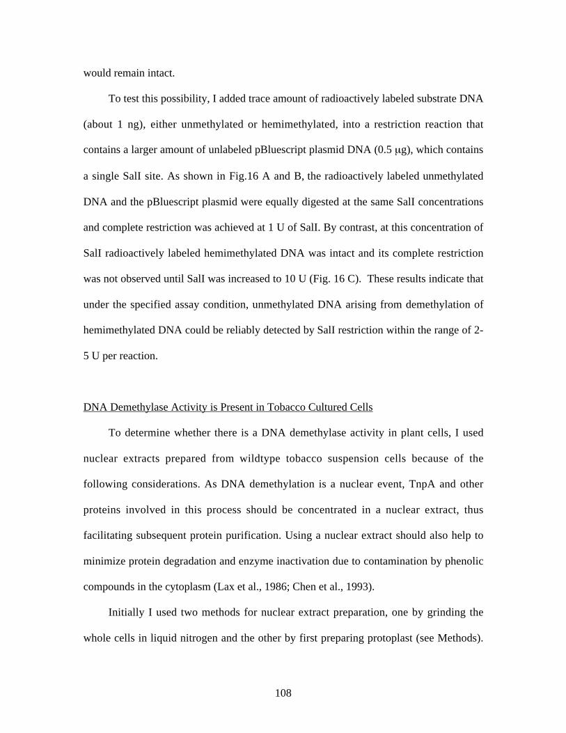
108
would remain intact.
To test this possibility, I added trace amount of radioactively labeled substrate DNA
(about 1 ng), either unmethylated or hemimethylated, into a restriction reaction that
contains a larger amount of unlabeled pBluescript plasmid DNA (0.5 µg), which contains
a single SalI site. As shown in Fig.16 A and B, the radioactively labeled unmethylated
DNA and the pBluescript plasmid were equally digested at the same SalI concentrations
and complete restriction was achieved at 1 U of SalI. By contrast, at this concentration of
SalI radioactively labeled hemimethylated DNA was intact and its complete restriction
was not observed until SalI was increased to 10 U (Fig. 16 C). These results indicate that
under the specified assay condition, unmethylated DNA arising from demethylation of
hemimethylated DNA could be reliably detected by SalI restriction within the range of 2-
5 U per reaction.
DNA Demethylase Activity is Present in Tobacco Cultured Cells
To determine whether there is a DNA demethylase activity in plant cells, I used
nuclear extracts prepared from wildtype tobacco suspension cells because of the
following considerations. As DNA demethylation is a nuclear event, TnpA and other
proteins involved in this process should be concentrated in a nuclear extract, thus
facilitating subsequent protein purification. Using a nuclear extract should also help to
minimize protein degradation and enzyme inactivation due to contamination by phenolic
compounds in the cytoplasm (Lax et al., 1986; Chen et al., 1993).
Initially I used two methods for nuclear extract preparation, one by grinding the
whole cells in liquid nitrogen and the other by first preparing protoplast (see Methods).

109
The quality of these preparations was then evaluated by assaying DNase activity. As
shown in Fig 17A, whereas very little DNase activity was detected in the nuclear extract
prepared by the first method, strong DNase activity was present in the nuclear extract
prepared using the protoplast protocol. For subsequent DNA demethylation assay, I have
therefore used the protoplast-based method for nuclear extract preparation.
The presence of strong DNase activity in the nuclear extract presents a problem
with the in vitro DNA demethylation assay, because DNA degradation interferes with
detection of demethylated DNA. Since Mg2+ alone is sufficient for the activity of most
enzymes, while both Mg2+ and Ca2+ are required for DNases (Latham, G. Ambion, inc.
unpublished data), I added the metal chelator EDTA to the DNA demethylation reaction
at a concentration significantly lower than that of Mg2+ but high enough to chelate
Figure 17. DNase activity in tobacco nuclear extracts and inhibition under the
conditions for the in vitro DNA demethylation assay. 2.5 µg pBluescript plasmid
DNA was incubated at 37 °C for 4 hrs with various amount of tobacco nuclear
extract prepared via protoplasting (NE2) in the absence (A) or presence (B) of
EDTA. The quality of the nuclear extract prepared by grinding in liquid nitrogen
(NE1) was compared by the DNase assay. M is 1kb DNA ladder.
µl
NE1 NE2
- 1 0 1 2 4 8 16
AM
BNE2
µl - 1 2 5
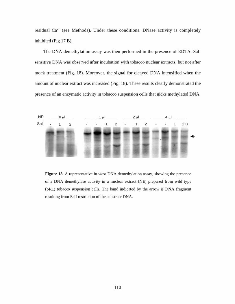
110
residual Ca2+ (see Methods). Under these conditions, DNase activity is completely
inhibited (Fig 17 B).
The DNA demethylation assay was then performed in the presence of EDTA. SalI
sensitive DNA was observed after incubation with tobacco nuclear extracts, but not after
mock treatment (Fig. 18). Moreover, the signal for cleaved DNA intensified when the
amount of nuclear extract was increased (Fig. 18). These results clearly demonstrated the
presence of an enzymatic activity in tobacco suspension cells that nicks methylated DNA.
1 µl 2 µl 4 µl .
SalI
NE
- 1 2 - - 1 2 - 1 2 - - 1 2 U
0 µl
Figure 18. A representative in vitro DNA demethylation assay, showing the presence
of a DNA demethylase activity in a nuclear extract (NE) prepared from wild type
(SR1) tobacco suspension cells. The band indicated by the arrow is DNA fragment
resulting from SalI restriction of the substrate DNA.

111
Discussion
A Sensitive in vitro DNA Demethylation Assay
To determine whether there is a DNA demethylase activity in plant cells, I
developed an in vitro DNA demethylation assay using hemimethylated DNA containing
the Spm promoter and the DCR as the substrate. This assay will permit the detection of
general DNA demethylase activities in plant cells, as well as an activity specific for
TnpA. Because TnpA has much higher binding affinity for the Spm promoter sequence
when hemimethylated than fully methylated (Chapter 5), using hemimethylated DNA
enhances the interaction between TnpA and its substrate DNA, thus increasing the
sensitivity for detection of TnpA-associated DNA demethylase activity.
Because fully methylated DNA is completely resistant to SalI restriction and
hemimethylated DNA is digestable at high enzyme concentrations, the in vitro DNA
demethylation assay can also be carried out with fully methylated DNA substrate by
increasing the amount of SalI in the reaction. This modified DNA demethylation assay
will be useful in characterizing the substrate specificity of plant DNA demethylases in
further studies.
Distinct DNA Demethylase Activities in Plant Cells
Although there is evidence suggesting the presence of DNA demethylase activities
in plant cells, the present study represents the first effort to directly test such a possibility.
My results showed that there is indeed a methylated DNA nicking activity in tobacco
suspension cells. The nicked DNA cannot be attributed to DNases in the nuclear extract,

112
because DNase activity was completely inhibited by the metal chelator EDTA. Although
it is still unknown whether this DNA nicking activity is specific to methylated DNA, it
clearly uses methylated DNA as substrate and is believed to be a DNA demethylase.
Since multiple DNA demethylases activities may be present in the crude nuclear extracts,
prior purification is necessary in further experiments to characterize the activity.
Simultaneously with this finding it was reported that the ROS1 protein in
Arabidopsis, a DNA glycosylase, is able to nick methylated DNA specifically (Gong et
al., 2002). Unlike the activity detected in this study, however, the ROS1 protein only
nicks DNA methylated with MspI methylase, which methylates the external cytosine in a
CCGG context, but not DNA methylated with SssI methylase that methylates the
cytosine in the CG dinucleotide context. Because the DNA substrate used in the present
study was methylated using the SssI methylase, it is very likely that the DNA
demethylase activity detected in our assay is distinct from ROS1. Interestingly, two ROS1
homologs have been identified in Arabidopsis. One of them is the DEMETER, which
encodes an embryo-specific DNA glycosylase but does not seem to have an in vivo
function in DNA demethylation (Choi et al., 2002). The function of the second homolog
has not been characterized, and therefore it would be interesting to see whether it encodes
a DNA glycosylase that catalyzes the removal of 5mC from the CG dinucleotide context.
Tissue-Specific and Temporal Expression of DNA Demethylase
As described previously (Chapter 5), TnpA-mediated DNA demethylation was not
observed in fully expanded leaves that have ceased to divide. This could be due to the
inability of TnpA to get access to methylated Spm sequence in the absence of DNA

113
replication, as DNA replication was shown to be required for TnpA to initiate DNA
demethylation (Chapter 5). Another possible cause is the lack of DNA demethylase in the
leaf cells. In animals, the 5-methylcytosine DNA glycosylase is exclusively expressed in
dividing cells (Jost and Jost, 1995; Schwarz et al., 2000). This observation raises the
interesting possibility that DNA demethylase activity in plants may also be expressed in a
tissue-specific or temporal manner. This may help to explain the phenotype of the mom
mutation of Arabidopsis that causes release of gene silencing but no change in DNA
methylation (Amedeo et al., 2000). The mom mutation is a weak allele affecting only a
transgene at later stage of development when cells have ceased dividing (Probst et al.,
2003).

114
References
Amedeo, P., Habu, Y., Afsar, K., Scheid, O.M., and Paszkowski, J. (2000). Disruptionof the plant gene MOM releases transcriptional silencing of methylated genes. Nature405, 203-206.
Bhattacharya, S.K., Ramchandani, S., Cervoni, N., and Szyf, M. (1999). Amammalian protein with specific demethylase activity for mCpG DNA. Nature 397,579-583.
Cervoni, N., Bhattacharya, S., and Szyf, M. (1999). DNA demethylase is a processiveenzyme. J. Biol. Chem. 274, 8363-8366.
Chen, Z., Ricigliano, J.W., and Klessig, D.F. (1993). Purification and characterizationof a soluble salicylic acid-binding protein from tobacco. Proc. Natl. Acad. Sci. USA90, 9533-9537.
Choi, Y., Gehring, M., Johnson, L., Hannon, M., Harada, J.J., Goldberg, R.B.,Jacobsen, S.E., and Fischer, R.L. (2002). DEMETER, a DNA glycosylase domainprotein, is required for endosperm gene imprinting and seed viability in arabidopsis.Cell 110, 33-42.
Cooke, R., and Penon, P. (1990). In vitro transcription from cauliflower mosaic viruspromoters by a cell-free extract from tobacco cells. Plant Mol Biol 14, 391-405.
Detich, N., Theberge, J., and Szyf, M. (2002). Promoter-specific activation anddemethylation by MBD2/demethylase. J. Biol. Chem. 277, 35791-35794.
Falnes, P.O., Johansen, R.F., and Seeberg, E. (2002). AlkB-mediated oxidativedemethylation reverses DNA damage in Escherichia coli. Nature 419, 178-182.
Gong, Z., Morales-Ruiz, T., Ariza, R.R., Roldan-Arjona, T., David, L., and Zhu,J.K. (2002). ROS1, a repressor of transcriptional gene silencing in Arabidopsis,encodes a DNA glycosylase/lyase. Cell 111, 803-814.
Hendrich, B., Hardeland, U., Ng, H.H., Jiricny, J., and Bird, A. (1999). The thymineglycosylase MBD4 can bind to the product of deamination at methylated CpG sites.Nature 401, 301-304.
Jost, J.P., and Jost, Y.C. (1995). Mechanism of active DNA demethylation duringembryonic development and cellular differentiation in vertebrates. Gene 157, 265-266.
Lax, S.R., Lauer, S.J., Browning, K.S., and Ravel, J.M. (1986). Purification andproperties of protein synthesis initiation and elongation factors from wheat germ.Methods Enzymol. 118, 109-128.
Ng, H.H., Zhang, Y., Hendrich, B., Johnson, C.A., Turner, B.M., Erdjument-Bromage, H., Tempst, P., Reinberg, D., and Bird, A. (1999). MBD2 is atranscriptional repressor belonging to the MeCP1 histone deacetylase complex. Nat.Genet. 23, 58-61.
Oakeley, E.J., and Jost, J.P. (1996). Non-symmetrical cytosine methylation in tobaccopollen DNA. Plant. Mol. Biol. 31, 927-930.
Petronzelli, F., Riccio, A., Markham, G.D., Seeholzer, S.H., Genuardi, M.,Karbowski, M., Yeung, A.T., Matsumoto, Y., and Bellacosa, A. (2000).Investigation of the substrate spectrum of the human mismatch-specific DNA N-

115
glycosylase MED1 (MBD4): fundamental role of the catalytic domain. J. CellPhysiol. 185, 473-480.
Probst, A.V., Fransz, P.F., Paszkowski, J., and Scheid, O.M. (2003). Two means oftranscriptional reactivation within heterochromatin. Plant J. 33, 743-749.
Ramchandani, S., Bhattacharya, S.K., Cervoni, N., and Szyf, M. (1999). DNAmethylation is a reversible biological signal. Proc. Natl. Acad. Sci. USA 96, 6107-6112.
Roberts, M.W., and Okita, T.W. (1991). Accurate in vitro transcription of plantpromoters with nuclear extracts prepared from cultured plant cells. Plant. Mol. Biol.16, 771-786.
Schwarz, S., Bourgeois, C., Soussaline, F., Homsy, C., Podesta, A., and Jost, J.P.(2000). A CpG-rich RNA and an RNA helicase tightly associated with the DNAdemethylation complex are present mainly in dividing chick embryo cells. Eur. J. CellBiol. 79, 488-494.
Trewick, S.C., Henshaw, T.F., Hausinger, R.P., Lindahl, T., and Sedgwick, B.(2002). Oxidative demethylation by Escherichia coli AlkB directly reverts DNA basedamage. Nature 419, 174-178.
Vairapandi, M., and Duker, N.J. (1996). Partial purification and characterization ofhuman 5-methylcytosine-DNA glycosylase. Oncogene 13, 933-938.
Wang, R.Y., Kuo, K.C., Gehrke, C.W., Huang, L.H., and Ehrlich, M. (1982). Heat-and alkali-induced deamination of 5-methylcytosine and cytosine residues in DNA.Biochim. Biophys. Acta. 697, 371-377.
Wu, P., Qiu, C., Sohail, A., Zhang, X., Bhagwat, A.S., and Cheng, X. (2003).Mismatch repair in methylated DNA. Structure and activity of the mismatch-specificthymine glycosylase domain of methyl-cpg-binding protein mbd4. J. Biol. Chem.278, 5285-5291.
Zhang, Y., Ng, H.H., Erdjument-Bromage, H., Tempst, P., Bird, A., and Reinberg,D. (1999). Analysis of the NuRD subunits reveals a histone deacetylase core complexand a connection with DNA methylation. Genes Dev. 13, 1924-1935.
Zhu, B., Zheng, Y., Hess, D., Angliker, H., Schwarz, S., Siegmann, M., Thiry, S., andJost, J.P. (2000). 5-methylcytosine-DNA glycosylase activity is present in a clonedG/T mismatch DNA glycosylase associated with the chicken embryo DNAdemethylation complex. Proc. Natl. Acad. Sci. USA 97, 5135-5139.

116
Chapter 7
DNA DEMETHYLASE ACTIVITY IS RECRUITED TO THE SPM
SEQUENCE AS A RESULT OF TRANSCRIPTIONAL ACTIVATION
Introduction
Although several proteins in animal cells have been shown to be capable of
converting 5mC to cytosine, whether and how they participate in sequence-specific DNA
demethylation still remain largely unknown. In some cases, DNA demethylases seem to
be targeted to DNA sequences by trans-acting factors through physical interactions. For
example, 5-methylcytosine DNA glycosylase, which could catalyze DNA demethylation
through a DNA repair pathway, has been shown recently to interact with the retinoid and
the estradiol receptors, both are known to promote sequence-specific DNA demethylation
(Zhu et al., 2001; Jost et al., 2002).
The previous findings that there is a DNA demethylase activity in plant cells and
that TnpA has a leucine-zipper domain in its C-terminus strongly suggest that a DNA
demethylase might be recruited to the Spm promoter sequence through direct or indirect
protein-protein interactions. To test this possibility, I purified TnpA and possible
interacting proteins from plant cells expressing the TnpA protein using a co-
immunoprecipitation approach. DNA demethylase activity was not detected in TnpA-
containing immunoprecipitate, however. The present study clearly ruled out the
117
possibility that TnpA is a sequence-specific DNA demethylase. Results from this
experiment and selective precipitation by ammonium sulfate also failed to show direct
interaction between TnpA and DNA demethylase activities. Since demethylation of the
Spm sequence was observed after induction of transcription from the Spm sequence under
the control of an inducible promoter, it is suggested that a DNA demethylase gains access
to the Spm sequence passively as a result of transcriptional activation by the TnpA
protein.
Materials and Methods
Plant Materials
Unless specified, all plant materials and treatments used in this study have been
described in previous chapters. Transgenic tobacco lines containing the pTA-U+D-Luc-
Spm3’ construct for inducible transcription activation was generated using the
Agribacterium-mediated leaf-disc transformation method as used for generation of other
transgenic lines.
Constructs
To clone pTA-Spm-LUC-Spm3’, the construct for inducible transcription through
the Spm sequence, the Spm-LUC reporter construct was first subcloned from pMS73 as
an approximately 4.7 kb fragment (Schläppi, 1994) into pBluescript II KS (+) and then

118
cloned as an Asp718 (filled-in with Klenow) to SpeI fragment into pTA7001 or pTA7002
(Aoyama and Chua, 1997) that was cut with XhoI and SpeI (XhoI filled-in with Klenow).
Immunoprecipitation
The FLAG-tagged TnpA protein and its truncated derivatives, FLAG-TnpA 540 and
FLAG-TnpA420, were pulled down using anti-FLAG M2-agarose affinity gel (A-2220,
Sigma-Aldrich, St. Louis, MO). Tobacco suspension cultures that express each protein
were first filtered through a layer of miracloth (Calbiochem-Novabiochem Corporation,
La Jolla, CA), weighed and grounded on ice in 3X volume of ice-cold extraction buffer,
which is composed of 50 mM Tris-HCl, pH 7.6 at 4 °C, 450 mM NaCl, 10 mM EDTA,
1% Triton X-100, 50 mM β-mercaptoethanol, 3 mM PMSF, 1X plant protease inhibitor
cocktail Sigma-Aldrich), 10% glycerol and 1% BSA. The extract was centrifuged at
15000 rpm, 4°C for 15 min, and 1 ml supernatant was transferred to a tube with 40 µl
anti-FLAG agarose beads, which had been pretreated following the vendor’s
recommendations. After incubation at 4 °C for 6 hrs on a shaker (Barnstead/thermolyne,
Dubuque, IOWA), the beads were washed 1-3 times with a buffer that contains 50 mM
Tris-HCl, pH 7.6 at 4°C, 150 mM NaCl, 10 mM EDTA, 50 mM β-mercaptoethanol, 1
mM PMSF, 1X plant protease inhibitor cocktail and 10% glycerol and 0.1%-1% Triton
X-100 depending on the required stringency. The gel was finally washed in washing
buffer that contains no Triton X-100.
For protein detection only, the gel was boiled in 40 µl 2X SDS protein sample
buffer and the supernatant was analyzed by a western blot assay. To release the FLAG-
tagged protein in its native form, the gel was incubated with 40 µl of 150 ng/µl FLAG

119
peptide solution at 4 °C for 45 min with shaking. The supernatant was collected after a
brief centrifugation at 10,000 rpm, 4°C. The elution was repeated once and the
supernatants were combined.
Ammonium Sulfate Precipitation
Selective ammonium sulfate precipitation of proteins from tobacco extract was
conducted following standard protocols (Lovrien and Matulis, 1997). Briefly, tobacco
suspension cultures were first filtered through a layer of miracloth (Calbiochem-
Novabiochem Corporation, La Jolla, CA). 50 g cells were weighed and ground in 50 ml
ice-cold extraction buffer (Tris-HCl, pH 7.6, 150 mM NaCl, 10% glycerol, 10 mM
EDTA, 50 mM 2-mercaptoethanol, 5 mM PMSF and 5mM NaF). The supernatant was
then collected after the homogenate was centrifuged at 40,000 rpm, 4 °C for 1 hr using a
Ti70 rotor (Beckman Ultracentrifuge). (NH4)2SO4 powder was added into the extract with
stirring to achieve the desired saturation. The slurry was stirred for additional 20 min,
then centrifuged at 17,500 rpm at 4 °C for 20 min. The supernatant was transferred to a
beaker and the process was repeated to precipitate proteins at higher (NH4)2SO4
concentrations.
The pellets from each fraction were dissolved in 2 ml protein extraction buffer and
centrifuged at 15000 rpm, 4 °C for 15 min to remove undissolved material. Each fraction
was dialyzed against 100X vol of dialysis buffer (20mM HEPES-KOH, pH7.9, 20%
glycerol, 100 mM KOAc, 0.2 mM EGTA, 0.1 mM EDTA, 2 mM DTT and 0.5 mM
PMSF) using cellulose ester tubing (Spectra/por CE, MWCO of 3500, CAT# 131162).
The buffer was changed every 4-6 hrs and this was repeated twice. Finally, the solution

120
was centrifuged at 15,000 rpm, 4 °C for 20 min and the supernatant was aliquoted and
stored at –80C.
DNA Demethylation Assay and Other Assays
DNA demethylase assay was performed as described in Chapter 6. In some cases,
hemimethylated DNA containing the DCR sequence alone was used as the substrate,
which was amplified by PCR as a 493 bp fragment from pDC131 using primer pairs KS
and SK (Raina, 1993). Silver staining of protein gels was performed using the Silver
Stain Plus kit (BioRad, Hercules, CA).
All other assays such as Western blots, gel mobility shift assays, RT-PCR and DNA
methylation assays were performed as described previously.
Results
Optimization of Conditions for TnpA Immunoprecipitation
To investigate the possibility that TnpA interacts physically with a DNA
demethylase activity, it is necessary to purify TnpA along with possible interacting
proteins from transgenic tobacco cells that express the TnpA protein. This was attempted
by co-immunoprecipitation (IP) using anti-FLAG antibody affinity gel (Methods), as
TnpA and its truncated derivatives TnpA540 and TnpA420 have been expressed as FLAG-
tagged fusion proteins in transgenic tobacco cells. Another advantage of this approach is
that, along with the use of proteomics tools, it would permit quick identification of the
proteins that participate in TnpA-mediated DNA demethylation.
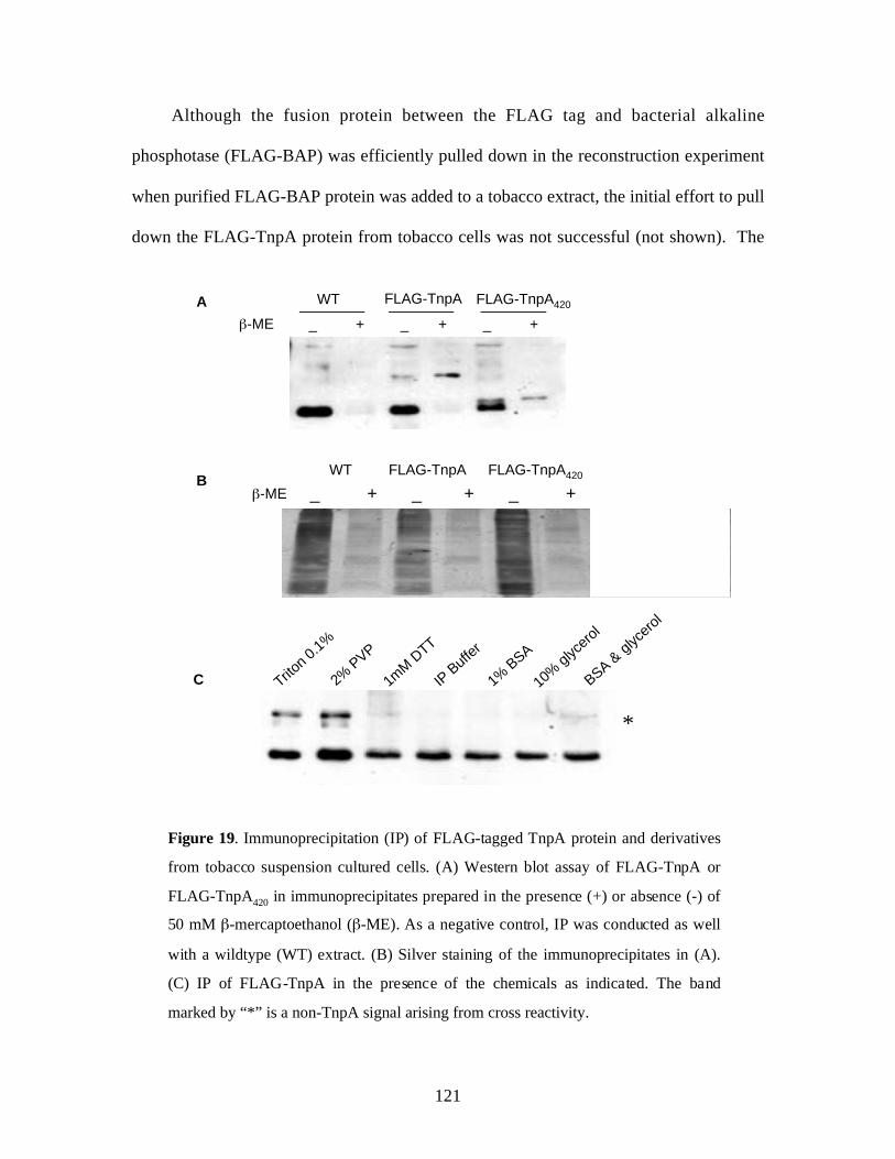
121
Although the fusion protein between the FLAG tag and bacterial alkaline
phosphotase (FLAG-BAP) was efficiently pulled down in the reconstruction experiment
when purified FLAG-BAP protein was added to a tobacco extract, the initial effort to pull
down the FLAG-TnpA protein from tobacco cells was not successful (not shown). The
Figure 19. Immunoprecipitation (IP) of FLAG-tagged TnpA protein and derivatives
from tobacco suspension cultured cells. (A) Western blot assay of FLAG-TnpA or
FLAG-TnpA420 in immunoprecipitates prepared in the presence (+) or absence (-) of
50 mM β-mercaptoethanol (β-ME). As a negative control, IP was conducted as well
with a wildtype (WT) extract. (B) Silver staining of the immunoprecipitates in (A).
(C) IP of FLAG-TnpA in the presence of the chemicals as indicated. The band
marked by “*” is a non-TnpA signal arising from cross reactivity.
A
β-ME _ + _ + _ +
WT FLAG-TnpA FLAG-TnpA420
2% P
VP
Triton 0
.1%
1% B
SA
IP B
uffer
1mM D
TT
BSA & gl
ycer
ol
10%
glyc
erol
*
C
BWT FLAG-TnpA
β-ME _ + _ + _ +FLAG-TnpA420

122
failure is partly due to some intrinsic property of the protein, because IP efficiency was
very low even with purified FLAG-TnpA in extraction buffer (not shown). Since no
TnpA protein was recovered at all when it was added to a plant extract, some components
in the tobacco extract may exacerbate this problem.
To optimize the conditions for TnpA immunoprecipitation, I therefore compared the
efficiency of immunopreciptation under various conditions, including different salt,
glycerol and detergent concentrations as well as in the presence of bovine serum albumin
(BSA) and polyvinylpolypyrrolidine (PVP). None of the tested conditions significantly
and consistently improved the IP efficiency (not shown). However, an inverse
relationship was observed between IP efficiency and the intensity of brown color,
suggesting that oxidation of polyphenolic compounds might interfere with
immunoprecipitation of the TnpA protein (Lax et al., 1986; Chen et al., 1993).
In order to prevent oxidation of phenolic compounds, I therefore increased the
concentration of the reducing reagent β-mercaptoethanol in the protein extraction buffer.
Remarkably, I was able to consistently pull down the FLAG-TnpA protein and its
truncated derivatives when 50 mM β-mercaptoethanol was used (Fig. 19A). Use of other
chemicals that supposedly increase specificity or stabilize protein complexes did not
further improve the efficiency of immunoprecipitation (Fig 19 C). Interestingly, it has
been reported previously that the presence of a reducing reagent, such β-mercaptoethanol
or DTT, is essential for maintaining the DNA binding ability of the TnpA protein
(Trentman, 1991). A likely explanation for this observation is that under the oxidative
condition proteins with protein-protein interaction domains tend to form functionally
inactive aggregate.

123
TnpA is not itself, nor does it Bind to, a DNA Demethylase
To be compatible with the in vitro DNA demethylation assay, the FLAG-tagged
TnpA protein was released from the affinity gel in its native form by incubation with
FLAG peptide solution. As a negative control, native FLAG-TnpA540 and FLAG-TnpA420
WT FLAG-TnpA FLAG-TnpA420
1 2 1 2 1 2 1 2
FLAG-TnpA420C
B
A
Figure 20. TnpA does not associate specifically with a DNA demethylase activity.
DNA demethylase activity in immunoprecipitates (2 µl each) prepared by (A) mild
washing or (B) extensive washing. The arrow indicates the position of SalI cleaved
DNA as a result of DNA demethylation. (C) Western blot analysis of the samples
used in (B).
SalI 0 2 3 0 2 3 0 2 3 0 2 3 U
WT FLAG-TnpA FLAG-TnpA420FLAG peptide
0 2 3 0 2 3 0 2 3 0 2 3 U
WT FLAG-TnpA FLAG-TnpA420FLAG peptide
Sal
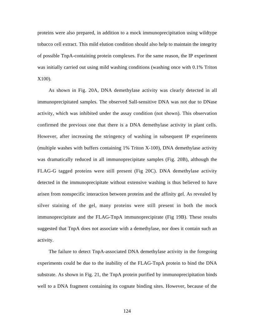
124
proteins were also prepared, in addition to a mock immunoprecipitation using wildtype
tobacco cell extract. This mild elution condition should also help to maintain the integrity
of possible TnpA-containing protein complexes. For the same reason, the IP experiment
was initially carried out using mild washing conditions (washing once with 0.1% Triton
X100).
As shown in Fig. 20A, DNA demethylase activity was clearly detected in all
immunoprecipitated samples. The observed SalI-sensitive DNA was not due to DNase
activity, which was inhibited under the assay condition (not shown). This observation
confirmed the previous one that there is a DNA demethylase activity in plant cells.
However, after increasing the stringency of washing in subsequent IP experiments
(multiple washes with buffers containing 1% Triton X-100), DNA demethylase activity
was dramatically reduced in all immunoprecipitate samples (Fig. 20B), although the
FLAG-G tagged proteins were still present (Fig 20C). DNA demethylase activity
detected in the immunoprecipitate without extensive washing is thus believed to have
arisen from nonspecific interaction between proteins and the affinity gel. As revealed by
silver staining of the gel, many proteins were still present in both the mock
immunoprecipitate and the FLAG-TnpA immunoprecipirate (Fig 19B). These results
suggested that TnpA does not associate with a demethylase, nor does it contain such an
activity.
The failure to detect TnpA-associated DNA demethylase activity in the foregoing
experiments could be due to the inability of the FLAG-TnpA protein to bind the DNA
substrate. As shown in Fig. 21, the TnpA protein purified by immunoprecipitation binds
well to a DNA fragment containing its cognate binding sites. However, because of the
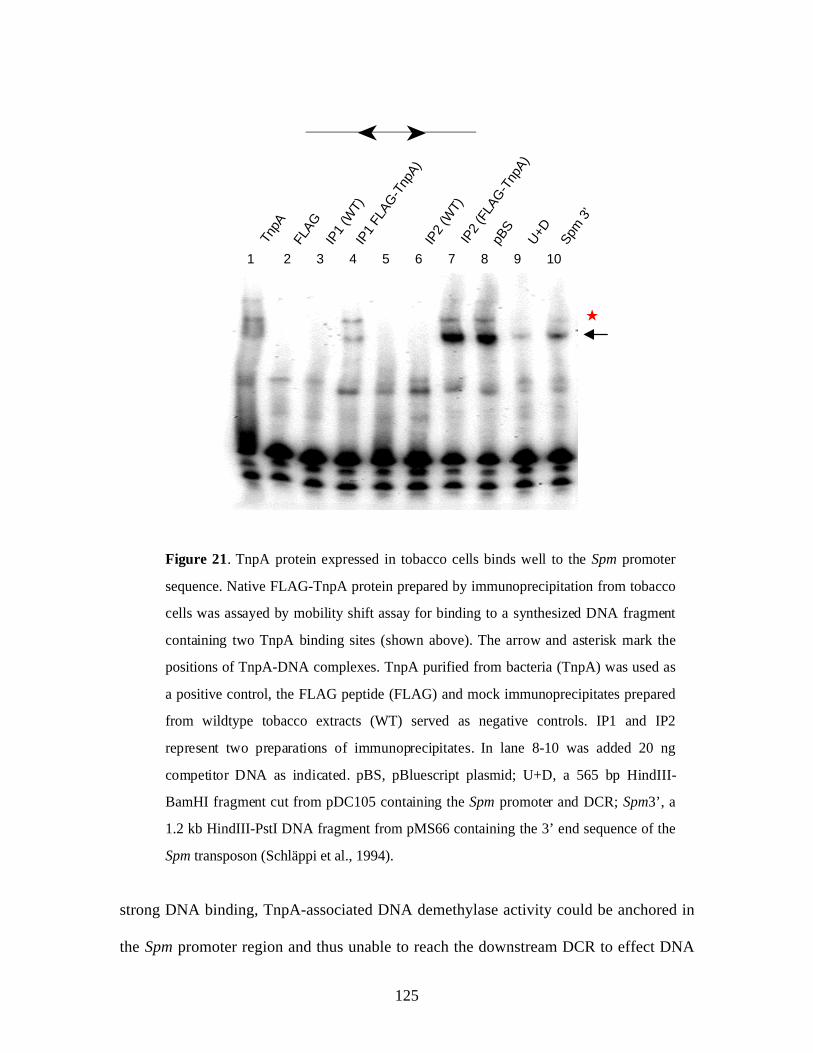
125
strong DNA binding, TnpA-associated DNA demethylase activity could be anchored in
the Spm promoter region and thus unable to reach the downstream DCR to effect DNA
Figure 21. TnpA protein expressed in tobacco cells binds well to the Spm promoter
sequence. Native FLAG-TnpA protein prepared by immunoprecipitation from tobacco
cells was assayed by mobility shift assay for binding to a synthesized DNA fragment
containing two TnpA binding sites (shown above). The arrow and asterisk mark the
positions of TnpA-DNA complexes. TnpA purified from bacteria (TnpA) was used as
a positive control, the FLAG peptide (FLAG) and mock immunoprecipitates prepared
from wildtype tobacco extracts (WT) served as negative controls. IP1 and IP2
represent two preparations of immunoprecipitates. In lane 8-10 was added 20 ng
competitor DNA as indicated. pBS, pBluescript plasmid; U+D, a 565 bp HindIII-
BamHI fragment cut from pDC105 containing the Spm promoter and DCR; Spm3’, a
1.2 kb HindIII-PstI DNA fragment from pMS66 containing the 3’ end sequence of the
Spm transposon (Schläppi et al., 1994).
➤
➤
IP2
(FLA
G-T
npA)
U+D
FLAG
TnpA
pBS
IP1
FLAG
-Tnp
A)
IP1
(WT)
IP2
(WT)
Spm
3’
★
1 2 3 4 5 6 7 8 9 10
126
demethylation. To check this possibility, I performed a DNA demethylation assay using a
hemimethylated DNA fragment containing the DCR sequence only as substrate
(Methods). Again, TnpA-associated DNA demethylase activity was not observed (Fig
22).
The foregoing experiments do not rule out the possibility that TnpA binds a 5-
methylcytosine DNA glycosylase (5MCDG). If a 5MCDG activity participates in TnpA-
mediated DNA demethylation, the resulting abasic sites at 5mC may become resistant to
SalI restriction, as is the case for EcoRII (Petrauskene et al., 1995), and thus evade
detection under the current assay conditions. To investigate this possibility, endonuclease
Figure 22. DNA demethylation assay using hemimethylated DNA containing DCR
only. (A) A diagram showing the primers KS and SK used for PCR amplification
of the DNA substrate and the expected DNA fragments after SalI restriction. (B)
DNA demethylase activity in immunoprecipitates as used for the experiment shown
in Figure 20B.
SalI 0 2 3 0 2 3 0 2 3 0 2 3 U
WT FLAG-TnpA FLAG-TnpA540 FLAG-TnpA420B
Sal I (GTCGAC)KS SK
93 bp 400 bp
DCR
A

127
IV, an apurinic/apyrimidinic (AP) endonuclease that nicks DNA specifically at abasic
sites (Kaboev et al., 1985; Masters et al., 1991), was used to initiate DNA nicking before
SalI restriction. No change in the amount of SalI sensitive DNA was observed, however,
indicating the absence of DNA glycosylase in the TnpA-containing immunoprecipitate
(Fig 23).
Another possibility for the lack of DNA demethylase activity in the TnpA
immunoprecipitate is that proteins that interact with TnpA might be washed off under the
more stringent IP condition. To address this question, I precipitated proteins from TnpA
0 2 3 0 2 0 2 3 0 2 0 2 3 0 2 U
WT FLAG-TnpAFLAG peptide
SalI
End IV 0 0 0 1 1 0 0 0 1 1 0 0 0 1 1 U
B
Figure 23. Detection of abasic sites in DNA using endonuclease IV.
(A) Diagram showing DNA demethylation through a DNA repair pathway involving
DNA glycosylase. (B) Using bacterial endonuclease IV (End IV) and SalI for
detection of DNA demethylase activity in the immunoprecipitates as used for the
experiment shown in Fig 20B.
A
5mC
5’p G3’
5’p Gp3’
CDNA
glycosylase
DNA Repair
Enzymes5’p G3’
5’p Gp3’C
C
5’p G3’
5’p Gp3’C

128
expressing tobacco cells at increasing concentrations of ammonium sulfate and
pBS
U+D#6
- + - - - - + - - - - + - - - + - - - - + - - - - +
- - - - - - - - + - - - -
30% 40% 50%30%+40%
40%+50%B
Figure 24. TnpA does not co-fractionate with DNA demethylase activity.
Mobility shift assay of precipitates at the concentrations of AmSO4 indicated (A) in the
absence or (B) presence of competitor DNA. The asterisk marks the position of DNA-
TnpA complexes. pBS and U+D, same DNA fragment as described in Figure 21. #6, a
134bp PCR-amplified DNA fragment containing 2 TnpA binding site, as described in
Chapter 4; The buffer used to dissolve the sample was used a control. 30%+40% and
40%+50%, mixtures of equal volumes of the 30% and 40% or 40% and 50% fractions
respectively. (C) DNA demethylase activity in different fractions of AmSO4
precipitation, as indicated by the band marked by the arrow.
A
30%
40%
50%
60%
Buffe
r
*
C 30% 40% 50% 60%
SalI 0 2 3 0 2 3 0 2 3 0 2 3 U
Fraction

129
determined their DNA demethylase activity. Due to the ‘salting out’ effect, proteins tend
to remain in their complex at high (NH4)2SO4 concentrations. If TnpA interacts with a
DNA demethylase activity, TnpA and DNA demethylase activity would be expected to
co-fractionate. While almost all FLAG-tagged TnpA protein was precipitated at 30%
saturation of (NH4)2SO4, as revealed by mobility shift assay (Fig. 24A and B), only a
small portion of DNA demethylase activity was present in this fraction (Fig. 24 C). By
contrast, most DNA demethylase activity seems to be in the fraction corresponding to
40% (NH4)2SO4 saturation, which contains little TnpA protein (Fig 24).
Taken together, all the experiments described above indicate that TnpA is unlikely to
be associated physically with a DNA demethylase.
Spm Demethylation after Transcriptional Activation
The finding that TnpA’s transcriptional activity is required for it to promote DNA
demethylation, as well as the failure to detect DNA demethylase activity in TnpA-
containing immunoprecipitates, strongly suggests an indirect mechanism for the
recruitment of DNA demethylase activity to the Spm sequence. To further investigate the
relationship between transcription activation and DNA demethylation, I generated
transgenic tobacco plants in which the Spm promoter and DCR along with a luciferase
reporter gene construct (Schläppi et al., 1994) are placed downstream of the
glucocorticoid-inducible (GR) promoter (Aoyama and Chua, 1997), the same vector as
used for controlling TnpA expression described in previous experiments (Fig 25A). This
will make it possible to assemble the transcription initiation complex on the GR promoter
upstream from the Spm promoter, then transcribe through the Spm sequence.

130
In most transgenic lines, the Spm sequence remained either methylated or
unmethylated after dexamethasone treatment to activate transcription (Not shown).
Figure 25. Spm demethyaltion after induction of transcription. (A) T-DNA region of the
binary vector used for controlling transcription through the Spm sequence. GVG, a hybrid
transcription factor consisting of the Gal4 binding domain (Gal4 BD), the viral VP16
activation domain (VP16 AD) and the glucocorticoid receptor domain (GR). Spm P, the
Spm promoter sequence; DCR, the promoter downstream sequence. The Spm P-DCR-Luc-
Spm 3’ sequence is transcribed as a single transcription unit. See the legend in Figure 1 for
detail. (B) DNA methylation in the Spm promoter (P) and DCR (D) sequence before (Ctl)
and after (Ind) induction treatment in two independent transgenic tabacco calli with the
construct shown in (A). (C) Spm methylation in transgenic tobacco calli, which have lost
methylation due to TnpA expression, 2 weeks after transfer to non-induction medium. SR1
is the line that does not have the TnpA-expression construct. In (B) and (C), the upper and
lower bands represent methylated and unmethylated DNA, respectively.
Dex
GVG E9 Nos P Hyg Nos T GR P3A35S P L BR B
LUC DCR Spm PSpm 3’
Gal4 BD VP16 AD
GRHP90
A
Gal4 BD VP16 AD GR
Dex
C
Callus #1
P D P D P D P D
Callus #2 SR1 Callus #3
BCallus #1 Callus #2
P D
Ctl Ind Ctl Ind
P D P D P D
131
However, two of 11 independent transgenic lines examined showed significant loss of
DNA methylation after 48 hrs of induction (Fig. 25B). As neither dexamethasone nor the
transcription factor GVG for inducible transcription interferes with DNA methylation in
the Spm sequence (Chapter 2), the present observation strongly supports a causal
connection between transcriptional activation and DNA demethylation.
Notably, none of the transgenic lines showed an increase in DNA methylation,
although there was clear evidence for the presence of de novo DNA methylase activity in
the cells under study. In transgenic lines that contain the construct for inducible TnpA
expression and have completely lost DNA methylation in the Spm sequence after
prolonged induction treatment, the Spm sequence regained methylation after depletion of
the inducing chemical (Fig. 25C),
Discussion
Recruitment by TnpA of DNA Demethylase Activity to the Spm Sequence
TnpA could promote active demethylation of the Spm sequence through one or a
combination of the following possible mechanisms. First, TnpA could itself be a
sequence-specific DNA demethylase. Although it has been shown that purified
recombinant TnpA protein overexpressed in bacteria does not have DNA demethylase
activity, the possibility that TnpA contains DNA demethylase activity has not been
formally ruled out, as some eukaryotic modifications might be essential for TnpA’s
ability to promote DNA demethylation. A more likely possibility is that TnpA recruits a
132
distinct protein that has DNA demethylase activity to the Spm sequence. Recruitment of
this DNA demethylase could be achieved through direct interaction with TnpA or
indirectly through a protein complex. Since the transcription activation domain of TnpA
has been shown to be indispensable for TnpA’s ability to promote DNA demethylation, a
third possible scenario is that DNA demethylases gains access to the Spm sequence as a
consequence of transcription activation, chromatin remodeling in particularly.
To distinguish these possibilities, I used co-immunoprecipitation to purify TnpA
and possible interacting proteins from tobacco cells that express TnpA as a FLAG tagged
protein and determined DNA demethylase activity in the immunoprecipitate using an in
vitro DNA demethylation assay. If TnpA itself is a DNA demethylase or it recruits such
an activity through protein-protein interactions, a DNA demethylase activity should be
detectable in the immunoprecipitate that contains TnpA, but not in ones containing its
truncated derivatives or in a mock immunoprecipitate from wildtype tobacco extract.
Although a DNA demethylase was detected in all immunoprecipitates under mild
washing conditions, no such activity was found to be specifically associated with the
TnpA protein regardless of the method used for detecting DNA demethylase activity.
This result clearly rules out the possibility that TnpA itself is a sequence-specific DNA
demethylase. The co-immunoprecipitation study does not support the hypothesis that
TnpA interacts with a DNA demethylase, either. It is not very likely that a TnpA-
associating DNA demethylase was washed off the affinity gel during the co-
immunoprecipitation experiment because TnpA and DNA demethylase did not co-
fractionate when proteins in whole cell extract was selectively precipitated by ammonium
sulfate. Results from these experiments and the inducible transcription approach

133
described below strongly suggest that DNA demethylase activities are recruited to the
Spm sequence passively as a consequence of transcriptional activation (see below).
DNA Demethylation in Response to Transcription Activation
A causal connection between transcription and DNA demethylation has been
identified in the present studies on TnpA (Chapter 2 and 3) and has gained support from
the observation that no direct interaction between TnpA and DNA demethylase activity
was revealed in the foregoing experiments. To further examine the role of transcription in
DNA demethylation, I used a glucocorticoid-inducible promoter to control transcriptional
activation from the Spm sequence. Demethylation of the Spm sequence was observed
soon after transcription induction (48 hrs), again suggesting an active process. Since
transcription activation was achieved by the binding of GVG, a chimeric transcription
factor consisting of the gal4 binding domain, the viral VP16 activation domain and the
glucocorticoid receptor domain, DNA demethylation cannot be attributed to the TnpA
protein. These results further suggest that TnpA promotes DNA demethylation through a
mechanism that is common with other transcriptional factors that are also able to promote
sequence-specific DNA demethylation.
One possible scenario is that DNA demethylase activity is recruited to actively
transcribing DNA sequences via the transcription machinery. There is evidence that the
animal G/T mismatch DNA glycosylase, which has been shown to be capable of
converting 5mC to normal cytosine through a DNA repair pathway (Zhu et al., 2000),
interacts with the transcriptional coactivator p300/CBP (Tini et al., 2002). As most
known DNA demethylation-promoting trans-acting factors are transcriptional activators

134
(Matsuo et al., 1998; Grange et al., 2001; Thomassin et al., 2001; Zhu et al., 2001; Jost et
al., 2002), it is likely that they recruit a DNA demethylase activity via the p300 protein or
similar coactivators. Interestingly, p300/CBP homologs and interacting proteins have
been identified in plants, which share a highly conserved structure to their animal
counterparts (Bordoli et al., 2001; Yuan and Giordano, 2002; Lafarge and Montane,
2003), implying that a similar DNA demethylation mechanism might have evolved in
plants.
DNA demethylation could result from transcription per se. It has long been known
that damaged nucleotides in the actively transcribed strand are preferentially repaired, a
process termed transcription-coupled repair (TCD) (Li and Bockrath, 1995), which has
been found in a wide range of living organisms including plants (Mellon et al., 1996;
Leadon and Avrutskaya, 1998; Sturm and Lienhard, 1998). A direct approach to test this
possibility is to activate transcription initiation while inhibiting transcription per se using
transcription inhibitors such as α-amanitin or cordycepin (Tumbar et al., 1999; Nye et al.,
2002). The present inducible transcription system would enable such an experiment,
because the transcriptional activator GVG, which activates transcription from the
inducible promoter, is constitutively expressed and its activation only requires the
binding of glucocorticoid ligands. Consequently, assembly of the transcription initiation
complex and chromatin decondensation will not be affected by α-amanitin or cordycepin.
Such an experiment is impossible with the inducible TnpA system described previously
(Chapter 2), as transcription activation from the Spm sequence requires the TnpA protein,
which requires de novo transcription.

135
There is accumulating evidence indicating that chromatin remodeling rather than
transcription per se plays a major role in facilitating the access of DNA demethylase to
methylated DNA. Although DNA demethylation mediated by the transcription factor NF-
κβ requires the presence of the general transcription machinery, it occurs even when
transcription is inhibited (Matsuo et al., 1998). It has also been suggested that
hyperacetylated histone may serve as a mark for targeting DNA demethylase (Cervoni
and Szyf, 2001). Probably the most telling evidence regarding the role of chromatin
structure in DNA demethylation is derived from studies of the mom1 and ddm1 mutations
in Arabidopsis. Both DDM1 and DDM1 proteins are SWI2/SNF2-like chromatin
remodeling protein (Amedeo et al., 2000; Brzeski and Jerzmanowski, 2003). Whereas
mutation in the DDM1 gene causes genome-wide reduction in DNA methylation
(Jeddeloh et al., 1999) and release of gene silencing (Jeddeloh et al., 1998), the mom1
mutant only shows derepression of gene silencing without a noticeable change in DNA
methylation (Amedeo et al., 2000).
Since transcription is generally associated with chromatin remodeling, these
observations seem to argue against a role of chromatin remodeling in DNA
demethylation. Remarkably, however, a recent study has revealed that the transgene
whose transcription is affected by the mom mutation is still in a heterochromatin context
(Probst et al., 2003). Since the ddm1 mutant is also associated with heterochromatin
decondensation in addition to histone hyperacetylation and loss of histone methylation
(Gendrel et al., 2002; Probst et al., 2003), heterochromatin structure could be the major
factor affecting the accessibility of DNA demethylase to methylated DNA.

136
The viral VP16 activation domain is among the best characterized to activate
transcription through increasing the level of histone acetylation and remodeling the
chromatin structure (Tumbar et al., 1999; Muller et al., 2001; Nye et al., 2002). The
present finding that Spm demethylation followed transcriptional activation by the GVG
transcription factor thus lends further support to the chromatin remodeling-based
mechanism. Moreover, this inducible system would permit a dynamic study of the
interrelationship between DNA demethylation and transcription activation, change in
histone methylation and acetylation as well as chromatin remodeling.
Transcriptional Gene Silencing and DNA Methylation
The experiment using an inducible promoter to control transcription through the
Spm sequence has also yielded information regarding the mechanism that controls de
novo methylation and gene silencing of the Spm sequence. DNA methylation can result
from gene silencing at the level of transcription (TGS) or posttranscription (PTGS) (Sijen
et al., 2001; Vaucheret and Fagard, 2001). In contrast to TGS, where transcription is
completely arrested, PTGS is characterized by active transcription from the promoter and
an insignificant steady-state level of full-length transcripts as a result of RNA
degradation. Since TGS and PTGS often occur to repetitive sequences, and dsRNA has
been found to be a potent silencer of gene expression (Matzke et al., 2000), it has been
hypothesized that silencing and methylation of transgenes may be due to aberrant RNA
derived from inverted repeats in the transgene (Muskens et al., 2000). If Spm methylation
results from such a PTGS mechanism, transcription activation should enhance DNA
methylation. However, this possibility is apparently not supported by our observation,

137
because none of the lines examined showed an increase in DNA methylation, regardless
of the initial methylation status of the Spm sequence.
Another interesting observation from this study is that inducible Spm demethylation
seems to depend on how the inducible Spm-LUC transgene is constructed. Only a small
Figure 26. Transgenes in close proximity are co-regulated.
(A) A diagramatic representation of the Spm sequence and the NPT II gene, which confers
resistance to kanamycin, in the vector used for tobacco transformation. (B) Three lines of
transgenic tobacco plants grown on MS medium containing 50 mg/L kanamycin, and (C)
DNA methylation of the Spm promoter (P) and the DCR (D) sequence. The upper and
lower bands in each lane represent methylated and unmethylated DNA, respectively.
SR1 SR83
SR87 SR/WT
A
C SR1 SR83 SR87
P D P D P D
B
NPT II Spm P DCR LUC Spm 3’
RB LB

138
number of transgenic lines showed inducible Spm demethylation when the 5’ end of the
Spm-LUC construct is placed adjacent to the border of the T-vector (facing inward, Fig
25A). By contrast, no such transgenic lines were obtained when the 5’ end of the Spm-
LUC construct is placed adjacent to the hygromycin expression cassette (facing outward).
One likely explanation for this observation is that the Spm sequence and the GVG
gene are more coregulated when they are in closer proximity. DNA methylation and
silencing of flanking sequences has been reported in animal studies (Pikaart et al., 1998)
and a similar phenomenon was noted in the present study of transgenic tobacco plants
with the Spm-LUC construct. As shown in Fig. 26, plants with more extensive DNA
methylation in the Spm sequence grew much slower than those with slight DNA
methylation when grown on medium containing kanamycin, resistance to which is
conferred by the NPT gene in the same vector. The transgene did not disrupt the normal
growth of the transgenic plants, however, when they were grown in normal medium. This
observation suggests that gene silencing or methylation in the Spm sequence may have
spread over to the flanking NPT gene.
A similar situation may occur in the transgenic lines with the inducible Spm-LUC
construct. One prediction from the hypothesis described above is that transcription was
efficiently activated in lines with unmethylated DNA, but was not in those with
methylated DNA. Such a correlation would lend further support to the foregoing
observation that transcriptional activation causes DNA demethylation. In transgenic lines
with the Spm-LUC transgene facing inwards, however, the GVG gene and the Spm
sequence are flanked by different sequences and thus may behave more independently.

139
To overcome the problem of cosilencing, the inducible transcription factor GVG can be
expressed using a different vector in lines with heavy DNA methylation.
References
Amedeo, P., Habu, Y., Afsar, K., Scheid, O.M., and Paszkowski, J. (2000). Disruptionof the plant gene MOM releases transcriptional silencing of methylated genes. Nature405, 203-206.
Aoyama, T., and Chua, N.H. (1997). A glucocorticoid-mediated transcriptionalinduction system in transgenic plants. Plant J. 11, 605-612.
Bordoli, L., Netsch, M., Luthi, U., Lutz, W., and Eckner, R. (2001). Plant orthologs ofp300/CBP: conservation of a core domain in metazoan p300/CBP acetyltransferase-related proteins. Nucleic Acids Res. 29, 589-597.
Brzeski, J., and Jerzmanowski, A. (2003). Deficient in DNA methylation 1 (DDM1)defines a novel family of chromatin-remodeling factors. J. Biol. Chem. 278, 823-828.
Cervoni, N., and Szyf, M. (2001). Demethylase activity is directed by histoneacetylation. J. Biol. Chem. 276, 40778-40787.
Chen, Z., Ricigliano, J.W., and Klessig, D.F. (1993). Purification and characterizationof a soluble salicylic acid-binding protein from tobacco. Proc. Natl. Acad. Sci. USA90, 9533-9537.
Gendrel, A.V., Lippman, Z., Yordan, C., Colot, V., and Martienssen, R.A. (2002).Dependence of heterochromatic histone H3 methylation patterns on the Arabidopsisgene DDM1. Science 297, 1871-1873.
Grange, T., Cappabianca, L., Flavin, M., Sassi, H., and Thomassin, H. (2001). Invivo analysis of the model tyrosine aminotransferase gene reveals multiple sequentialsteps in glucocorticoid receptor action. Oncogene 20, 3028-3038.
Jeddeloh, J.A., Bender, J., and Richards, E.J. (1998). The DNA methylation locusDDM1 is required for maintenance of gene silencing in Arabidopsis. Genes Dev. 12,1714-1725.
Jeddeloh, J.A., Stokes, T.L., and Richards, E.J. (1999). Maintenance of genomicmethylation requires a SWI2/SNF2-like protein. Nat. Genet. 22, 94-97.
Jost, J.P., Thiry, S., and Siegmann, M. (2002). Estradiol receptor potentiates, in vitro,the activity of 5-methylcytosine DNA glycosylase. FEBS Lett. 527, 63-66.
Kaboev, O.K., Luchkina, L.A., and Kuziakina, T.I. (1985). Apurinic and apyrimidinicDNA endonuclease of extremely thermophilic Thermothrix thiopara. J. Bacteriol.164, 878-881.

140
Lafarge, S., and Montane, M.H. (2003). Characterization of Arabidopsis thalianaortholog of the human breast cancer susceptibility gene 1: AtBRCA1, stronglyinduced by gamma rays. Nucleic Acids Res. 31, 1148-1155.
Lax, S.R., Lauer, S.J., Browning, K.S., and Ravel, J.M. (1986). Purification andproperties of protein synthesis initiation and elongation factors from wheat germ.Methods Enzymol. 118, 109-128.
Leadon, S.A., and Avrutskaya, A.V. (1998). Requirement for DNA mismatch repairproteins in the transcription- coupled repair of thymine glycols in Saccharomycescerevisiae. Mutat. Res. 407, 177-187.
Li, B.H., and Bockrath, R. (1995). Benefit of transcription-coupled nucleotide excisionrepair for gene expression in u.v.-damaged Escherichia coli. Mol. Microbiol. 18, 615-622.
Lovrien, R.E., and Matulis, D. (1997). Selective Precipitation of Proteins. In "CurrentProtocols in Protein Science". Edited by Coligan, JE, Dun, BM, Ploegh HL, Speicher,DW and Wingfield, PT 4.5.1-4.5.36.
Masters, C.I., Moseley, B.E., and Minton, K.W. (1991). AP endonuclease and uracilDNA glycosylase activities in Deinococcus radiodurans. Mutat. Res. 254, 263-272.
Matsuo, K., Silke, J., Georgiev, O., Marti, P., Giovannini, N., and Rungger, D.(1998). An embryonic demethylation mechanism involving binding of transcriptionfactors to replicating DNA. EMBO J. 17, 1446-1453.
Matzke, M.A., Mette, M.F., and Matzke, A.J. (2000). Transgene silencing by the hostgenome defense: implications for the evolution of epigenetic control mechanisms inplants and vertebrates. Plant Mol. Biol. 43, 401-415.
Mellon, I., Rajpal, D.K., Koi, M., Boland, C.R., and Champe, G.N. (1996).Transcription-coupled repair deficiency and mutations in human mismatch repairgenes. Science 272, 557-560.
Muller, W.G., Walker, D., Hager, G.L., and McNally, J.G. (2001). Large-scalechromatin decondensation and recondensation regulated by transcription from anatural promoter. J. Cell Biol. 154, 33-48.
Muskens, M.W., Vissers, A.P., Mol, J.N., and Kooter, J.M. (2000). Role of invertedDNA repeats in transcriptional and post-transcriptional gene silencing. Plant Mol.Biol. 43, 243-260.
Nye, A.C., Rajendran, R.R., Stenoien, D.L., Mancini, M.A., Katzenellenbogen, B.S.,and Belmont, A.S. (2002). Alteration of large-scale chromatin structure by estrogenreceptor. Mol. Cell. Biol. 22, 3437-3449.
Petrauskene, O.V., Gromova, E.S., Romanova, E.A., Volkov, E.M., Oretskaya, T.S.,and Shabarova, Z.A. (1995). DNA duplexes containing methylated bases or non-nucleotide inserts in the recognition site are cleaved by restriction endonucleaseR.EcoRII in presence of canonical substrate. Gene 157, 173-176.
Pikaart, M.J., Recillas-Targa, F., and Felsenfeld, G. (1998). Loss of transcriptionalactivity of a transgene is accompanied by DNA methylation and histone deacetylationand is prevented by insulators. Genes Dev. 12, 2852-2862.
Probst, A.V., Fransz, P.F., Paszkowski, J., and Scheid, O.M. (2003). Two means oftranscriptional reactivation within heterochromatin. Plant J. 33, 743-749.

141
Schläppi, M., Raina, R., and Fedoroff, N. (1994). Epigenetic regulation of the maizeSpm transposable element: novel activation of a methylated promoter by TnpA. Cell77, 427-437.
Sijen, T., Vijn, I., Rebocho, A., van Blokland, R., Roelofs, D., Mol, J.N., and Kooter,J .M. (2001). Transcriptional and posttranscriptional gene silencing aremechanistically related. Curr. Biol. 11, 436-440.
Sturm, A., and Lienhard, S. (1998). Two isoforms of plant RAD23 complement a UV-sensitive rad23 mutant in yeast. Plant J. 13, 815-821.
Thomassin, H., Flavin, M., Espinas, M.L., and Grange, T. (2001). Glucocorticoid-induced DNA demethylation and gene memory during development. EMBO J. 20,1974-1983.
Tini, M., Benecke, A., Um, S.J., Torchia, J., Evans, R.M., and Chambon, P. (2002).Association of CBP/p300 acetylase and thymine DNA glycosylase links DNA repairand transcription. Mol. Cell 9, 265-277.
Tumbar, T., Sudlow, G., and Belmont, A.S. (1999). Large-scale chromatin unfoldingand remodeling induced by VP16 acidic activation domain. J. Cell Biol. 145, 1341-1354.
Vaucheret, H., and Fagard, M. (2001). Transcriptional gene silencing in plants: targets,inducers and regulators. Trends Genet. 17, 29-35.
Yuan, L.W., and Giordano, A. (2002). Acetyltransferase machinery conserved inp300/CBP-family proteins. Oncogene 21, 2253-2260.
Zhu, B., Zheng, Y., Hess, D., Angliker, H., Schwarz, S., Siegmann, M., Thiry, S., andJost, J.P. (2000). 5-methylcytosine-DNA glycosylase activity is present in a clonedG/T mismatch DNA glycosylase associated with the chicken embryo DNAdemethylation complex. Proc. Natl. Acad. Sci. USA 97, 5135-5139.
Zhu, B., Benjamin, D., Zheng, Y., Angliker, H., Thiry, S., Siegmann, M., and Jost,J.P. (2001). Overexpression of 5-methylcytosine DNA glycosylase in humanembryonic kidney cells EcR293 demethylates the promoter of a hormone-regulatedreporter gene. Proc. Natl. Acad. Sci. USA 98, 5031-5036.

142
Chapter 8
CONCLUSION
Mechanism of TnpA-Mediated DNA Demethylation
Programmed DNA methylation and demethylation are essential for the normal
growth and development of both animals and plants. Although much has been learned
about the mechanisms that control de novo and maintenance DNA methylation (Bestor,
2000; Finnegan and Kovac, 2000; Kishimoto et al., 2001; Lindroth et al., 2001; Cao and
Jacobsen, 2002a, b), our understanding of DNA demethylation is still rudimentary.
Genome-wide demethylation, followed by the imposition of a gender- or tissue-specific
methylation pattern, is likely to be one mechanism by which differential epigenetic
control is achieved. Differential gene expression is also achieved in development by
targeted demethylation of different subgroups of genes in different tissues. In animals,
some trans-acting factors are known to promote demethylation of DNA sequences to
which they bind. Several proteins have also been found to be capable of converting 5-
methylcytosine into cytosine by different routes. However, whether and how these
enzymes participate in the sequence-specific DNA demethylation mediated by trans-
acting factors is largely unknown. Virtually nothing is known in plants, although there is
evidence that DNA demethylation occurs in plants as well.

143
In this study, I carried out experiments aimed at dissecting the mechanism by which
the maize Spm transposon-encoded TnpA protein promotes demethylation of the Spm
promoter and its downstream GC-rich sequence. Using an inducible promoter to control
TnpA expression in transgenic tobacco cells that harbor a methylated Spm transgene, I
was able to follow the process of DNA demethylation during DNA replication. I found
that TnpA-mediated DNA demethylation is an active process, much more rapid than can
be explained by blockage of DNA remethylation. This observation suggests that TnpA is
either a sequence-specific DNA demethylase or it recruits such an enzyme to the Spm
sequence. To further understand the underlying mechanism, I developed an in vitro DNA
demethylation assay that is designed specifically for sensitive detection of TnpA-
associated DNA demethylase activity. Indeed, I detected DNA demethylase activity in
nuclear extracts prepared from tobacco suspension cells. By an immunoprecipitation
approach, I purified native TnpA and its truncated derivatives from tobacco cells that
overexpress these proteins. However, no direct interactions between TnpA and DNA
demethylase activity was detected. Since the purified TnpA protein still binds well to the
Spm promoter sequence, my results clearly rule out the possibility that TnpA itself is a
DNA demethylase. They further suggest that a DNA demethylase is recruited to the Spm
sequence through a mechanism that does not involve direct protein-protein interaction.
A prominent feature of TnpA-mediated DNA demethylation is that not only the
Spm promoter, which contains multiple TnpA binding sites, but also its downstream
sequence, gets demethylated. In further experiments, I found that the TnpA protein is a
transcriptional activator specific to the Spm promoter and this activity appears to be
essential for TnpA to promote DNA demethylation. On the one hand, truncated TnpA

144
derivatives with a deletion of the C-terminal 80 or 190 amino acids lack both the abilities
to activate transcription and to promote Spm demethylation. On the other hand, the fusion
protein between the truncated TnpA540 protein and the viral VP16 activation domain
promotes Spm demethylation more efficiently than the full-length TnpA protein.
Demethylation of the Spm sequence was also observed when transcription from the Spm
sequence was activated from an upstream inducible promoter by a VP16 activation
domain-containing transcription factor. These findings strongly suggest that a DNA
demethylase activity is recruited to the Spm sequence as a consequence of transcriptional
activation. Since most other trans-acting factors that promote sequence-specific DNA
demethylation are known transcriptional activators, the present finding may have
revealed a common mechanism of active DNA demethylation. Conceivably, a DNA
demethylase could be targeted to actively transcribing sequences through the
transcription initiation complex or the transcription elongation complex. Alternatively, a
DNA demethylase may gain access to methylated DNA passively as a result of chromatin
remodeling.
Although TnpA-mediated DNA demethylation is very rapid, it occurs at a rather
slow rate in the initial stage of TnpA-DNA interaction. In agreement with this
observation, the present study demonstrated that DNA replication is required for TnpA to
initiate the demethylation event. The requirement for an additional step lies in the fact
that TnpA’s binding is much weaker to fully methylated DNA compared with
hemimethylated DNA. Based on these observations, a two-step mechanism is proposed
for TnpA-mediated DNA demethylation: 1) An initial passive process where TnpA binds
to post replicative, unmethylated DNA and prevents its remethylation; 2) A subsequent

145
active mechanism, which follows transcription activation by TnpA and involves a DNA
demethylase activity.
Significance of the Present Findings
The present study has revealed two essential elements of sequence-specific active
DNA demethylation mediated by trans-acting factors. Whereas the DNA-binding ability
defines target specificity, the transcriptional activation domain appears to facilitate the
access of DNA demethylases to methylated DNA, which is otherwise embedded in
compact heterochromatin. When DNA binding is affected by DNA methylation in the
target sequence, DNA replication is required for the trans-acting factors to bind
hemimethylated DNA. Of course, another critical prerequisite for active DNA
demethylation is that DNA demethylase activity must be available when trans-acting
factor is expressed.
This understanding can account for several distinct DNA demethylation patterns
mediated by other trans-acting factors. For example, a one-step active mechanism has
been reported for DNA demethylation by transcription factors such as the retinoid and
estradiol receptors, as well as the glucocorticoid receptor (Grange et al., 2001; Thomassin
et al., 2001; Zhu et al., 2001; Jost et al., 2002), whose binding to DNA is through other
proteins that are already anchored in the target DNA and thus is not affected by DNA
methylation. In the case of TnpA, however, an initial passive process is required for
146
TnpA to bind to hemimethylated DNA and usher in the subsequent step of active
mechanism.
A similar two-step DNA demethylation mechanism has been reported for the
transcriptional activator NF-kappa B (Matsuo et al., 1998) and the Epstein-Barr virus
latent replication origin protein EBNA-1 (Hsieh, 1999). Although the EBNA-1 protein
has not been associated with transcriptional activity, it is well known that DNA
replication is generally accompanied by transcription and histone hyperacetylation
(Vogelauer et al., 2002). Notably, DNA demethylation due to the EBNA-1 protein is
restricted to the region where the protein binds. This is in sharp contrast to that mediated
by transcriptional factors such as TnpA, which promote demethylation of sequences well
beyond the binding sites. The LacI repressor represents another type of trans-acting
factors. Although it has been shown to be able to maintain its binding sites unmethylated,
there is no evidence that it can promote active DNA demethylation (Lin et al., 2000; Lin
and Hsieh, 2001). In this case, the observed DNA demethylation is probably a result of
maintenance of DNA unmethylation rather than loss of DNA methylation. Similar
activity has been reported for the CTCF protein, a multifunctional protein (Ohlsson et al.,
2001) that can prevent DNA methylation of its binding site in the imprinting center (Hark
et al., 2000; Chao et al., 2002). Unlike the LacI protein, however, CTCF binds to its
binding sites only in the maternal X chromosome that will undergo inactivation.
Importantly, binding of CTCF to its cognate sequence is inhibited by DNA methylation
and demethylation in the paternal X-chromosome has not been observed (Hark et al.,
2000). Although CTCF can be a transcriptional activator or repressor dependent on the
locus as well as protein modifications or interaction partners, it appears to be a

147
transcriptional repressor in the imprinting locus because it interacts with a histone
deacetylase (Chernukhin et al., 2000; Lutz et al., 2000; Klenova et al., 2001). This
observation underscores the importance of the transcriptional domain of trans-acting
factors in promoting DNA demethylation.
The present findings have practical implications as well. Genetic engineering,
which entails the expression of exogenous genes to confer desired traits in plants, has
become a powerful tool for crop improvement. However, a major difficulty that has
surfaced in this approach is that transgenes are often transcriptionally silenced and in
many cases become methylated (Vaucheret et al., 1998; Matzke et al., 2000).
Understanding how genes are selectively demethylated is likely to have practical
applications in improving our ability to maintain transgenes in an active state.
Another practical application of understanding targeted gene demethylation is to
enhance our ability to use homologous recombination to alter gene sequences in situ.
DNA methylation not only interferes with transcription, but also with recombination,
possibly due to the formation of compact chromatin structures around methylated DNA.
This has been shown in the fungus Ascobolus (Maloisel and Rossignol, 1998) and is very
likely to be true in other organisms with extensive DNA methylation, particularly plants
(Puchta, 2002). The ability to minimize homologous ectopic recombination is
undoubtedly an important mechanism for maintaining stability in genomes with large
populations of transposons and retrotransposons (Fedoroff, 1999). But the extremely low
frequency of homologous recombination in plants presents a major obstacle to the
successful use of homologous recombination to inactive genes, as well as to modify gene
sequences for improved agricultural performance or nutritional quality. Targeted DNA

148
demethylation could therefore be useful to break the barrier that prevents homologous
recombination.
The present study on the mechanism by which the Spm-encoded TnpA protein
promotes Spm demethylation has not only filled a significant gap in our understanding of
epigenetic regulation of gene expression, but may also allow us to manipulate the
expression of agronomically important genes. Because the principle of DNA
demethylation appears similar in plants and animals, the knowledge obtained from this
study could be useful as well in designing new strategies for gene therapy.
Future Directions
The main question that remains to be answered in future studies is how DNA
demethylase activity is recruited to the Spm sequence. Specifically, it is necessary to
determine whether DNA demethylase activity is present in the transcription initiation
complex or the transcription elongation complex or whether it reaches methylated DNA
as a result of chromatin remodeling. Moreover, there is also a need to identify and
characterize the DNA demethylase activity that participates in TnpA-mediated Spm
demethylation.
Although a DNA demethylase activity was detected in the nuclear extracts prepared
from tobacco suspension cultured cells, no physical interaction between this enzyme and
the TnpA protein was revealed in the present study. However, a DNA demethylase that
weakly interacts with TnpA might have been lost during the stringent washing step in
149
preparing the TnpA-immunoprecipitate. To address this question, native blue-gel
electrophoresis (BN-PAGE) can be used to compare the apparent molecular size of TnpA
in cell extract and the TnpA-containing immunoprecipitate. The BN-PAGE method is a
modified 2-D gel electrophoresis technique that was originally developed for
characterization of protein complexes in mitochondria and later found wide applications
in proteomics (Schagger and von Jagow, 1991; Schagger, 2001; Brookes et al., 2002;
Nijtmans et al., 2002). If TnpA turns out to be in a protein complex, this approach can
help in optimizing the conditions for immunoprecipitation.
As a transcriptional activator, TnpA could facilitate the binding of a DNA
demethylase to the Spm sequence through the transcription machinery or by exposing
binding sites in a chromatin context. TnpA may interact with other proteins only when
bound to DNA. For example, conformational changes and increased protein-protein
interaction induced by DNA binding have been reported for the Fos and Jun proteins,
which form heterodimers through a leucine-zipper domain (Patel et al., 1990). Thus, it
would be interesting to determine whether a DNA demethylase is recruited to the Spm
promoter sequence in the presence of TnpA.
It has been demonstrated that TnpA binds strongly to the Spm promoter sequence
when unmethylated and hemimethylated, but not fully methylated. By forming
homodimers, TnpA also creates large intermolecular DNA-protein complexes (Raina et
al., 1998). To facilitate purification of such complexes, biotinylated hemimethylated Spm
promoter fragments will be used to pull down TnpA and DNA-binding proteins whose
binding to DNA depends on TnpA-mediated transcriptional activation. Use of
150
biotinylated DNA would allow easy recovery of the DNA-protein complex using
paramagnetic streptavidin beads (Kroeger and Abraham, 1997).
Another question that remains to be answered is whether transcription per se is
required for Spm demethylation. This can be investigated by using the inducible
transcription system, as has been discussed in Chapter 7.
In the light of the recent revelation about the interplay between histone
modifications, chromatin structure and DNA demethylation (Richards and Elgin, 2002)
as well as the current finding that transcriptional activation leads to Spm demethylation, it
would be very interesting to examine changes in these elements during the course of
DNA demethylation. Changes in histone modification from methylation to
hyperacetylation has recently been reported during transcription activation at the
dihydrofolate reductase promoter (Nicolas et al., 2003). In this regard, both the TnpA
inducible system and the inducible Spm transcription system would be useful because
they permit a dynamic study of the DNA demethylation process.
The inducible system for controlling TnpA expression or transcription at the Spm
sequence has yielded much information that leads to the current understanding of the
mechanism of TnpA-mediated DNA demethylation. Still, there are many more questions
that can be addressed using the inducible system. For example, in combination with
bisulfite sequencing (Clark et al., 1994), a technique for mapping all cytosine sites, it
would permit one to examine the dynamic change in DNA methylation in the course of
TnpA expression. Such a study would address a range of interesting questions. For
example, do the two strands get demethylated differentially? Does demethylation occur
distributively or progressively from the TnpA binding sites? Does 5mC in every context

151
(CpG, CpNpG and non-asymmetric position) get demethylated with the same efficiency?
Gain of DNA methylation has been observed in transgenic tobacco calli after withdrawal
of the inducing chemical. This will allow a detailed and dynamic study of the process of
de novo methylation.
Dissecting the mechanism by which the Spm sequence gets methylated is also
important for us to understand how TnpA reverses this epigenetic mark. One of the
efforts that have been made is to reproduce the Spm epigenetic phenomenon (DNA
methylation and demethylation) in Arabidopsis, so as to take advantage of the available
rich genetic resources. Among hundreds of transgenic plants examined, however, none
show a trace of methylation in the Spm sequence. This observation reflects some
fundamental difference between tobacco and Arabidopsis. One possible reason for the
lack of DNA methylation in the Spm sequence in transgenic Arabidopsis plants is the lack
of trans-acting factors that recognize the Spm sequence for de novo methylation. As
histone deacetylation and methylation have been shown to be the determinants of DNA
methylation, it would be interesting to test whether fusion of a histone deacetylase or
methyltransferase domain to truncated TnpA derivatives would bring about de novo
methylation of the Spm sequence. Alternatively, the Spm sequence could remain
unmethylated because of factors that bind and maintain an active status of the Spm
transgene. These possibilities need to be examined as well in future studies.
Lastly, but not least importantly, the DNA demethylase activity that participates in
TnpA-mediated DNA demethylation needs to be identified and characterized. If it turns
out that this activity is not in the transcription complex, it might be necessary to purify
DNA demethylase activities from cell extracts by other separation methods. After the

152
corresponding genes are cloned, their roles in TnpA-mediated Spm demethylation can be
determined by examining TnpA’s ability to promote Spm demethylation in transgenic
plants with the respective gene silenced.
References
Bestor, T.H. (2000). The DNA methyltransferases of mammals. Hum. Mol. Genet. 9,2395-2402.
Brookes, P.S., Pinner, A., Ramachandran, A., Coward, L., Barnes, S., Kim, H., andDarley-Usmar, V.M. (2002). High throughput two-dimensional blue-nativeelectrophoresis: A tool for functional proteomics of mitochondria and signalingcomplexes. Proteomics 2, 969-977.
Cao, X., and Jacobsen, S.E. (2002a). Role of the Arabidopsis DRM Methyltransferasesin De Novo DNA Methylation and Gene Silencing. Curr. Biol. 12, 1138-1144.
Cao, X., and Jacobsen, S.E. (2002b). Locus-specific control of asymmetric and CpNpGmethylation by the DRM and CMT3 methyltransferase genes. Proc. Natl. Acad. Sci.USA.
Chao, W., Huynh, K.D., Spencer, R.J., Davidow, L.S., and Lee, J.T. (2002). CTCF, acandidate trans-acting factor for X-inactivation choice. Science 295, 345-347.
Chernukhin, I.V., Shamsuddin, S., Robinson, A.F., Carne, A.F., Paul, A., El-Kady,A.I., Lobanenkov, V.V., and Klenova, E.M. (2000). Physical and functionalinteraction between two pluripotent proteins, the Y-box DNA/RNA-binding factor,YB-1, and the multivalent zinc finger factor, CTCF. J. Biol. Chem. 275, 29915-29921.
Clark, S.J., Harrison, J., Paul, C.L., and Frommer, M. (1994). High sensitivitymapping of methylated cytosines. Nucleic Acids Res. 22, 2990-2997.
Finnegan, E.J., and Kovac, K.A. (2000). Plant DNA methyltransferases. Plant Mol.Biol. 43, 189-201.
Grange, T., Cappabianca, L., Flavin, M., Sassi, H., and Thomassin, H. (2001). Invivo analysis of the model tyrosine aminotransferase gene reveals multiple sequentialsteps in glucocorticoid receptor action. Oncogene 20, 3028-3038.
Hark, A.T., Schoenherr, C.J., Katz, D.J., Ingram, R.S., Levorse, J.M., andTilghman, S.M. (2000). CTCF mediates methylation-sensitive enhancer-blockingactivity at the H19/Igf2 locus. Nature 405, 486-489.
Hsieh, C.L. (1999). Evidence that protein binding specifies sites of DNA demethylation.Mol. Cell. Biol. 19, 46-56.
Jost, J.P., Thiry, S., and Siegmann, M. (2002). Estradiol receptor potentiates, in vitro,the activity of 5-methylcytosine DNA glycosylase. FEBS Lett. 527, 63-66.
Kishimoto, N., Sakai, H., Jackson, J., Jacobsen, S.E., Meyerowitz, E.M., Dennis,E.S., and Finnegan, E.J. (2001). Site specificity of the Arabidopsis METI DNA
153
methyltransferase demonstrated through hypermethylation of the superman locus.Plant Mol. Biol. 46, 171-183.
Klenova, E.M., Chernukhin, I.V., El-Kady, A., Lee, R.E., Pugacheva, E.M.,Loukinov, D.I., Goodwin, G.H., Delgado, D., Filippova, G.N., Leon, J., Morse,H.C., 3rd, Neiman, P.E., and Lobanenkov, V.V. (2001). Functionalphosphorylation sites in the C-terminal region of the multivalent multifunctionaltranscriptional factor CTCF. Mol. Cell. Biol. 21, 2221-2234.
Kroeger, K.M., and Abraham, L.J. (1997). Magnetic bead purification of specifictranscription factors using mutant competitor oligonucleotides. Anal. Biochem. 250,127-129.
Lin, I.G., Tomzynski, T.J., Ou, Q., and Hsieh, C.L. (2000). Modulation of DNAbinding protein affinity directly affects target site demethylation. Mol. Cell. Biol. 20,2343-2349.
Lin, I.G., and Hsieh, C.L. (2001). Chromosomal DNA demethylation specified byprotein binding. EMBO Rep. 2, 108-112.
Lindroth, A.M., Cao, X., Jackson, J.P., Zilberman, D., McCallum, C.M., Henikoff,S., and Jacobsen, S.E. (2001). Requirement of CHROMOMETHYLASE3 formaintenance of CpXpG methylation. Science 292, 2077-2080.
Lutz, M., Burke, L.J., Barreto, G., Goeman, F., Greb, H., Arnold, R., Schultheiss,H., Brehm, A., Kouzarides, T., Lobanenkov, V., and Renkawitz, R. (2000).Transcriptional repression by the insulator protein CTCF involves histonedeacetylases. Nucleic Acids Res. 28, 1707-1713.
Maloisel, L., and Rossignol, J.L. (1998). Suppression of crossing-over by DNAmethylation in Ascobolus. Genes Dev. 12, 1381-1389.
Matsuo, K., Silke, J., Georgiev, O., Marti, P., Giovannini, N., and Rungger, D.(1998). An embryonic demethylation mechanism involving binding of transcriptionfactors to replicating DNA. EMBO J. 17, 1446-1453.
Matzke, M.A., Mette, M.F., and Matzke, A.J. (2000). Transgene silencing by the hostgenome defense: implications for the evolution of epigenetic control mechanisms inplants and vertebrates. Plant Mol. Biol. 43, 401-415.
Nicolas, E., Roumillac, C., and Trouche, D. (2003). Balance between Acetylation andMethylation of Histone H3 Lysine 9 on the E2F-Responsive Dihydrofolate ReductasePromoter. Mol. Cell. Biol. 23, 1614-1622.
Nijtmans, L.G., Henderson, N.S., and Holt, I.J. (2002). Blue Native electrophoresis tostudy mitochondrial and other protein complexes. Methods 26, 327-334.
Ohlsson, R., Renkawitz, R., and Lobanenkov, V. (2001). CTCF is a uniquely versatiletranscription regulator linked to epigenetics and disease. Trends Genet. 17, 520-527.
Patel, L., Abate, C., and Curran, T. (1990). Altered protein conformation on DNAbinding by Fos and Jun. Nature 347, 572-575.
Puchta, H. (2002). Gene replacement by homologous recombination in plants. PlantMol. Biol. 48, 173-182.
Raina, R., Schläppi, M., Karunanandaa, B., Elhofy, A., and Fedoroff, N. (1998).Concerted formation of macromolecular Suppressor-mutator transposition complexes.Proc. Natl. Acad. Sci. USA 95, 8526-8531.
Richards, E.J., and Elgin, S.C. (2002). Epigenetic codes for heterochromatin formationand silencing: rounding up the usual suspects. Cell 108, 489-500.

154
Schagger, H., and von Jagow, G. (1991). Blue native electrophoresis for isolation ofmembrane protein complexes in enzymatically active form. Anal. Biochem. 199, 223-231.
Schagger, H. (2001). Blue-native gels to isolate protein complexes from mitochondria.Methods Cell Biol. 65, 231-244.
Thomassin, H., Flavin, M., Espinas, M.L., and Grange, T. (2001). Glucocorticoid-induced DNA demethylation and gene memory during development. EMBO J. 20,1974-1983.
Vaucheret, H., Beclin, C., Elmayan, T., Feuerbach, F., Godon, C., Morel, J.B.,Mourrain, P., Palauqui, J.C., and Vernhettes, S. (1998). Transgene-induced genesilencing in plants. Plant J. 16, 651-659.
Vogelauer, M., Rubbi, L., Lucas, I., Brewer, B.J., and Grunstein, M. (2002). Histoneacetylation regulates the time of replication origin firing. Mol. Cell 10, 1223-1233.
Zhu, B., Benjamin, D., Zheng, Y., Angliker, H., Thiry, S., Siegmann, M., and Jost,J.P. (2001). Overexpression of 5-methylcytosine DNA glycosylase in humanembryonic kidney cells EcR293 demethylates the promoter of a hormone-regulatedreporter gene. Proc. Natl. Acad. Sci. USA 98, 5031-5036.

Curriculum Vitae
Hongchang Cui
EducationPh.D., Plant Physiology, Penn State University. 2003.MS in Botany, Department of Biology, Peking University, Beijing. 1990.BS in Botany, Department of Biology, Peking University, Beijing. 1987.
Awards and Fellowships1. The Alumni Association Dissertation Award, Penn State University. 2003.2. Keystone conference scholarship for the meeting “Epigenetics in Development and
Disease.” Taos, New Mexico. 20023. Nina Fedoroff Teaching Assistant Award, Penn State University. 2000.4. Life Sciences Consortium Fellowship, Penn State University.1997-99.5. Third prize for Advancement in Science & Technology, Beijing Municipal
government, 1995.6. First Prize for academic achievement, Beijing Academy of Agriculture & Forestry, 1994.7. Second prize for Advancement in Science & Technology, Beijing Municipal
government, 1993.8. “Guanghua” Award for Excellence in Graduate Studies, Peking University. 1990.9. Award for Academic Excellence in Undergraduate Studies, Peking University. 1987.
Major Publications1. Cui, H. and N.V. Fedoroff, 2002. Inducible DNA demethylation mediated by the
maize Spm transposon-encoded TnpA protein. Plant Cell, 14 (11): 2883-99.2. Cui, H., J. Liu, and S. Sun, 1999. Improved conditions for the purification of
phosphoglucomutase (PGM) isoenzymes from Chinese cabbage and studies on theirmolecular properties. Acta Agriculture Borealis-Sinica, 14(1):134-140.
3. Cui, H., L. Li, X. Zheng and S. Sun, 1998. Purification of the heat-tolerance relatedisozyme of phosphoglucomutase from Chinese cabbage. Acta Agriculture Borealis-Sinica, 13(4): 86-92.
4. Cui, H., 1996. Investigation of the major browning inhibitory factors in honey. ActaAgriculture Borealis-Sinica, 11(4): 125-128.
5. Cui, H. and X. Zheng, 1993. Application of HPLC method to seed varietyidentification of Chinese cabbage. Seed, 6: 53-58.
6. Cui, H., 1993. Optimization of HPLC conditions for the separation of water-solubleproteins in Chinese cabbage seeds. Seed, 4: 7-12.
7. Cui, H. and Z. Li. Studies on the regeneration of the vascular system in new bark ofBroussonetia paperyferus Vent. after girdling. Collection of Graduate Theses ofPeking University, 1990.