dissection and detailed anatomy of an · pdf filedissection and detailed anatomy of an...
TRANSCRIPT

Third mandibular molar impaction Rev Arg de Anat Clin; 2013, 5 (3): 229-234__________________________________________________________________________________________
Todos los derechos reservados. Reg. Nº: 5104953 www.anatclinar.com.ar229
Case Report
DISSECTION AND DETAILED ANATOMY OF AN IMPACTED MANDIBULAR THIRD MOLAR
Radu C. Ciuluvică, Mugurel C. Rusu
Discipline of Anatomy, Faculty of Dental Medicine, University of Medicine and Pharmacy “Carol Davila”, Bucharest, Romania
RESUMEN
Los trastornos neurosensoriales del nervio alveolar inferior (IAN) o el nervio lingual (LN) se reportan comúnmente en casos del tercer molar inferior impactado. Los casos están documentados usualmente mediante estudios imagenológicos. Pruebas de disección de la anatomía detallada en estos casos son raras, sino inexistentes. Se informa aquí sobre un estudio de disección en un caso de un tercer molar inferior impactado (ángulo bucal), en un cadáver de un hombre adulto, 68 años de edad. Más allá de las “clásicas” relaciones del diente impactado con el IAN y el LN, también han sido encontradas estrechas relaciones con las ramas de LN, el ganglio de debajo de la mandíbula, el nervio milohioideo (MN) y la anastomosis del LN y MN. Estos detalles anatómicos deben considerarse también cuando tales casos están documentados en pacientes antes de procedimientos quirúrgicos. Los efectos de los daños del ganglio de debajo de la mandíbula deben ser analizados más.
Palabras clave: Diente impactado; nervio lingual; nervio milohioideo; ganglio de debajo de la mandíbula.
ABSTRACT
Neurosensory disturbances of the inferior alveolar nerve (IAN), or the lingual nerve (LN), are commonly reported in cases of third mandibular molar impaction. Cases are usually documented by use of imagistic methods. Dissection proofs of the detailed anatomy in such cases are rare, if not absent. It is reported here a dissection study in a case of an impacted (bucco-angular) third mandibular molar, in an adult male cadaver, 68 years old. Beyond the “classical” relations of the impacted tooth with the IAN and the LN, close
relations were also found with the LN branches, submandibular ganglion, mylohyoid nerve (MN), and the anastomosis of the LN and the MN. These anatomical details should be also considered when such cases are documented in patients before surgical procedures. Effects of the submandibular ganglion damage should be further explored.
Key words: impacted tooth; lingual nerve; mylohyoid nerve; submandibular ganglion
INTRODUCTION
In dental medical practice care should be taken when impacted teeth are approached, in order to avoid damage of immediately lying structures (Bataineh, 2001, Grandini et al, 1993;Janakiraman et al, 2010; Neves et al, 2012, Sivolella et al, 2012). Third molar impaction is a frequent condition (over 55%), the impacted mandibular molars being encountered more frequently than the maxillary ones (Hashemipour et al, 2013). The mandibular third molar impaction is associated with delayed root development of the tooth (Lauesen et al, 2013).
_________________________________________________
* Correspondence to: Rusu Mugurel Constantin, “Carol Davila” University of Medicine and Pharmacy, 8 Eroilor Sanitari Blvd., RO-76241, Bucharest, Romania. [email protected]
Received: 2 July, 2013. Revised: 28 July, 2013. Accepted: 16 August, 2013.

Third mandibular molar impaction Rev Arg de Anat Clin; 2013, 5 (3): 229-234__________________________________________________________________________________________
Todos los derechos reservados. Reg. Nº: 5104953 www.anatclinar.com.ar230
The lingual nerve (LN) is the sensory nerve of the anterior two-thirds of tongue (Rusu et al, 2008). During third mandibular molar surgery, performing the lingual flap retraction may lead to LN damage in 9.1% of patients (Gomes et al, 2005). Also, the inferior alveolar nerve (IAN) is at risk during extraction of impacted mandibular third molars; the risk of neurosensory impairment
is however low if the roots lie above the mandibular canal (Xu et al, 2013). A case study is reported here, bringing rare dissection evidence of the detailed anatomy of nerves coursing near or adjacent to an impacted third mandibular molar. This piece of evidence may be important for accurate interpretations ofimagistic proofs during treatment planning.
Figure 1- Right side dissection of a formalin-fixed human adult specimen, at the level of the third mandibular molar region. 1. maxillary sinus, buccal artery, buccal nerve; 2. temporal muscle, coronoid process of mandible; 3. lingual nerve; 4. impacted mandibular third molar, inferior alveolar nerve (intramandibular); 5. right angle of mandible; 6. facial artery.

Third mandibular molar impaction Rev Arg de Anat Clin; 2013, 5 (3): 229-234__________________________________________________________________________________________
Todos los derechos reservados. Reg. Nº: 5104953 www.anatclinar.com.ar231
Figure 2- Second step of dissection in figure 1, after removal of the impacted molar. 1. lingual nerve; 2. dehiscent bottom of the impacted molar alveolus, contacting the submandibular ganglion; 3. inferior alveolar nerve.
CASE REPORT
During a routine anatomical dissection in an adult male cadaver, 68 years old, at the level of the right hemimandible, after the masseter muscle was removed from the mandibular ramus, a
partly impacted mandibular third molar was found (Fig.1). It was a buccoangular impaction, which was better observed after the coronoid process with the temporal muscle tendon were cut and the anterior half of the mandibular ramus was partly removed (Fig.1). At this stage of dissection,
Third mandibular molar impaction Rev Arg de Anat Clin; 2013, 5 (3): 229-234__________________________________________________________________________________________
Todos los derechos reservados. Reg. Nº: 5104953 www.anatclinar.com.ar232
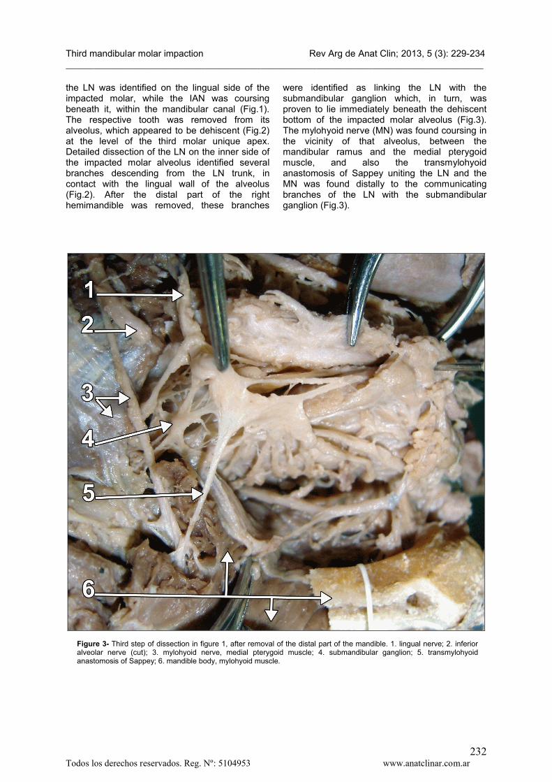
the LN was identified on the lingual side of the impacted molar, while the IAN was coursing beneath it, within the mandibular canal (Fig.1). The respective tooth was removed from its alveolus, which appeared to be dehiscent (Fig.2) at the level of the third molar unique apex. Detailed dissection of the LN on the inner side of the impacted molar alveolus identified several branches descending from the LN trunk, in contact with the lingual wall of the alveolus (Fig.2). After the distal part of the right hemimandible was removed, these branches
were identified as linking the LN with the submandibular ganglion which, in turn, was proven to lie immediately beneath the dehiscent bottom of the impacted molar alveolus (Fig.3). The mylohyoid nerve (MN) was found coursing in the vicinity of that alveolus, between the mandibular ramus and the medial pterygoid muscle, and also the transmylohyoid anastomosis of Sappey uniting the LN and the MN was found distally to the communicating branches of the LN with the submandibular ganglion (Fig.3).
Figure 3- Third step of dissection in figure 1, after removal of the distal part of the mandible. 1. lingual nerve; 2. inferior alveolar nerve (cut); 3. mylohyoid nerve, medial pterygoid muscle; 4. submandibular ganglion; 5. transmylohyoid anastomosis of Sappey; 6. mandible body, mylohyoid muscle.

Third mandibular molar impaction Rev Arg de Anat Clin; 2013, 5 (3): 229-234__________________________________________________________________________________________
Todos los derechos reservados. Reg. Nº: 5104953 www.anatclinar.com.ar233
DISCUSSION
In oral and maxillofacial surgery one of the most performed procedures is the extraction of impacted teeth. During surgery, mandibular third molars can be accidentally displaced to neighboring anatomic spaces. Distolingual angulated lower third molars are the most prone to be displaced (Ortakoğlu, 2002). Nerve injuries (IAN, LN) are frequently reported as related to third mandibular molars surgery. A previous study reported permanent sensory loss of the IAN and LN to be 0.6% and 1.1%, respectively, after surgical removal of impacted third molars (Jerjes et al, 2010). Although the figures are low, they may have legal implications. Patients should be warned regarding the risks of third molar surgery, including possible damage to the IAN and LN. Therefore, informed consent must be obtained before the procedure (Jerjes et al, 2010).From an anatomical viewpoint, the LN and IAN are not the only nerves closely related to an impacted mandibular third molar. The branches of the LN at this level, the MN, or an occasional anastomosis of Sappey should also be considered when local specific anatomy is discussed. The anastomosis of Sappey was found in 33.3% in the lateral sulcus of tongue, and it was described as “mylohyoid or sublingual curl” (Racz and Maros, 1981). This anastomosis was reported twice, in two different journals (Potu et al, 2010, Potu et al, 2009), and it was considered, due to its close relation with the third molar, more susceptible to injury during third mandibular molar surgery (Potu et al, 2010).Bleeding is a common and potential serious complication of lower third molar removal. Excessive bleeding was most encountered when both bone removal and tooth division were performed (Smith, 2013). The IAN passes through the inferior alveolar canal in the mandible, being accompanied by the inferior alveolar vessels. Anatomically, the inferior alveolar vein is the most superior structure in the canal. When rotary instruments are used, the bleeding will alert the surgeon that the superior aspect of the bony canal has been broken and the vein is injured. More profuse bleeding usually indicates damage to the inferior alveolar artery, which lies underneath the vein and superior to the nerve (Jerjes et al, 2010).Submandibular gland function was found altered after damage of chorda tympani (Chilla et al, 1982). However, we did not find reports on the submandibular gland secretory function damage in cases of removal of impacted mandibular third molars. When the submandibular ganglion is closely related to an impacted mandibular third
molar, morphofunctional changes should be assumed before taking any decision regarding surgical interventions. Further studies are thus needed to explore this direction.
REFERENCES
Bataineh AB. 2001. Sensory nerve impairment following mandibular third molar surgery. J Oral Maxillofac Surg 59: 1012-17.
Chilla R, Nicklatsch J, Arglebe C. 1982. Late sequelae of iatrogenic damage to chorda tympani nerve. Acta Otolaryngol 94: 461-65.
Gomes AC, Vasconcelos BC, de Oliveira e Silva ED, da Silva LC. 2005. Lingual nerve damage after mandibular third molar surgery: a randomized clinical trial. J Oral Maxillofac Surg 63: 1443-46.
Grandini SA, Barros VM, Salata LA, Rosa AL, Soares UN. 1993. Complications in exodontia--accidental dislodgment to adjacent anatomical areas. Braz Dent J 3: 103-12.
Hashemipour MA, Tahmasbi-Arashlow M, Fahimi-Hanzaei F. 2013. Incidence of impacted mandibular and maxillary third molars: a radiographic study in a Southeast Iran population. Med Oral Patol Oral Cir Bucal 18: e140-15.
Janakiraman EN, Alexander M, Sanjay P. 2010. Prospective analysis of frequency and contributing factors of nerve injuries following third-molar surgery. J Craniofac Surg 21: 784-6.
Jerjes W, Upile T, Shah P, Nhembe F, Gudka D, Kafas P. 2010. Risk factors associated with injury to the inferior alveolar and lingual nerves following third molar surgery-revisited. Oral Surg Oral Med Oral Pathol Oral Radiol Endod 109: 335-45.
Lauesen SR, Andreasen JO, Gerds TA, Christensen SS, Borum M, Hillerup S. 2013. Association between third mandibular molar impaction and degree of root development in adolescents. Angle Orthod 83: 3-9.
Neves FS, de Almeida SM, Boscolo FN, Haiter-Neto F, Alves MC, Crusoe-Rebello I. 2012. Risk assessment of inferior alveolar neuro-vascular bundle by multidetector computed tomography in extractions of third molars. Surg Radiol Anat 34: 619-24.
Ortakoğlu K, Okcu, K.M., Karasu, H.A., Günaydin, Y. 2002. Accidental Displacement of Impacted Third Molar into Lateral Pharyngeal Space. Turk J Med Sci 32: 431-33.
Potu BK, D'Silva SS, Thejodhar P, Jattanna NC.2010. An unusual communication between the mylohyoid and lingual nerves in man: its

Third mandibular molar impaction Rev Arg de Anat Clin; 2013, 5 (3): 229-234__________________________________________________________________________________________
Todos los derechos reservados. Reg. Nº: 5104953 www.anatclinar.com.ar234
significance in lingual nerve injury. Indian J Dent Res 21: 141-42.
Potu BK, Pulakunta T, Ray B, Rao MS, Bhat KM, D'Silva SS. 2009. Unusual communication between the lingual nerve and mylohyoid nerves in a South Indian male cadaver: its clinical significance. Rom J Morphol Embryol 50: 145-46.
Racz L, Maros T. 1981. The anatomic variants of the lingual nerve in human. Anat Anz 149: 64-71.
Rusu MC, Nimigean V, Podoleanu L, Ivascu RV, Niculescu MC. 2008. Details of the intralingual topography and morphology of the lingual nerve. Int J Oral Maxillofac Surg 37: 835-39.
Sivolella S, Boccuzzo G, Gasparini E, De Conti G, Berengo M. 2012. Assessing the need for computed tomography for lower-third-molar extraction: a survey among 322 dentists. Radiol Med 117: 112-24.
Smith WP. 2013. The relative risk of neuro-sensory deficit following removal of mandibular third molar teeth: the influence of radiography and surgical technique. Oral Surg Oral Med Oral Pathol Oral Radiol 115: 18-24.
Xu GZ, Yang C, Fan XD, Yu CQ, Cai XY, Wang Y. 2013. Anatomic relationship betweenimpacted third mandibular molar and the mandibular canal as the risk factor of inferior alveolar nerve injury. Br J Oral Maxillofac Surg;10.1016/j.bjoms. 2013.01.011