dissertation - othes.univie.ac.atothes.univie.ac.at/13818/1/2011-01-10_0003810.pdf · 3 summary...
TRANSCRIPT
DISSERTATION
Titel der Dissertation
Generation of a polymimotope vaccine against malignant melanoma
angestrebter akademischer Grad
Doktor/in der Naturwissenschaften (Dr. rer.nat.) Verfasserin / Verfasser: Mag. Sonja Gaier
Dissertationsgebiet (lt. Stu-dienblatt):
Genetik – Mikrobiologie (Stzw)
Betreuerin / Betreuer: ao. Univ.-Prof. Dr. Heimo Breiteneder
Wien, im Jänner 2011

Danksagung
Der experimentelle Teil dieser Arbeit wurde in der Arbeitsgruppe von Priv. Doz. Dr.
Stefan Wagner an der Abteilung von ao. Univ.-Prof. Dr. Heimo Breiteneder am Institut
für Pathophysiologie und Allergieforschung des Zentrum für Pathophysiologie,
Infektiologie und Immunologie der Medizinischen Universität Wien durchgeführt.
Ao. Univ.-Prof. Dr. Heimo Breiteneder und PD Dr. Stefan Wagner danke ich für die
Ermöglichung meiner Arbeit an diesem Institut und für die wissenschaftliche Betreuung
meiner Dissertation.
Bei der Österreichischen Akademie der Wissenschaften (ÖAW) bedanke ich mich für
die finanzielle Unterstützung meiner Dissertation durch ein DOC-fFORTE Stipendium.
Ich danke auch ao. Univ.-Prof. DI Dr. Reingard Grabherr und PD DDr. Angelika
Riemer für die Begutachtung dieser Arbeit.
Ein ganz großes Dankeschön an meine LaborkollegInnen des „Lab 16“!
Danke Nina für deine Unterstützung in der Zellkultur und bei den allgemeinen
Belangen des Laboralltages, Gerlinde für deine grenzenlose Geduld (v.a. mit mir) bei
den Mausexperimenten, und natürlich Christian und Merima für euren wissenschaft-
lichen Beistand.
Ein riesiges DANKE an Ursula und Julia! Danke, dass ich mit all meinen kleinen und
großen Problemchen zu euch kommen kann.
DANKE an meine Familie und meine Freunde! Danke, dass ihr alle für mich da seid,
mich unterstützt und mir Kraft gebt!

Table of Contents Zusammenfassung..................................................................................... 1 Summary .................................................................................................... 3 Chapter I .................................................................................................... 5 Background
Malignant melanoma ......................................................................................................7 Melanoma associated antigens........................................................................................10 High molecular weight-melanoma associated antigen ...................................................11 Therapy of melanoma .....................................................................................................15 Monoclonal antibodies in clinical use ............................................................................17 Monoclonal antibodies directed to HMW-MAA............................................................19 Cancer vaccines – from peptide to mimotope vaccines..................................................21 Multi-epitope vaccines....................................................................................................25 Aim of this thesis ............................................................................................................26 References.......................................................................................................................27 Chapter II................................................................................................... 35 Successful selection of mimotopes from phage-displayed libraries strongly depends on the selection strategy
Abstract...........................................................................................................................37 Introduction.....................................................................................................................39 Materials and Methods....................................................................................................41 Results.............................................................................................................................47 Discussion.......................................................................................................................51 Tables..............................................................................................................................55 Figures ............................................................................................................................57 References.......................................................................................................................65 Chapter III ................................................................................................. 69 Specificity of mimotope-induced anti-high molecular weight-melanoma associated antigen (HMW-MAA) antibodies does not ensure biological activity
Abstract...........................................................................................................................71 Introduction.....................................................................................................................73 Materials and Methods....................................................................................................75 Results.............................................................................................................................81 Discussion.......................................................................................................................85 Figures ............................................................................................................................89 References.......................................................................................................................95

Chapter IV Concluding Remarks..................................................................................................101 References ....................................................................................................................104 Appendix Curriculum Vitae .......................................................................................................107 List of Publications.....................................................................................................109
1
Zusammenfassung “Peptidmimics” von konformationellen Epitopen eines Tumorantigens werden auch
als Mimotope bezeichnet und stellen vielversprechende Kandidaten für die Entwicklung
von Tumorvakzinen dar. Derartige Mimotopvakzine induzieren eine Tumorantigen-
spezifische Immunantwort. Kürzlich konnte für das maligne Melanom gezeigt werden,
dass durch Vakzinierung von Kaninchen mit einem an Tetanustoxoid gekoppelten
Mimotop des HMW-MAA (“high molecular weight-melanoma associated antigen”)
Antikörper gebildet wurden, die das Tumorwachstum in einem Melanommausmodell
hemmen konnten. HMW-MAA wurde als Zielantigen gewählt, da es von vielen
Melanomzellen exprimiert wird und in normalen Geweben fast nicht vorkommt.
Das Ziel dieser Dissertation war die Entwicklung einer Polymimotopvakzine, die
eine starke humorale Immunantwort gegen mehrere Epitope des HMW-MAA
induzieren sollte. Verschiedene Studien haben gezeigt, dass Vakzine, die gegen mehr
als ein Epitop eines Tumorantigens gerichtet sind, eine höhere Erfolgsrate aufweisen.
Der Effekt der induzierten Antikörper sollte sowohl in vitro (Tumorproliferation,
antikörperabhängige, zellvermitteltete Zytotoxizität, komplementabhängige Zyto-
toxizität) als auch in vivo (Melanommausmodell: C57BL/6 Mäuse mit einem
subkutanen Tumor der HMW-MAA transfizierten Mausmelanomzelllinie B16F10)
untersucht werden.
Mittels einer linearen pIII-12mer Phagenpeptidbank wurden Peptidliganden für eine
Reihe von anti-HMW-MAA monoklonalen Antikörpern (mAbs VT80.12, VF1-TP43,
VF1-TP34, 149.53 und 225.28S F(ab')2) selektiert. Diese Antikörper erkennen
unterschiedliche Epitope des HMW-MAA, weshalb sie sich gegenseitig in ihrer
Bindung nicht inhibieren. Es wurden verschiedene Panningstrategien angewendet
(Panning mit Oberflächenimmobilisierung und in Lösung mit Protein G- bzw.
Streptavidin-Bindung). Für jeden mAb wurde mindestens ein Peptidligand identifiziert.
Es waren aber nur die Peptide, die für die mAbs VT80.12 und VF1-TP43 identifiziert
wurden, auch in der Lage, die Bindung der entsprechenden mAbs an das HMW-MAA
zu inhibieren. Daher wurden diese beiden Peptide jeweils an KLH („keyhole limpet
hemocyanin“) als immunogenen Träger gekoppelt. Die in Kaninchen induzierten
Antikörper zeigten einen starken Hintergrund verursacht durch die anti-KLH
Antikörper. Durch die Immunisierung von BALB/c Mäusen konnte dieses Problem
gelöst werden. Beide Peptide induzierten Peptid-spezifische Antikörper, aber nur das
2
VT80.12-Peptid induzierte auch anti-HMW-MAA Antikörper. Diese Antikörper waren
jedoch nicht in der Lage, das Wachstum der HMW-MAA exprimierenden humanen
Melanomzelllinie 518A2 in vitro zu inhibieren.
In dieser Arbeit konnten sehr viele neue Informationen für die Selektion von
Mimotopen gewonnen werden, jedoch konnten noch keine idealen Mimotopkandidaten
für die Polymimotopvakzine identifiziert und im Melanommausmodell getestet werden.
3
Summary Peptide mimics, also called mimotopes, of conformational epitopes of tumor antigens
are promising candidates for the design of anti-tumor vaccines. Such mimotope
vaccines stimulate a tumor antigen specific immune response. For melanoma, it was
recently demonstrated that vaccination of rabbits with a mimotope of the high molecular
weight-melanoma associated antigen (HMW-MAA) coupled to tetanus toxoid induced
antibodies that suppressed tumor growth in a melanoma xenotransplant severe
combined immunodeficiency mouse model. The HMW-MAA was selected as target
antigen because it is highly expressed on melanoma cells and has a restricted
distribution in normal tissues.
The aim of this thesis was to develop a polymimotope vaccine that would induce a
strong humoral immune response against several epitopes of the HMW-MAA. Several
studies have demonstrated that vaccines directed against multiple epitopes of a tumor
antigen have a higher success rate. The effect of the induced antibodies were then to be
tested in vitro (tumor proliferation, antibody-dependent cell-mediated cytotoxicity,
complement-dependent cytotoxicity) and in vivo (melanoma mouse model: C57BL/6
mice harboring a subcutaneous tumor of the mouse melanoma cell line B16F10
transfected with the HMW-MAA).
Peptide ligands for a panel of anti-HMW-MAA monoclonal antibodies (mAbs)
– VT80.12, VF1-TP43, VF1-TP34, 149.53, and 225.28S F(ab')2 – which recognize
distinct epitopes on the HMW-MAA and do not cross-inhibit each other were selected
using a linear pIII-12mer phage display peptide library by surface panning as well as
solution-phase panning with protein G or streptavidin capture. Peptide ligands were
identified for each mAb, but only one peptide identified for each of the mAbs VT80.12
and VF1-TP43 was able to inhibit the binding of the respective mAb to the HMW-
MAA. These two peptides were then coupled to keyhole limpet hemocyanin (KLH) as
immunogenic carrier and used for immunization of rabbits. Due to the high background
of the KLH-induced antibodies, further immunizations were performed in BALB/c
mice. Both peptides induced a peptide specific immune response but only the VT80.12-
peptide induced anti-HMW-MAA antibodies. However, these antibodies did not
suppress the proliferation of the HMW-MAA expressing human melanoma cell line
518A2 in vitro.

4
In summary, we have produced a lot of new methodologic know-how on the
selection of mimotopes but could not yet identify ideal mimotope candidates which
could be tested as parts of a polymimotope vaccine in the melanoma mouse model.

5
Chapter I
Background

6
Chapter I – Background
7
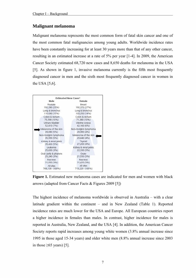
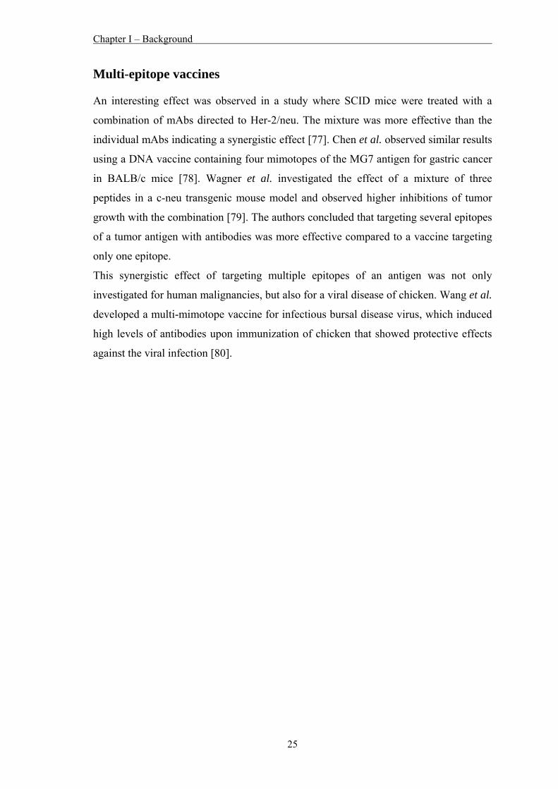
Malignant melanoma Malignant melanoma represents the most common form of fatal skin cancer and one of
the most common fatal malignancies among young adults. Worldwide incidence rates
have been constantly increasing for at least 30 years more than that of any other cancer,
resulting in an estimated increase at a rate of 5% per year [1-4]. In 2009, the American
Cancer Society estimated 68,720 new cases and 8,650 deaths for melanoma in the USA
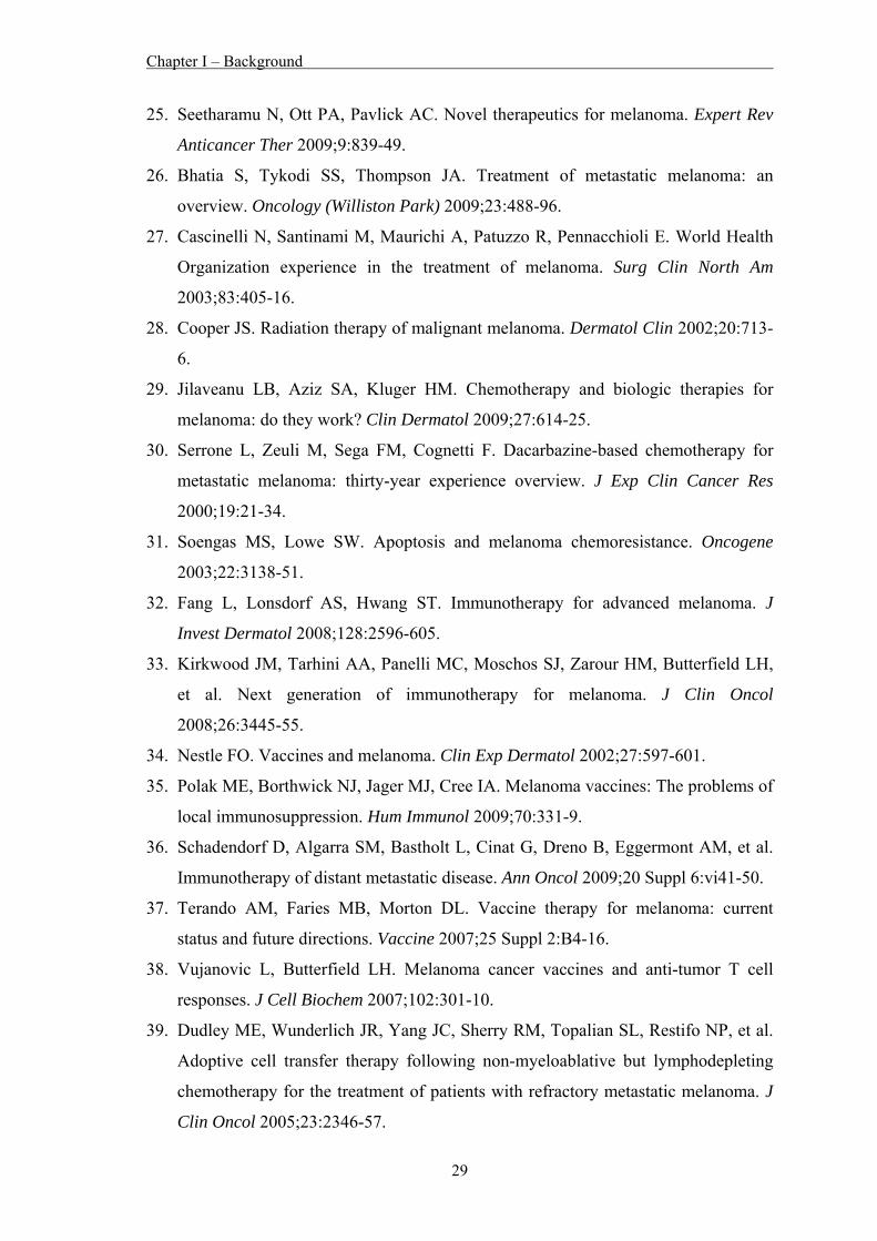
[5]. As shown in figure 1, invasive melanoma currently is the fifth most frequently
diagnosed cancer in men and the sixth most frequently diagnosed cancer in women in
the USA [5,6].
Figure 1. Estimated new melanoma cases are indicated for men and women with black
arrows (adapted from Cancer Facts & Figures 2009 [5])
The highest incidence of melanoma worldwide is observed in Australia – with a clear
latitude gradient within the continent – and in New Zealand (Table 1). Reported
incidence rates are much lower for the USA and Europe. All European countries report
a higher incidence in females than males. In contrast, higher incidence for males is
reported in Australia, New Zealand, and the USA [4]. In addition, the American Cancer
Society reports rapid increases among young white women (3.8% annual increase since
1995 in those aged 15-34 years) and older white men (8.8% annual increase since 2003
in those ≥65 years) [5].
Chapter I – Background
8
Table 1. Comparative melanoma incidence for selected states and countries worldwide
for the time period 1998-2002 (adapted from MacKie et al. [4])
Incidence (per 105 subjects) Country Male Female
Australia Queensland 55.8 41.1 New South Wales 38.5 26.5 Victoria 27.3 23.4 New Zealand 34.8 31.4 US SEER 14 registries 19.4 14.4 Switzerland, Vaud 16.6 19.6 Norway 14.2 14.6 Sweden 11.9 12.1 Denmark 11.9 14.1 Latvia 3.2 4.2 Lithuania 3.7 5.2 Estonia 5.3 6.6 Belarus 2.7 3.5 Serbia 3.8 4.8
Although diagnosis of melanoma has significantly improved [7], leading to an increase
in the percentage of patients being diagnosed with “thin” melanomas (≤1 mm) and a
decrease in the percentage of patients diagnosed with “thick” melanomas (>4 mm) [8],
the mortality rate for melanoma has been continually increasing over the past decade
[1,4].
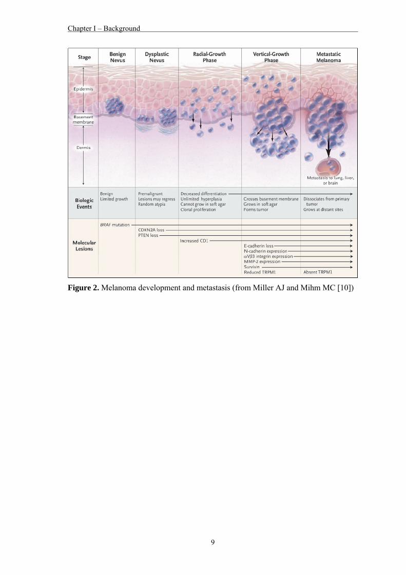
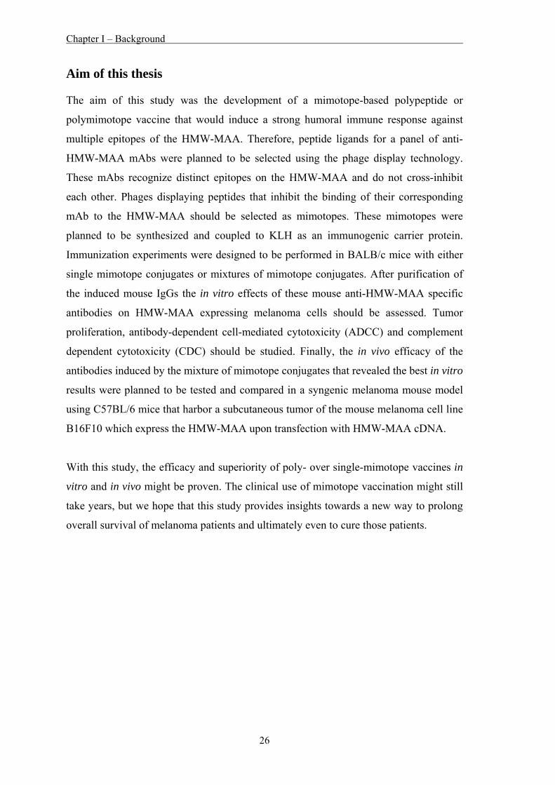
Melanomas generally originate from benign nevi, which are clonally expanded
melanocytes that proliferate abnormally but do not progress [9]. The overcoming of
senescence leads to a dysplastic nevus, which can progress to a stage of spreading with
low invasive potential (radial growth phase). The next stage (vertical growth phase)
often results in metastases to local lymph nodes and eventually to distant sites,
including other skin sites, pleura, lungs, liver and brain [9]. A schematic view of this
process is shown in figure 2.
Chapter I – Background
9
Figure 2. Melanoma development and metastasis (from Miller AJ and Mihm MC [10])
Chapter I – Background
10
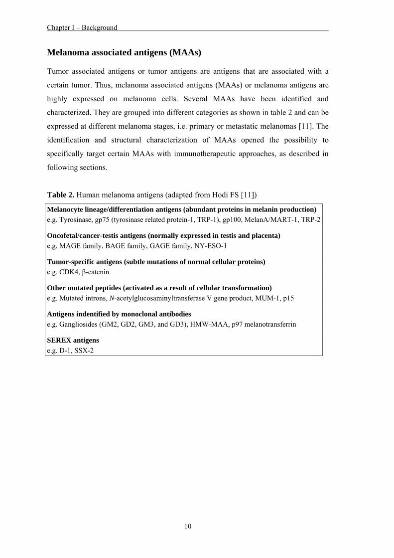
Melanoma associated antigens (MAAs) Tumor associated antigens or tumor antigens are antigens that are associated with a
certain tumor. Thus, melanoma associated antigens (MAAs) or melanoma antigens are
highly expressed on melanoma cells. Several MAAs have been identified and
characterized. They are grouped into different categories as shown in table 2 and can be
expressed at different melanoma stages, i.e. primary or metastatic melanomas [11]. The
identification and structural characterization of MAAs opened the possibility to
specifically target certain MAAs with immunotherapeutic approaches, as described in
following sections.
Table 2. Human melanoma antigens (adapted from Hodi FS [11])
Melanocyte lineage/differentiation antigens (abundant proteins in melanin production) e.g. Tyrosinase, gp75 (tyrosinase related protein-1, TRP-1), gp100, MelanA/MART-1, TRP-2
Oncofetal/cancer-testis antigens (normally expressed in testis and placenta) e.g. MAGE family, BAGE family, GAGE family, NY-ESO-1
Tumor-specific antigens (subtle mutations of normal cellular proteins) e.g. CDK4, β-catenin
Other mutated peptides (activated as a result of cellular transformation) e.g. Mutated introns, N-acetylglucosaminyltransferase V gene product, MUM-1, p15
Antigens indentified by monoclonal antibodies e.g. Gangliosides (GM2, GD2, GM3, and GD3), HMW-MAA, p97 melanotransferrin
SEREX antigens e.g. D-1, SSX-2
Chapter I – Background
11
High molecular weight-melanoma associated antigen (HMW-MAA) One of the MAAs is the high molecular weight-melanoma associated antigen (HMW-
MAA), also known as the chondroitin sulfate proteoglycan 4 (CSPG4) or melanoma
chondroitin sulfate proteoglycan (MCSP). This MAA was originally identified with
murine monoclonal antibodies (mAbs) on the surface of human melanoma cells at the
beginning of the 1980’s [12,13].
The HMW-MAA gene is located on human chromosome 15 [14] and encodes a 2322
amino acid (aa) long protein (UniProt accession number: Q6UVK1). A schematic view
of the HMW-MAA sequence is shown in figure 3. The first 29 aa residues display a
signal peptide and the remaining aa the core protein which contains three major domain
structures: a large extracellular domain (1), a short hydrophobic transmembrane region
(2), and a short cytoplasmic tail (3). There are 15 potential N-linked glycosylation sites
throughout the extracellular domain [1].
The expression of HMW-MAA seems to be regulated by a classical 5’ CpG island
promoter that precedes the ten exons comprising the human HMW-MAA gene [15].
The level of methylation of the promoter predicts HMW-MAA expression in vivo.
Unmethylated promoter DNA leads to HMW-MAA expression in human melanoma
cells, whereas methylation of promoter DNA results in the absence of HMW-MAA
expression in normal human lymphocytes [15].
Figure 3. Analysis of the full-length aa sequence of the HMW-MAA protein (from
Campoli et al. [1])
Chapter I – Background
12
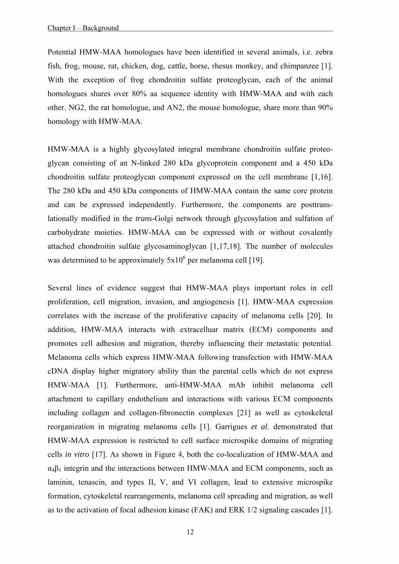
Potential HMW-MAA homologues have been identified in several animals, i.e. zebra
fish, frog, mouse, rat, chicken, dog, cattle, horse, rhesus monkey, and chimpanzee [1].
With the exception of frog chondroitin sulfate proteoglycan, each of the animal
homologues shares over 80% aa sequence identity with HMW-MAA and with each
other. NG2, the rat homologue, and AN2, the mouse homologue, share more than 90%
homology with HMW-MAA.
HMW-MAA is a highly glycosylated integral membrane chondroitin sulfate proteo-
glycan consisting of an N-linked 280 kDa glycoprotein component and a 450 kDa
chondroitin sulfate proteoglycan component expressed on the cell membrane [1,16].
The 280 kDa and 450 kDa components of HMW-MAA contain the same core protein
and can be expressed independently. Furthermore, the components are posttrans-
lationally modified in the trans-Golgi network through glycosylation and sulfation of
carbohydrate moieties. HMW-MAA can be expressed with or without covalently
attached chondroitin sulfate glycosaminoglycan [1,17,18]. The number of molecules
was determined to be approximately 5x10⁶ per melanoma cell [19].
Several lines of evidence suggest that HMW-MAA plays important roles in cell
proliferation, cell migration, invasion, and angiogenesis [1]. HMW-MAA expression
correlates with the increase of the proliferative capacity of melanoma cells [20]. In
addition, HMW-MAA interacts with extracelluar matrix (ECM) components and
promotes cell adhesion and migration, thereby influencing their metastatic potential.
Melanoma cells which express HMW-MAA following transfection with HMW-MAA
cDNA display higher migratory ability than the parental cells which do not express
HMW-MAA [1]. Furthermore, anti-HMW-MAA mAb inhibit melanoma cell
attachment to capillary endothelium and interactions with various ECM components
including collagen and collagen-fibronectin complexes [21] as well as cytoskeletal
reorganization in migrating melanoma cells [1]. Garrigues et al. demonstrated that
HMW-MAA expression is restricted to cell surface microspike domains of migrating
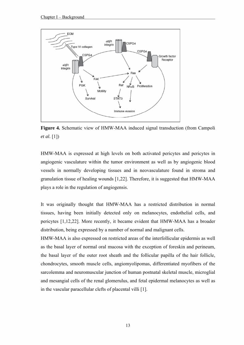
cells in vitro [17]. As shown in Figure 4, both the co-localization of HMW-MAA and
α4β1 integrin and the interactions between HMW-MAA and ECM components, such as
laminin, tenascin, and types II, V, and VI collagen, lead to extensive microspike
formation, cytoskeletal rearrangements, melanoma cell spreading and migration, as well
as to the activation of focal adhesion kinase (FAK) and ERK 1/2 signaling cascades [1].
Chapter I – Background
13
Figure 4. Schematic view of HMW-MAA induced signal transduction (from Campoli
et al. [1])
HMW-MAA is expressed at high levels on both activated pericytes and pericytes in
angiogenic vasculature within the tumor environment as well as by angiogenic blood
vessels in normally developing tissues and in neovasculature found in stroma and
granulation tissue of healing wounds [1,22]. Therefore, it is suggested that HMW-MAA
plays a role in the regulation of angiogensis.
It was originally thought that HMW-MAA has a restricted distribution in normal
tissues, having been initially detected only on melanocytes, endothelial cells, and
pericytes [1,12,22]. More recently, it became evident that HMW-MAA has a broader
distribution, being expressed by a number of normal and malignant cells.
HMW-MAA is also expressed on restricted areas of the interfollicular epidermis as well
as the basal layer of normal oral mucosa with the exception of foreskin and perineum,
the basal layer of the outer root sheath and the follicular papilla of the hair follicle,
chondrocytes, smooth muscle cells, angiomyolipomas, differentiated myofibers of the
sarcolemma and neuromuscular junction of human postnatal skeletal muscle, microglial
and mesangial cells of the renal glomerulus, and fetal epidermal melanocytes as well as
in the vascular paracellular clefts of placental villi [1].
Chapter I – Background
14
HMW-MAA has been found to be expressed on more than 90% of surgically removed
benign nevi and melanoma lesions, with a limited degree of intra- and interlesional
heterogeneity [1]. HMW-MAA expression has also been found in astrocytomas,
gliomas, neuroblastomas, squamous cell carcinoma of the head and neck, basal breast
cancer, mesothelioma, pancreatic carcinoma, some types of renal cell carcinoma,
chordoma, chondrosarcoma, soft tissue sarcomas, and hematologic malignancies
(expression on blast cells in both childhood and adult acute lymphoblastic leukemia and
childhood acute myeloid leukemia) as well as cancer stem cells [1].
Chapter I – Background
15
Therapy of melanoma While most early stage melanomas (thin primary tumors; stage 0-II) are highly curable
with optimal surgical excision, patients with advanced stage melanoma (lymph node
involvement and metastases; stage III-IV) have a poor prognosis and often succumb,
due to failure of metastasis control [1,3,23-25].
Metastatic melanoma is highly resistant to conventional therapy, including standard
chemotherapy and radiotherapy. The response rate for conventional agents, like
cisplatin, dacarbazine or temozolomide, as single agent or in combination, is only 15-
25% [26-31]. Therefore, the need to develop and apply novel and improved therapeutic
strategies for the treatment of melanoma has been emphasized.
As melanoma is among the most immunogenic of all solid caners, a lot of different
immunotherapeutic approaches to treat melanoma have been investigated in preclinical
and clinical studies and have been the focus of several review articles in the recent past
[3,9,25,26,32-38]. An overview describing the general mechanisms of the investigated
strategies is shown in table 3.
Table 3. Approaches for the treatment of melanoma (adapted from Nestle FO [34])
Active (= Vaccination) Whole cell vaccines (e.g. autologous or allogeneic melanoma cell lines)Antigen-based vaccines (e.g. peptides, protein, DNA, RNA) Adjuvant-based vaccines (e.g. dendritic cells)
Passive Adoptive cell transfer (e.g. tumor-infiltrating lymphocytes (TILs)) Antibodies (e.g. anti-CTLA-4) Inhibitors (targeting molecules in signal transduction pathways) Adjuvants (e.g. Toll-like receptor agonists) Cytokines (e.g. IL-2, IFN-α)
So far, only the immune-modulating agents interferon-α (IFN-α) and interleukin-2 (IL-
2) are approved by the US Food and Drug Administration (FDA). IFN-α is administered
for the treatment of high-risk melanoma patients. IL-2 is used for the treatment of
patients with metastatic melanoma [25,32,33]. For both cytokines, the overall response
rates remain low and the benefits must be balanced against several serious side-effects,
Chapter I – Background
16
such as flu-like symptoms, anorexia, fatigue, depression, thyroid dysfunction, skin rash,
altered blood cell counts, and liver toxicity [9,33].
Noteworthy, the development of a vaccine that would show significant clinical benefit
in melanoma has not been successful by now, as clinical responses remain at a low rate.
However, the extent of research activity in the field and a number of novel approaches
indicate that such an approach remains attractive [36].
Encouraging results were obtained by using immunostimulatory mAbs directed to
immune-receptor molecules to increase immune responses. MAbs of this type (e.g. anti-
CTLA-4, anti-CD137 and anti-CD40) are currently studied in clinical trials [25].
Nevertheless, complications such as autoimmunity and systemic inflammation are
problematic side effects associated with these therapeutics.
In addition, adoptive cell transfer of ex vivo expanded autologous tumor reactive
lymphocytes in combination with lymphodepletion or myeloablation by the use of
chemotherapy or radiochemotherapy resulted in 50-70% objective clinical response
[39,40]. However, this are time-consuming and cost-intensive treatments, which are
therefore at the moment not routinely applicable in the general clinical practice.
Chapter I – Background
17
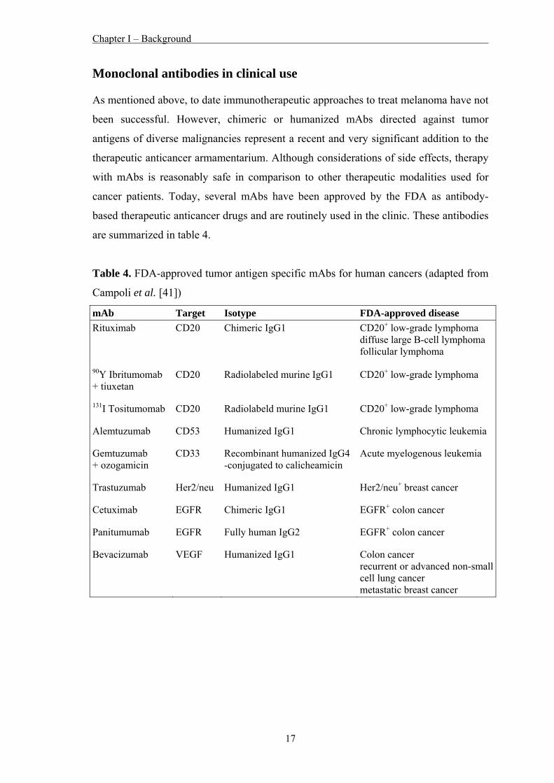
Monoclonal antibodies in clinical use As mentioned above, to date immunotherapeutic approaches to treat melanoma have not
been successful. However, chimeric or humanized mAbs directed against tumor
antigens of diverse malignancies represent a recent and very significant addition to the
therapeutic anticancer armamentarium. Although considerations of side effects, therapy
with mAbs is reasonably safe in comparison to other therapeutic modalities used for
cancer patients. Today, several mAbs have been approved by the FDA as antibody-
based therapeutic anticancer drugs and are routinely used in the clinic. These antibodies
are summarized in table 4.
Table 4. FDA-approved tumor antigen specific mAbs for human cancers (adapted from
Campoli et al. [41])
mAb Target Isotype FDA-approved disease Rituximab CD20 Chimeric IgG1 CD20+ low-grade lymphoma
diffuse large B-cell lymphoma follicular lymphoma
90Y Ibritumomab + tiuxetan
CD20 Radiolabeled murine IgG1 CD20+ low-grade lymphoma
131I Tositumomab CD20 Radiolabeld murine IgG1 CD20+ low-grade lymphoma
Alemtuzumab CD53 Humanized IgG1 Chronic lymphocytic leukemia
Gemtuzumab + ozogamicin
CD33 Recombinant humanized IgG4 -conjugated to calicheamicin
Acute myelogenous leukemia
Trastuzumab Her2/neu Humanized IgG1 Her2/neu+ breast cancer
Cetuximab EGFR Chimeric IgG1 EGFR+ colon cancer
Panitumumab EGFR Fully human IgG2 EGFR+ colon cancer
Bevacizumab VEGF Humanized IgG1 Colon cancer recurrent or advanced non-small cell lung cancer metastatic breast cancer
Chapter I – Background
18
The huge costs of the production of mAbs are the greatest disadvantage of this kind of
therapy. Doses used in humans are typically in the mg per kg body weight range, and
most regimens involve repetitive applications over longer time periods. Therefore,
expensive GMP produced batches are required for quality control, toxicology and
clinical trials. An example for the high costs is given in a recent publication where
clinicians from the Norfolk and Norwich University Hospital estimated that more than
€ 2.9 millions were necessary per year to make Herceptin® (Trastuzumab) available to
75 patients who might be eligible for the treatment [42].
Chapter I – Background
19
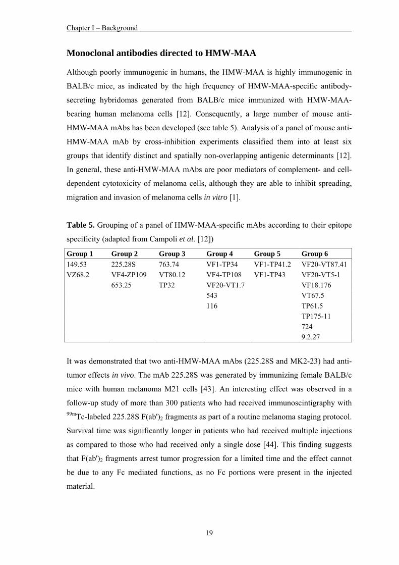
Monoclonal antibodies directed to HMW-MAA Although poorly immunogenic in humans, the HMW-MAA is highly immunogenic in
BALB/c mice, as indicated by the high frequency of HMW-MAA-specific antibody-
secreting hybridomas generated from BALB/c mice immunized with HMW-MAA-
bearing human melanoma cells [12]. Consequently, a large number of mouse anti-
HMW-MAA mAbs has been developed (see table 5). Analysis of a panel of mouse anti-
HMW-MAA mAb by cross-inhibition experiments classified them into at least six
groups that identify distinct and spatially non-overlapping antigenic determinants [12].
In general, these anti-HMW-MAA mAbs are poor mediators of complement- and cell-
dependent cytotoxicity of melanoma cells, although they are able to inhibit spreading,
migration and invasion of melanoma cells in vitro [1].
Table 5. Grouping of a panel of HMW-MAA-specific mAbs according to their epitope
specificity (adapted from Campoli et al. [12])
Group 1 Group 2 Group 3 Group 4 Group 5 Group 6 149.53 225.28S 763.74 VF1-TP34 VF1-TP41.2 VF20-VT87.41 VZ68.2 VF4-ZP109 VT80.12 VF4-TP108 VF1-TP43 VF20-VT5-1 653.25 TP32 VF20-VT1.7 VF18.176 543 VT67.5 116 TP61.5 TP175-11 724 9.2.27
It was demonstrated that two anti-HMW-MAA mAbs (225.28S and MK2-23) had anti-
tumor effects in vivo. The mAb 225.28S was generated by immunizing female BALB/c
mice with human melanoma M21 cells [43]. An interesting effect was observed in a
follow-up study of more than 300 patients who had received immunoscintigraphy with 99mTc-labeled 225.28S F(ab')2 fragments as part of a routine melanoma staging protocol.
Survival time was significantly longer in patients who had received multiple injections
as compared to those who had received only a single dose [44]. This finding suggests
that F(ab')2 fragments arrest tumor progression for a limited time and the effect cannot
be due to any Fc mediated functions, as no Fc portions were present in the injected
material.
Chapter I – Background
20
Furthermore, Hafner et al. were able to demonstrate that the mAb 225.28S was able to
suppress tumor growth in a human melanoma xenotransplant model in severe combined
immunodeficiency (SCID) mice [45].
In addition, stage IV melanoma patients had a significant increase of survival
prolongation upon immunization with the anti-idiotypic mAb MK2-23, which mimics
the epitope of the anti-HMW-MAA mAb 763.74 [46,47].
Chapter I – Background
21
Cancer vaccines – from peptide to mimotope vaccines Passive administration with tumor antigen specific mAbs, like the treatment with the
Herceptin® antibody, has produced promising results in the clinics. However, a number
of concerns remains such as repeated treatments and associated costs, limited duration
of therapeutic effectiveness, and possible undesired immunogenicity. Therefore, a
therapeutic approach capable of inducing active specific immunity would offer
sustained protection at a lower cost, preventive therapy and long term immunity.
One of the great problems in developing cancer vaccines is that tumor associated self-
antigens do not induce an immune response, since the corresponding reactive B and T
cell clones have been eliminated during the establishment of self-tolerance. Especially
for melanoma, this kind of malignancy seems to be very efficient regarding tumor
escape/evasion of the host immune responses via tumor-induced immunosuppression.
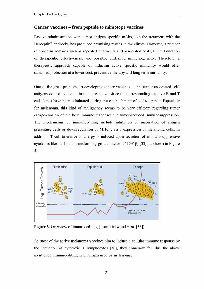
The mechanisms of immunoediting include inhibition of maturation of antigen
presenting cells or downregulation of MHC class I expression of melanoma cells. In
addition, T cell tolerance or anergy is induced upon secretion of immunosuppressive
cytokines like IL-10 and transforming growth factor-β (TGF-β) [33], as shown in Figure
5.
Figure 5. Overview of immunoediting (from Kirkwood et al. [33])
As most of the active melanoma vaccines aim to induce a cellular immune response by
the induction of cytotoxic T lymphocytes [38], they somehow fail due the above
mentioned immunoediting mechanisms used by melanoma.
Chapter I – Background
22
Therefore, and since some tumor antigen specific mAbs are successfully used in the
clinics to treat different cancers, several strategies have been developed to overcome
this problem via focusing on B cells. As an example, research groups have predicted
putative B cell epitopes of the tumor antigen Her-2/neu and could demonstrate that
some of these B cell epitopes coupled to an immunogenic carrier protein induced a
humoral immune response directed to the tumor antigen with anti-tumor activity
[48,49].
Cancer vaccines designed to elicit an antibody response that targets antigenic sites on a
tumor antigen must closely mimic the three-dimensional structure of the corresponding
region on the antigen. Studies of the three-dimensional structures of antigen-antibody
complexes showed that antigenic epitopes are conformational and vaccine design
should consider such parameters to elicit antibodies of high affinity.
Molecular mimicry provides a way to generate vaccine components that elicit and/or
enhance an immune response against tumor antigens that are mostly non-mutated self-
antigens and therefore poorly or non immunogenic in patients.
The benefits of synthetic peptides and the knowledge of the effects of epitope mimicry
led to the development of mimotopes which are peptides that mimic the structure of the
epitope of an antigen that is defined by an antibody. The term “mimotope” was
introduced by Geysen and his colleagues in the 1980’s [50]. Easy identification of
mimotopes was made feasible by the application of the so called “phage display”
technology, which was developed by George P. Smith [51].
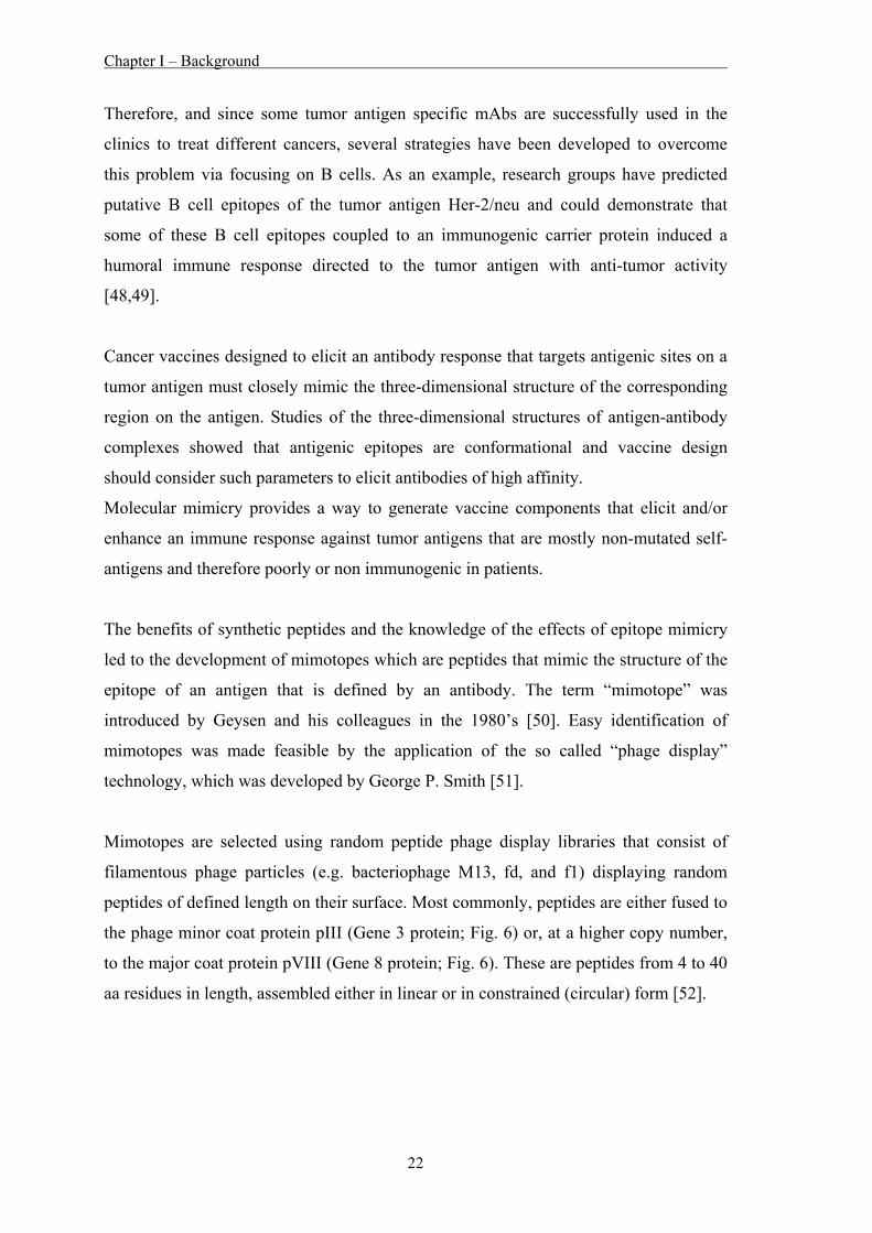
Mimotopes are selected using random peptide phage display libraries that consist of
filamentous phage particles (e.g. bacteriophage M13, fd, and f1) displaying random
peptides of defined length on their surface. Most commonly, peptides are either fused to
the phage minor coat protein pIII (Gene 3 protein; Fig. 6) or, at a higher copy number,
to the major coat protein pVIII (Gene 8 protein; Fig. 6). These are peptides from 4 to 40
aa residues in length, assembled either in linear or in constrained (circular) form [52].
Chapter I – Background
23
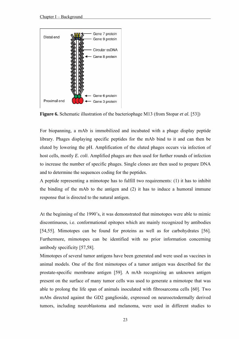
Figure 6. Schematic illustration of the bacteriophage M13 (from Stopar et al. [53])
For biopanning, a mAb is immobilized and incubated with a phage display peptide
library. Phages displaying specific peptides for the mAb bind to it and can then be
eluted by lowering the pH. Amplification of the eluted phages occurs via infection of
host cells, mostly E. coli. Amplified phages are then used for further rounds of infection
to increase the number of specific phages. Single clones are then used to prepare DNA
and to determine the sequences coding for the peptides.
A peptide representing a mimotope has to fulfill two requirements: (1) it has to inhibit
the binding of the mAb to the antigen and (2) it has to induce a humoral immune
response that is directed to the natural antigen.
At the beginning of the 1990’s, it was demonstrated that mimotopes were able to mimic
discontinuous, i.e. conformational epitopes which are mainly recognized by antibodies
[54,55]. Mimotopes can be found for proteins as well as for carbohydrates [56].
Furthermore, mimotopes can be identified with no prior information concerning
antibody specificity [57,58].
Mimotopes of several tumor antigens have been generated and were used as vaccines in
animal models. One of the first mimotopes of a tumor antigen was described for the
prostate-specific membrane antigen [59]. A mAb recognizing an unknown antigen
present on the surface of many tumor cells was used to generate a mimotope that was
able to prolong the life span of animals inoculated with fibrosarcoma cells [60]. Two
mAbs directed against the GD2 ganglioside, expressed on neuroectodermally derived
tumors, including neuroblastoma and melanoma, were used in different studies to
Chapter I – Background
24
generate mimotopes of the antigen. The vaccine-induced antibodies exhibited protection
against human GD2-positive melanoma growth in the SCID mouse xenograft model
[61] or induced a reduction of spontaneous liver metastases [62].
MAbs used in the clinic also served as sources to generate mimotopes of tumor
antigens. Mimotopes of Her-2/neu were generated using trastuzumab [63,64], of
epidermal growth factor receptor (EGFR) using cetuximab [65,66], and of CD20 using
rituximab [67-70]. Some of these mimotopes [65-67] induced a humoral immune
response able to inhibit tumor cell growth in vitro and are good candidates for active
immunotherapy.
For melanoma, Hafner et al. described mimotopes of the melanoma cell-adhesion
molecule (Mel-CAM), which induced Mel-CAM specific antibodies in BALB/c mice.
These antibodies showed an enhanced proliferation of the Mel-CAM positive human
melanoma cell line MelJuSo in vitro. However, these antibodies mediated low but
specific cell lysis of MelJuSo cells due to complement dependent cytotoxicity [71,72].
In regard to HMW-MAA, mimotopes were successfully identified using the mAb
225.28S [73,74] and the mAb 763.74 [75], respectively. These mimotopes induced
HMW-MAA specific antibodies that showed anti-tumor effects in vitro [73-75] as well
as in vivo in a human melanoma xenotransplant SCID mouse model [76].
Chapter I – Background
25
Multi-epitope vaccines An interesting effect was observed in a study where SCID mice were treated with a
combination of mAbs directed to Her-2/neu. The mixture was more effective than the
individual mAbs indicating a synergistic effect [77]. Chen et al. observed similar results
using a DNA vaccine containing four mimotopes of the MG7 antigen for gastric cancer
in BALB/c mice [78]. Wagner et al. investigated the effect of a mixture of three
peptides in a c-neu transgenic mouse model and observed higher inhibitions of tumor
growth with the combination [79]. The authors concluded that targeting several epitopes
of a tumor antigen with antibodies was more effective compared to a vaccine targeting
only one epitope.
This synergistic effect of targeting multiple epitopes of an antigen was not only
investigated for human malignancies, but also for a viral disease of chicken. Wang et al.
developed a multi-mimotope vaccine for infectious bursal disease virus, which induced
high levels of antibodies upon immunization of chicken that showed protective effects
against the viral infection [80].
Chapter I – Background
26
Aim of this thesis The aim of this study was the development of a mimotope-based polypeptide or
polymimotope vaccine that would induce a strong humoral immune response against
multiple epitopes of the HMW-MAA. Therefore, peptide ligands for a panel of anti-
HMW-MAA mAbs were planned to be selected using the phage display technology.
These mAbs recognize distinct epitopes on the HMW-MAA and do not cross-inhibit
each other. Phages displaying peptides that inhibit the binding of their corresponding
mAb to the HMW-MAA should be selected as mimotopes. These mimotopes were
planned to be synthesized and coupled to KLH as an immunogenic carrier protein.
Immunization experiments were designed to be performed in BALB/c mice with either
single mimotope conjugates or mixtures of mimotope conjugates. After purification of
the induced mouse IgGs the in vitro effects of these mouse anti-HMW-MAA specific
antibodies on HMW-MAA expressing melanoma cells should be assessed. Tumor
proliferation, antibody-dependent cell-mediated cytotoxicity (ADCC) and complement
dependent cytotoxicity (CDC) should be studied. Finally, the in vivo efficacy of the
antibodies induced by the mixture of mimotope conjugates that revealed the best in vitro
results were planned to be tested and compared in a syngenic melanoma mouse model
using C57BL/6 mice that harbor a subcutaneous tumor of the mouse melanoma cell line
B16F10 which express the HMW-MAA upon transfection with HMW-MAA cDNA.
With this study, the efficacy and superiority of poly- over single-mimotope vaccines in
vitro and in vivo might be proven. The clinical use of mimotope vaccination might still
take years, but we hope that this study provides insights towards a new way to prolong
overall survival of melanoma patients and ultimately even to cure those patients.
Chapter I – Background
27
References 1. Campoli M, Ferrone S, Wang X. Functional and clinical relevance of chondroitin
sulfate proteoglycan 4. Adv Cancer Res 2010;109:73-121.
2. Chang AE, Karnell LH, Menck HR. The National Cancer Data Base report on
cutaneous and noncutaneous melanoma: a summary of 84,836 cases from the past
decade. The American College of Surgeons Commission on Cancer and the
American Cancer Society. Cancer 1998;83:1664-78.
3. Jandus C, Speiser D, Romero P. Recent advances and hurdles in melanoma
immunotherapy. Pigment Cell Melanoma Res 2009;22:711-23.
4. MacKie RM, Hauschild A, Eggermont AM. Epidemiology of invasive cutaneous
melanoma. Ann Oncol 2009;20 Suppl 6:vi1-7.
5. American Cancer Society. Cancer Facts & Figures 2009. Atlanta: American
Cancer Society; 2009.
6. Rigel DS, Russak J, Friedman R. The evolution of melanoma diagnosis: 25 years
beyond the ABCDs. CA Cancer J Clin 2010;60:301-16.
7. Terando A, Sabel MS, Sondak VK. Melanoma: adjuvant therapy and other
treatment options. Curr Treat Options Oncol 2003;4:187-99.
8. Reintgen C, Shivers S, Reintgen M, Giuliano R, Reintgen D. The changing face of
malignant melanoma. J Surg Oncol 2010;101:443-6.
9. Burke S, Lakshmikanth T, Colucci F, Carbone E. New views on natural killer cell-
based immunotherapy for melanoma treatment. Trends Immunol 2010;31:339-45.
10. Miller AJ, Mihm MC, Jr. Melanoma. N Engl J Med 2006;355:51-65.
11. Hodi FS. Well-defined melanoma antigens as progression markers for melanoma:
insights into differential expression and host response based on stage. Clin Cancer
Res 2006;12:673-8.
12. Campoli MR, Chang CC, Kageshita T, Wang X, McCarthy JB, Ferrone S. Human
high molecular weight-melanoma-associated antigen (HMW-MAA): a melanoma
cell surface chondroitin sulfate proteoglycan (MSCP) with biological and clinical
significance. Crit Rev Immunol 2004;24:267-96.
13. Wilson BS, Imai K, Natali PG, Ferrone S. Distribution and molecular
characterization of a cell-surface and a cytoplasmic antigen detectable in human
melanoma cells with monoclonal antibodies. Int J Cancer 1981;28:293-300.
Chapter I – Background
28
14. Rettig WJ, Real FX, Spengler BA, Biedler JL, Old LJ. Human melanoma
proteoglycan: expression in hybrids controlled by intrinsic and extrinsic signals.
Science 1986;231:1281-4.
15. Luo W, Wang X, Kageshita T, Wakasugi S, Karpf AR, Ferrone S. Regulation of
high molecular weight-melanoma associated antigen (HMW-MAA) gene
expression by promoter DNA methylation in human melanoma cells. Oncogene
2006;25:2873-84.
16. Ross AH, Cossu G, Herlyn M, Bell JR, Steplewski Z, Koprowski H. Isolation and
chemical characterization of a melanoma-associated proteoglycan antigen. Arch
Biochem Biophys 1983;225:370-83.
17. Garrigues HJ, Lark MW, Lara S, Hellstrom I, Hellstrom KE, Wight TN. The
melanoma proteoglycan: restricted expression on microspikes, a specific
microdomain of the cell surface. J Cell Biol 1986;103:1699-710.
18. Spiro RC, Freeze HH, Sampath D, Garcia JA. Uncoupling of chondroitin sulfate
glycosaminoglycan synthesis by brefeldin A. J Cell Biol 1991;115:1463-73.
19. Burchiel SW, Martin JC, Imai K, Ferrone S, Warner NL. Heterogeneity of HLA-
A,B, Ia-like, and melanoma-associated antigen expression by human melanoma cell
lines analyzed with monoclonal antibodies and flow cytometry. Cancer Res
1982;42:4110-5.
20. Burg MA, Grako KA, Stallcup WB. Expression of the NG2 proteoglycan enhances
the growth and metastatic properties of melanoma cells. J Cell Physiol
1998;177:299-312.
21. Harper JR, Reisfeld RA. Inhibition of anchorage-independent growth of human
melanoma cells by a monoclonal antibody to a chondroitin sulfate proteoglycan. J
Natl Cancer Inst 1983;71:259-63.
22. Schlingemann RO, Rietveld FJ, de Waal RM, Ferrone S, Ruiter DJ. Expression of
the high molecular weight melanoma-associated antigen by pericytes during
angiogenesis in tumors and in healing wounds. Am J Pathol 1990;136:1393-405.
23. Balch CM, Soong SJ, Gershenwald JE, Thompson JF, Reintgen DS, Cascinelli N,
et al. Prognostic factors analysis of 17,600 melanoma patients: validation of the
American Joint Committee on Cancer melanoma staging system. J Clin Oncol
2001;19:3622-34.
24. Eggermont AM. European approach to the treatment of malignant melanoma. Curr
Opin Oncol 2002;14:205-11.
Chapter I – Background
29
25. Seetharamu N, Ott PA, Pavlick AC. Novel therapeutics for melanoma. Expert Rev
Anticancer Ther 2009;9:839-49.
26. Bhatia S, Tykodi SS, Thompson JA. Treatment of metastatic melanoma: an
overview. Oncology (Williston Park) 2009;23:488-96.
27. Cascinelli N, Santinami M, Maurichi A, Patuzzo R, Pennacchioli E. World Health
Organization experience in the treatment of melanoma. Surg Clin North Am
2003;83:405-16.
28. Cooper JS. Radiation therapy of malignant melanoma. Dermatol Clin 2002;20:713-
6.
29. Jilaveanu LB, Aziz SA, Kluger HM. Chemotherapy and biologic therapies for
melanoma: do they work? Clin Dermatol 2009;27:614-25.
30. Serrone L, Zeuli M, Sega FM, Cognetti F. Dacarbazine-based chemotherapy for
metastatic melanoma: thirty-year experience overview. J Exp Clin Cancer Res
2000;19:21-34.
31. Soengas MS, Lowe SW. Apoptosis and melanoma chemoresistance. Oncogene
2003;22:3138-51.
32. Fang L, Lonsdorf AS, Hwang ST. Immunotherapy for advanced melanoma. J
Invest Dermatol 2008;128:2596-605.
33. Kirkwood JM, Tarhini AA, Panelli MC, Moschos SJ, Zarour HM, Butterfield LH,
et al. Next generation of immunotherapy for melanoma. J Clin Oncol
2008;26:3445-55.
34. Nestle FO. Vaccines and melanoma. Clin Exp Dermatol 2002;27:597-601.
35. Polak ME, Borthwick NJ, Jager MJ, Cree IA. Melanoma vaccines: The problems of
local immunosuppression. Hum Immunol 2009;70:331-9.
36. Schadendorf D, Algarra SM, Bastholt L, Cinat G, Dreno B, Eggermont AM, et al.
Immunotherapy of distant metastatic disease. Ann Oncol 2009;20 Suppl 6:vi41-50.
37. Terando AM, Faries MB, Morton DL. Vaccine therapy for melanoma: current
status and future directions. Vaccine 2007;25 Suppl 2:B4-16.
38. Vujanovic L, Butterfield LH. Melanoma cancer vaccines and anti-tumor T cell
responses. J Cell Biochem 2007;102:301-10.
39. Dudley ME, Wunderlich JR, Yang JC, Sherry RM, Topalian SL, Restifo NP, et al.
Adoptive cell transfer therapy following non-myeloablative but lymphodepleting
chemotherapy for the treatment of patients with refractory metastatic melanoma. J
Clin Oncol 2005;23:2346-57.
Chapter I – Background
30
40. Dudley ME, Yang JC, Sherry R, Hughes MS, Royal R, Kammula U, et al. Adoptive
cell therapy for patients with metastatic melanoma: evaluation of intensive
myeloablative chemoradiation preparative regimens. J Clin Oncol 2008;26:5233-9.
41. Campoli M, Ferris R, Ferrone S, Wang X. Immunotherapy of malignant disease
with tumor antigen-specific monoclonal antibodies. Clin Cancer Res 2010;16:11-
20.
42. Barrett A, Roques T, Small M, Smith RD. How much will Herceptin really cost?
BMJ 2006;333:1118-20.
43. Imai K, Molinaro GA, Ferrone S. Monoclonal antibodies to human melanoma-
associated antigens. Transplant Proc 1980;12:380-3.
44. Bender H, Grapow M, Schomburg A, Reinhold U, Biersack HJ. Effects of
diagnostic application of monoclonal antibody on survival in melanoma patients.
Hybridoma 1997;16:65-8.
45. Hafner C, Breiteneder H, Ferrone S, Thallinger C, Wagner S, Schmidt WM, et al.
Suppression of human melanoma tumor growth in SCID mice by a human high
molecular weight-melanoma associated antigen (HMW-MAA) specific monoclonal
antibody. Int J Cancer 2005;114:426-32.
46. Mittelman A, Chen ZJ, Liu CC, Hirai S, Ferrone S. Kinetics of the immune
response and regression of metastatic lesions following development of humoral
anti-high molecular weight-melanoma associated antigen immunity in three patients
with advanced malignant melanoma immunized with mouse antiidiotypic
monoclonal antibody MK2-23. Cancer Res 1994;54:415-21.
47. Mittelman A, Chen ZJ, Yang H, Wong GY, Ferrone S. Human high molecular
weight melanoma-associated antigen (HMW-MAA) mimicry by mouse anti-
idiotypic monoclonal antibody MK2-23: induction of humoral anti-HMW-MAA
immunity and prolongation of survival in patients with stage IV melanoma. Proc
Natl Acad Sci U S A 1992;89:466-70.
48. Dakappagari NK, Douglas DB, Triozzi PL, Stevens VC, Kaumaya PT. Prevention
of mammary tumors with a chimeric HER-2 B-cell epitope peptide vaccine. Cancer
Res 2000;60:3782-9.
49. Jasinska J, Wagner S, Radauer C, Sedivy R, Brodowicz T, Wiltschke C, et al.
Inhibition of tumor cell growth by antibodies induced after vaccination with
peptides derived from the extracellular domain of Her-2/neu. Int J Cancer
2003;107:976-83.
Chapter I – Background
31
50. Geysen HM, Rodda SJ, Mason TJ. A priori delineation of a peptide which mimics a
discontinuous antigenic determinant. Mol Immunol 1986;23:709-15.
51. Smith GP. Filamentous fusion phage: novel expression vectors that display cloned
antigens on the virion surface. Science 1985;228:1315-7.
52. Smith GP, Petrenko VA. Phage Display. Chem Rev 1997;97:391-410.
53. Stopar D, Spruijt RB, Wolfs CJ, Hemminga MA. Protein-lipid interactions of
bacteriophage M13 major coat protein. Biochim Biophys Acta 2003;1611:5-15.
54. Felici F, Luzzago A, Folgori A, Cortese R. Mimicking of discontinuous epitopes by
phage-displayed peptides, II. Selection of clones recognized by a protective
monoclonal antibody against the Bordetella pertussis toxin from phage peptide
libraries. Gene 1993;128:21-7.
55. Luzzago A, Felici F, Tramontano A, Pessi A, Cortese R. Mimicking of
discontinuous epitopes by phage-displayed peptides, I. Epitope mapping of human
H ferritin using a phage library of constrained peptides. Gene 1993;128:51-7.
56. Devlin JJ, Panganiban LC, Devlin PE. Random peptide libraries: a source of
specific protein binding molecules. Science 1990;249:404-6.
57. Cwirla SE, Peters EA, Barrett RW, Dower WJ. Peptides on phage: a vast library of
peptides for identifying ligands. Proc Natl Acad Sci U S A 1990;87:6378-82.
58. Riemer AB, Jensen-Jarolim E. Mimotope vaccines: epitope mimics induce anti-
cancer antibodies. Immunol Lett 2007;113:1-5.
59. Zhu ZY, Zhong CP, Xu WF, Lin GM, Ye GQ, Ji YY, et al. PSMA mimotope
isolated from phage displayed peptide library can induce PSMA specific immune
response. Cell Res 1999;9:271-80.
60. Popkov M, Sidrac-Ghali S, Alakhov V, Mandeville R. Epitope-specific antibody
response to HT-1080 fibrosarcoma cells by mimotope immunization. Clin Cancer
Res 2000;6:3629-35.
61. Bolesta E, Kowalczyk A, Wierzbicki A, Rotkiewicz P, Bambach B, Tsao CY, et al.
DNA vaccine expressing the mimotope of GD2 ganglioside induces protective GD2
cross-reactive antibody responses. Cancer Res 2005;65:3410-8.
62. Fest S, Huebener N, Weixler S, Bleeke M, Zeng Y, Strandsby A, et al.
Characterization of GD2 peptide mimotope DNA vaccines effective against
spontaneous neuroblastoma metastases. Cancer Res 2006;66:10567-75.
Chapter I – Background
32
63. Jiang B, Liu W, Qu H, Meng L, Song S, Ouyang T, et al. A novel peptide isolated
from a phage display peptide library with trastuzumab can mimic antigen epitope of
HER-2. J Biol Chem 2005;280:4656-62.
64. Riemer AB, Klinger M, Wagner S, Bernhaus A, Mazzucchelli L, Pehamberger H,
et al. Generation of Peptide mimics of the epitope recognized by trastuzumab on the
oncogenic protein Her-2/neu. J Immunol 2004;173:394-401.
65. Hartmann C, Muller N, Blaukat A, Koch J, Benhar I, Wels WS. Peptide mimotopes
recognized by antibodies cetuximab and matuzumab induce a functionally
equivalent anti-EGFR immune response. Oncogene 2010;29:4517-27.
66. Riemer AB, Kurz H, Klinger M, Scheiner O, Zielinski CC, Jensen-Jarolim E.
Vaccination with cetuximab mimotopes and biological properties of induced anti-
epidermal growth factor receptor antibodies. J Natl Cancer Inst 2005;97:1663-70.
67. Li M, Yan Z, Han W, Zhang Y. Mimotope vaccination for epitope-specific
induction of anti-CD20 antibodies. Cell Immunol 2006;239:136-43.
68. Perosa F, Favoino E, Caragnano MA, Dammacco F. CD20 mimicry by a mAb
rituximab-specific linear peptide: a potential tool for active immunotherapy of
autoimmune diseases. Ann N Y Acad Sci 2005;1051:672-83.
69. Perosa F, Favoino E, Vicenti C, Guarnera A, Racanelli V, De Pinto V, et al. Two
structurally different rituximab-specific CD20 mimotope peptides reveal that
rituximab recognizes two different CD20-associated epitopes. J Immunol
2009;182:416-23.
70. Perosa F, Favoino E, Vicenti C, Merchionne F, Dammacco F. Identification of an
antigenic and immunogenic motif expressed by two 7-mer rituximab-specific cyclic
peptide mimotopes: implication for peptide-based active immunotherapy. J
Immunol 2007;179:7967-74.
71. Hafner C, Samwald U, Wagner S, Felici F, Heere-Ress E, Jensen-Jarolim E, et al.
Selection of mimotopes of the cell surface adhesion molecule Mel-CAM from a
random pVIII-28aa phage peptide library. J Invest Dermatol 2002;119:865-9.
72. Hafner C, Wagner S, Jasinska J, Allwardt D, Scheiner O, Wolff K, et al. Epitope-
specific antibody response to Mel-CAM induced by mimotope immunization. J
Invest Dermatol 2005;124:125-31.
Chapter I – Background
33
73. Riemer AB, Hantusch B, Sponer B, Kraml G, Hafner C, Zielinski CC, et al. High-
molecular-weight melanoma-associated antigen mimotope immunizations induce
antibodies recognizing melanoma cells. Cancer Immunol Immunother 2005;54:677-
84.
74. Wagner S, Hafner C, Allwardt D, Jasinska J, Ferrone S, Zielinski CC, et al.
Vaccination with a human high molecular weight melanoma-associated antigen
mimotope induces a humoral response inhibiting melanoma cell growth in vitro. J
Immunol 2005;174:976-82.
75. Luo W, Hsu JC, Tsao CY, Ko E, Wang X, Ferrone S. Differential immunogenicity
of two peptides isolated by high molecular weight-melanoma-associated antigen-
specific monoclonal antibodies with different affinities. J Immunol 2005;174:7104-
10.
76. Wagner S, Krepler C, Allwardt D, Latzka J, Strommer S, Scheiner O, et al.
Reduction of human melanoma tumor growth in severe combined immunodeficient
mice by passive transfer of antibodies induced by a high molecular weight
melanoma-associated antigen mimotope vaccine. Clin Cancer Res 2008;14:8178-
83.
77. Spiridon CI, Ghetie MA, Uhr J, Marches R, Li JL, Shen GL, et al. Targeting
multiple Her-2 epitopes with monoclonal antibodies results in improved antigrowth
activity of a human breast cancer cell line in vitro and in vivo. Clin Cancer Res
2002;8:1720-30.
78. Chen Y, Wu K, Guo C, Liu C, Han S, Lin T, et al. A novel DNA vaccine
containing four mimicry epitopes for gastric cancer. Cancer Biol Ther 2005;4:308-
12.
79. Wagner S, Jasinska J, Breiteneder H, Kundi M, Pehamberger H, Scheiner O, et al.
Delayed tumor onset and reduced tumor growth progression after immunization
with a Her-2/neu multi-peptide vaccine and IL-12 in c-neu transgenic mice. Breast
Cancer Res Treat 2007;106:29-38.
80. Wang YS, Fan HJ, Li Y, Shi ZL, Pan Y, Lu CP. Development of a multi-mimotope
peptide as a vaccine immunogen for infectious bursal disease virus. Vaccine
2007;25:4447-55.

34
35
Chapter II
Successful selection of mimotopes from phage-displayed
libraries strongly depends on the selection strategy
Sonja Gaier1, Nina Balazs1, Christian Radauer1, Soldano Ferrone2, Christine Hafner1,3,
Heimo Breiteneder1, Stefan Wagner1
1Department of Pathophysiology and Allergy Research, Medical University of Vienna,
Vienna, Austria 2Departments of Surgery, of Immunology and of Pathology, University of Pittsburgh
Cancer Institute, Pittsburgh, Pennsylvania, USA 3Karl Landsteiner Institute for Dermatological Research, St. Poelten, Austria
Manuscript submitted to Molecular Biotechnology

36
Chapter II Successful selection of mimotopes from phage-displayed libraries strongly depends on the selection strategy
37
Abstract
The phage display technology has been successfully applied to study epitopes of a
variety of antigens. Different libraries displaying random peptides fused to
bacteriophage coat proteins have been developed. Peptide ligands selected from these
libraries using monoclonal antibodies (mAbs) can mimic conformational epitopes which
are recognized by the antibodies. In the recent past, such peptide mimics, also called
mimotopes, have been used to develop antigen specific cancer vaccines. We have
focused our research on the high molecular weight-melanoma associated antigen
(HMW-MAA) which is highly expressed on melanoma cells. We screened five anti-
HMW-MAA monoclonal antibodies with different epitope specificities using a linear
pIII-12mer phage display peptide library. Peptide ligands were selected by three
different panning strategies: immobilization of the mAb (surface panning), protein G
capture of the mAb, or streptavidin capture of the biotinylated mAb (solution-phase
panning). Peptide ligands for each mAb could only be identified with the surface
panning strategy. We therefore conclude that surface panning is superior to solution-
phase panning concerning a straight forward way to identify peptide ligands.

Chapter II Successful selection of mimotopes from phage-displayed libraries strongly depends on the selection strategy
38
Chapter II Successful selection of mimotopes from phage-displayed libraries strongly depends on the selection strategy
39
Introduction
The phage display technology uses both filamentous (e.g. M13, fd, f1) and, more
recently, lytic bacteriophages (e.g. T4, T7, λ) to display foreign peptides or proteins on
the phage surfaces (1). In the case of filamentous phages, the minor coat proteins pIII,
pVI, pVII, and pIX as well as the major coat protein pVIII have been successfully used
for peptide presentation (1). The coat proteins pIII and pVIII are commonly used to
display random peptides fused to their N-terminus. Peptides are 4 to 40 amino acid (aa)
residues in length, displayed either in linear or in constrained (circular) form (2). As
large foreign peptides can disrupt the structural stability of the phages by hampering
assembly and infection as a result of interference with pIII or pVIII function, they can
only be displayed as hybrid phage particles (3,4). Therefore, phage display systems
have been classified according to the arrangement of the coat protein genes into three
different types: (1) type 3, (2) type 33, and (3) type 3+3 (the same applies to pVIII as
well as pVI). Type 3 systems display foreign peptides on every copy of pIII. In type 33
vectors, the phage genome bears two genes encoding for the wild-type and recombinant
molecule. Type 3+3 systems have two genes for pIII on separate genomes, the wild-type
version on a so called helper phage, the recombinant version on a phagemid (4). The
latter two system types supply mosaic phages.
Epitope study and other relevant research fields have been successfully investigated by
using phage display (5-7). One attractive application of this technology is the
identification of mimotopes which are small peptides that structurally mimic a given
antibody-binding site but are composed of different amino acids (8). Mimotopes are
able to mimic conformational epitopes both of protein and carbohydrate antigens (9),
and should induce antibodies against the target antigen upon immunization. Therefore,
they have been used in vaccines to induce immune responses against bacterial
polysaccharides or tumor antigens.
Regarding tumor antigen specific vaccines, several monoclonal antibodies (mAbs)
directed against diverse tumor antigens have been used to identify mimotopes which
were then investigated as vaccines. For malignant melanoma, the high molecular
weight-melanoma associated antigen (HMW-MAA) has presented an interesting target
antigen in the recent past. Mimotopes were identified using different phage display
peptide libraries by screening several anti-HMW-MAA mAbs. The mAb 763.74 was
screened with both a linear pVIII-15mer and a cyclic pVIII-12mer (XCX8CX) library
(10), the mAb 149.53 with a linear pVIII-15mer library (11), and the mAb 225.28S with
Chapter II Successful selection of mimotopes from phage-displayed libraries strongly depends on the selection strategy
40
a linear pVIII-9mer (12,13), a linear pVIII-15mer (11) as well as with a cyclic pIII-
12mer (CX10C) library (14). All studies followed a surface panning strategy, i.e.
immobilization of the mAb by coating to a plastic surface, resulting in the identification
of peptide ligands, except for the cyclic pVIII-12mer.
Our intent was to identify peptide ligands for five different anti-HMW-MAA mAbs
using surface as well as solution-phase panning strategies to evaluate under which
conditions tight-binding peptides can be selected. Therefore, we have applied a
commercially available phage display peptide library from New England BioLabs
(Ipswich, MA, USA), which expresses 12-mer peptides at the N-terminus of the minor
coat protein pIII of bacteriophage M13 (Ph.D.-12™). This library has been successfully
used not only for the generation of mimotopes of tumor antigens (8,15) but also for
epitope mapping of antibodies (16-18) as well as diverse other antigens (19-22).
Chapter II Successful selection of mimotopes from phage-displayed libraries strongly depends on the selection strategy
41
Materials and Methods
Monoclonal anti-HMW-MAA antibodies
The mAbs VT80.12, VF1-TP34, 149.53, 225.28S F(ab')2,VF1-TP43 and TP61.5 were
developed and characterized as described elsewhere (23-26).
Biotinylation of mAbs
NHS-LC-Biotin (Pierce, Rockford, IL, USA) was diluted in dimethylformamide at a
concentration of 40 mg/ml. Five microliters of this solution was added to 1 mg/ml mAb
in PBS and incubated for 45 min at room temperature (RT). Excess NHS-LC-Biotin
was removed by dialysis against PBS. Successful biotinylation was proven by
streptavidin detection in a dot blot assay. Briefly, biotinylated mAb was dotted onto a
nitrocellulose membrane (Whatman, Dassel, Germany) and incubated with alkaline
phosphatase (AP)-conjugated streptavidin (GE Healthcare, Little Chalfont, UK). Color
development was done with 5-bromo-4-chloro-3-indolyl phosphate/nitroblue
tetrazolium.
Cell lines
The human melanoma cell line 518A2 which expresses high levels of HMW-MAA and
M14, the human melanoma cell line with no detectable expression of HMW-MAA,
were maintained in RPMI 1640 medium (Lonza, Verviers, Belgium). The medium was
supplemented with 10% (v/v) FCS and 1% (v/v) antibiotic-antimycotic mix (both from
Gibco, Paisley, UK). Both cell lines were cultured in a humidified atmosphere
containing 5% CO2 and 95% ambient air at 37°C.
Preparation of cell lysates
A total of 5x10⁷ melanoma cells was suspended in 1 ml of lysis buffer (50 mM Tris-
HCl, pH 7.4; 150 mM NaCl; 1% (v/v) Triton X-100; 1x complete EDTA-free protease
inhibitor mix (Roche, Mannheim, Germany)), extensively vortexed and incubated on ice
for 15 min. After disruption, samples were centrifuged 10 min at 800 x g at 4°C.
Supernatants were removed from cell debris and stored at -20°C until use. Protein
concentration was determined using a bicinchoninic acid (BCA) protein assay (Pierce).
Chapter II Successful selection of mimotopes from phage-displayed libraries strongly depends on the selection strategy
42
Microsomal preparations
Cells (~5x10⁷) were disrupted in 1 ml lysis buffer (50 mM Na-phosphate, pH 7.4; 2 mM
EDTA; 250 mM saccharose; 1x complete EDTA-free protease inhibitor mix (Roche))
using a dounce tissue grindler. Unbroken nuclei were separated by centrifugation at
1000 x g. Thereafter, supernatants were centrifuged at 32000 x g for 1 h at 4°C. Pellets
were solubilized in buffer containing 100 mM Na-phosphate, pH 7.4; 2 mM EDTA; 500
mM NaCl and 1% Triton X-100. Samples were stored at 4°C until use. Protein
concentration was determined using a BCA protein assay (Pierce).
Biopanning
The Ph.D.-12™ library that expresses linear 12mer peptides fused to the pIII minor coat
protein of bacteriophage M13 was purchased from New England Biolabs (Ipswich, MA,
USA). Biopanning protocols for surface and solution-phase panning were executed
following the manufacturer’s instructions with some minor modifications.
Surface panning (direct coating).
MaxiSorp immunotubes (Nunc, Roskilde, Denmark) were coated with 10 µg mAb in 1
ml 50 mM Na-carbonate buffer, pH 9.6, overnight (o/n) at 4°C. Unspecific binding sites
were blocked with TBST (50 mM Tris-HCl, pH 7.5; 150 mM NaCl; 0.1% (v/v) Tween-
20) containing 3% (w/v) milk powder for 1 h at RT. Afterwards, the immobilized mAb
was incubated with 1x10¹¹ phages and 10 µg isotype control (BD Biosciences, Franklin
Lakes, NJ, USA) in 1 ml TBST for 1 h at RT with agitation. Unbound phages and
phage-isotype complexes were removed by extensively washing with TBST. Bound
phages were eluted with 1 ml elution buffer (0.2 M Glycine-HCl, pH 2.2). The eluate
was neutralized by adding 150 µl 1 M Tris-HCl, pH 9.1.
Solution-phase panning with surface protein G capture.
MaxiSorp immunotubes (Nunc) were coated with 20 µg protein G (AbD Serotec,
Düsseldorf, Germany) in 1 ml 100 mM NaHCO3, pH 8.6 o/n at 4°C. Unspecific binding
sites were blocked with 100 mM NaHCO3, pH 8.6 containing 5 mg/ml BSA for 1 h at
4°C. Meanwhile, 1x10¹¹ phages were preincubated with 10 µg mAb in TBST at RT.
Protein G capture of the phage-mAb complex was allowed for 1 h at RT. Bound phages
were eluted with 1 ml elution buffer (0.2 M Glycine-HCl, pH 2.2; 1 mg/ml BSA) and
neutralized by adding 150 µl 1 M Tris-HCl, pH 9.1. An incubation step of phages with
the isotype control was included for the 2nd as well as for the 3rd round of selection.
Chapter II Successful selection of mimotopes from phage-displayed libraries strongly depends on the selection strategy
43
Solution-phase panning with surface streptavidin capture.
MaxiSorp immunotubes (Nunc) were coated with 100 µg streptavidin in 1 ml 100 mM
NaHCO3, pH 8.6 o/n at 4°C. Unspecific binding sites were blocked with 100 mM
NaHCO3, pH 8.6 containing 5 mg/ml BSA and 0.1 µg/ml streptavidin for 1 h at 4°C.
Meanwhile, 1x10¹¹ phages were preincubated with 10 µg biotinylated mAb in TBST at
RT. Streptavidin capture of the phage-biotinylated mAb complex was allowed for 30
min at RT. Bound phages were eluted as described above for the solution-phase panning
with surface protein G capture.
For all three panning strategies, phages were amplified in E. coli ER2738. The
amplified phages were purified by precipitation with 20% polyethylene glycol (PEG)
6000, 2.5 M NaCl and used in the next round. Three rounds of selection were performed
with increasing Tween-20 concentrations (1st round: 0.1%; 2nd round: 0.3%; 3rd round:
0.5%). After that, individual plaques were picked up randomly of the unamplified 3rd
round eluate to screen for specific single phage clones.
Phage ELISA
MaxiSorp immunoplates (Nunc) were coated with 5 µg/ml mAb in 50 mM Na-
carbonate buffer, pH 9.6 o/n at 4°C. Nonspecific binding sites were blocked with PBS
containing 3% milk powder (3% MPBS). Phage precipitates were diluted in 3% MPBS,
added to mAb-coated plates, and incubated for 1 h at RT. After washing with PBS,
bound phages were detected using a horse radish peroxidase (HRP)-conjugated mouse
anti-M13 antibody (GE Healthcare) diluted 1:5000 in 3% MPBS. Color development
was carried out with o-phenylendiamin (Fast o-Phenylendiamin HCl; Sigma, St. Louis,
MO, USA). The reaction was stopped by addition of 0.18 M H2SO4 and the absorbance
was measured at 450 nm.
ssDNA isolation and sequencing
Single phage clones that bound specifically to the respective mAb were amplified in
E.coli ER2738 (o/n-culture 1:100 diluted) for 4.5 h at 37°C with vigorous shaking.
Bacterial cells were pelleted by centrifugation and single stranded DNA from each
phage clone was isolated from 2 ml phage containing supernatant using the QIAprep
Spin M13 Kit (Qiagen, Hilden, Germany) according to the manufacturer’s instructions.
Agarose gel electrophoresis (1.2%) was carried out to check ssDNA integrity. PCR was
Chapter II Successful selection of mimotopes from phage-displayed libraries strongly depends on the selection strategy
44
performed using the SequiTherm EXCEL II DNA Sequencing Kit (Epicentre
Biotechnologies, Madison, WI, USA) with an IRD-800 labeled -96 gIII sequencing
primer (5´- CCC TCA TAG TTA GCG TAA CG -3´; MWG Biotech, High Point, NC,
USA). DNA sequence analysis was performed with an automatic LI-COR fluorescent
sequencer 4000 L (LI-COR, Lincoln, NE, USA) and the AlignIR V2.0 software (LI-
COR).
Competitive ELISA
MaxiSorp immunoplates (Nunc) were coated with 5 µg/ml mAb in 50 mM Na-
carbonate buffer, pH 9.6 o/n at 4°C. Nonspecific binding sites were blocked with 3%
MPBS. Phage particles (1011 pfu/well) and cell lysates (0.5, 1, and 1.5 mg/ml) were
added simultaneously and incubated for 2 h at RT. After washing with PBS, bound
phages were detected as described for phage ELISA.
Synthesis of peptides
The peptides NHLDTVMSLRLRC (80.12p3), NYQDLQRTHFKSGPGPGC (43.12p3),
and an unrelated peptide AEGEFTRTQPGRFPGGGGGC (control peptide) were
synthesized using F-moc strategy by piCHEM (Graz, Austria). The purity of the
peptides was ~95%, as assessed by HPLC.
ELISA inhibition assay
Maxisorp immunoplates (Nunc) were coated o/n at 4°C with 4 µg/ml of mAb T61.5 in
coating buffer (50 mM Na-carbonate, pH 9.6). Ten nanograms of biotinylated mAb was
incubated with increasing concentrations (0, 10, 50, and 100 µg/ml) of synthetic peptide
in TBST (0.5% Tween-20) containing 1% (w/v) BSA o/n at 4°C. The next day,
microtiter plates were blocked with TBST/3% milk powder and incubated for 3 h at RT
with 100 µg/ml microsomal preparations diluted in TBST/1% BSA. After washing,
mAb preincubated with peptides was added and incubation was continued for one
additional hour at RT. Bound biotinylated mAb was detected using AP-conjugated
streptavidin (GE Healthcare), followed by addition of p-nitrophenylphosphate (Sigma).
Absorbance was measured at 405 nm. Percentage of inhibition was calculated as
follows: 100 – (OD (inhibited)/OD (uninhibited) x 100).
Chapter II Successful selection of mimotopes from phage-displayed libraries strongly depends on the selection strategy
45
Sequence and structural alignments
Identified peptide ligands were aligned with the HMW-MAA aa sequence (UniProt
accession no. Q6UVK1) using the pairwise local alignment BLAST (27).
Using the ModBase Database of Comparative Protein Structure Models (28), four
models spanning the extracellular domain of HMW-MAA were chosen for mapping of
mimotope residues onto the molecular surface of HMW-MAA using MIMOX, a Web
Tool for Phage Display Analysis (29). Laminin alpha 2 chain LG4-5 domain pair (PDB
accession no. 1dykA) was used as template to model aa 27-380 of HMW-MAA, the
crystal structure of mammalian fatty acid synthase (PDB accession no. 2vz8B) for aa
260-1733, the C-cadherin ectodomain (PDB accession no. 1l3wA) for aa 1259-1832,
and the crystal structure of mammalian fatty acid synthase in complex with NADP
(PDB accession no. 2vz9A) for aa 680-2234.

Chapter II Successful selection of mimotopes from phage-displayed libraries strongly depends on the selection strategy
46
Chapter II Successful selection of mimotopes from phage-displayed libraries strongly depends on the selection strategy
47
Results
To select the positive clones that bind to the anti-HMW-MAA mAbs, a random 12-mer
phage display peptide library composed of 1x10¹¹ independent phage clones was
incubated with the mAbs. For each biopanning, the phage titer of the amplified eluate
was determined in plaque forming units (pfu) for inputs and outputs to determine the
degree of selection.
Surface panning with the mAb VT80.12
After three rounds of selection, the total number of phages that bound to the mAb
VT80.12 was increased from 3x10⁵ pfu/ml in the 1st round to 2x10⁹ pfu/ml in the 3rd
round (Fig. 1A). Twenty-four individual phage clones were tested by phage ELISA for
their binding ability to the mAb VT80.12 (Fig. 1B). Twenty-two positive clones were
subjected to DNA sequencing yielding two predominant DNA sequences that encoded
the aa sequences NHLDTVMSLRLR (“80.12p3”; eight phage clones) and
HFYQFSLLNDMQ (“pVT80-2”; ten phage clones). Phage particles, expressing the two
sequences, were co-incubated with a cell lysate from the HMW-MAA expressing
human melanoma cell line 518A2 (HMW-MAApos) or the HMW-MAA negative human
melanoma cell line M14 (HMW-MAAneg) on ELISA plate-immobilized mAb VT80.12.
Only phages displaying the 80.12p3-peptide were competitively removed in a
concentration-dependent way up to 35% (Fig. 2A). Therefore, the peptide 80.12p3 was
chemically synthesized with an additional C-terminal cysteine residue for conjugation
purposes. This peptide inhibited the binding of the biotinylated mAb VT80.12 to the
HMW-MAA only up to 23% in an ELISA inhibition experiment (Fig. 2B).
Surface panning with the mAb VF1-TP43
Three rounds of selection were performed with the mAb VF1-TP43. The phage titer
was increased from 1x10⁵ pfu/ml in the 1st round to 3x10⁹ pfu/ml in the 3rd round (Fig.
3A). Thirty individual phage clones were tested for their specificity to the mAb VF1-
TP43 (Fig. 3B). Twenty positive clones were subjected to DNA sequencing yielding
one DNA sequence encoding the aa sequence NYQDLQRTHFKS (“43.12p3”). In a
competitive ELISA experiment, binding of the phage-displayed peptide was reduced up
to 33% by a HMW-MAA containing cell lysate (Fig. 4A). The peptide 43.12p3 was
synthesized with an GPGPG-linker and an additional C-terminal cysteine residue. It was
Chapter II Successful selection of mimotopes from phage-displayed libraries strongly depends on the selection strategy
48
able to inhibit the binding of the biotinylated mAb VF1-TP43 to the HMW-MAA in a
dose-dependent way up to 92% (Fig. 4B).
Surface panning with the mAb VF1-TP34
Three rounds of selection were performed with the mAb VF1-TP34 showing a distinct
increase of the phage titer from 1x10⁷ pfu/ml to 1x10⁹ pfu/ml (Fig. 5A). Thereafter, 141
individual clones were picked up randomly and tested in phage ELISA for their
specificity. Only nine phage clones were able to bind specifically to the mAb VF1-
TP34. DNA sequencing yielded the two deduced aa sequences IICHPYPKRCVN
(“pTP34-1”; eight individual clones) and SSTIQKHLETRR (“pTP34-2”; one clone).
Both peptides (as phage-displayed peptides) were not competitively removed by a
HMW-MAA containing cell lysate (data not shown).
Surface panning with the mAb 149.53
Four rounds of selection were performed with the mAb 149.53, because the increase of
the phage titer was not sufficient after three rounds (Fig. 5B). Thereafter, the phage titer
was increased from 2x10⁶ pfu/ml to 1x10⁹ pfu/ml. Overall, 118 individual clones of the
fourth round were tested for their binding ability to the mAb 149.53. Twenty-six
positive clones were subjected to DNA sequencing resulting in the one deduced aa
sequence YVCPPMIHLCYS (“p149.53”). The binding of this phage-displayed peptide
to the mAb 149.53 was not inhibited by a cell lysate from HMW-MAApos cells.
Surface panning with the mAb 225.28S F(ab')2
After three rounds of selection, the phage titer was increased from 2x10⁶ pfu/ml in the
1st round to 2x10⁸ pfu/ml in the 3rd round (Fig. 5C). Twenty-four indiviudal phage
clones were tested by phage ELISA for their binding ability to the F(ab')2-fragment of
the mAb 225.28S. Twenty-three positive clones were subjected to DNA sequencing
yielding one DNA sequence that encoded the aa sequence SWIHWNQADKLF
(“p225Fab”). The binding of this phage-displayed peptide to the mAb 225.28S F(ab')2
was not inhibited by a cell lysate from HMW-MAApos cells.
Chapter II Successful selection of mimotopes from phage-displayed libraries strongly depends on the selection strategy
49
Identification of peptide ligands by solution-phase panning
Due to the fact that direct coating of an antibody may alter its native 3-dimensional
structure – although antibodies should bind preferentially with their Fc-part to the
surface of microtiter plates – phage displayed peptides that could mimic conformational
epitopes, which are recognized by the variable regions of an antibody, might not be
identified. We therefore decided to perform the panning procedure under more native
conditions for the mAbs, i.e. as solution-phase panning and used two different
strategies.
First, as protein G binds antibodies of the IgG1 isotype specifically via their Fc-part,
this possibility seemed to be a very elegant one of identifying specific phages for our
mAbs VT80.12, VF1-TP34, and 149.53. Therefore, protein G was immobilized and
incubated with the mAb-phage complex. There was no increase of phage titer from the
1st to the 3rd round (~107 pfu/ml for each round) and the amplified, precipitated phages
from each round were negative for the respective mAb in phage ELISA experiments.
In a second solution-phase panning procedure, streptavidin was immobilized to catch
the biotinylated mAbs VT80.12 and VF1-TP34, respectively. Despite increasing phage
titer (from ~106 pfu/ml in the 1st round to ~109 pfu/ml in the 3rd round), the amplified,
precipitated rounds were not specific for the mAbs.
Sequence and structure alignments
The peptides 43.12p3 and p225Fab could be aligned to the extracellular domain of
HMW-MAA at position 387-397 and 445-449, respectively, by BLAST (Fig. 6). No
significant similarities were found for the other peptides.
The peptide 80.12p3 could be located at position 570-527 of HMW-MAA using
MIMOX (Table 1). Locations of the peptides 43.12p3, p225Fab, and 80.12p3 on the
molecular surface of the HMW-MAA are illustrated in Fig. 7.

Chapter II Successful selection of mimotopes from phage-displayed libraries strongly depends on the selection strategy
50

Chapter II Successful selection of mimotopes from phage-displayed libraries strongly depends on the selection strategy
51
Discussion
We have performed biopannings of five anti-HMW-MAA mAbs that recognize
different epitopes of HMW-MAA described by lacking cross-reactivity to each other,
using the Ph.D.-12™ library from New England BioLabs. Biopannings were performed
in three ways: (1) surface panning/direct coating of the mAb, (2) solution-phase panning
with surface protein G capture of the mAb, and (3) solution-phase panning with surface
streptavidin capture of the biotinylated mAb, yielding different results regarding
selection of peptide ligands for each panning strategy.
At least one peptide sequence for each mAb was obtained with the surface panning
protocol. Phages binding specifically to the mAb VT80.12 were easily obtained,
resulting in two peptide ligands. Whereas the peptide 80.12p3 inhibited the binding of
the mAb VT80.12 to the HMW-MAA (Fig. 2), the other peptide (pVT80-2) did not,
indicating that the latter peptide did not mimic the antigen binding site of the mAb
properly. As inhibitions obtained with the synthetic peptide 80.12.p3 were lower than
the ones obtained with the phage-associated peptide, it is possible that the phage particle
provides some inhibitory effects for this peptide. This can be explained by the fact that
the interaction between the antibody and the phage-displayed peptide depends on the
peptide’s microenvironment that is provided by the phage particle during the affinity
selection (30).
One peptide ligand was identified for the mAb VF1-TP43. All sequenced phage clones
both from the 2nd (data not shown) and 3rd round (Fig. 3) displayed only this aa
sequence indicating that this was the dominant peptide ligand for the mAb VF1-TP43.
The peptide 43.12p3 inhibited the binding of HMW-MAA to the mAb VF1-TP43 both
as phage-displayed peptide (Fig. 4A) and as synthetic peptide (Fig. 4B), excluding the
possibility that the conformation of the phage-displayed peptide was constrained by the
structural context of the bacteriophage surface. Furthermore, the additional GPGPG-
linker of the synthetic peptide might stabilize the peptide’s conformation leading to a
higher inhibition obtained with the synthetic peptide than the phage-displayed one.
These results clearly indicate that the synthetic peptide adopts a conformation in
solution that is comparable with that on the phage surface (14). This proves that this
mimotope is a genuine antigenic mimic of the HMW-MAA-epitope recognized by the
mAb VF1-TP43. Hence, the peptide 43.12p3 will be considered as a mimotope
candidate for immunization experiments to induce HMW-MAA specific antibodies.
Chapter II Successful selection of mimotopes from phage-displayed libraries strongly depends on the selection strategy
52
Biopanning of the mAb VF1-TP34 was difficult yielding only ~6% positive individual
phage clones displaying two peptide sequences. Peptide pTP34-1 contains two cysteine
residues at position 3 and 10, which are likely to form a disulfide bridge, thus
transforming the linear peptide into a circular one displayed on the phage particle. In
our case, regarding coupling the synthetic peptide to an immunogenic carrier protein via
an additionally introduced C-terminal cysteine residue for vaccination purposes, the
chemical synthesis and the coupling process would only be possible with great effort.
Due to this “unpreferable” sequence, the peptide pTP34-1 would be excluded for further
examinations. As this peptide was more frequently found than the other peptide (pTP34-
2), the first seemed to be the dominant ligand obtained with the library used. However,
both peptides did not inhibit the binding of HMW-MAA to the mAb VF1-TP34,
indicating that these peptide ligands do not mimic the epitope.
For the mAb 149.53 four rounds of selection were needed to obtain sufficient phage
titers (Fig. 5B). After that, one peptide sequence containing two cysteine residues at
position 3 and 10 could be obtained for all of the sequenced phage inserts. Therefore,
the same conclusions can be made as for the peptide pTP34-1.
With the F(ab')2-fragment of the mAb 225.28S one peptide sequence was obtained.
Comparing this sequence to already described mimotopes identified with the mAb
225.28S (11-14) revealed no sequence similarities. Peptide ligands obtained by Ferrone
et al. (11), Hafner et al. (14), and Wagner et al. (13) yielded the same consensus
sequence. We suggest that the F(ab')2-fragment, which binds properly to HMW-MAA,
has a slightly different three-dimensional structure due to the missing Fc portion
compared to the mAb 225.28S, thus preferring different types of peptide constraints.
All identified peptides have one to four aromatic aa residues as well as many
hydrophobic aa side chains, suggesting that they might partially mimic carbohydrate
moieties, which would be in line with the fact that HMW-MAA is a highly glycosylated
integral membrane chondroitin sulfate proteoglycan. The presence of both aromatic and
hydrophobic residues is often a characteristic of peptides that mimic carbohydrates,
resembling sugar moieties in their size and cyclic shape (31). However, local alignment
or mimotope mapping of the peptides with the HMW-MAA sequence yielded matches
for the peptides 43.12p3, p225Fab, and 80.12p3 (Fig. 6+7; Table 1), that could be
allocated to surface-exposed residues of the extracellular domain of HMW-MAA.

Chapter II Successful selection of mimotopes from phage-displayed libraries strongly depends on the selection strategy
53
Both solution-phase panning strategies either by protein G or streptavidin capture did
not yield any specific peptide ligand. This might be attributed to restrictions regarding
concentrations of either mAb or the catching molecules, incubation time, washing
conditions as well as elution conditions, which affect the two important parameters of
affinity selection, i.e. stringency and yield.
We therefore conclude that screening a panel of several peptide libraries rather than just
one and optimization of screening conditions should lead to the identification of high-
affinity peptides. It is possible that a single library will not provide tight-binding
peptides for a lot of antibodies as the preference of an antibody for particular peptide
ligands is usually not known in advance.
Acknowledgments
We thank Hubert Pehamberger for financial support.

Chapter II Successful selection of mimotopes from phage-displayed libraries strongly depends on the selection strategy
54
Chapter II Successful selection of mimotopes from phage-displayed libraries strongly depends on the selection strategy
55
Tables
Table 1 Mimotope mapping. The peptide 80.12p3 was mapped to the HMW-MAA
surface using the conservative match mode of the web tool MIMOX (29). The crystal
structure of mammalian fatty acid synthase (PDB accession no. 2vz8B) was used to
model aa 260-1733 of HMW-MAA.
aa position 570 568 566 567 569 565 563 561 541 536 534 527
HMW-MAA Q H L E T I M S L R L R 80.12p3 N H L D T V M S L R L R

Chapter II Successful selection of mimotopes from phage-displayed libraries strongly depends on the selection strategy
56
Chapter II Successful selection of mimotopes from phage-displayed libraries strongly depends on the selection strategy
57
Figures
Fig. 1 Specificity of phages selected for the mAb VT80.12. Amplified panning rounds
(A) and 24 selected single phage clones (B) were tested for their specificity to the mAb
VT80.12 ( ) or a mouse IgG1 isotype control ( ) in phage ELISA experiments.
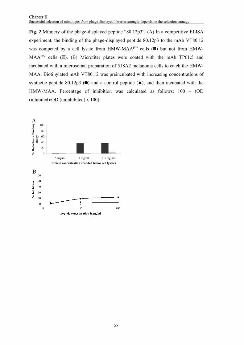
Chapter II Successful selection of mimotopes from phage-displayed libraries strongly depends on the selection strategy
58
Fig. 2 Mimicry of the phage-displayed peptide “80.12p3”. (A) In a competitive ELISA
experiment, the binding of the phage-displayed peptide 80.12p3 to the mAb VT80.12
was competed by a cell lysate from HMW-MAApos cells ( ) but not from HMW-
MAAneg cells ( ). (B) Microtiter plates were coated with the mAb TP61.5 and
incubated with a microsomal preparation of 518A2 melanoma cells to catch the HMW-
MAA. Biotinylated mAb VT80.12 was preincubated with increasing concentrations of
synthetic peptide 80.12p3 ( ) and a control peptide ( ), and then incubated with the
HMW-MAA. Percentage of inhibition was calculated as follows: 100 – (OD
(inhibited)/OD (uninhibited) x 100).
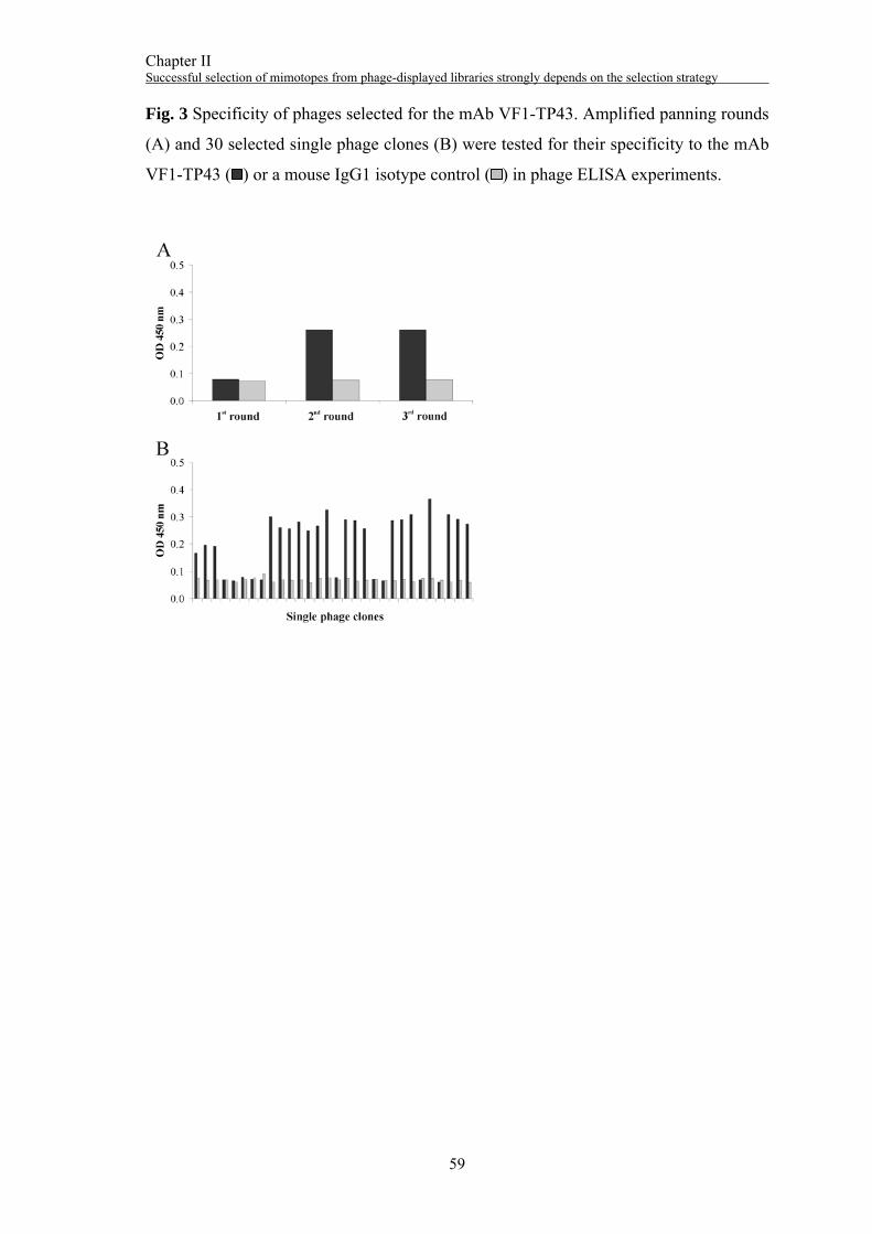
Chapter II Successful selection of mimotopes from phage-displayed libraries strongly depends on the selection strategy
59
Fig. 3 Specificity of phages selected for the mAb VF1-TP43. Amplified panning rounds
(A) and 30 selected single phage clones (B) were tested for their specificity to the mAb
VF1-TP43 ( ) or a mouse IgG1 isotype control ( ) in phage ELISA experiments.
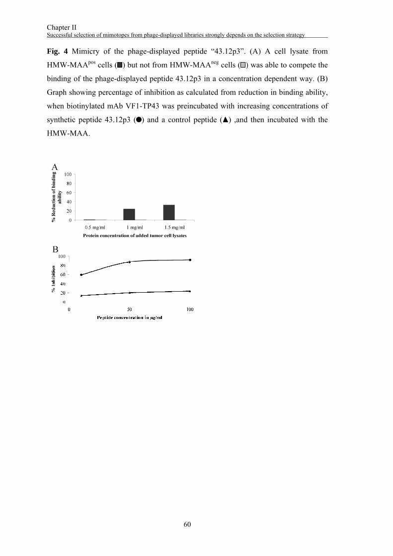
Chapter II Successful selection of mimotopes from phage-displayed libraries strongly depends on the selection strategy
60
Fig. 4 Mimicry of the phage-displayed peptide “43.12p3”. (A) A cell lysate from
HMW-MAApos cells ( ) but not from HMW-MAAneg cells ( ) was able to compete the
binding of the phage-displayed peptide 43.12p3 in a concentration dependent way. (B)
Graph showing percentage of inhibition as calculated from reduction in binding ability,
when biotinylated mAb VF1-TP43 was preincubated with increasing concentrations of
synthetic peptide 43.12p3 ( ) and a control peptide ( ) ,and then incubated with the
HMW-MAA.
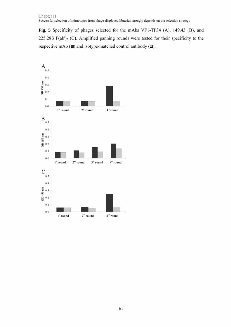
Chapter II Successful selection of mimotopes from phage-displayed libraries strongly depends on the selection strategy
61
Fig. 5 Specificity of phages selected for the mAbs VF1-TP34 (A), 149.43 (B), and
225.28S F(ab')2 (C). Amplified panning rounds were tested for their specificity to the
respective mAb ( ) and isotype-matched control antibody ( ).

Chapter II Successful selection of mimotopes from phage-displayed libraries strongly depends on the selection strategy
62
Fig. 6 Local alignment of identified peptide ligands with HMW-MAA. The sequences
of the peptides 43.12p3 (magenta) and p225Fab (cyan) could be aligned with HMW-
MAA (UniProt accession no. Q6UVK1) using BLAST (27). Identical residues are
indicated in bold, similar ones in italics. The length of the whole peptide sequences is
indicated with colored solid lines. The signal peptide and the transmembrane region of
HMW-MAA are indicated in bold and underlined letters at the N-terminus and C-
terminus, respectively.
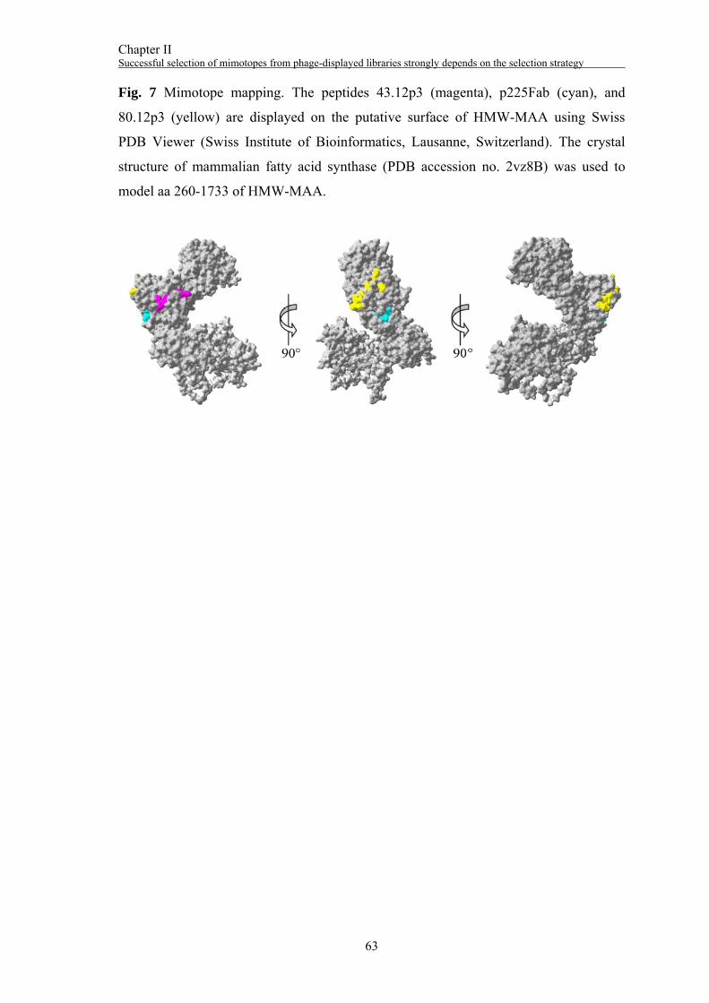
Chapter II Successful selection of mimotopes from phage-displayed libraries strongly depends on the selection strategy
63
Fig. 7 Mimotope mapping. The peptides 43.12p3 (magenta), p225Fab (cyan), and
80.12p3 (yellow) are displayed on the putative surface of HMW-MAA using Swiss
PDB Viewer (Swiss Institute of Bioinformatics, Lausanne, Switzerland). The crystal
structure of mammalian fatty acid synthase (PDB accession no. 2vz8B) was used to
model aa 260-1733 of HMW-MAA.

Chapter II Successful selection of mimotopes from phage-displayed libraries strongly depends on the selection strategy
64
Chapter II Successful selection of mimotopes from phage-displayed libraries strongly depends on the selection strategy
65
References
1. Bratkovic, T. (2010). Progress in phage display: evolution of the technique and its
application. Cell Mol Life Sci, 67, 749-767.
2. Smith, G. P., Petrenko, V. A. (1997). Phage Display. Chem Rev, 97, 391-410.
3. Makowski, L. (1993). Structural constraints on the display of foreign peptides on
filamentous bacteriophages. Gene, 128, 5-11.
4. Smith, G. P. (1993). Preface - Surface display and peptide libraries. Gene, 128, 1-2.
5. Cortese, R., Felici, F., Galfre, G., Luzzago, A., Monaci, P., Nicosia, A. (1994).
Epitope discovery using peptide libraries displayed on phage. Trends Biotechnol,
12, 262-267.
6. Cortese, R., Monaci, P., Luzzago, A., Santini, C., Bartoli, F., Cortese, I., et al.
(1996). Selection of biologically active peptides by phage display of random
peptide libraries. Curr Opin Biotechnol, 7, 616-621.
7. Meola, A., Delmastro, P., Monaci, P., Luzzago, A., Nicosia, A., Felici, F., et al.
(1995). Derivation of vaccines from mimotopes. Immunologic properties of human
hepatitis B virus surface antigen mimotopes displayed on filamentous phage. J
Immunol, 154, 3162-3172.
8. Yang, L., Jiang, H., Shi, B., Wang, H., Li, J., Wang, H., et al. (2010). Identification
and characterization of Ch806 mimotopes. Cancer Immunol Immunother, 59, 1481-
1487.
9. Devlin, J. J., Panganiban, L. C., Devlin, P. E. (1990). Random peptide libraries: a
source of specific protein binding molecules. Science, 249, 404-406.
10. Luo, W., Hsu, J. C., Tsao, C. Y., Ko, E., Wang, X., Ferrone, S. (2005). Differential
immunogenicity of two peptides isolated by high molecular weight-melanoma-
associated antigen-specific monoclonal antibodies with different affinities. J
Immunol, 174, 7104-7110.
11. Ferrone, S., Wang, X. (2001). Active specific immunotherapy of malignant
melanoma and peptide mimics of the human high-molecular-weight melanoma-
associated antigen. Recent Results Cancer Res, 158, 231-235.
12. Riemer, A. B., Hantusch, B., Sponer, B., Kraml, G., Hafner, C., Zielinski, C. C., et
al. (2005). High-molecular-weight melanoma-associated antigen mimotope
immunizations induce antibodies recognizing melanoma cells. Cancer Immunol
Immunother, 54, 677-684.
Chapter II Successful selection of mimotopes from phage-displayed libraries strongly depends on the selection strategy
66
13. Wagner, S., Hafner, C., Allwardt, D., Jasinska, J., Ferrone, S., Zielinski, C. C., et al.
(2005). Vaccination with a human high molecular weight melanoma-associated
antigen mimotope induces a humoral response inhibiting melanoma cell growth in
vitro. J Immunol, 174, 976-982.
14. Hafner, C., Wagner, S., Allwardt, D., Riemer, A. B., Scheiner, O., Pehamberger,
H., et al. (2005). Cross-reactivity of mimotopes with a monoclonal antibody against
the high molecular weight melanoma-associated antigen (HMW-MAA) does not
predict cross-reactive immunogenicity. Melanoma Res, 15, 111-117.
15. Jiang, B., Liu, W., Qu, H., Meng, L., Song, S., Ouyang, T., et al. (2005). A novel
peptide isolated from a phage display peptide library with trastuzumab can mimic
antigen epitope of HER-2. J Biol Chem, 280, 4656-4662.
16. Mohammadi, M., Rasaee, M. J., Rajabibazl, M., Paknejad, M., Zare, M.,
Mohammadzadeh, S. (2007). Epitope mapping of PR81 anti-MUC1 monoclonal
antibody following PEPSCAN and phage display techniques. Hybridoma
(Larchmt), 26, 223-230.
17. Hu, S., Zhu, Z., Li, L., Chang, L., Li, W., Cheng, L., et al. (2008). Epitope mapping
and structural analysis of an anti-ErbB2 antibody A21: Molecular basis for tumor
inhibitory mechanism. Proteins, 70, 938-949.
18. Zheng, L., Li, B., Qian, W., Zhao, L., Cao, Z., Shi, S., et al. (2008). Fine epitope
mapping of humanized anti-IgE monoclonal antibody omalizumab. Biochem
Biophys Res Commun, 375, 619-622.
19. Li, Y., Ning, Y. S., Wang, Y. D., Hong, Y. H., Luo, J., Dong, W. Q., et al. (2007).
Production of mouse monoclonal antibodies against Helicobacter pylori Lpp20 and
mapping the antigenic epitope by phage display library. J Immunol Methods, 325,
1-8.
20. Li, Y., Ning, Y. S., Wang, Y. D., Luo, J., Wang, W., Dong, W. Q., et al. (2008).
Production of mouse monoclonal antibodies against Helicobacter pylori Catalase
and mapping the antigenic epitope by phage display library. Vaccine, 26, 1263-
1269.
21. Xie, H. L., Wang, Z., Cui, S. J., Zhang, C. F., Cui, Y. D. (2010). The epitope of the
VP1 protein of porcine parvovirus. Virol J, 7, 161.
Chapter II Successful selection of mimotopes from phage-displayed libraries strongly depends on the selection strategy
67
22. Lin, M., McRae, H., Dan, H., Tangorra, E., Laverdiere, A., Pasick, J. (2010). High-
resolution epitope mapping for monoclonal antibodies to the structural protein Erns
of classical swine fever virus using peptide array and random peptide phage display
approaches. J Gen Virol, 91, 2928-2940.
23. Giacomini, P., Natali, P., Ferrone, S. (1985). Analysis of the interaction between a
human high molecular weight melanoma-associated antigen and the monoclonal
antibodies to three distinct antigenic determinants. J Immunol, 135, 696-702.
24. Temponi, M., Fawwaz, R. A., Kekish, U., Wang, T. S., Ferrone, S. (1991).
Improvement by affinity chromatography on antiidiotypic monoclonal antibodies
(MAbs) of immunoreactivity and in vivo targeting of radiolabelled anti-HMW-
MAA MAb TP61.5 in nude mice bearing human melanoma lesions. Int J Cancer,
49, 624-630.
25. Temponi, M., Gold, A. M., Ferrone, S. (1992). Binding parameters and idiotypic
profile of the whole immunoglobulin and Fab' fragments of murine monoclonal
antibody to distinct determinants of the human high molecular weight-melanoma
associated antigen. Cancer Res, 52, 2497-2503.
26. Campoli, M. R., Chang, C. C., Kageshita, T., Wang, X., McCarthy, J. B., Ferrone,
S. (2004). Human high molecular weight-melanoma-associated antigen (HMW-
MAA): a melanoma cell surface chondroitin sulfate proteoglycan (MSCP) with
biological and clinical significance. Crit Rev Immunol, 24, 267-296.
27. http://blast.ncbi.nlm.nih.gov/.
28. http://modbase.compbio.ucsf.edu/modbase-cgi/index.cgi.
29. http://immunet.cn/mimox/.
30. Galfre, G., Monaci, P., Nicosia, A., Luzzago, A., Felici, F., Cortese, R. (1996).
Immunization with phage-displayed mimotopes. Methods Enzymol, 267, 109-115.
31. Dharmasena, M. N., Jewell, D. A., Taylor, R. K. (2007). Development of peptide
mimics of a protective epitope of Vibrio cholerae Ogawa O-antigen and
investigation of the structural basis of peptide mimicry. J Biol Chem, 282, 33805-
33816.

68
69
Chapter III
Specificity of mimotope-induced anti-high molecular weight-
melanoma associated antigen (HMW-MAA) antibodies
does not ensure biological activity
Sonja Gaier1*, Julia Latzka1*, Gerlinde Hofstetter1, Nina Balazs1, Soldano Ferrone2,
Otto Scheiner1, Heimo Breiteneder1, Hubert Pehamberger3, Stefan Wagner1
* These authors contributed equally to this work and are listed in alphabetical order. 1Department of Pathophysiology and Allergy Research, Medical University of Vienna,
Vienna, Austria 2Departments of Surgery, of Immunology and of Pathology, University of Pittsburgh
Cancer Institute, Pittsburgh, Pennsylvania, USA 3Department of Dermatology, Medical University of Vienna, Vienna, Austria
Manuscript submitted to Vaccine

70
Chapter III Specificity of mimotope-induced anti-HMW-MAA antibodies does not ensure biological activity
71
Abstract
Vaccines based on peptide mimics (mimotopes) of conformational tumor antigen
epitopes have been investigated for a variety of human tumors including breast cancer,
tumors expressing the carcinoembryonic antigen, B cell lymphoma, neuroblastoma, and
melanoma. In our previous work, we designed a vaccine based on a mimotope of the
high molecular weight-melanoma associated antigen (HMW-MAA) that elicited HMW-
MAA specific antibodies with anti-tumor activity in vitro and in vivo. In this study, we
aimed to identify additional HMW-MAA mimotopes with distinct epitope specificities
as potential components of a polymimotope melanoma vaccine.
Random peptide phage libraries were screened with the anti-HMW-MAA monoclonal
antibodies (mAbs) VT80.12 and VF1-TP43 yielding one peptide ligand for each mAb.
Both peptides confirmed epitope mimicry in inhibition ELISAs and were coupled to the
carrier protein keyhole limpet hemocyanin (KLH) for immunization experiments.
Although both HMW-MAA mimotopes elicited peptide specific antibodies in rabbits or
BALB/c mice, HMW-MAA specific antibodies were only obtained in mice upon
immunization with the mimotope defined by the mAb VT80.12. However, despite being
cross-reactive to HMW-MAA these antibodies had no effect on the in vitro tumor
proliferation of HMW-MAA expressing human melanoma cells.
This work describes constraints related to mimotopes generated by phage display and
discusses the potential relevance of mimotope-selecting mAbs and applied
immunization models on the outcome of mimotope vaccination. From our results we
conclude that these aspects should be considered when using mimotopes as vaccine
components.

Chapter III Specificity of mimotope-induced anti-HMW-MAA antibodies does not ensure biological activity
72
Chapter III Specificity of mimotope-induced anti-HMW-MAA antibodies does not ensure biological activity
73
1. Introduction
For at least two decades, the high molecular weight-melanoma associated antigen
(HMW-MAA) has been the focus of several studies to implement effective
immunotherapy for melanoma. Originally identified with murine monoclonal antibodies
(mAbs) on the surface of human melanoma cells, HMW-MAA has been found to be
overexpressed in at least 80% of primary and metastatic melanomas and more recently,
in several other tumors as well as cancer stem cells 1,2. However, HMW-MAA
expression is not restricted to transformed cells, as it has been found on various cells of
normal tissue including melanocytes, hairfollicles and basal cells of the epidermis as
well as endothelial cells, chondrocytes and pericytes 1. Its expression on both activated
and resting pericytes in tumor vessels has suggested that HMW-MAA is critical to
tumor angiogenesis 2. Furthermore, HMW-MAA is known to contribute to the
malignant phenotype of melanoma by activating several signaling cascades (e.g. Rho
GTPases, p130cas, FAK) that are involved in adhesion, migration and spreading of
melanoma cells 3.
The idea of HMW-MAA-directed immunotherapy has been primarily supported by
preclinical studies that showed prolonged survival rates of melanoma patients in
association with anti-HMW-MAA specific antibodies (Abs) induced by active
immunization 4-6. Since then, a number of antibody- as well as T cell-based
immunotherapies have been investigated. Recently, Maciag et al. generated a Listeria
monocytogenes (Lm)-based vaccine against HMW-MAA 7. Immunization of C57BL/6
mice bearing B16F10-HMW-MAA melanomas induced HMW-MAA specific CD8+
and CD4+ T cells that were equally required for tumor inhibition, as in vivo depletion of
each of these cells resulted in uncontrolled tumor growth 7. Noteworthy, this study
provided the first syngenic melanoma mouse model for HMW-MAA as well as novel
information on the contribution of T cells in anti-HMW-MAA immunity.
In contrast, we emphasized the induction of humoral anti-HMW-MAA immunity by
using peptide mimics (mimotopes) as immunogens. Mimotopes are small peptides that
mimic conformational B cell epitopes of antigens and can be selected by screening
random peptide phage libraries with a mAb of interest. As mimotopes do not
necessarily share the identical amino acid sequence with the original antigen, they are
ideal antigen surrogates able to overcome immunotolerance 8-11. Regarding melanoma,

Chapter III Specificity of mimotope-induced anti-HMW-MAA antibodies does not ensure biological activity
74
this is of particular interest as most melanoma associated antigens including the HMW-
MAA are known to be self-antigens 1,12.
In previous studies, we identified a linear HMW-MAA mimotope (225D9.2+) which
mimics an epitope recognized by the anti-HMW-MAA mAb 225.28S. Immunization
with this mimotope coupled to tetanus toxoid induced HMW-MAA specific Abs in
rabbits that inhibited tumor cell proliferation in vitro 13. Passive administration of these
Abs in a xenogenic melanoma SCID mouse model inhibited tumor growth up to 40% in
a therapeutic setting and up to 62% in a prophylactic setting 14.
As epitope loss is commonly found in melanoma 15 and several studies indicate that
targeting multiple epitopes of a tumor antigen has synergistic effects and correlates with
higher success rates, efficacy of a melanoma vaccine might be improved by the co-
application of several peptides 16-18.
In this study, we report the selection of additional HMW-MAA mimotopes with distinct
epitope specificities and discuss constraints related to mimotope vaccines regarding
their immunogenicity and antigenicity.
Chapter III Specificity of mimotope-induced anti-HMW-MAA antibodies does not ensure biological activity
75
2. Materials and methods
2.1. Monoclonal anti-HMW-MAA antibodies
The mAbs VT80.12, VF1-TP43, and TP61.5 were developed and characterized as
described elsewhere 19-21.
2.2. Biotinylation of Abs (mAbs, rabbit IgGs)
NHS-LC-Biotin (Pierce, Rockford, IL, USA) was diluted in dimethylformamide at a
concentration of 40 mg/ml. Five microliters of this solution was added to 1 mg/ml mAb
or rabbit IgGs in PBS and incubated for 45 min at room temperature (RT). Excess NHS-
LC-Biotin was removed by dialysis against PBS.
2.3. Cell lines
The human melanoma cell line 518A2 which expresses high levels of HMW-MAA and
M14, a human melanoma cell line with no detectable expression of HMW-MAA, were
maintained in RPMI 1640 medium (Lonza, Verviers, Belgium). The medium was
supplemented with 10% (v/v) FCS and 1% (v/v) antibiotic-antimycotic mix (both from
Gibco, Paisley, UK). Both cell lines were cultured in a humidified atmosphere
containing 5% CO2 and 95% ambient air at 37°C.
2.4. Microsomal preparations
Microsomal preparations were performed using ~5x10⁷ cells according to the protocol
described elsewhere 22. Protein concentration was determined using a bicinchoninic acid
(BCA) protein assay (Pierce).
2.5. Phage display, affinity selection and sequence analysis
Peptide ligands for the mAb VT80.12 were selected from a pVIII-15mer phage display
peptide library 23. Therefore, the mAb VT80.12 was immobilized and incubated with
~1010 phages. Phages displaying peptides that bound to the mAb VT80.12 were eluted
and amplified in E. coli TG1.
Peptide ligands for the mAb VF1-TP43 were selected using the Ph.D.-12™ Phage
Display Peptide Library, a pIII-12mer library, purchased from New England Biolabs
(Ipswich, MA, USA). Biopanning was executed following the manufacturer’s
instructions.
Chapter III Specificity of mimotope-induced anti-HMW-MAA antibodies does not ensure biological activity
76
After three rounds of selection, single phage clones that bound to the respective mAb
were identified by phage ELISA and subsequently subjected for DNA sequencing.
2.6. Synthesis of peptides
The peptides GRQYYEGRKPDYRAAC (15/3/6) and NYQDLQRTHFKSGPGPGC
(43.12p3) were synthesized using F-moc strategy by piCHEM (Graz, Austria). The
purity of the peptides was ~95%, as assessed by HPLC.
2.7. ELISA inhibition assay
MaxiSorp immunoplates (Nunc, Rosklide, Denmark) were coated overnight (o/n) at 4°C
with 4 µg/ml of mAb T61.5 in 50 mM Na-carbonate buffer, pH 9.6. Ten nanograms of
biotinylated mAb were incubated with increasing concentrations (0, 10, 50, 100, and
500 µg/ml) of synthetic peptides in TBST (0.5% (v/v) Tween-20) containing 1% (w/v)
BSA o/n at 4°C. After blocking with TBST/3% (w/v) milk powder, plates were
incubated for 3 h at RT with 100 µg/ml microsomal preparations diluted in TBST/1%
BSA. After washing, the mAb preincubated with peptides was added and incubation
was continued for an additional hour at RT. Bound biotinylated mAb was detected
using alkaline phosphatase (AP)-conjugated streptavidin (GE Healthcare, Little
Chalfont, UK), followed by addition of p-nitrophenylphosphate (Sigma). Absorbance
was measured at 405 nm. Percentage of inhibition was calculated as follows: 100 – (OD
(inhibited)/OD (uninhibited) x 100).
2.8. Conjugation of peptides
Peptides were coupled to the carrier protein keyhole limpet hemocyanin (KLH; Sigma,
St. Louis, MO, USA) or bovine serum albumin (BSA; Pierce) using the
heterobifunctional crosslinker reagent m-maleimidobenzoyl-N-hydroxysuccinimide
(MBS; Pierce) as described previously 13. Conjugation of the peptides to the carrier
proteins was verified in a dot blot assay. Therefore, peptide conjugates were dotted onto
nitrocellulose (NC) membrane (Whatman, Dassel, Germany). After blocking with
PBST (0.5% Tween-20) containing 3% milk powder, NC strips were incubated with
biotinylated mAb, followed by AP-conjugated streptavidin (GE Healthcare). Color
development was done with 5-bromo-4-chloro-3-indolyl phosphate/nitroblue
tetrazolium.
Chapter III Specificity of mimotope-induced anti-HMW-MAA antibodies does not ensure biological activity
77
2.9. Immunizations
New Zealand white rabbits were immunized at Charles River Laboratories (Kisslegg,
Germany). Immunizations were done thrice in 14- to 28-day intervals (day 1, 29, and
43) each with 200 µg of the peptide-KLH conjugates or KLH alone adsorbed to CFA
(first immunization) or IFA (second and third immunization). Serum samples were
taken before treatment (preimmune serum) and ten days after the first (day 11) and
second immunization (day 39). Rabbits were sacrificed two weeks after the third
immunization (day 57).
Female BALB/c (4 groups, n = 3; 6-8 weeks old; Charles River Laboratories, Sulzfeld,
Germany) were immunized i.p. three times in 14-day intervals (day 1, 15, and 29) each
with 15 µg of the peptide-KLH conjugates or KLH alone adsorbed to aluminium
hydroxide (Alum; Serva, Heidelberg, Germany) in a total volume of 150 µl PBS
solution. Sham-treated mice received PBS buffered alum only. Sera were taken from the
tail vein on day 0 (preimmune serum), 22, and 41. Mice were treated according to
European Union Rules of Animal Care, with permission 66.009/152-II/10b/2009 from
the Austrian Ministry of Science.
2.10. Purification of rabbit or mouse IgG Abs
Total IgG from sera of rabbits were purified according to the protocol described
elsewhere 13.
For purification of mouse IgG Abs, serum samples taken after the third immunization or
after sacrifice of mice were pooled for each group and diluted with an equal volume of
binding buffer (20 mM Na-phosphate, pH 7.0). The HiTrap Protein G HP column (GE
Healthcare) was equilibrated with binding buffer and the sample applied. Bound Abs
were eluted with 100 mM glycine-HCl, pH 2.7 and neutralized by addition of 1 M Tris-
HCl, pH 9.0.
Purification of rabbit or mouse IgG was monitored using nonreducing 8% SDS-PAGE.
Protein concentration was determined by a BCA protein assay (Pierce).
Chapter III Specificity of mimotope-induced anti-HMW-MAA antibodies does not ensure biological activity
78
2.11. Peptide specific Ab response
MaxiSorp immunoplates (Nunc) were coated with 10 µg/ml peptide-BSA conjugates or
KLH or BSA in 50 mM Na-carbonate buffer, pH 9.6, o/n at 4°C. Nonspecific binding
sites were blocked with TBST/3% milk powder.
Purified rabbit Abs were diluted at concentrations of 0.05, 0.1, 0.5, 1, 5, and 10 µg/ml in
TBST/0.5% BSA, added to antigen-coated plates, and incubated for 2 h at RT. After
washing, bound Abs were detected using AP-conjugated swine anti-rabbit IgG (Dako,
Glostrup, Denmark) diluted 1:1000 in TBST/0.5% BSA. Color development was
performed as described for the ELISA inhibition assay.
Mouse sera were diluted 1:1000 in TBST/0.5% BSA and incubated for 2 h at RT. AP-
conjugated rabbit anti-mouse IgG + IgM (Jackson ImmunoResearch, West Grove, PA,
USA) diluted 1:5000 in TBST/0.5% BSA was used as a second step Ab. Visualization
of bound mouse Abs was performed as described above.
2.12. HMW-MAA specific Ab response
2.12.1. ELISA protocol
For the detection of the HMW-MAA specific Ab response, HMW-MAA was purified
from microsomal preparations as described for the ELISA inhibition assay. After
blocking and catching, plates were incubated for 2 h at RT with increasing
concentrations (12.5, 50, and 200 µg/ml) of purified rabbit Abs diluted in TBST/1%
BSA. Bound IgG was detected as described for the peptide-specific Ab response.
2.12.2. FACS protocol
Flow cytometric analysis of melanoma cells stained with sera from immunized mice or
purified Abs from immunized rabbits was performed as previously described 24. Briefly,
5x10⁵ cells were incubated for 1 h on ice with a 1:10 dilution of pooled mouse sera for
each group or 100 µg purified rabbit Abs in 100 µl PBS/1% BSA. One microgram of
mAb VT80.12 served as positive control staining of HMW-MAA. Cells were then
washed twice with PBS/0.5% BSA and incubated for an additional 30 min on ice with
FITC-labeled goat anti-mouse IgG or FITC-labeled goat anti-rabbit IgG (both diluted
1:2000 in PBS/1% BSA; AbD Serotec, Düsseldorf, Germany). After washing, cells
were resuspended in PBS/0.5% BSA and 20,000 gated events were analyzed by flow
cytometry on a BD FACScanto using BD FACSDiva software (BD Biosciences,
Chapter III Specificity of mimotope-induced anti-HMW-MAA antibodies does not ensure biological activity
79
Franklin Lakes, NJ, USA). Histogram overlays were done applying FlowJo software
(Tree Star, Ashland, OR, USA).
2.12.3. Immunohistochemistry (IHC) protocol
Immunohistochemical staining of established 518A2 tumor tissues was performed as
described by Wagner et al. 14. Briefly, tumor sections were incubated with either
blocking buffer (negative control), 30 µg/ml biotinylated mAb VT80.12 (positive
control) or 100 µg/ml biotinylated purified Abs of rabbits either immunized with
15/3/6-KLH conjugate or KLH. After washing with TBST, slides were incubated with
StreptABComplex/HRP (Dako) for 30 min. Specific Ab binding was visualized by a
DAB chromogen solution (Dako) following hematoxylin counterstaining. Stained slides
were viewed by an Olympus Vanox AHBT3 microscope and photographed with a Zeiss
AxioCam MRc5 camera.
2.13. Inhibition of tumor cell growth in vitro
Tumor cells were seeded in 96-well tissue culture plates (Costar; Corning, NY, USA) at
1500 cells/well. Cells were allowed to adhere o/n at 37°C. Purified Abs from mice
immunized with either the 15/3/6-KLH conjugate or KLH were added at increasing
concentrations (0, 0.01, 0.1, and 1 mg/ml) and incubation was continued for an
additional 72 h at 37°C. Cells were pulsed with 0.5 µCi of [methyl-3H]thymidine/well
(GE Healthcare) for another 6 h at 37°C and then harvested. Incorporated [3H]thymidine
was measured by a Wallac MicroBeta TriLux 1450 counter (PerkinElmer, Waltham,
MA, USA). Percentage of inhibition of proliferation was calculated by comparing the
cpm values of treated cells with those of untreated cells, which were set at 100%.

Chapter III Specificity of mimotope-induced anti-HMW-MAA antibodies does not ensure biological activity
80
Chapter III Specificity of mimotope-induced anti-HMW-MAA antibodies does not ensure biological activity
81
3. Results
3.1. Identification of peptide ligands
Biopanning of the mAb VT80.12 was performed with a linear pVIII-15mer phage
display peptide library. After three rounds of selection, the total number of phages that
bound to the mAb VT80.12 was increased from 1x10⁵ CFU/ml in the 1st round to 1x10⁹
CFU/ml in the 3rd round. Twenty individual phage clones of each the 2nd and 3rd round
were tested for their ligand specificity in phage ELISA. Among these, eighteen (2nd
round) and nineteen (3rd round) phage clones bound to the mAb VT80.12. DNA
sequencing of these 37 phage clones yielded one peptide sequence
GRQYYEGRKPDYRAA (“15/3/6”). Phages (106-1013 CFU/ml) displaying this
sequence showed a concentration-dependent inhibition up to 70% in ELISA
experiments (data not shown). Therefore, the peptide 15/3/6 was chemically synthesized
with an additional C-terminal cysteine residue for conjugation purposes. In ELISA
inhibition experiments, the peptide inhibited the binding of the mAb VT80.12 to the
HMW-MAA up to 93% (Fig. 1A).
Biopanning of the mAb VF1-TP43 was performed with a linear pIII-12mer phage
display peptide library. Three rounds of selection yielded an increase of phage titer from
1x10⁵ pfu/ml (1st round) to 3x10⁹ pfu/ml (3rd round). Among 30 tested phage clones, 20
were specifically recognized by the mAb VF1-TP43 in phage ELISA. DNA sequencing
yielded one peptide sequence NYQDLQRTHFKS (“43.12p3”). In a competitive ELISA
experiment, binding of the phage displayed peptide to the mAb VF1-TP43 was reduced
up to 33% by a HMW-MAA containing cell lysate (data not shown). The peptide
43.12p3 was synthesized with a GPGPG-linker and an additional C-terminal cysteine
residue. In subsequent inhibition experiments, this peptide inhibited the binding of the
mAb VF1-TP43 to the HMW-MAA up to 100% (Fig. 1B).
3.2. Peptide specific Ab response in rabbits
The peptide 15/3/6 was coupled to KLH or BSA as carrier protein. Conjugation to the
carrier protein was confirmed in a dot blot assay (data not shown). New Zealand white
rabbits were immunized with the conjugate 15/3/6-KLH or the carrier protein KLH
alone. After three immunizations, Abs were purified using a HiTrap protein A HP
column. Twenty milliliters of serum yielded ~20 mg of IgG Abs. The purity was greater
than 95% as confirmed by SDS-PAGE (data not shown). Peptide specific Abs were
Chapter III Specificity of mimotope-induced anti-HMW-MAA antibodies does not ensure biological activity
82
determined by ELISA after incubation of coated peptide-BSA conjugate, BSA or KLH
with purified IgG Abs. Both, 15/3/6- and KLH-specific Abs were already detectable at a
concentration of 0.05 µg/ml and increased in a dose- dependent manner (Fig. 2). Abs
purified from the KLH-immunized rabbit did not bind to the peptide. No Abs directed
against BSA could be determined.
3.3. HMW-MAA specific Ab response in rabbits
HMW-MAA specificity of rabbit Abs was determined in ELISA by testing their binding
ability to the HMW-MAA. Binding to the HMW-MAA negative cell line M14 was
detected for both, the peptide-conjugate and the KLH-induced Abs (Fig. 2). Even when
the HMW-MAA catching mAb was directly incubated with purified rabbit Abs and
subsequently detected with AP-conjugated swine anti-rabbit IgG, high background
levels were measured (data not shown).
In a second approach, HMW-MAA specificity of rabbit Abs was investigated by FACS
analysis. The HMW-MAA positive human melanoma cell line 518A2 (HMW-MAApos)
or the HMW-MAA negative human melanoma cell line M14 (HMW-MAAneg) were
incubated with each 100 µg purified rabbit Abs and detected with a FITC-conjugated
goat anti-rabbit IgG. As expected, the mAb VT80.12 specifically bound to the HMW-
MAApos, but not to the HMW-MAAneg cells. However, HMW-MAA specificity of the
15/3/6-KLH conjugate induced Abs could not be observed, as Abs induced by KLH
showed a similar staining pattern to HMW-MAApos as well as to HMW-MAAneg cells
(Fig. 3A).
In a third approach, we used solid 518A2 tumors established in C.B.17 SCID/SCID
mice 14 to detect HMW-MAA reactive rabbit Abs by IHC. Whereas staining of
melanoma cells was observed with the biotinylated mAb VT80.12, none was observed
with either an isotype-matched control mAb (data not shown) or PBS buffer. Also in
this setup, we detected a high background staining of anti-KLH Abs on 518A2 tumor
tissue, and therefore no HMW-MAA specific anti-15/3/6 Abs (Fig. 3B).
Chapter III Specificity of mimotope-induced anti-HMW-MAA antibodies does not ensure biological activity
83
3.4. Peptide specific Ab response in BALB/c mice
The peptides 15/3/6 or 43.12p3 were coupled each to KLH or BSA. Female BALB/c
mice (4 groups, n = 3) were immunized with 15 µg of the peptide-KLH conjugates,
KLH or were sham-treated with PBS buffered alum. Serum samples of each group were
tested for the presence of peptide specific Abs in ELISA. Peptide specificity was
confirmed for both anti-15/3/6-KLH and anti-43.12p3-KLH Abs as they bound to the
corresponding peptide-BSA conjugates (Fig. 4). As expected, KLH specific Abs were
detected in the KLH-group (Fig. 5) as well as in the groups immunized with the
peptide-KLH conjugates (data not shown). Sham-treated mice did not develop Abs
against the peptides 15/3/6 or 43.12p3, KLH or BSA (data not shown).
3.5. HMW-MAA specific Ab response in BALB/c mice
Preimmune serum samples or serum samples after the third immunization of individual
mice were pooled for each group. Serum pools were diluted 1:10 and incubated with
HMW-MAApos or HMW-MAAneg cells. Bound Abs were detected with a FITC-labeled
goat anti-mouse IgG by FACS analysis. None of the mouse Abs of preimmune serum
pools showed reactivity to HMW-MAApos or HMW-MAAneg cells (data not shown).
Immunization with the 15/3/6-KLH conjugate induced HMW-MAA specific Abs,
whereas the 43.12p3-KLH conjugate did not (Fig. 5). KLH-induced Abs showed
negligible background staining on HMW-MAApos as well as HMW-MAAneg cells.
3.6. Inhibition of tumor cell growth
The effect of anti-15/3/6-KLH Abs on tumor growth in vitro was determined by a
[3H]thymidine proliferation assay. HMW-MAApos or HMW-MAAneg cells were
incubated with increasing concentrations of purified mouse Abs. Upon treatment with
anti-15/3/6 Abs, proliferation of HMW-MAApos cells was inhibited to a maximum of
~17% (Fig. 6). Almost identical results were obtained, when either HMW-MAApos cells
were treated with anti-KLH Abs or HMW-MAAneg cells were incubated with anti-
15/3/6 or anti-KLH Abs. Results given in Fig. 7 represent mean values of 3 independent
experiments.

Chapter III Specificity of mimotope-induced anti-HMW-MAA antibodies does not ensure biological activity
84
Chapter III Specificity of mimotope-induced anti-HMW-MAA antibodies does not ensure biological activity
85
4. Discussion
In this study we aimed to select two mimotopes defined by the anti-HMW-MAA mAbs
VT80.12 and VF1-TP43 that should serve together with our previously described
225D9.2+ mimotope 13,14 as components of a polymimotope vaccine against melanoma.
We therefore screened two linear phage display peptide libraries (pVIII-15mer with
mAb VT80.12, pIII-12mer with mAb VF1-TP43) yielding one peptide ligand for each
mAb. Both peptides (15/3/6: ligand to VT80.12; 43.12p3: ligand to VF1-TP43)
inhibited binding of the respective mAb to the HMW-MAA up to 100%, thereby
proofing epitope mimicry and their definition as mimotopes. Upon immunization of
either rabbits or BALB/c mice, each mimotope coupled to the carrier protein KLH
demonstrated immunogenicity by inducing mimotope specific Abs. However, only the
15/3/6-KLH conjugate induced HMW-MAA specific Abs in BALB/c mice, but these
Abs failed to inhibit tumor cell proliferation in vitro.
Epitope mimicry to HMW-MAA was confirmed for both peptides 15/3/6 and 43.12p3
in ELISA inhibition experiments (Fig. 1). Thus, constraints referring to phage display
were preliminary excluded.
An important issue of this study was the applied animal model in the context of the
carrier protein used for vaccination. Initially, immunization experiments were
performed in rabbits as high amounts of antibodies can be obtained from the blood for
subsequent in vitro tests. Although mimotope specific Abs were induced for the tested
mimtope, these showed no HMW-MAA specificity in ELISA experiments (Fig. 2).
Instead, we observed a high background staining of KLH specific Abs on the HMW-
MAA-positive or -negative cell lysate, as well as on the HMW-MAA catching mAb.
Similarly, FACS staining (Fig. 3) as well as immunohistochemistry (Fig. 4) failed to
confirm HMW-MAA specific Abs. Immunizations were then repeated in BALB/c mice
to assess whether the results from the rabbit immunizations were attributed to the carrier
protein KLH or the applied animal model. Using this model, FACS analysis confirmed
HMW-MAA specificity of mimotope-induced Abs (Fig. 6), indicating that the
undesired binding of rabbit anti-KLH Abs was likely attributed to the rabbit Abs and
not to the carrier protein KLH. However, we cannot exclude a possible contribution of
the used adjuvants (CFA/IFA in rabbits vs. alum in BALB/c mice) to the unspecific
binding of rabbit anti-KLH Abs, as adjuvants are known to differentially deliver antigen
Chapter III Specificity of mimotope-induced anti-HMW-MAA antibodies does not ensure biological activity
86
and activate the immune system 25. We therefore conclude that the choice of carrier
protein has an impact on the outcome of vaccination and might depend on the used
immunization model.
Although the 43.12p3-KLH conjugate induced mimotope specific mouse Abs, these
failed to cross-react with the natural antigen HMW-MAA (Fig. 6), indicating that
immunogenicity of mimotopes does not necessarily correlate with their specificity. The
discrepancy between epitope specificity of an anti-HMW-MAA mAb and epitope
specificity of HMW-MAA mimotope-induced Abs is best described by previous studies
of Wagner et al. 13 and Hafner et al. 26. Both studies deal with the identification of
distinct HMW-MAA mimotopes which were defined by the same mAb (225.28S) and
coupled to the same carrier protein (tetanus toxoid, TT) for subsequent immunization
experiments in rabbits. Using the identical immunization protocol, a HMW-MAA
specific Ab response was only obtained upon immunization with the mimotope-TT
conjugate described by Wagner et al. 13. These findings clearly indicate that – despite
conforming affinity selections as well as conforming immunization protocols – not
every mimotope truly mimics the relevant epitope of the original antigen. On the one
hand, such constraints might originate during affinity selection as phage displaying
peptides may bind to residues of the mAb that are not part of the paratope. Although
such peptides might inhibit the binding of their defined mAb to the original epitope,
these will fail to induce a specific immune response to the original antigen. In addition,
B cell epitopes are known to be conformational in nature 27. Hence, it is possible that
mAbs recognize certain conformations of epitopes which are not displayed in the same
structural context by the natural antigen. The above-described constraints probably
outline the major pitfall of the phage display technique that complicates the selection of
“true“ mimotopes, solely based on their binding characteristics to mAbs. On the other
hand, it is to state that immunization with mimotopes does not guarantee the induction
of Abs that display the appropriate paratope to the natural antigen, emphazising also a
possible contribution of the used animal model to the outcome of mimotope
vaccination. This might be of particular relevance for other investigators who apply
mimotopes for vaccination strategies.
Immunization of BALB/c mice with the 15/3/6 mimotope conjugate resulted in the
induction of mimotope- and HMW-MAA specific Abs (Fig. 5 and 6). However, these
Abs showed no anti-tumor activity in in vitro proliferation assays (Fig. 7). Importantly,
the mAb VT80.12 itself does not inhibit tumor cell proliferation neither directly nor
Chapter III Specificity of mimotope-induced anti-HMW-MAA antibodies does not ensure biological activity
87
indirectly by mediating ADCC or CDC 28. We therefore have to question whether anti-
tumor activity in vivo of the mimotope defining mAb should be a prerequisite to induce
antibodies with tumor inhibiting properties. As a matter of fact, certain mAbs (e.g.
Rituximab 29-32, Cetuximab 33,34, Trastuzumab 11,35) which are currently applied for
cancer therapies and successfully served as sources to generate mimotope vaccines with
anti-tumor activity in vitro fulfill this requirement even if in vivo results for the these
mimotope vaccines are not available yet. As reported by Hafner et al., passive
administration of the anti-HMW-MAA mAb 225.28S significantly reduced tumor
volume in a human melanoma xenotransplant SCID mouse model, although this mAb
had no effect on human melanoma cells in vitro 36. This supports the hypothesis of
using mAbs with in vivo rather than in vitro anti-tumor activity for the selection of
mimotopes.
To date, only one mimotope-based vaccine targeting the HMW-MAA has demonstrated
anti-tumor activity in vitro 13 and in vivo 14. The identification of additional HMW-
MAA mimotopes as components of an effective polymimotope vaccine might depend
on the in vivo anti-tumor activity of the selecting mAbs per se but also on the used
animal model as well as on the paratope conformation of the mimotope-induced Abs. In
addition, we suggest that the efficacy of every mimotope based vaccine should be
evaluated in an animal model by active immunization.
Acknowledgments
We thank Christine Hafner for providing the linear pVIII-15mer phage display peptide
library.

Chapter III Specificity of mimotope-induced anti-HMW-MAA antibodies does not ensure biological activity
88
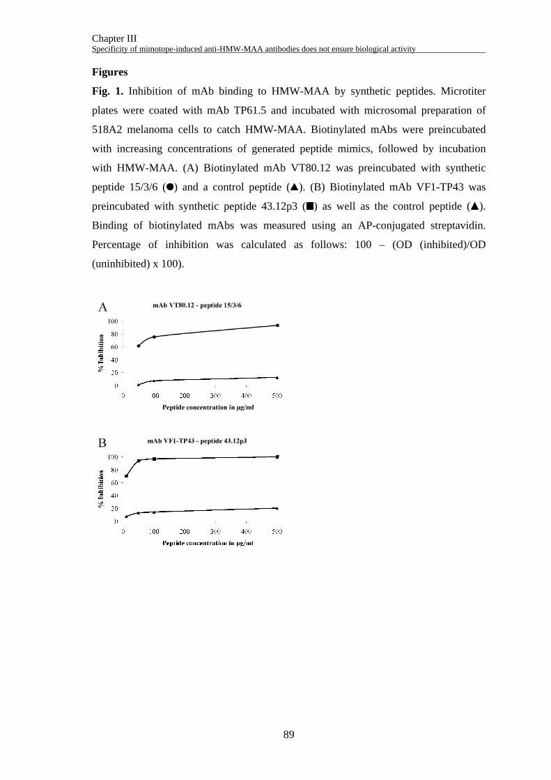
Chapter III Specificity of mimotope-induced anti-HMW-MAA antibodies does not ensure biological activity
89
Figures
Fig. 1. Inhibition of mAb binding to HMW-MAA by synthetic peptides. Microtiter
plates were coated with mAb TP61.5 and incubated with microsomal preparation of
518A2 melanoma cells to catch HMW-MAA. Biotinylated mAbs were preincubated
with increasing concentrations of generated peptide mimics, followed by incubation
with HMW-MAA. (A) Biotinylated mAb VT80.12 was preincubated with synthetic
peptide 15/3/6 ( ) and a control peptide ( ). (B) Biotinylated mAb VF1-TP43 was
preincubated with synthetic peptide 43.12p3 ( ) as well as the control peptide ( ).
Binding of biotinylated mAbs was measured using an AP-conjugated streptavidin.
Percentage of inhibition was calculated as follows: 100 – (OD (inhibited)/OD
(uninhibited) x 100).
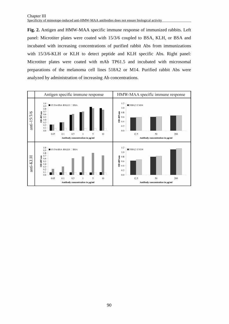
Chapter III Specificity of mimotope-induced anti-HMW-MAA antibodies does not ensure biological activity
90
Fig. 2. Antigen and HMW-MAA specific immune response of immunized rabbits. Left
panel: Microtiter plates were coated with 15/3/6 coupled to BSA, KLH, or BSA and
incubated with increasing concentrations of purified rabbit Abs from immunizations
with 15/3/6-KLH or KLH to detect peptide and KLH specific Abs. Right panel:
Microtiter plates were coated with mAb TP61.5 and incubated with microsomal
preparations of the melanoma cell lines 518A2 or M14. Purified rabbit Abs were
analyzed by administration of increasing Ab concentrations.
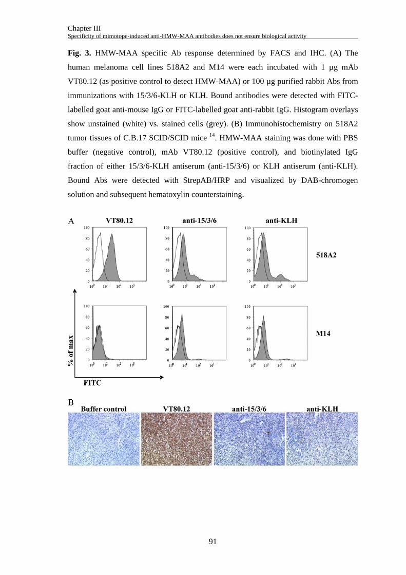
Chapter III Specificity of mimotope-induced anti-HMW-MAA antibodies does not ensure biological activity
91
Fig. 3. HMW-MAA specific Ab response determined by FACS and IHC. (A) The
human melanoma cell lines 518A2 and M14 were each incubated with 1 µg mAb
VT80.12 (as positive control to detect HMW-MAA) or 100 µg purified rabbit Abs from
immunizations with 15/3/6-KLH or KLH. Bound antibodies were detected with FITC-
labelled goat anti-mouse IgG or FITC-labelled goat anti-rabbit IgG. Histogram overlays
show unstained (white) vs. stained cells (grey). (B) Immunohistochemistry on 518A2
tumor tissues of C.B.17 SCID/SCID mice 14. HMW-MAA staining was done with PBS
buffer (negative control), mAb VT80.12 (positive control), and biotinylated IgG
fraction of either 15/3/6-KLH antiserum (anti-15/3/6) or KLH antiserum (anti-KLH).
Bound Abs were detected with StrepAB/HRP and visualized by DAB-chromogen
solution and subsequent hematoxylin counterstaining.
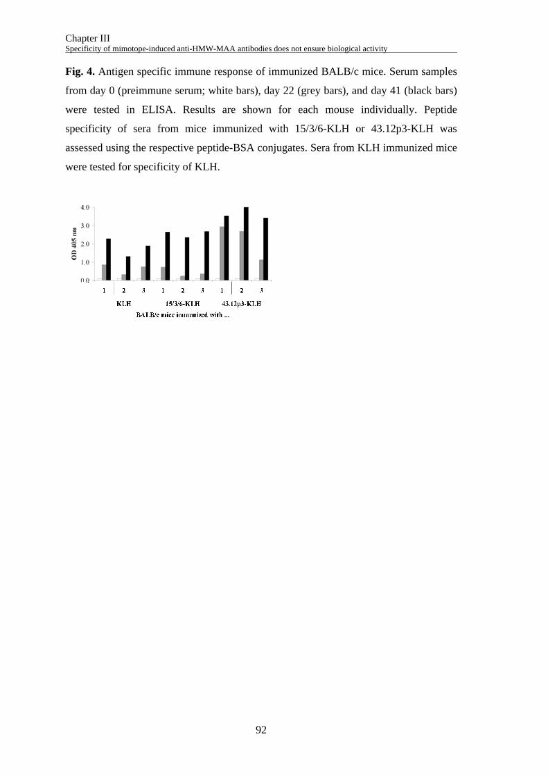
Chapter III Specificity of mimotope-induced anti-HMW-MAA antibodies does not ensure biological activity
92
Fig. 4. Antigen specific immune response of immunized BALB/c mice. Serum samples
from day 0 (preimmune serum; white bars), day 22 (grey bars), and day 41 (black bars)
were tested in ELISA. Results are shown for each mouse individually. Peptide
specificity of sera from mice immunized with 15/3/6-KLH or 43.12p3-KLH was
assessed using the respective peptide-BSA conjugates. Sera from KLH immunized mice
were tested for specificity of KLH.
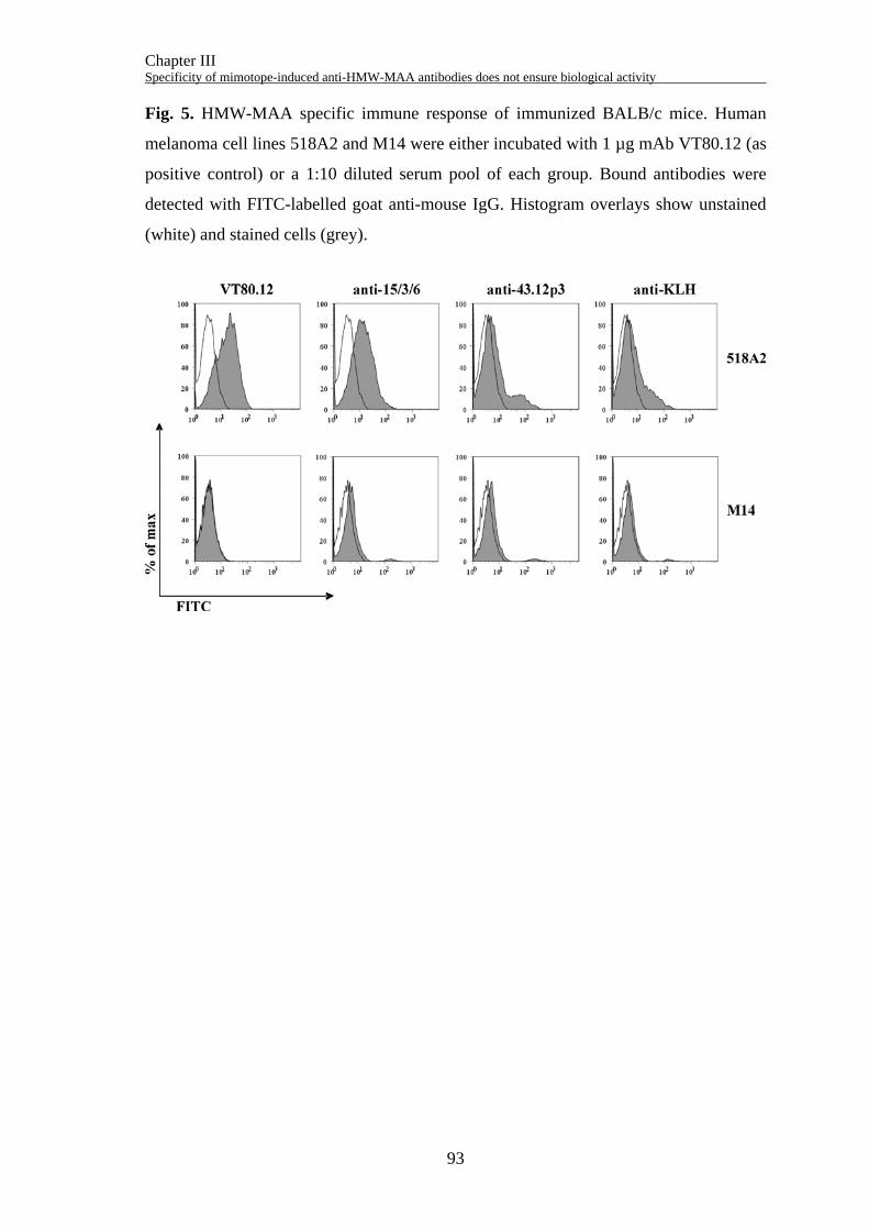
Chapter III Specificity of mimotope-induced anti-HMW-MAA antibodies does not ensure biological activity
93
Fig. 5. HMW-MAA specific immune response of immunized BALB/c mice. Human
melanoma cell lines 518A2 and M14 were either incubated with 1 µg mAb VT80.12 (as
positive control) or a 1:10 diluted serum pool of each group. Bound antibodies were
detected with FITC-labelled goat anti-mouse IgG. Histogram overlays show unstained
(white) and stained cells (grey).
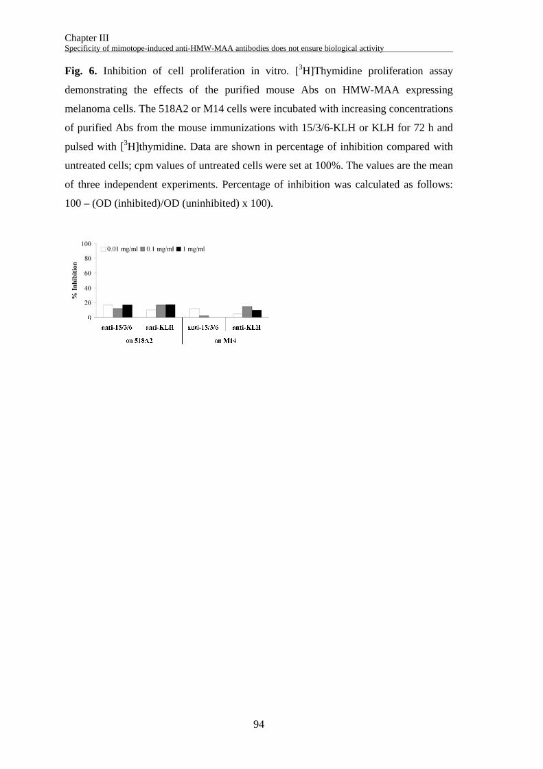
Chapter III Specificity of mimotope-induced anti-HMW-MAA antibodies does not ensure biological activity
94
Fig. 6. Inhibition of cell proliferation in vitro. [3H]Thymidine proliferation assay
demonstrating the effects of the purified mouse Abs on HMW-MAA expressing
melanoma cells. The 518A2 or M14 cells were incubated with increasing concentrations
of purified Abs from the mouse immunizations with 15/3/6-KLH or KLH for 72 h and
pulsed with [3H]thymidine. Data are shown in percentage of inhibition compared with
untreated cells; cpm values of untreated cells were set at 100%. The values are the mean
of three independent experiments. Percentage of inhibition was calculated as follows:
100 – (OD (inhibited)/OD (uninhibited) x 100).
Chapter III Specificity of mimotope-induced anti-HMW-MAA antibodies does not ensure biological activity
95
References
1. Campoli MR, Chang CC, Kageshita T, Wang X, McCarthy JB, Ferrone S. Human
high molecular weight-melanoma-associated antigen (HMW-MAA): a melanoma
cell surface chondroitin sulfate proteoglycan (MSCP) with biological and clinical
significance. Crit Rev Immunol 2004;24:267-96.
2. Wang X, Wang Y, Yu L, Sakakura K, Visus C, Schwab JH, et al. CSPG4 in cancer:
multiple roles. Curr Mol Med 2010;10:419-29.
3. Chang CC, Campoli M, Luo W, Zhao W, Zaenker KS, Ferrone S. Immunotherapy
of melanoma targeting human high molecular weight melanoma-associated antigen:
potential role of nonimmunological mechanisms. Ann N Y Acad Sci 2004;1028:340-
50.
4. Bender H, Grapow M, Schomburg A, Reinhold U, Biersack HJ. Effects of
diagnostic application of monoclonal antibody on survival in melanoma patients.
Hybridoma 1997;16:65-8.
5. Mittelman A, Chen ZJ, Liu CC, Hirai S, Ferrone S. Kinetics of the immune
response and regression of metastatic lesions following development of humoral
anti-high molecular weight-melanoma associated antigen immunity in three patients
with advanced malignant melanoma immunized with mouse antiidiotypic
monoclonal antibody MK2-23. Cancer Res 1994;54:415-21.
6. Mittelman A, Chen ZJ, Yang H, Wong GY, Ferrone S. Human high molecular
weight melanoma-associated antigen (HMW-MAA) mimicry by mouse anti-
idiotypic monoclonal antibody MK2-23: induction of humoral anti-HMW-MAA
immunity and prolongation of survival in patients with stage IV melanoma. Proc
Natl Acad Sci U S A 1992;89:466-70.
7. Maciag PC, Seavey MM, Pan ZK, Ferrone S, Paterson Y. Cancer immunotherapy
targeting the high molecular weight melanoma-associated antigen protein results in
a broad antitumor response and reduction of pericytes in the tumor vasculature.
Cancer Res 2008;68:8066-75.
8. Zhao L, Liu Z, Fan D. Overview of mimotopes and related strategies in tumor
vaccine development. Expert Rev Vaccines 2008;7:1547-55.
9. Bramswig KH, Knittelfelder R, Gruber S, Untersmayr E, Riemer AB, Szalai K, et
al. Immunization with mimotopes prevents growth of carcinoembryonic antigen
positive tumors in BALB/c mice. Clin Cancer Res 2007;13:6501-8.
Chapter III Specificity of mimotope-induced anti-HMW-MAA antibodies does not ensure biological activity
96
10. Riemer AB, Hantusch B, Sponer B, Kraml G, Hafner C, Zielinski CC, et al. High-
molecular-weight melanoma-associated antigen mimotope immunizations induce
antibodies recognizing melanoma cells. Cancer Immunol Immunother 2005;54:677-
84.
11. Riemer AB, Klinger M, Wagner S, Bernhaus A, Mazzucchelli L, Pehamberger H,
et al. Generation of Peptide mimics of the epitope recognized by trastuzumab on the
oncogenic protein Her-2/neu. J Immunol 2004;173:394-401.
12. Engelhard VH, Bullock TN, Colella TA, Sheasley SL, Mullins DW. Antigens
derived from melanocyte differentiation proteins: self-tolerance, autoimmunity, and
use for cancer immunotherapy. Immunol Rev 2002;188:136-46.
13. Wagner S, Hafner C, Allwardt D, Jasinska J, Ferrone S, Zielinski CC, et al.
Vaccination with a human high molecular weight melanoma-associated antigen
mimotope induces a humoral response inhibiting melanoma cell growth in vitro. J
Immunol 2005;174:976-82.
14. Wagner S, Krepler C, Allwardt D, Latzka J, Strommer S, Scheiner O, et al.
Reduction of human melanoma tumor growth in severe combined immunodeficient
mice by passive transfer of antibodies induced by a high molecular weight
melanoma-associated antigen mimotope vaccine. Clin Cancer Res 2008;14:8178-
83.
15. Ray S, Chhabra A, Mehrotra S, Chakraborty NG, Ribas A, Economou J, et al.
Obstacles to and opportunities for more effective peptide-based therapeutic
immunization in human melanoma. Clin Dermatol 2009;27:603-13.
16. Pilla L, Rivoltini L, Patuzzo R, Marrari A, Valdagni R, Parmiani G. Multipeptide
vaccination in cancer patients. Expert Opin Biol Ther 2009;9:1043-55.
17. Spiridon CI, Ghetie MA, Uhr J, Marches R, Li JL, Shen GL, et al. Targeting
multiple Her-2 epitopes with monoclonal antibodies results in improved antigrowth
activity of a human breast cancer cell line in vitro and in vivo. Clin Cancer Res
2002;8:1720-30.
18. Wagner S, Jasinska J, Breiteneder H, Kundi M, Pehamberger H, Scheiner O, et al.
Delayed tumor onset and reduced tumor growth progression after immunization
with a Her-2/neu multi-peptide vaccine and IL-12 in c-neu transgenic mice. Breast
Cancer Res Treat 2007;106:29-38.
Chapter III Specificity of mimotope-induced anti-HMW-MAA antibodies does not ensure biological activity
97
19. Giacomini P, Veglia F, Cordiali Fei P, Rehle T, Natali PG, Ferrone S. Level of a
membrane-bound high-molecular-weight melanoma-associated antigen and a
cytoplasmic melanoma-associated antigen in surgically removed tissues and in sera
from patients with melanoma. Cancer Res 1984;44:1281-7.
20. Matsui M, Temponi M, Ferrone S. Characterization of a monoclonal antibody-
defined human melanoma-associated antigen susceptible to induction by immune
interferon. J Immunol 1987;139:2088-95.
21. Wilson BS, Imai K, Natali PG, Ferrone S. Distribution and molecular
characterization of a cell-surface and a cytoplasmic antigen detectable in human
melanoma cells with monoclonal antibodies. Int J Cancer 1981;28:293-300.
22. Jasinska J, Wagner S, Radauer C, Sedivy R, Brodowicz T, Wiltschke C, et al.
Inhibition of tumor cell growth by antibodies induced after vaccination with
peptides derived from the extracellular domain of Her-2/neu. Int J Cancer
2003;107:976-83.
23. Felici F, Castagnoli L, Musacchio A, Jappelli R, Cesareni G. Selection of antibody
ligands from a large library of oligopeptides expressed on a multivalent exposition
vector. J Mol Biol 1991;222:301-10.
24. Holmes K, Lantz LM, Fowlkes BJ, Schmid I, Giorgi JV. Preparation of cells and
reagents for flow cytometry. Curr Protoc Immunol 2001;Chapter 5:Unit 5.3.
25. Brunner R, Jensen-Jarolim E, Pali-Scholl I. The ABC of clinical and experimental
adjuvants--a brief overview. Immunol Lett 2010;128:29-35.
26. Hafner C, Wagner S, Allwardt D, Riemer AB, Scheiner O, Pehamberger H, et al.
Cross-reactivity of mimotopes with a monoclonal antibody against the high
molecular weight melanoma-associated antigen (HMW-MAA) does not predict
cross-reactive immunogenicity. Melanoma Res 2005;15:111-7.
27. Yang X, Yu X. An introduction to epitope prediction methods and software. Rev
Med Virol 2009;19:77-96.
28. Campoli M, Ferrone S, Wang X. Functional and clinical relevance of chondroitin
sulfate proteoglycan 4. Adv Cancer Res 2010;109:73-121.
29. Li M, Yan Z, Han W, Zhang Y. Mimotope vaccination for epitope-specific
induction of anti-CD20 antibodies. Cell Immunol 2006;239:136-43.
30. Perosa F, Favoino E, Caragnano MA, Dammacco F. CD20 mimicry by a mAb
rituximab-specific linear peptide: a potential tool for active immunotherapy of
autoimmune diseases. Ann N Y Acad Sci 2005;1051:672-83.
Chapter III Specificity of mimotope-induced anti-HMW-MAA antibodies does not ensure biological activity
98
31. Perosa F, Favoino E, Vicenti C, Guarnera A, Racanelli V, De Pinto V, et al. Two
structurally different rituximab-specific CD20 mimotope peptides reveal that
rituximab recognizes two different CD20-associated epitopes. J Immunol
2009;182:416-23.
32. Perosa F, Favoino E, Vicenti C, Merchionne F, Dammacco F. Identification of an
antigenic and immunogenic motif expressed by two 7-mer rituximab-specific cyclic
peptide mimotopes: implication for peptide-based active immunotherapy. J
Immunol 2007;179:7967-74.
33. Hartmann C, Muller N, Blaukat A, Koch J, Benhar I, Wels WS. Peptide mimotopes
recognized by antibodies cetuximab and matuzumab induce a functionally
equivalent anti-EGFR immune response. Oncogene 2010;29:4517-27.
34. Riemer AB, Kurz H, Klinger M, Scheiner O, Zielinski CC, Jensen-Jarolim E.
Vaccination with cetuximab mimotopes and biological properties of induced anti-
epidermal growth factor receptor antibodies. J Natl Cancer Inst 2005;97:1663-70.
35. Jiang B, Liu W, Qu H, Meng L, Song S, Ouyang T, et al. A novel peptide isolated
from a phage display peptide library with trastuzumab can mimic antigen epitope of
HER-2. J Biol Chem 2005;280:4656-62.
36. Hafner C, Breiteneder H, Ferrone S, Thallinger C, Wagner S, Schmidt WM, et al.
Suppression of human melanoma tumor growth in SCID mice by a human high
molecular weight-melanoma associated antigen (HMW-MAA) specific monoclonal
antibody. Int J Cancer 2005;114:426-32.

99
Chapter IV
Concluding Remarks

100
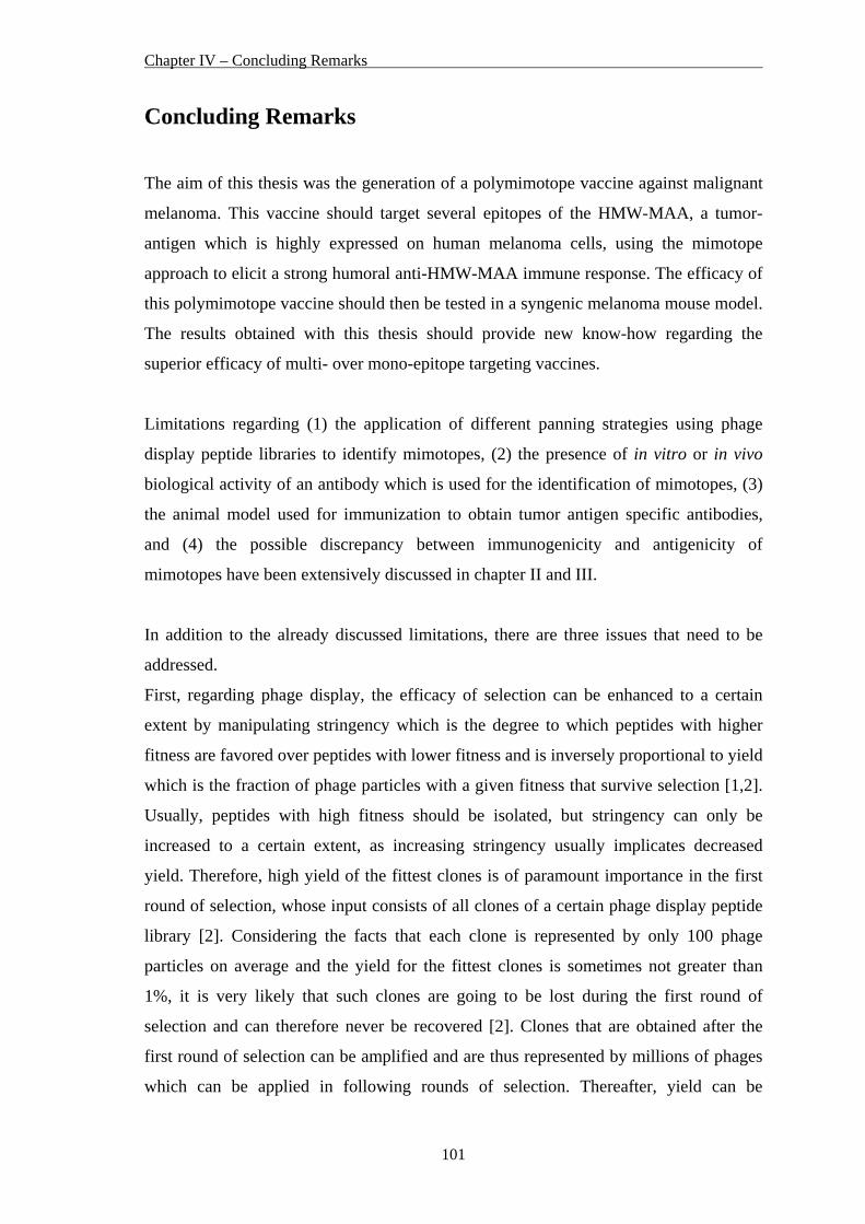
Chapter IV – Concluding Remarks
101
Concluding Remarks
The aim of this thesis was the generation of a polymimotope vaccine against malignant
melanoma. This vaccine should target several epitopes of the HMW-MAA, a tumor-
antigen which is highly expressed on human melanoma cells, using the mimotope
approach to elicit a strong humoral anti-HMW-MAA immune response. The efficacy of
this polymimotope vaccine should then be tested in a syngenic melanoma mouse model.
The results obtained with this thesis should provide new know-how regarding the
superior efficacy of multi- over mono-epitope targeting vaccines.
Limitations regarding (1) the application of different panning strategies using phage
display peptide libraries to identify mimotopes, (2) the presence of in vitro or in vivo
biological activity of an antibody which is used for the identification of mimotopes, (3)
the animal model used for immunization to obtain tumor antigen specific antibodies,
and (4) the possible discrepancy between immunogenicity and antigenicity of
mimotopes have been extensively discussed in chapter II and III.
In addition to the already discussed limitations, there are three issues that need to be
addressed.
First, regarding phage display, the efficacy of selection can be enhanced to a certain
extent by manipulating stringency which is the degree to which peptides with higher
fitness are favored over peptides with lower fitness and is inversely proportional to yield
which is the fraction of phage particles with a given fitness that survive selection [1,2].
Usually, peptides with high fitness should be isolated, but stringency can only be
increased to a certain extent, as increasing stringency usually implicates decreased
yield. Therefore, high yield of the fittest clones is of paramount importance in the first
round of selection, whose input consists of all clones of a certain phage display peptide
library [2]. Considering the facts that each clone is represented by only 100 phage
particles on average and the yield for the fittest clones is sometimes not greater than
1%, it is very likely that such clones are going to be lost during the first round of
selection and can therefore never be recovered [2]. Clones that are obtained after the
first round of selection can be amplified and are thus represented by millions of phages
which can be applied in following rounds of selection. Thereafter, yield can be
Chapter IV – Concluding Remarks
102
decreased in favor of high stringency for the subsequent rounds. However, there is a
limit to stringency, because in practice selection techniques are imperfect and there is an
unavoidable background yield of all phages regardless of their fitness [2]. If stringency
is set too high, the yield of a specifically selected phage will fall far below the
background of a nonspecifically isolated phage, and all power of discrimination in favor
of high fitness is lost [2].
Second, regarding the fact that a peptide which binds its selector antibody (thus
qualifying it as an antigenic mimic) is not able to elicit antibodies directed against the
target antigen when used to immunize naive animals (thus failing as an immunogenic
mimic), there are additional considerations which should be mentioned [2].
Small peptides are often flexible and might adopt a certain conformation when binding
the selector antibody but other conformations when eliciting new antibodies upon
immunization [3,4]. Therefore, only few of these antibodies, if any, would cross-react
with the authentic epitope. Furthermore, a peptide might be an antigenic mimic without
being a true structural mimic. Such a peptide would bind the selector antibody in an
entirely different way than does the original authentic epitope, via altogether different
interactions [2].
However, the induction of mimotope specific antibodies is a much more difficult
process and varies considerably from one mimotope to another. This may be reasonable
since the immune system can differentiate different antigens and produces very different
responses [5].
Third, as short synthetic peptides representing B cell epitopes, continuous/linear or
discontinuous/conformational, are generally poor immunogens, they require conjugation
to a carrier protein providing T helper cell epitopes and usually the co-administration of
an adjuvant in order to induce an antibody response [6-8].
Because it is now difficult to obtain carriers such as tetanus toxoid (TT) or diphtheria
toxoid, it seems to be a good and cheap alternative to use KLH, as it is a large (>2000
kDa), highly glycosylated protein frequently used as a carrier protein in animals and
humans due to its potent immunogenicity [9,10]. Nevertheless, there are epitopes on the
surface of KLH that cross-react with a variety of different carbohydrate antigens [10].
May et al. immunized mice with mimotopes, which were identified for the
glucuronoxylomannan (GXM) component of the capsular polysaccharide of
Chapter IV – Concluding Remarks
103
Cryptococcus neoformans, conjugated to KLH as carrier. They observed that KLH
alone induced antibodies that cross-reacted with GXM, but did not confer protection
against cryptococcal infection, suggesting that the cross-reactive epitope on KLH was
not protective [10].
Additionally, different carriers may influence the immunogenicity of a mimotope
conjugate. In this concern, Li et al. showed that antibodies induced upon immunization
of mice with a CD20 mimotope coupled to KLH were capable of mediating more
efficient complement killing of CD20+ cells when compared to the antibodies from mice
vaccinated with the respective TT-conjugate [11]. Wondimu et al. observed the opposite
when using GD2 ganglioside mimotopes coupled to KLH or synthesized as MAP
(multiple-antigen peptides). Here, the KLH-conjugate was less efficient than the MAP
[12].
In conclusion, the immunogenicity of a mimotope conjugate can be increased by using a
correct carrier and caution needs to be taken when creating vaccines for carbohydrate
antigens using KLH as carrier protein [10-12].
Chapter IV – Concluding Remarks
104
References 1. Bratkovic T. Progress in phage display: evolution of the technique and its
application. Cell Mol Life Sci 2010;67:749-67.
2. Smith GP, Petrenko VA. Phage Display. Chem Rev 1997;97:391-410.
3. Folgori A, Tafi R, Meola A, Felici F, Galfre G, Cortese R, et al. A general strategy
to identify mimotopes of pathological antigens using only random peptide libraries
and human sera. Embo J 1994;13:2236-43.
4. Motti C, Nuzzo M, Meola A, Galfre G, Felici F, Cortese R, et al. Recognition by
human sera and immunogenicity of HBsAg mimotopes selected from an M13
phage display library. Gene 1994;146:191-8.
5. Zhu ZY, Zhong CP, Xu WF, Lin GM, Ye GQ, Ji YY, et al. PSMA mimotope
isolated from phage displayed peptide library can induce PSMA specific immune
response. Cell Res 1999;9:271-80.
6. Charalambous BM, Feavers IM. Mimotope vaccines. J Med Microbiol
2001;50:937-9.
7. Partidos CD, Stanley CM, Steward MW. Immune responses in mice following
immunization with chimeric synthetic peptides representing B and T cell epitopes
of measles virus proteins. J Gen Virol 1991;72:1293-9.
8. Srinivasan M, Domanico SZ, Kaumaya PT, Pierce SK. Peptides of 23 residues or
greater are required to stimulate a high affinity class II-restricted T cell response.
Eur J Immunol 1993;23:1011-6.
9. Harris JR, Markl J. Keyhole limpet hemocyanin (KLH): a biomedical review.
Micron 1999;30:597-623.
10. May RJ, Beenhouwer DO, Scharff MD. Antibodies to keyhole limpet hemocyanin
cross-react with an epitope on the polysaccharide capsule of Cryptococcus
neoformans and other carbohydrates: implications for vaccine development. J
Immunol 2003;171:4905-12.
11. Li M, Yan Z, Han W, Zhang Y. Mimotope vaccination for epitope-specific
induction of anti-CD20 antibodies. Cell Immunol 2006;239:136-43.
12. Wondimu A, Zhang T, Kieber-Emmons T, Gimotty P, Sproesser K, Somasundaram
R, et al. Peptides mimicking GD2 ganglioside elicit cellular, humoral and tumor-
protective immune responses in mice. Cancer Immunol Immunother 2008;57:1079-
89.

105
Appendix

106
Appendix
107
Curriculum Vitae
Name: Mag. Sonja Gaier
Date of Birth: April 1, 1982
Place of Birth: Steyr, Austria
Nationality: Austria
Adress: Dornbacherstraße 12/11, 1170 Vienna, Austria
Contact: email: [email protected]
Education:
09/1988 – 07/1992 Elementary School, Gleink, Austria 09/1992 – 06/2000 Grammar School, Steyr, Austria general qualification for university entrance on June 21, 2000 10/2000 – 09/2006 Studies in Genetics/Microbiology, University of Vienna, Austria 05/2005 – 08/2006 Diploma thesis at the Department of Pathophysiology, Medical University of Vienna, Austria September 5, 2006 Received Masters degree (Mag. rer. nat.) 09/2006 – 12/2007 Project management and laboratory work within “EuroPrevall” (“The Prevalence, Cost and Basis of Food Allergy in Europe” – a European Commission funded project within the 6th framework programme, FOOD-CT-2005-514000) and a Biomay AG (Vienna, Austria) sponsored project on recombinant allergens at the Department of Pathophysiology and Allergy Research, Medical University of Vienna, Austria 10/2006 – current PhD studies in Genetics/Microbiology, University of Vienna, Austria PhD thesis at the Department of Pathophysiology and Allergy Research, Medical University of Vienna, Austria 05/2007 Research visit at the Paul Ehrlich Institute, Langen, Germany 01/2008 – 12/2010 Recipient of a DOC-fFORTE-fellowship of the Austrian Academy of Sciences

Appendix
108
Appendix
109
List of Publications
PAPERS as first author Gaier S*, Marsh J*, Oberhuber C*, Rigby NM, Lovegrove A, Alessandri S, Briza P,
Radauer C, Zuidmeer L, van Ree R, Hemmer W, Sancho AI, Mills C, Hoffmann-
Sommergruber K, Shewry PR. Purification and Structural Stability of the Peach
Allergens Pru p 1 and Pru p 3. Mol. Nutr. Food Res. 2008; 52(Suppl 2): 220-9
Gaier S, Oberhuber C, Hemmer W, Radauer C, Rigby NM, Marsh JT, Mills CE,
Shewry PR, Hoffmann-Sommergruber K. Pru p 3 as a marker for symptom severity
for peach allergic patients in a birch pollen environment. J Allergy Clin Immunol.
2009; 124(1): 166-7
Gaier S, Balazs N, Radauer C, Ferrone S, Hafner C, Breiteneder H, Wagner S.
Successful selection of mimotopes from phage-displayed libraries strongly depends
on the selection strategy. Manuscript submitted to Molecular Biotechnology
Gaier S*, Latzka J*, Hofstetter G, Balazs N, Ferrone S, Scheiner O, Breiteneder H,
Pehamberger H, Wagner S. Specificity of mimotope-induced anti-high molecular
weight-melanoma associated antigen (HMW-MAA) antibodies does not ensure
biological activity. Manuscript submitted to Vaccine
PAPERS as coauthor Oberhuber C, Bulley SM, Ballmer-Weber BK, Bublin M, Gaier S, DeWitt AM, Briza
P, Hofstetter G, Lidholm J, Vieths S, Hoffmann-Sommergruber K. Characterization of
Bet v 1 related allergens from kiwifruit using sera from patients with combined
kiwifruit and birch pollen allergy. Mol. Nutr. Food Res. 2008; 52(Suppl 2): 230-40
Appendix
110
Mandalari G, Adel-Patient K, Barkholt V, Baro C, Bennett L, Bublin M, Gaier S,
Graser G, Ladics GS, Mierzejewska D, Vassilopoulou E, Vissers YM, Zuidmeer L,
Rigby NM, Salt LJ, Defernez M, Mulholland F, Mackie AR, Wickham MS, Mills EN.
In vitro digestibility of beta-casein and beta-lactoglobulin under simulated human
gastric and duodenal conditions: A multi-laboratory evaluation. Regul Toxicol
Pharmacol. 2009; 55(3): 372-81
PRESENTATIONS at national and international congress meetings as presenting author Gaier S, Oberhuber C, Scheiner O, Hoffmann-Sommergruber K. Molecular
characterisation of Pru p 1, the Bet v 1 homologue from peach. Poster presentation
at the Annual Meeting of the Austrian Society of Allergology and Immunology (ÖGAI),
December 2005, Graz, Austria
Gaier S, Oberhuber C, Scheiner O, Hoffmann-Sommergruber K. Pru p 1, a major
food allergen from peach (Prunus persica). Poster presentation at the vfwf –
university lecture at the Medical University of Vienna, May 2006, Vienna, Austria
Gaier S, Oberhuber C, Rigby N, Marsh J, Hemmer W, Mills C, Shewry P, Scheiner O,
Hoffmann-Sommergruber K. IgE reactivity patterns of peach allergic patients in
Austria. Poster presentation at the Pirquet Symposium, December 2006, Vienna,
Austria
Gaier S, Oberhuber C, Rigby N, Marsh J, Mills C, Shewry P, Scheiner O, Hoffmann-
Sommergruber K. The effect of thermal processing and in vitro gastrointestinal
digestion on the stability of the peach allergens, Pru p 1 and Pru p 3. Poster
presentation at the Annual Meeting of the Austrian Society of Allergology and
Immunology (ÖGAI), December 2007, Alpbach, Austria
Appendix
111
Gaier S, Oberhuber C, Hemmer W, Rigby N, Marsh J, Mills C, Shewry P, Scheiner O,
Hoffmann-Sommergruber K. LTP – a relevant allergen for Austrian Rosaceae
allergic patients?. Poster presentation at the Annual Meeting of the Austrian Society of
Allergology and Immunology (ÖGAI), December 2007, Alpbach, Austria
Gaier S, Oberhuber C, Rigby N, Marsh J, Hemmer W, Mills C, Shewry P, Scheiner O,
Hoffmann-Sommergruber K. Allergenicity of peach allergens Pru p 1 and Pru p 3
related with protein stability. Poster presentation at the 27th EAACI Congress, June
2008, Barcelona, Spain
Gaier S, Oberhuber C, Rigby N, Marsh J, Hemmer W, Mills C, Shewry P, Scheiner O,
Hoffmann-Sommergruber K. pH conditions exert effects on the stability of the peach
allergens Pru p 1 and Pru p 3. Poster presentation at the Joint Annual Meeting of
Immunology of the Austrian and German Societies (ÖGAI, DGfI), September 2008,
Vienna, Austria
Gaier S, Latzka J, Ferrone S, Pehamberger H, Scheiner O, Breiteneder H, Wagner S.
Selection of HMW-MAA mimotopes for the generation of a polymimotope vaccine
against malignant melanoma. Poster presentation at the Joint Annual Meeting of
Immunology of the Austrian and German Societies (ÖGAI, DGfI), September 2008,
Vienna, Austria
Gaier S, Ferrone S, Hafner C, Pehamberger H, Breiteneder H, Wagner S. HMW-MAA
mimotopes for the generation of a polymimotope vaccine against malignant
melanoma. Poster presentation at the Joint Meeting of the 7th World Congress on
Melanoma and 5th Congress of the European Association of Dermato-Oncology
(EADO), May 2009, Vienna, Austria
Gaier S, Ferrone S, Hafner C, Pehamberger H, Breiteneder H, Wagner S. Design of a
polymimotope vaccine against malignant melanoma using HMW-MAA
mimotopes. Poster presentation at the 2nd European Congress of Immunology (ECI),
September 2009, Berlin, Germany
Appendix
112
OTHER PRESENTATIONS at national and international meetings Hoffmann-Sommergruber K, Sancho A, Holzhauser T, Skov P, Alessandri S, Wal J,
Rigby N, Marsh J, Bernard H, Gaier S, Lauer I, Ma Y, Oberhuber C, Barkholt V,
Griesmeier U, Bublin M, Radauer C, Scheurer S, Reese G, Zuidmeer L, Akkerdaas J,
Neubauer A, Fernández-Rivas M, Ballmer-Weber B, Vieths S, van Ree R, Shewry P,
Mills C. The Europrevall allergen library: a collection of high quality purified
natural and recombinant allergens for in vitro diagnosis. Poster at the 26th EAACI
Congress, June 2007, Göteborg, Sweden
Oberhuber C, Gaier S, Untersmayr-Elsenhuber E, Hafner C, Hoffmann-Sommergruber
K. Adverse reactions to dark chocolate – a case report. Poster at the World Allergy
Congress, December 2007, Bangkok, Thailand
Forstenlechner P, Ma Y, Gaier S, Oberhuber C, Scheiner O, Hoffmann-Sommergruber
K. Cloning of wheat profilin. Poster presentation at the Annual Meeting of the
Austrian Society of Allergology and Immunology (ÖGAI), December 2007, Alpbach,
Austria
Bruckmüller MU, Oberhuber C, Gaier S, Vázquez-Cortéz S, Fernández-Rivas M,
Hoffmann-Sommergruber K. Melon is a relevant elicitor of food allergy in Southern
Europe. Poster at the Joint Annual Meeting of Immunology of the Austrian and
German Societies (ÖGAI, DGfI), September 2008, Vienna, Austria