dissertation submitted for branch – i.m.s. (general
TRANSCRIPT
THE TAMIL NADU DR. M.G.R. MEDICAL UNIVERSITY
CHENNAI
DIAGNOSTIC APPROACH TO PALPABLE BREAST LUMPS-
THE MODIFIED TRIPLE TEST
DISSERTATION SUBMITTED FOR
BRANCH – I.M.S. (GENERAL SURGERY)
SEPTEMBER – 2006
ACKNOWLEDGEMENT
I Whole heartedly thank with gratitude Prof. Dr.
M.Kalyanasundaram M.S., FICS., Professor and Head of Department of Surgery
Govt. Rajaji Hospital and Madurai Medical College, Madurai for encouragement,
advice and guidance in the preparation of this study.
My special thanks goes to Prof. Dr. V.Seetharaman M.S., my unit chief who
had been a constant Source of encouragement and inspiration for the completion of
this study.
I am grateful to our Dean Dr. Saraswathy for her kind permisision to utilize
the clinical material necessary for conducting this study.
I am grateful to my Asst. Professors Dr. S.P.Ramanthan M.S., DNB., Dr.
P.Amutha M.S., Dr. M.Sekaran M.S., for their immense help and guidance.
Last but not the least, I thank all the patients for their kind cooperation in
carrying out the study successfully.
DECLARATION
This is consolidated report on “ DIAGNOSTIC APPROACH TO PALPABLE BREAST LUMPS THE MODIFIED TRIPLE TEST”, a study conducted at Department of General Surgery, Govt. Rajaji Hospital Madurai, during the period July 2003 to Jan 2006.
This is submitted to TN Dr. M.G.R. Medical University, Chennai in partial fulfillment of the rules and regulations for the MS Degree Examination in General Surgery.
Govt. Rajaji Hospital Dr. M.Lakshmi Narayanan
Madurai.
CONTENTS
Page
1. INTRODUCTION 1
2. REVIEW OF LITERATURE 3
3. DISCUSSION 6
4. CLINICAL EXAMINATION 9
5. BREAST ULTRASOUND 13
6. FINE NEEDLE ASPIRATION CYTOLOGY 30
7. CORE NEEDLE BIOPSY 38
8. AIM OF STUDY 40
9. MATERIALS AND METHODS 41
10. ANALYSIS OF DATA 45
11. CONCLUSION 52
12. PROFORMA
13. BIBLIOGRAPHY
14. MASTER CHART
15. ABREVIATIONS

INTRODUCTION
Until a few years ago, it was generally believed that breast tumor should be excised and
histologically examined to determine its nature with certainty – because the preoperative physical
assessment alone was associated with too much uncertainty.
Eventually, with the advent of mammography, a radiological tool became available to the
surgeons to make a pre-operative diagnosis of the breast with a reasonable degree of accuracy.
However, it was the introduction of FNAC that changed the entire outlook to the matter. The
combination of Physical examination, Mammography and FNAC came to be called upon as the “Triple
Test” for assessment of breast lumps and has now become the gold standard in the work-up of the
same.
Breast Ultrasound has now become available at higher resolutions, and is proving to be a highly
useful adjunct to mammography. The wide acceptance of ultrasound as a diagnostic modality has been
documented extensively in literature. However, opinions vary about the usefulness of USG breast in the
evaluation of masses, and, surgeons are cautioned to be aware of its attributes as well as its
deficiencies.
The ‘Modified Triple Test’ utilizes-Physical Examination, Ultrasonography of the breast as the
radiological method, and FNAC for the diagnosis of palpable breast lumps; and it is gaining acceptance
with the recent advances in technology and refinements in the interpretative criteria of sonographically
characterized masses.
Today, the studies concentrate on whether a benign result of the tests mentioned above makes
excision biopsy unnecessary. Most often, this question arises in connection with localized changes in
breasts with Fibrocystic diseases. For these tests to be adopted they must however, have the same
degree of accuracy as excisional biopsy – because, non-excision of a malignant tumor is unacceptable.
REVIEW OF LITERATURE
Medical literature abounds with studies of evaluation of breast lumps that emphasizes that the
statement “every palpable mass must be assessed and clarified”.
Hermansen C. et.al4 in 1987 prospectively studied 650 breast tumors and applied the term
‘Triple test’ to the traid of physical examination; mammography and FNAC used to diagnose them. He
concluded that the diagnostic accuracy of the triple test is comparable to that of histological
examination. Hardy JR. et. Al6. assessed 143 patients with palpable breast modules with clinical
examination; FNAC mammography; ultrasonography and magnetic resonance imaging (MRI) and
concluded that the combination of cytology and ultrasound was best at correctly diagnosing
malignancy.
Lawrence N Bassett et. Al.7 assessed the usefulness of mammography and sonography in
women less than 35years of age (1016 women) during a 8 year period. This study found that
mammography was not useful in women less than 35 years. However sonography was useful in
avoiding unnecessary biopsies and for this reason was the intial examination in younger women. But it
was not useful in detecting nonpalpable carcinomas or in differentiating benign from malignant solid
masses.
Vetto JJ et al in 1996 studied 55 women below the recommended age of screening
mammography with the 3 elements of ‘Modified Triple Test’
(C/E, Usg; FNAC/CNB). The test had a specificity and negative predictive value of 100% for
malignancy. They concluded that use of MTT for diagnosis of palpable breast lesions in younger
women yields high diagnostic accuracy without the need for routine open biopsy, resulting in overall
reduction patient charges.
Purasri P et. al3. retrospectively assessed 603 patients with breast lumps using the ‘Quadruple
test’ – C/E/USG/Mammography/FNAC. A stepwise logistic discriminant analysis was used to derive a

novel diagnostic index. This predicted the diagnosis in 98% of women <35 years correctly.
Hatada T et. al5. retrospectively studied 114 lesions and compared diagnoses obtained by
standard FNAC and that of ultrasound guided FNAC with surgical findings and found the accuracy to
be 65% and 86% respectively. They concluded that Usg-guided FNAC improves the preoperative
diagnosis especially in patients with tumor less than 2 cm.
Heiken TT et al conducted a prospective analysis of office-bases breast ultrasound, on 660
breast lesions and found that suspicious lesions determined by USG had a 75% chance of being
malignant; however 5% of lesions characteristic of fibroadenoma turned out to be malignancies.
Jill S Montrey8 attempted to determine the usefulness of ultrasound as a screening tool for
breast cancer in women <35 years, with indeterminate mammography, persistent symptoms and high
risk history.
Literary review of Breast Usg for screening :-
Carcinoma detected
Frazier et al 1982 (3) 3/135 (2.2%)
Kopens et al 1985 (2) 3/127 (2.4%)
Basset et al 1987 (1) 1/612 (0.2%)
Frazier et al 1985 (4) 36/600 (6%)
Thus breast ultrasonography when used correctly can be effective, but is not fool proof,
especially in lesion <1cm.

DISCUSSION
Breast lumps are an enormous source of anxiety to patients and a stratified approach has to be
adopted to properly (chart I) evaluate and treat them. Though benign breast diseases are more
common; there is a preoccuption with malignancy (undue but understandable) amongst the patients and
physicians alike.
The ultimate diagnosis and treatment of a patient with a dominant breast mass rests with the
clinician. A dominant breast mass is defined as a solid or cystic lump that persists throughout the
menstrual cycle.
The pre op assessment of a breast mass begins with a through history and systematic physical
examination. However it is generally accepted that this alone can be inadequate for an accurate
diagnosis. The breast is naturally multiondular and hence the difficulty in appreciating small nodules
amidst the normal lumpiness is apparent. Apart from this is the varied expertise of the examiner in
breast palpation. Thus imaging of the breast using various physical imaging modalities is important.
The earlist recorded case of breast imaging is that of SALOMON, a German pathologist who in
1913 reported the use of the recently developed X-ray to visualize breast structure in amputated breasts.
He demonstrated the irregular mass density and microcalcification which are still in use as the most
important benchmarks to identify possible breast malignancy. Even today mammography remains the
method of choice for detecting the occult,
CHART – 1
COMMON BREAST SYMPTOMS

LUMP NIPPLE DISCHARGE PAIN
NODULATIRY LUMP CYCLICAL ONCYCLICAL
DIFFUSE >30YEARS
SYMMETRICAL LOCALIZED
ASSYMETRICAL
REASSURE
& TRIPLE TEST
DISCHARGE
PHYSICAL IMAGING TISSUE
EXAMINATION DIAGNOSIS
Flow chart show diagnostic protocol for patients with breast complaints
nonpalpable lesion and remains the standard for imaging of breast against which other modalities
advocated now are measured.
Other imaging modalities have now been introduced for
• Visualizing lesion missed by mammography
• To replace mammography with another efficient, more economic device that would alsobe used

by non-radiologists.
The diagnostic utility of ultrasonogram was documented in 1950s; but it was only with the
development of “grey scaling” by Korsakoff and associates in Sweden years later that better issue
representation was obtained. Recent technical advances in ultrasonography have expanded the potential
utility of this modality in the evaluation of breast lesions far beyond distinguishing solid abnormalities
from cystic ones.
Tissue diagnosis remains the most reliable confirmatory tool for breast lumps. In 1912, Ward
used fine needle aspiration (FNAC) Cytology to examine lymph nodes for lymphoma. Eventually the
procedure was attempted on patients with breast lumps in 1962 by Martin and Ellis at New York.
Cytological interpretation requires skill particularly in view of cell types and pathologies encountered,
but high levels of accuracy can be obtained with experience.
CLINICAL EXAMINATION
A palpable breast mass may be identified when it becomes it becomes sufficiently large to be
differentiated from surrounding breast tissue physically by an examiner – usually the patient or the
physician; or is perceived on an imaging examination. Determining by physical examination whether a
mass is present can be difficult, as, all breasts are variable in the combination of glandular tissue,
fibrosis and fat. True masses are generally asymmetric in relation to the other breast; distinct from the
surrounding tissue and three dimensional.
The underlying cause of complaints about the breast proves to be benign in the overwhelming
majority of cases. Breast symptoms, however, induce such a great anxiety in the patient that
malignancy needs to be excluded as speedily as possible.

The first steps in this are the classic ones of history and physical examination.
Essential points in the history include :
Age
Menstrual status
Family and reproductive history
Lactational history
Radiation to the chest
H/o benign breast disease
In physical examination of patients with a breast with a breast complaint-seclusion, warmth and
privacy are particularly important in the examination of the breast. This avoids discomfort and
embarrassment to the patient. Good lighting enables detection of minor abnormalities. The patient sits
stripped to the waist. Attention is paid to
GENERAL APPEARANCE :
Nourishment, colour, mucous membranes, palms.
Inspection from bed end :
1. Arms by side
2. Arms elevated. Looked for
Symmetry of nipples and breasts.
a. Niple abnormalities
b. Vascularity
c. Indrawing and prominence of skin
d. Tethering
e. Peau d’ orange
f. Skin nodules
g. Ulceration

Palpation :
1. Supraclavicular fossae
2. Breasts :
a. Lightly
b. Deeply systematically from the areola concentrically outwards including the
axillary tail.
3. Axillae :
1. Taking the weight of the patient’s forearm on the examiner’s.
2. Both axilae simultaneously from behind the patient.
3. Abdomen for ascites, hepatomegaly or abnormal masses.
Particularly valuable in practice are
a) inspection with arms firstly dependent, then elevated.
b) Intial light touch over the breats prior to systematic deepar palpation in
concentric circles, working outwards.
c) Simulatancous examination of the axillae performed from behind the patient.
Atypical cancer may be firm and have indistinct borders and attachments to the skin or fascia
with dimpling or nipple retraction. Benign lesions typically have discrete borders, well defind margins
and are mobile. Cysts can be differentiated from solid lesions by palpation.
Rosner et al reported that physical examination can correctly identify only 58% of 66 patients
with cysts. Significant discordance among experienced examiners may occur. In one study, surgeons
performed physical examination independently and agreed on the need for biopsy in only 73% of 15
masses subsequently proved malignant. (Boyd et al).
Somers et al studied certain palpable abnormalities defined as areas of thickening,
predominence of tenderness without an associated dominant mass on physical examination; no
suspicious mammographic lesion; and rubbery, firm, cystic soft mass, needle sensation by FNA. The
incidence of malignancy in these ‘suspicious’ group was less than 1% (1/06), leading to the conclusion
that this subset of patients with palpable abnormalities did not require surgical biopsy.
Nevertheless, the physical findings or benign disease and malignancy in its earliest stages may
overlap; and without the use of FNA and/or breast imaging, some palpable malignant lesions may be
followed up inappropriately; leading to serious consequences for both the patient and the physician.
Although some masses exhibit distinct physical findings, an imaging evaluation is required in
almost all cases to characterize the palpable lesion, search for ipsilateral multifocal or multicentric
carinoma, and screen the contralateral breast. A negative imaging evaluation, however, should never
over rule a strongly suspiscious finding on physical examination or vice versa.
********
ULTRASOUND OF THE BREST
Although diagnostic ultrasound equipment has been available since the 1950s. it is only in
relatively recent years that the widespread use of this technique has been accepted by radiologists and
then by surgeons. A lot of this has related to difficulties with earlier machines.
HISTORY
The transducers that produce the ultrasonic signal were initially quite crude devices that
operated with low frequencies. Although penetration of the signal is better at these frequencies,
resolution of tissue abnormalities is not as good. It was not until the development of ‘gray scaling’ by
Korsakoff and associates in Sydney that better tissue representation was obtained. As the name of the
technique implies, the images now obtained were in shapes of gray and this shading was corresponding
to tissue changes and particularly to areas of localized pathology. Improvements in transducer design
followed, allowing various frequencies to be used, with the higher frequencies used in areas such as the
breast, thyroid and testis.
Even with all the advances, it must be stated and emphasized that the use of USG equipment is
very operator dependant and interpretation of the images requires a large amount of practical training.
Skilled interpreters of ultrasonic images, do, however, follow 3 golden rules –
1. Never make an interpretation on a single image, superimpose the ultrasonic images mentally
to formulate a 3 dimensional image of the scanned tissues and to ensure that the displayed
feature is consistent with the 3D image.
2. Because a feature is displayed it is not necessarily real – always rule out artifacts.
3. Because a feature is not displayed it is not necessarily not there.
The original use of USG was to determine whether a breast mass was a cyst or solid lesion.
Advances in technology and refinements in interpretative criteria have expanded the role of USG in
characterizing masses as having benign, malignant or equivocal features.
BREST ULTRASOUND TECHNIQUE :
Confirm the location of the mass noted on physical examination; a diagram from the referring
physicians to demonstrate the lesions’ position.
A high-resolution small parts transducer 7.5 – 10 mhz is used.
Evaluate the Region of Interest (ROI) to assess whether the clinical abnormality is
corresponding to the ultrasound findings.
Patient positioning: The patient is positioned in an oblique manner with a pillow placed under
the shoulder of the breast to be examined.
The degree of obliquity is determined by the position of the breast – aiming to have the breast
spread evenly over the wall, with the nipple pointing to the ceiling. The arm should be elevated
over the patient’s head to facilitate even distribution of the breast tissue, but should not be so
elevated that the breast is retracted superiorly. A little care exercised in positioning makes the USG
localization of the lesion less of a problem.
a. Lumps felt better in the upright position may be scanned in that position.
b. Fluid in cystic masses may be confirmed by changing to upright/decubits
position.
Region of interest is trapped with the examining finger and the transducer is placed directly
over the abnormality.
Examine in overlapping radial/antiradial planes – to determine relation of lesion to ducts and
avoid errors as mistaking fat islands for solid masses.
The entire periphery of the lesion must be evaluated in multiple planes through a 180 degree are
to determine the nature of margins, its shape and appearance of surrounding tissue.
Compression of tissue with the transducer is helpful in spreading apart breast tissues, flattening
islands of fatty tissue, and eliminating artifictual shadowing.
The rest of the breast tissue is systematically imaged from periphery to the nipple.
The retroareolar area can be evaluated with the transducer angled into the area in multiple
planes.
LABELING OF LESIONS :
The lesion is labelled on a clock face.
Its distance form nipple is given in centimeters.
The greatest diameter should be measured.
The height (AP diameter) is determined to obtain a height/width ratio.
NORMAL APPEARANCE OF THE BREAST :
Knowledge of normal variations of breast architecture is essential for the detection and accurate
diagnosis of abnormalities.
SKIN :
The skin is imaged as an echogenic layer of approximately 3mm or less in thickness, often a
hypoechnoic central line. Large areas of skin thickening are difficult to recognize without comparing
the ROI to the opposite breast or to a normal area within the same breast. A stand-off pad may be used
to detect subtle abnormalities.
SUBCUTANEOUS TISSUE :

The subcutaneous fat layer is a hypoechoic layer situated between the skin line and the breast
parenchyma. Cooper’s ligaments are imaged as curvilinear lines extending from the breast tissue to the
superficial fasical layer-producing a scalloped appearance.
Breast cancer does not arise in the subcutaneous but may involve it by direct extension.
Focal increased echotexture: Malignancy, inflammatory lesions, edema, fat necrosis, or biopsy
scar.
Diffuse increased reflectivity : edema of any cause (e.g., Heart failure), diffuse from of breast
cancer, inflammatory breast cancer, inflammatory mastitis, or radiation therapy.
Lesions indigenious to this plane are sebaceous cysts, epidermoid inclusion cysts,
hemangiomas, and rarely smooth muscle and fibrous tissue tumors.
NIPPLE AND AREOLA :
This is difficult area to visualize because of difficulties with transducer contact by nipple
protrusion or inversion, producing air trapping which is a source of shadowing. Compression of the
nipple and imaging with multiple planes angled toward the subareolar region can be of help. Ducts
can be visualized and traced upon the breast tissue, echogenic areas within the ducts usually represent
debris. Visualization of a mass within a dilated duct may indicate a papilloma, carcinoma or other
lesion.
BREAST PARENCHYMAL LAYER :
The breast tissue is more dense and uniform in the younger patient, age and parity however, are
not good predictors for the USG appearance for any one individual.
The breast parenchyma consists of ductal, lobular and fibrous tissue in varying proportions. The
normal appearance of the breast can range form almost completely fatty with only a few echogenic
fibroglandular tissue with little or no fat.
Whatever the relative composition of fibroglandular tissue and fat, the fibroglandular tissue
should have a regular appearance of echogenic structures arranged in multiple layers parallel to the
chest wall without distortion or swirled or pulled appearance.
Most benign and malignant lesions are imaged as nodules hypoechoic relative to paraenchyma
and are seen most easily in homogenously echogenic breasts, which do not deform with compression.
However, the most useful maneuver is imaging in multiple planes, which will demonstrate the
continuity of fat island with other areas of fat.
RETROMAMMARY AREA :
It is a hypoechoic fat plane, deep to the superficial fascia, pectoralis. Usually well demonstated
deep to the echogenic glandular tissue.
LYMPHNODES, ARTERIES, AND VEINS
Lymphonods are frequently imaged within the breast and axilla as oval nodules with an
eccentric hyperechoic hilum a surrounding less echogenic rim of tissue.
Benign nodes – large in size but maintain their morphology.
Malignant nodes – only gross abnormalities can be detected., when they appear as lobulated
hypoechoiec nodules.

Arteries and veins are visualized as tubular anechoic structures within the breast. Pulsation will
confirm the arterial nature of a vassel. Color Doppler can be used to demonstrate vascular structure
within the breast and tumors.
********
CHARACTERIZATION OF BREAST MASSES
CYSTS :
The first step in characterizing breast masses is cyst/solid differentiation. When the breast
masses meet all of the diagnostic criteria for cysts the USG accuracy approaches 100%.
The criteria include :
1. Round, oval lobulated shape.
2. Anechoic
3. Well-defined posterior border
4. Increased through and through transmission

5. No alteration of surrounding parenchyma.
Unfortunately, not all cysts display these criteria. The resolution of USG for cystic lesions is up to
5mm with 100% accuracy.
The presence of internal echoes is the most common diagnostic difficulty in cyst/solid
differentiation. Cysts have internal echoes due to
Presence of cholesterol or calcium
Hemorrhage
Infection
FOLLOW UP OF CYSTIC LESIONS
C hart – (2)
SIMPLE CYST
COMPLEX CYST
Confirm benign nature and observe
Follow-up Usg at 4-6 months
No malignant or indeterminate features
Observe
Large complex/indeterminate cysts
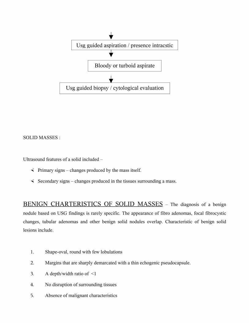
SOLID MASSES :
Ultrasound features of a solid included –
Primary signs – changes produced by the mass itself.
Secondary signs – changes produced in the tissues surrounding a mass.
BENIGN CHARTERISTICS OF SOLID MASSES – The diagnosis of a benign
nodule based on USG findings is rarely specific. The appearance of fibro adenomas, focal fibrocystic
changes, tubular adenomas and other benign solid nodules overlap. Characteristic of benign solid
lesions include.
1. Shape-oval, round with few lobulations
2. Margins that are sharply demarcated with a thin echogenic pseudocapsule.
3. A depth/width ratio of <1
4. No disruption of surrounding tissues
5. Absence of malignant characteristics
Usg guided aspiration / presence intracstic
Bloody or turboid aspirate
Usg guided biopsy / cytological evaluation

6. Homogenous low level internal echoes are usually present.
FIBROADENOMAS :
They are most common benign solid nodules. They are most commonly detected in the younger
age group. The USG appearance is determined by the relative amounts of fibrous and epithelial tissue.
c. Enhanced through and through transmission
d. Posterior attenuation in relation to the fibrous component
e. Coarse calcifications in degenerating fibroadenomas producing shadowing.
f. Tubular structures seen with lactating and juvenile types.
g. Different from fat lobules by their non compressible characteristics.
LIPMAS AND FIBROADENOMYOLIPOMA :
1. Lobulated masses that do not distort surrounding tissues.
2. Presence of internal due to fat
FAT NECECROSIS :
1. Focal hyperechoic nodule with a central lucency.
MALIGNANT CHARTISTICS OF BREAST LESIONS

16. Stellate masses
17. Circumscribed masses
18. Diffuse edema changes
19. Calcifications
Stellate masses : Desmoplastic reaction produces contraction of the breast tissue towards the mass,
disrupting the normal parallel soft tissue planes.
These masses have an irregular hypoechoic lesion with distortion of the surrounding breast tissue.
Disruption of normal trabecular structures.
Extension along the plane of the ducts.
Posterior acoustic shadowing.
Increased reflectivity of the subcutaneous tissue.
DD of stellate lesions :
Carcinoma ( most common )
Radial scar
Sclerosing adenosis
Post surgical scarring
Circumscribed masses : Breast cancer also commonly presents as hypoechoic masses
that appear to displace breast tissue.
Oval, round

Multilobulated
Height / width ratio > 1
>3 lobulations
reactive halo surrounding the mass
DD of circumscribed masses :
Carnioma – Usually the high grade NOS variety of ductal carcinoma & medullary,
colloid, and papillary ca.,
Metastatic lesions – from lymphona, melanoma, leukemia, sarcoma, adencarcinoma
from other sites.
Inflammation and abscesses.
Hematoma.
Phylloides tumour
Fibroadenoma.
Diffuse edema changes : A difficult type of carcinoma to distinguish is one that does
not produce a mass but only diffuse changes in echotexture and parenchymal pattern.
• Subtle increased reflectivity of parenchyma and subcutaneous fat.
• Loss of normal orientation of tissue planes.
• Poor differentiation in the fat-parenchyma interface.
DD of diffuse edema :
Lobular and ductal carcinoma, inflammatory carcinoma.
Breast contusion.

Congestive cardiac failure ( usually bilateral).
Breast / chest wall irradiation.
Superior vena cava obstruction.
Axillary Lymphatic obstruction.
Calcifications : Breast carcinoma may produce calcification both with and without
mass. Calcification of sufficient size and clustered variety in a homogenous tissue can be
picked up by USG.
Intracystic carcinomas : Rare lesions, usually papillary carcinoma that has obstructed a
duct and bleed producing a blood filled cyst.
DD of intracystic mass :
Papillary carcinoma
Necrotic solid tumor
Cysts containing hemorrhage, debirs
Abscess
Hematoma
With tha aid of the above criteria USG can come to a reliable diagnosis of breast
lumps, depending upon the size of the lesions. The minimum size of cystic lesions
picked up by USG are 2-3 mm. Solid lesions > 0.5 cm are usually categorized with
accuracy, 80% of solid lesions >2 cms and, 20% of lumps> 1-2cms 5% of lumps <1cm
is the diagnostic accuracy of USG.
*****

INDICATIONS FOR BREAST ULTRASOUND
From the above discussion we can summarise the now standardized indications
for Breast Ultrasound as follows :
1. Evaluation of a palpable lump or mammographic abnormality.
2. To confirm the existence of a lesion and its nature.
3. Single imaging modality in our patients or in pregnant or lactating patients
who present with symptoms.
4. To differentiate solid from cystic lesions.
5. For pathological examination of indeterminate lesions-guided FNA/CNB can
be carried out.
6. Pre operative localization of lesions with needle placements, documentation of
lesion removal by Usg of biopsy specimen.
7. Follow up of surgically altered breast – by tracing it through the breast and
subcutaneous layer to the biopsy scar on skin, as well as for guidance for FNA/
CNB.
8. Follow up after augmentation mammoplasty to detect extracapsular rupture /
contour abnormalities of the prosthesis.
9. Follow up of probably benign lesions that have not been biopsied.
10. Adjunctive to mammography as a screening tool for Carcinoma breast.
*****
ADVANTAGES OF USG OVER BREST MAMMOGRAPHY
1. No risk of radiation and its inherent consequences.
2. Can quality a lesion-as size, shape, echogenicity and relation to surrounding
tissues as against a mammographic report of a ‘density’ which is less specific.
3. Sonographically guided biopsies are more accurate than streotactic biopsies as
the needle can be visualized in real time throughout the entire procedure. This
adds confidence that the biopsy sample was obtained from the lesion.
4. Mammographic positioning for pre-operative localization of the masses is
cumbersome and time consuming and less accurate. Hence USG is the
modality of choice for guidance.
5. Surgically altered breast has confusing findings and is non-informative on
mammography.
6. Palpable lumps following extracapsular rupture of a prosthesis may be
impossible to image by mammography.
7. Long term follow-up of benign nodules not excised is associated with risk of
radiation on mammography and hence best performed by USG.
*****
LIMITATIONS OF BREAST ULTRASOUND
Breast ultrasound usage is avoided in certain situations where no efficacy has been
documented:
1. Routine evaluation of post operative breast.
2. Cancer Screening – as USG cannot detect all the non-palpable cancers that are
also missed on mammography.
3. Cannot detect microcalcification well; small solid lesions in fatty or mixed breast
tissue are not sonographically visible.
4. High false negative rate for non palpable cancers, (23%) which are smaller,
clinically occult.
5. Substantial false positive rate-sonogram may pick up ‘lesions’ which are rarely
clinically significant.
6. Unreliable in lesions<1 cm.
7. Less accurate in dense breasts.
These data suggest that breast ultrasound can be useful both as a single imaging
modality and as a adjunct of mammography in evaluation of patients with breast Jumps.
In addition, ultrasound facilitates preoperative needle biopsy of non-palpable
abnormalities, permitting timely and cost effective patient care.
*****
FINE NEEDLE ASPIRATION CYTOLOGY
The advent of cytology and availability of experienced cytologists has diminished
the diagnostic role of excision biopsy especially with regard to breast lumps.
FNAC can play a significant role in the early diagnosis of breast lumps to rule out
malignancy, as an attractive alternative to open biopsy. It is our responsibility to further
the integrity of this important procedure by understanding the merits and pitfalls of
breasts FNAC and adhering to the defined guidelines.
HISTORY :
Martin and Ellis in 1926, New York first introduced the concept of FNAC in the
midst of controversies regarding the credibility of the procedure. Breast FNAC is a
simple complex procedure that is influenced by many variables. Accurate interpretation
of breast FNAC requires clear guidelines for specimen acquisition, staining and
preparation.
Bamforth in 1966 defined cytology as; “The examination of cells obtained by
needle or drill biopsy in solid organs or tissue masses or form cut surface of such
materials freshly removed by surgical biopsy:.
Innumerable studies have now shown that cytological examination of cells obtained
this way is a rapid and reliable method of diagnosing malignant diseases of the breast.
REQUIRMENTS :
1. Spirit swab to clean the skin.
2. Disposable 10ml or 20ml syringe with fine needle 22G/23G.
3. A number of 76mm x 26mm microscopic Slides.
4. Fixatives like cytofix solution which is a commercial preparation, containing
absolute alcohol slides.
5. Transport box for prepared slides.

6. Complete lab. Request form giving patient’s particulats
PROCEDURE :
The Procedure is usually performed without anaesthesia. Very rarely local
anaesthesia is used. Local anaesthesia is generally not necessary because it is painful and
may obsure the mass.
1. The skin over the lump is wiped with antiseptic and the lump is fixed between
the fingers and the thumb and held steadily for needling. Then needle is guided
through the skin into the lesion.
2. Once the needle is felt to enter the lesion, the plunger is retracted to create
vacuum. Should the lesion prove to be cystic the fluid should be aspirated in its
entirety and expressed into a universal container and sent for cytological
examination.
3. If the lump is found to be solid, the needle is gently moved back and forth into
the substance of the tumor three or four times and is inserted into a different
part of the tumor each time to dislodge the cells from the tumor, so that the
aspirate will contain the sufficient material for cytological studies.
4. Throughout the technique a negative pressure must be sustained in the syringe.
Only after the tumor has been repeatedly probed, should suction be released
and the pressure in the syringe allowed to return gradually to atmospheric
pressure and the needle is withdrawn from the breast. The pressure in the
syringe must be adjusted in a way, so that the aspirated cells are retained in the
lumen of the needle, sustained suction as the needle is withdrawn result in, the
cells being withdrawn into the barrel of the syringe rendering them
inaccessible for processing.

PREPARATION OF ASPIRATE :
It is possible to make smear directly from the aspirate in the outpatient
department. Alternatively, the aspirated cells can be suspended in fixative and
transferred to cytologic laboratory for processing.
PREPARATION OF SMEAR :
After aspiration the needle and syringe are withdrawn from the skin.
Although nothing is visible in the syringe cells should have collected inside the
bore of the needle.
The needle is disconnected from the barrel, the syringe is filled with air.
A clean glass slide should be ready and the needle is reconnected to the barrel and
the aspirated material is blown out into the slide.
Any large tissue fragments which do not spread easily should be crushed between
the cover slip and the slide.
The smear is air dried and fixed in cytofix solution. Then the slide is duly
labelled.
FINATIVES :
A number of fixatives are used in cytology –
• 95% Ethyl alcohol
• 95% Ethyl Alcohol with 3% glacial acetic acid – to improve the nucleoprotein
fixing properties.
STAINING PROCEDURES :
Some cytopathologists prefer to interpret these slides stained
Papanicolaou method – when available this may be preferred in cytology for its
superior display of nuclear morphology and clear translucent demonstration of
cytoplasm.
Hematoxylin and Eosin method.
*****
NORMAL CYTOLOGY OF THE BREAST
The mammary gland is a compound gland made up of independent units
containing their own ducto-alveolar systems.
The alveoli are lined by simple cuboidal or low cuboidal epithelium which may
alter in size depending on the secretary status of the breast. The ducts are also similar in
lining, becoming pseudostratified along the main lactiferous and stratified as they
approach the nipple.
The typical cytological features in different breast condition are given below.
BENIGN :
FYBROADENOMA :
Duct cells are seen in large group and sheets surrounded by “stripped” nucleus.
A small degree of nuclear pleomorphism of duct cells is usually present.
Their high cellularity and moderate pleomorphism can be mistaken for a
malignancy.
FIBROCYSTIC DISEASE :

Numerous small foam cells and large metaplastic apocrine cells with acidophilic
cytoplasm.
Dark pyknotic nuclei,
Sheets and clusters of uniform elongated cell; with transparent cytoplasm and having
dendrite process.
PLASMA CELL MASTITIS :
Foam cells and sheets of depenerate epithelial cells are common.
Lipid containing macrophages and occasional foreign body giant cells.
Diagnostic feature is presence of large amounts of plasmacytes, Lymphyocytes
and esinophils and few neutrophils.
MALIGNANCY :
There are structured criteria for the diagnosis of malignancy by cytology, which
are stratified into :
Structural alterations in the cells.
Changes in inter relationship of cells in cell clusters.
Indirect criteria.
Structural modifications :
Alterations of nuclear cytoplasmic ratio with disproportionate enlargement of
nuclei.
Hyperchromasia due to increased chromosomal content, aberrant chromatin
pattern.
Increased number of nucleoli beyond the normal.
Multinucleation with nuclear atypia, abnormal mitotic figures.
Marked thickening of nuclear membrane.
Cytoplasmic changes enhanced by staining, such as pronounced
basophilia/acidophilia.
Presence of cytoplasmic inclusions like pigment granules, leukocytes and cellular
debris.
Atypical vacuolation especially in adenorcarcinoma.
Cell –Cell changes :
Enlargement of cells beyond normal shape.
Aberrant forms with associated nuclear atypia.
Degenerative or necrotic changes.
Lack of uniform orientation of cells and nuclei.
Anisocytosis/anisokaryosis – with marked variation in size of cells/nuclei
within the same cell cluster.
Loss of distinct cell boundaries.
Dense grouping of and crowding of cells and nuclei.
Engulfment of one cell by another.
Cells grouping into characteristic patterns.
Indirect Changes :
Presence of blood.
Increased Lymphocytes.
Prominent histiocytes and polymorphonuclear cells.

*****
ADVANTAGES OF FNAC :
1. Does not require anaesthesia.
2. Tolerated by patients as an OP procedure.
3. No secialized equipment required.
4. No damage to breast tissue that may occur with open biopsy.
5. Though small hematomas may be encountered, neither hemorrhage nor sepsis is
noted.
6. Remote chances of tumor seeding along needle tract.
7. Lymphatic / vascular dissemination of tumor is virtually unknown.
8. Can easily diagnose cysts / abscesses and treat them.
9. Eliminated need for open biopsy when a diagnosis of carcinoma is made that is
the incidence of false positive in malignancy is almost nil.
10. Can be easily repeated.
LIMITATIONS
1. Cannot qualify the type of malignancy.
2. False negative reporting when
a. Small tumors <1 cm.
b. Sclerotic lesions.
c. Deep seated tumors.
d. Large/pendulous breasts.
e. Lesions beyond the length of the needle.
*****
CORE NEEDLE BIOPSY (CNB)
Dominant lumps >2 cm in diameter can be approached by a wide bore cutting
needle; such as Tru-Cut needle that the advantage of producing a core of tissue which
can be paraffin embedded and used as a normal histological speciman with a modern
spring loaded device, with a local amaesthesia to the skin; a series of high quality cores
can be obtained rapidly and almost painlessly.
ADVANTAGES :
Ability to obtain a core of tissue that is sufficiently large for HPE
Providing more details of tumor structure.
Ability to distinguish between invasive and IDCA.
Any pathologist can interpret the results obviating the need for special skill of a
cytopathologist.
Large amount of prognostic information can be obtained including.
• Biological grade
• Receptor level
• DNA analysis
DISADVANTAGES :
Risk of seeding the needle tract with tumor cells.
Biopsy should be adjusted to avoid carrying potentially malignant tissue into the chest
wall.
False positive incase of radial scars.
False positive incase of hard lesions, deflecting the needle into surrounding fat.
Thus CNB can be adopted as an alternative office biopsy technique to FNAC
carrying sensitivity of 89%-100%.
*****
AIM OF STUDY
The study is conducted with the objective of assessing the combined and
individual reliablility of the Modified Triple Test in making a pre-procedural diagnosis
of palpable breast lumps.
The components of the Modified Triple Test are :
1. Clinical Examination (C/E)
2. Breast Ultrasonogram (USG)
3. Fine Needle Aspiration Cytology/Core Needle Biopsy
(FNAC/CNB)
*****
MATERIALS AND METHODS
A prospective cross sectional study of 100 female patients attending the out
patient department at the Department of General Surgery, Government Rajaji Hospital,
Madurai, with the complaint of a palpable lump/lumps in the breast was undertaken.
Male patients and female patients with advanced Breast Cancer that makes
diagnosis obvious were excluded from the study (n=32).
The inclusion criteria were :
1. Female >30 years.
2. C/o breast lump – clinically palpable as a localized lesion differing from the
surrounding breast tissue.
Each patient was put through the Modified Triple Test. On the basis of a systematic
clinical examination, the lumps were grouped as – Malignant, Benign or inconclusive.
The lesions were classified as Malignant, Benign or lnconclusive based on the
following features.
a. Malignant – ill defined, heterogenous mass with sharp angulations; presence of
microcalcifications.
b. Benign lesions were either cystic or solid
Cysts – Round, oval, anechoic, well defined with through and through transmission in
simple cysts. Abscesses show low level internal echoes.
Solid : 1. Fibroadenoma - Round, oval
Bi/Trilobulated
Well defined with pseudocapsule
Homogenous internal echos
2. Fibroadenosis - Generalized increase in the fibroglandular
elements of breast.
Hyperechoeic shadows with cystic areas
No distortion of breast architecture
Ultrasound examination also included the contralateral breast, axillae, internal
mammary lymph nodes. For deeply seated lesions Sono-guidance was utilized to
aspirate cysts and to take biopsies.
After this, FNAC/CNB was performed by the attending surgeon and sent for
cytological/Histopathological examination.
CHART – 3
FLOW CHART DEPICTING THE STUDY PROTOCOL
SAMPLE SIZE
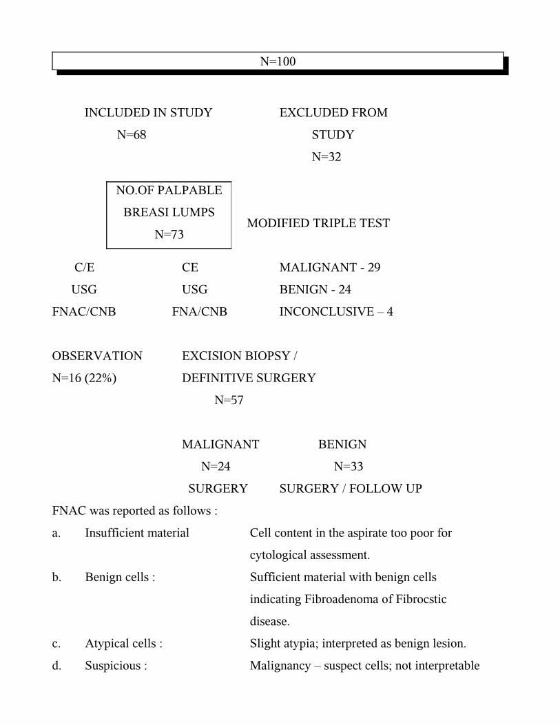
N=100
INCLUDED IN STUDY EXCLUDED FROM
N=68 STUDY
N=32
MODIFIED TRIPLE TEST
C/E CE MALIGNANT - 29
USG USG BENIGN - 24
FNAC/CNB FNA/CNB INCONCLUSIVE – 4
OBSERVATION EXCISION BIOPSY /
N=16 (22%) DEFINITIVE SURGERY
N=57
MALIGNANT BENIGN
N=24 N=33
SURGERY SURGERY / FOLLOW UP
FNAC was reported as follows :
a. Insufficient material Cell content in the aspirate too poor for
cytological assessment.
b. Benign cells : Sufficient material with benign cells
indicating Fibroadenoma of Fibrocstic
disease.
c. Atypical cells : Slight atypia; interpreted as benign lesion.
d. Suspicious : Malignancy – suspect cells; not interpretable
NO.OF PALPABLE
BREASI LUMPS
N=73
as carcinoma with certainity.
e. Carcinoma : Cells indicative of malignancy.
Results were interpreted as follows :
a-repeat FNAC/CNB
b,c-benign
d-inconclusive
c-malignant
The results of the modified Triple Test were then analysed individually and as a
combination. Any component indicating a malignant report were taken as malignancy.
Inconclusive reports were subject to excision biopsy on an inpatient basis. Patients with
malignancy were treated with definitive surgery. The post procedural histopathological
reports were compared to the results of the Modified Triple Test.
*****
DATA ANALYSIS
Of the patients randomly referred for the study (n=100), patients not fitting the
inclusion criteria were excluded (n=32). Thus 68 patients with 73breast lumps were
inducted into the study.
The MTT obviated excision biopsy in 16 patients (22%) and this is the potential
advantage of this test. Thus the final study group (n=57) underwent the MTT followed
by excision biopsy, the results of which were available for comparison.
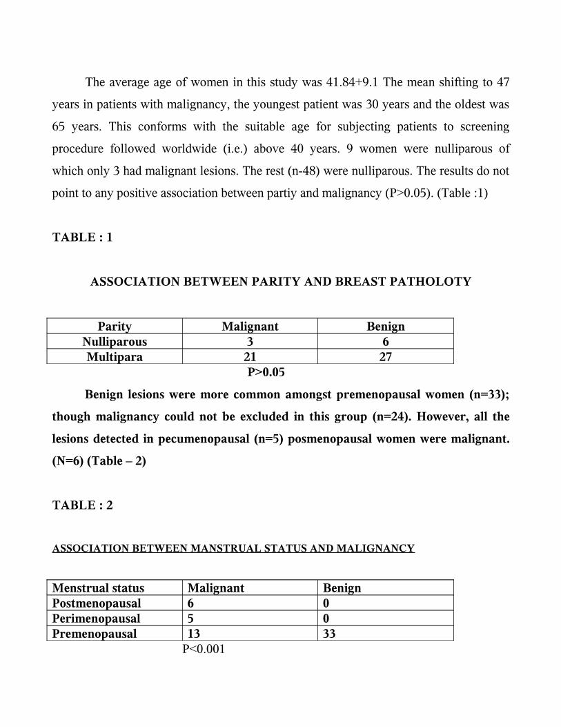
The average age of women in this study was 41.84+9.1 The mean shifting to 47
years in patients with malignancy, the youngest patient was 30 years and the oldest was
65 years. This conforms with the suitable age for subjecting patients to screening
procedure followed worldwide (i.e.) above 40 years. 9 women were nulliparous of
which only 3 had malignant lesions. The rest (n-48) were nulliparous. The results do not
point to any positive association between partiy and malignancy (P>0.05). (Table :1)
TABLE : 1
ASSOCIATION BETWEEN PARITY AND BREAST PATHOLOTY
Parity Malignant BenignNulliparous 3 6Multipara 21 27
P>0.05
Benign lesions were more common amongst premenopausal women (n=33);
though malignancy could not be excluded in this group (n=24). However, all the
lesions detected in pecumenopausal (n=5) posmenopausal women were malignant.
(N=6) (Table – 2)
TABLE : 2
ASSOCIATION BETWEEN MANSTRUAL STATUS AND MALIGNANCY
Menstrual status Malignant BenignPostmenopausal 6 0Perimenopausal 5 0Premenopausal 13 33
P<0.001

On excision biopsy, the 57 lumps were confirmed histopathologically as cither
Malignant (n=24) and Benign (n=33).
The pathological reports were :
Total of malignancies = 24
Intra Ductal Carcinoma = 20
Lobular Carcinoma = 1
Medullary Carcinoma = 1
Carcino Sarcoma = 1
Malignant cystosarcoma
Phylloides = 1
Total no. of Benign lesions = 33
Fibroadenoma = 14
Fibrocystic Disease = 10
Breast Abscess = 6
Benign cystosarcoma
Phylloides = 3
The associations between each component of MTT and the combined MTT with
the biopsy report were subjected to computer generated analysis.
Clinical examination finding of a hard, irregular lump was more like to be
malignant, while a soft or firm mass with regular surface suggested benign lesions
(P>0.001). Benign lesions most often confused with malignancy were Benign
cystosarcoma phylloides & Fibrocystic disease. (Table-3)
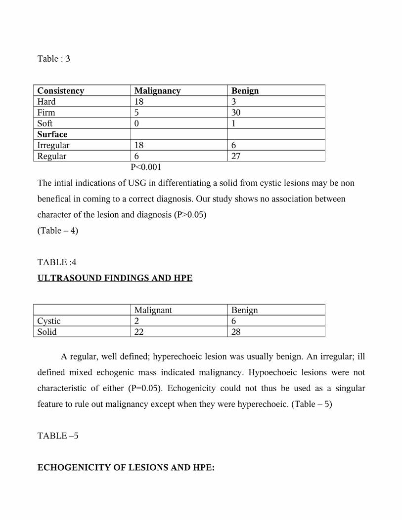
Table : 3
Consistency Malignancy BenignHard 18 3Firm 5 30Soft 0 1SurfaceIrregular 18 6Regular 6 27
P<0.001
The intial indications of USG in differentiating a solid from cystic lesions may be non
benefical in coming to a correct diagnosis. Our study shows no association between
character of the lesion and diagnosis (P>0.05)
(Table – 4)
TABLE :4
ULTRASOUND FINDINGS AND HPE
Malignant BenignCystic 2 6Solid 22 28
A regular, well defined; hyperechoeic lesion was usually benign. An irregular; ill
defined mixed echogenic mass indicated malignancy. Hypoechoeic lesions were not
characteristic of either (P=0.05). Echogenicity could not thus be used as a singular
feature to rule out malignancy except when they were hyperechoeic. (Table – 5)
TABLE –5
ECHOGENICITY OF LESIONS AND HPE:

Echogenicity Malignant BenignMixed 10 6Hypoechoeic 14 23Hyperchoeic 0 4
P = 0.05
Ultrasound detected Axillary nodes in all patients (n=12) found to be clinically
node positive; however it could not distinguish whether the node had malignant
infiltration or not.
Ultrasound guided FNA/CNB was done in 6 cases; 2 of which were cystic lesions
with a residual mass on aspiration of the cysts. Both turned out to malignancies missed
on routine FNAC.
Amongst the individual tests; clinical examination was more likely to miss a
malignancy (Sensitivity 75%); as against ultrasound (Sensitivity 92%) or FNA / CNB
(Sensitivity 100%). FNA/CNB correctly identified malignancy in all 24 cases; while
ultrasound misinterpreted 1 case as malignant (a case of Benign cystosarcoma
phylloides) with specificities of 100% and 85% respectively.
The MTT was 85% specific with malignant lesions. But 5 cases were
misdiagnosed as malignancies and turned out in 3 cases to be Fibrocystic Disease and 2
cases were Benign cystosarcoma phylloides – both benign lesions. Inconclusive results
(n=4) on MTT were also confirmed to be benign lesions. Thus MTT, though had false
positives with respect to malignancy but no false negatives (i.e.,) a negative predictive
value of 100%. These data are comparable to the original triple test with its sensitivity
(65%-96%) and specificity (55%-98%) as reported in various studies (Tables – 6-10).
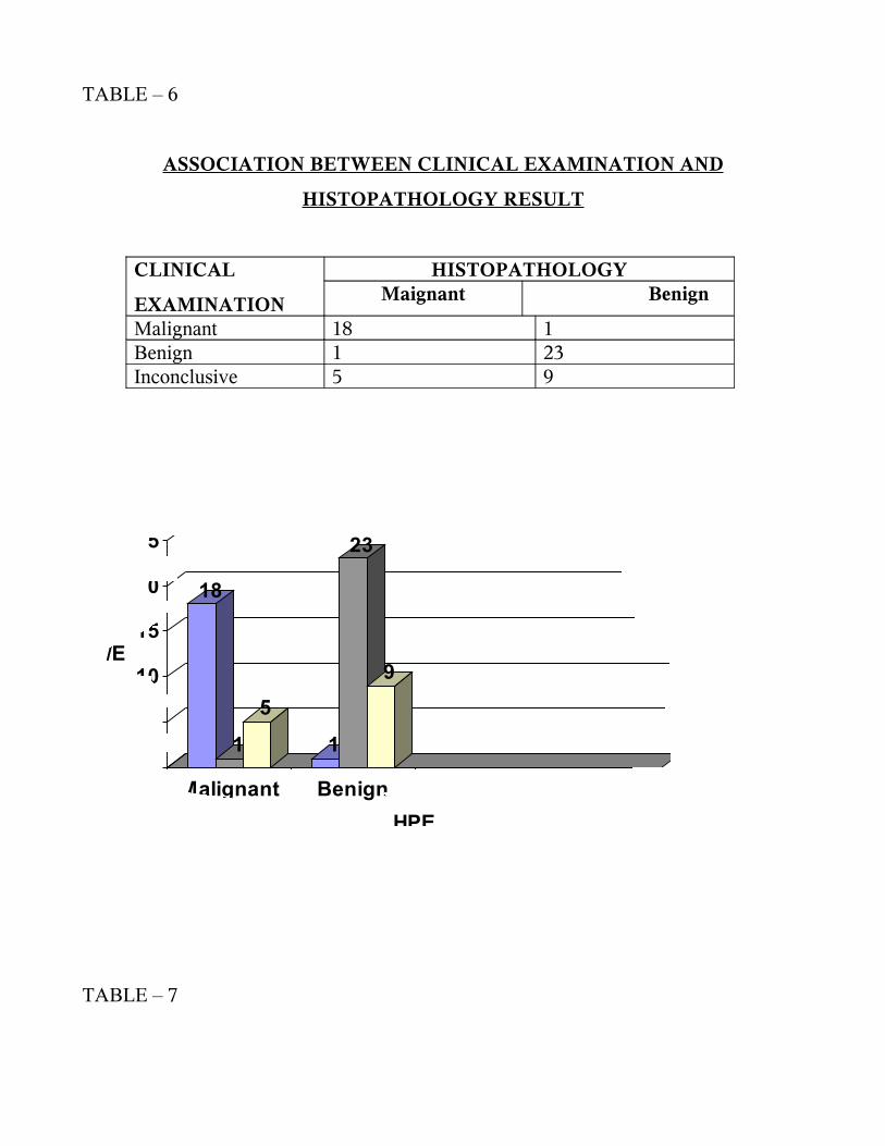
TABLE – 6
ASSOCIATION BETWEEN CLINICAL EXAMINATION AND
HISTOPATHOLOGY RESULT
CLINICAL
EXAMINATION
HISTOPATHOLOGYMaignant Benign
Malignant 18 1Benign 1 23Inconclusive 5 9
TABLE – 7
18
1
5
1
23
9
0
5
10
15
20
25
C/E
Malignant Benign
HPE

ASSOCIATION BETWEEN ULTRASONOGRAM AND HPE
ULTRASONOGRAMHPE
Maignant Benign
Malignant 22 3Benign 1 28Inconclusive 1 2
0
5
10
15
20
25
30
Malignant Benign
TABLE – 8
ASSOCIATION OF FNA/CNB WITH HPE

CLINICAL
EXAMINATION
HISTOPATHOLOGY
Maignant Benign
Malignant 18 1Benign 1 23Inconclusive 5 9
0
27
0
5
10
15
20
25
30
FNA/CNB
Malignant
HPE
East
West
North
TABLE – 9
ASSOCIATION MODIFIED TRIPLE TESTS WITH HPE
MTT HPE
Maignant Benign
Malignant 24 5

Benign 0 24Inconclusive 0 4
24
0 0
5
24
4
0
5
10
15
20
25
MTT
Malignant
HPE
TABLE – 10
STATISTICAL ANALYSIS OF THE STUDY
C/E USG FNA/CNB MTTSensitivity 75%
(53-80%)
92%
(72-99%)
100%
(83-100%)
100%
(83-100%)Specificity 97%
(82-100%)
85%
(67-94%)
100%
(87-100%)
85%
(67-94%)

Positive
Predictive Value
95%
(70-99%)
81%
(61-93%)
100%
(83-100%)
83%
(64-93%)Negative
Predictive Value
84%
(64-90%)
93%
(76-99%)
100%
(87-100%)
100%
(85-100%)‘P’ Value <0.001 <0.001 <0.001 <0.001
FNA / CNB as a single test was a superior diagnostic test than the other two tests,
but only when complemented by them could the lesion be characterized in all
dimensions for the chosen interventional procedure.
When an inconclusive was reported the likelihood of malignancy increases in
ascending order from FNA/CN, USG to C/E (n=o, n=1, n=5) respectively. All 4 cases
deemed inconclusive after MTT were later diagnosed as Fibrocystic disease.
CONCLUSION
Detection and management of a breast mass requires an optimal environment for
interpretation, relevant use of clinical information, technically excellent imaging
procedures, proper interpretation of finding and patient recommendations.
Our results show that the diagnostic accuracy of combined physical examination
breast USG and FNA/CNB is comparable to that of histological examination.
A fine collaboration between experienced radiologists, cytologist and the
Clinician is required.
Ultrasound when replacing mammography serve as effective an imaging modality
in palpable breast lumps and is more comprehensive.
Ultrasound breast aids biopsy techniques by guidance to the representative area
than increasing yield.
CNB is a suitable alternative when FNA is inconclusive and may offer additional
information.
Thus the use of MTT to complement findings in differential diagnosis of a lesion
in a symptomatic women seeking medical care deserves acceptance and further
evalution. This may lead to less delay in treatment when malignancy is suspected and to
avoidance of surgical exploration when a benign nature of lesion is suspected.
