dissociation fromthat a-i selective to - pnas · proc. natl.acad. sci. usa vol. 80, pp. 5435-5439,...
TRANSCRIPT

Proc. Natl. Acad. Sci. USAVol. 80, pp. 5435-5439, September 1983Medical Sciences
Dissociation of tissue uptake of cholesterol ester from that ofapoprotein A-I of rat plasma high density lipoprotein:Selective delivery of cholesterol ester to liver,adrenal, and gonad
(cholesterol ether/tyramine cellobiose/lipoprotein turnover)
CHRISTOPHER GLASS, RAY C. PITTMAN, DAVID B. WEINSTEIN, AND DANIEL STEINBERGDivision of Metabolic Disease, Department of Medicine, University of California, San Diego, La Jolla, California 92093
Contributed by Daniel Steinberg, May 26, 1983
ABSTRACT The metabolic fate of homologous high densitylipoprotein (HDL) was studied in the rat, tracing the apoproteinA-I (apo A-I) and cholesterol ester moieties simultaneously. Theapo A-I was labeled with covalently linked "MI-labeled tyraminecellobiose, which accumulates in the cells degradingthe apopro-tein; [3H]cholesterol ethers, which cannot be hydrolyzed or mo-bilized after uptake, were incorporated into the lipid core of re-constituted HDL to reflect the fate of the cholesterol esters. Severallines of evidence, including direct comparison with biologicallylabeled HDL, are presented to support the validity of this ap-proach. The liver was the major organ of cholesterol ether up-take, accounting for 65% of the total; the adrenal gland and ovarywere the most active organs per gram (wet) of weight. Uptake ofcholesterol ether was 7-fold-greater than that-of apo A-I in ad-renal, 4-fold greater in the ovary, and >2-fold greater in the liver.The remaining tissues took up apo A-I and cholesterol ethers atmore nearly equal rates. Transfer of HDL-associated cholesterolethers and 'RI-labeled apo A-I to other lipoprotein fractions wasnot observed; thus, the results reflect direct uptake from HDLitself. Whereas uptake of low density lipoprotein appears to in-volve endocytosis of intact particles, uptake of HDL in at leastsome rat tissues involves additional, more complex, transfermechanisms.
The increasingly impressive evidence that risk of coronary heartdisease correlates inversely with plasma high density lipopro-tein (HDL) levels (1) has stimulated a great deal of research onHDL metabolism. This protective effect may be related to HDL-mediated transport of cholesterol from extrahepatic tissues tothe liver, as postulated by Glomset (2), but evidence for such"reverse cholesterol transport" in vivo is largely indirect andalmost no quantitative data are available. HDL can also, at leastin the rat, deliver cholesterol to steroidogenic tissues (3, 4). Thisdelivery may be receptor mediated although the targeting.ofHDL for such uptake is poorly understood.We have used nondegradable markers to determine simul-
taneously the uptake of HDL-associated apoprotein A-I (apo A-I) and cholesterol esters by various tissues in vivo. Apo A-I wastraced by covalently attaching to it "2I-labeled tyramine-cel-lobiose (lasI-TC). The TC moiety, which can be radioiodinatedto high specific activity, remains trapped in tissues after uptakeand degradation of the labeled protein because it cannot be de-graded by mammalian cells and does not readily cross cellmembranes (5). The fate of cholesterol esters was determinedby using [3H]cholesteryl linoleyl ether as a marker, as de-scribed (6). Since cholesterol ethers cannot be hydrolyzed by
mammalian cells, they remain trapped in tissues after uptakein the same manner as the nondegradable-protein marker. Cho-lesterol ethers were introduced' into HDL by a modification ofthe procedure for reconstituting low density lipoprotein (LDL)(7).The rat was chosen for these studies in part to evaluate fur-
ther the special role of HDL in this animal for delivering cho-lesterol to the adrenal gland and gonads and in part because therat has little or no cholesterol ester exchange protein in its plasma(8). In the rabbit-and probably in other species with plasmacholesterol ester-exchange protein-cholesterol ethers, likecholesterol esters, exchange rapidly among lipoprotein frac-tions (9). This makes it very difficult to be certain which li-poprotein fraction is responsible for observed tissue uptake. Inthe present studies in the rat, HDL-associated cholesterol ethersdid not redistribute to other lipoproteins after injection. Thus,accumulation of cholesterol ethers by tissues resulted from de-livery by HDL itself.
MATERIALS AND METHODS
Lipoproteins. HDL was isolated in the density interval 1.09-1.21 g/ml from serum of large female Sprague-Dawley rats(retired breeders) by using sequential ultracentrifugation ac-cording to standard techniques (10). Apo A-I was purified andlabeled with '"I-TC as described (11). '"I-TC-labeled apo A-I (125I-TC-apo A-I) was reassociated with HDL by incubationfor 1 hr at 370C in phosphate-buffered saline (pH 7.4) (Pi/NaCl)at a ratio of 1 mol of '"I-TC-apo A-I per 20 mol of HDL. Un-bound apo A-I was removed by flotation of the HDL at a saltdensity of 1.21 g/ml.To biologically label rat HDL in the cholesterol ester moiety,
rats were fasted overnight and then fed [la,2a-3H]cholesterol(Amersham) in 0.5 ml of corn oil delivered through a gastrictube. Serum was recovered 22 hr later and HDL was isolatedby sequential ultracentrifugation. At this time, 80% of.the 3Hlabel was associated with cholesterol esters and 20%, with freecholesterol. To reduce the fraction of 3H in free cholesterol,this preparation was injected into a donor animal and, 1 hr later,blood was drawn and HDL was reisolated. This procedure de-creased the radioactivity in free cholesterol to <5% of total.
Incorporation of [3H]Cholesterol Ethers into HDL. Radio-labeled cholesteryl linoleyl etherwas prepared from [la, 2a-3H]-cholesterol and linoleyl methanesulfonate (12) as described (13).
Abbreviations: HDL, high density lipoprotein; LDL, low density li-poprotein; apo A-I, apoprotein A-I; TC, tyramine cellobiose; 125I-TC,125I-labeled TC; FCR, fractional catabolic rate; Pi/NaCl, phosphate-buffered saline (pH 7.4).
5435
The publication costs of this article were defrayed in part by page chargepayment. This article must therefore be hereby marked "advertise-nent" in accordance with 18 U.S.C. §1734 solely to indicate this fact.
5436 Medical Sciences: Glass et al.
Rat HDL was brought to a concentration of 2 mg/ml in Pi/NaCl,and aliquots (2 mg of HDL protein) were Iyophilized onto 25mg of purified potato starch (Sigma) in siliconized 15-ml ex-
traction tubes. The HDL was partially delipidated by extractionwith 5 ml of heptane at -200C for 1 hr. The starch was thenpelleted by low-speed centrifugation and the heptane phase was
removed. The samples were extracted two more times withheptane at -20'C for 30 min. Exogenous lipids consisting of2 mg of unlabeled cholesteryl linoleate, 0.5-5 x 10⁸ dpm of[3H]cholesteryl linoleyl ether (<1 ug total), and 0. 1 mg of un-
labeled free cholesterol were added to each sample in 200 /1of heptane and incubated at room temperature for 1 hr. Theheptane was then evaporated under nitrogen until the starchappeared powder dry. HDL was solubilized from the starch byaddition of Pi/NaCl/0.02% sodium azide and incubated over-
night at 370C with gentle agitation. The starch was then sedi-mented at 10,000 x g for 20 min and the resulting supernatewas filtered through an 0.8-,um filter (Unipore). The resultingHDL containing [3H]cholesteryl linoleyl ether was turbid.Equilibrium density gradient ultracentrifugation resolved theproduct into two peaks of radioactivity: the first, recovered ina density range similar to that of native HDL, was associatedwith HDL apoproteins; the second, which accounted for 30-40% of recovered radioactivity, had a density of <1.02 g/mland was unassociated with protein. This light fraction ac-
counted for all of the turbidity observed in the starting ma-
terial. We routinely reisolated the reconstituted HDL in thedensity interval 1.07-1.21 g/ml by sequential ultracentrifu-gation to remove this light component. The resulting prepa-
ration is referred to below as "reconstituted" HDL. The in-franate of the reconstituted HDL reisolated at 1.07 g/ml was
dialyzed against Pi/NaCl and reassociated with l25I-TC-apo A-I as described above.
In Vivo Studies. Sprague-Dawley female rats (175-225 gm)were fasted overnight prior to and throughout the course. ofturnover studies but were allowed free access to water. On themorning of the study, animals were anesthetized and silasticcatheters were placed in the left jugular vein. After a 3-hr re-
covery period, radiolabeled samples were injected and plasmadecay data were obtained by drawing periodic blood samplesof 100-125 ,u. Plasma decay data were fitted to a biexponentialfunction and fractional catabolic rates (FCR) were calculatedusing a simple kinetic model (14). Tissues were collected 24 hrafter injection and homogenized as described (11). In some ex-
periments, hepatocytes and nonparenchymal cells were iso-lated after collagenase perfusion of the liver (15).
Radioassay Procedures. Samples containing 1251 or 3H were
radioassayed using standard techniques. To assay 3H in tissuehomogenates containing both "2I and 3H, samples were broughtto a volume of 1 ml with waters and extracted using the methodof Dole (16). This resulted in nearly quantitative recovery ofcholesterol ethers in the nonpolar phase (93.5 2.7%) with vir-tually no contamination by 125I-TC-labeled protein or its deg-radation products (<0.5% of -total 125I).
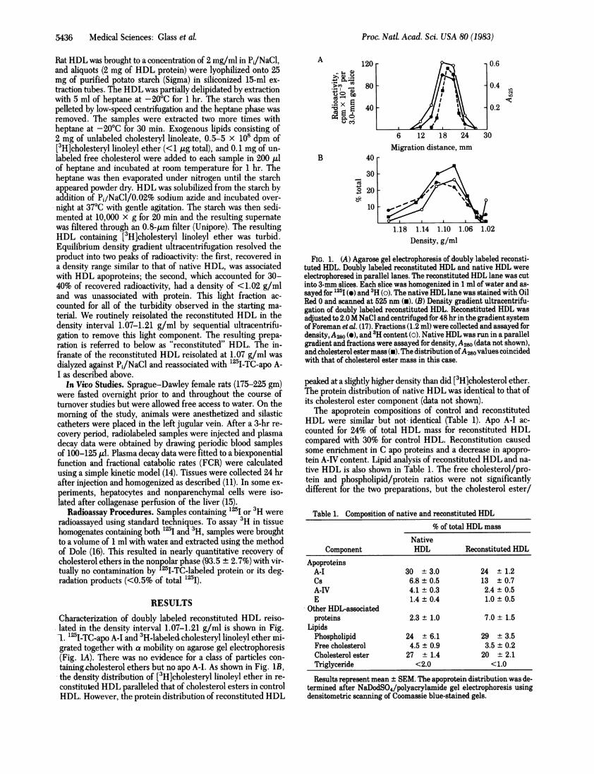
RESULTSCharacterization of doubly labeled reconstituted HDL reiso-lated in the density interval 1.07-1.21 g/ml is shown in Fig.1. 125I-TC-apo A-I and 3H-labeledcholesteryl linoleyl ether mi-grated together with a mobility on agarose gel electrophoresis(Fig. 1A). There was no evidence for a class of particles con-
taining.cholesterol ethers but no apo A-I. As shown in Fig. 1B,the density distribution of [3H]cholesteryl linoleyl ether in re-
constituted HDL paralleled that of cholesterol esters in controlHDL. However, the protein distribution of reconstituted HDL
A
B
120
5- _ 80
0X E 4A0 E P. 4
40
-"30
CZ4 2010
10
I'
I
6 12 18 24
Migration distance, mm
0.6
0.4
e
0.2
30
1.18 1.14 1.10 1.06 1.02Density, g/ml
FIG. 1. (A) Agarose gel electrophoresis of doubly labeled reconsti-tuted HDL. Doubly labeled reconstituted HDL and native HDL were
electrophoresed in parallel lanes. The reconstituted HDL lane was cutinto 3-mm slices. Each slice was homogenized in 1 ml of water and as-
sayed for 125I () and 3H (0). The native HDL lane was stained with OilRed 0 and scanned at 525 nm (i). (B) Density gradient ultracentrifu-gation of doubly labeled reconstituted HDL. Reconstituted HDL was
adjusted to 2.0 M NaCl and centrifuged for 48 hr in the gradient systemof Foreman et al. (17). Fractions (1.2 ml) were collected and assayed fordensity, A20 (e), and 'H content (o). Native HDL was run in a parallelgradient and fractions were assayed for density, A2w (data not shown),and cholesterol ester mass (W). The distribution ofA280values coincidedwith that of cholesterol ester mass in this case.
peaked at a slightly higher density than did [3H]cholesterol ether.The protein distribution of native HDL was identical to that ofits cholesterol ester component (data not shown).The apoprotein compositions of control and reconstituted
HDL were similar but not identical (Table 1). Apo A-I ac-
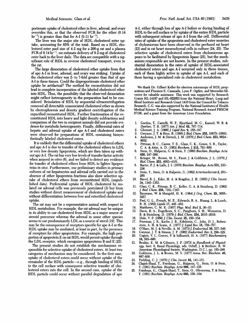
counted for 24% of total HDL mass for reconstituted HDLcompared with 30% for control HDL. Reconstitution causedsome enrichment in C apo proteins and a decrease in apopro-tein A-IV content. Lipid analysis of reconstituted HDL and na-
tive HDL is also shown in Table 1. The free cholesterol/pro-tein and phospholipid/protein ratios were not significantlydifferent for the two preparations, but the cholesterol ester/
Table 1. Composition of native and reconstituted HDL% of total HDL mass
NativeComponent HDL Reconstituted HDL
ApoproteinsA-I 30 ± 3.0 24 ± 1.2Cs 6.8 ± 0.5 13 ± 0.7A-IV 4.1 ± 0.3 2.4 ± 0.5E 1.4 ± 0.4 1.0 ± 0.5
Other HDL-associatedproteins 2.3 ± 1.0 7.0 ± 1.5
LipidsPhospholipid 24 ± 6.1 29 ± 3.5Free cholesterol 4.5 ± 0.9 3.5 ± 0.2Cholesterol ester 27 ± 1.4 20 ± 2.1Triglyceride <2.0 <1.0
Results represent mean ± SEM. The apoprotein distribution was de-termined after NaDodSO4/polyacrylamide gel electrophoresis usingdensitometric scanning of Coomassie blue-stained gels.
Proc. Natl. Acad. Sci. USA 80 (1983)

Proc. NatL Acad. Sci. USA 80 (1983) 5437
protein ratio of reconstituted HDL was 25-35% less than thatof control HDL. Electron microscopy of negatively stainedpreparations of native and reconstituted HDL showed that mostof the reconstituted HDL particles were spherical, with di-ameters not significantly different from that of control HDL(data not shown). There was, in addition, a population of dis-coidal particles, identified on the basis of their formation ofrouleaux, that accounted for 15-20% of the total particle num-ber. This subclass, presumably incompletely reconstituted, mayaccount for the slight difference in the density distribution ofprotein and lipid components of reconstituted HDL mentionedabove and for its overall lower cholesterol ester/protein ratio.
In studies to be reported elsewhere, the effect of reconsti-tution on HDL-cell interaction was tested in cultured rat he-patocytes. Uptake of TC-labeled native HDL was identical tothat of TC-labeled reconstituted HDL. Unlabeled native HDLcompeted equally for uptake of both labeled preparations.The plasma decay kinetics of reconstituted HDL were com-
pared with those of cholesterol esters in a native particle. Re-constituted doubly labeled HDL was biologically screened bypassage through a donor animal for 1 hr. Serum from the donorwas then injected into recipient animals. Plasma decay curves
for 125I-TC-apo A-I and [3H]cholesteryl linoleyl ether and a rep-
resentative plasma decay curve for rat HDL biosyntheticallylabeled with cholesterol esters are shown in Fig. 2. The FCRof 125I-TC-apo A-I in the reconstituted HDL was 0.11 ± 0.01hr-'. The FCR for the [3H]cholesterol ether component was
0.14 ± 0.01 hr-' (n = 4), compared with the FCR for biolog-ically labeled HDL cholesterol esters of 0.14 ± 0.02 hr-' (n =7).
Tissues were assayed for radioactivity 24 hr after injection ofthe biologically screened reconstituted HDL. At that time, >90%of the initial serum "2I and 3H had left the circulation. Thefraction of total recovered extravascular radioactivity found inselected tissues is shown in Table 2. The liver was the pre-
dominant organ of cholesterol ether uptake and was also themajor organ of apo A-I uptake. Isolation of hepatocytes andnonhepatocytes showed that 93-96% of both trapped labels wasassociated with hepatocytes. The kidney exhibited a high rateof apo A-I uptake, as in previous studies (11), but took up <1%of the injected dose of cholesterol ethers. Recoveries of 1"Iand 3H at 24 hr ranged from 78% to 90% of the injected dose.Redistribution or leakage of the two labels was not examineddirectly in these experiments but has previously been shown tobe insignificant for up to 48 hr in the case of 125I-TC-apo A-I(11) and for up to 6 days after uptake of [3H]cholesterol ethers(6).
b._
a1) 4)
c b03 Z
5 10 15 20 25Time, hr
FIG. 2. Representative plasma decay curve ofbiologically screeneddoubly labeled reconstituted HDL. *, 125I-TC-apo A-I; o, [3H]choles-teryl linoleyl ether. Also shown is a representative plasma decay curveof biosynthetically labeled HDL cholesterol esters (u).
Table 2. Tissue uptake of 1"I-TC-apo A-I and [3H]cholesteryllinoleyl ether from reconstituted HDL
% of total body uptakeOrgan 1"I-TC-apo A-I [3H]Cholesteryl etherLiver 37 ± 2.9 64 ± 2.3Kidney 18 ± 2.6 0.5 ± 0.1Adrenal 0.6 ± 0.1 2.8 ± 0.3Ovary 0.9±0.3 2.9±0.6Other 43 ± 7.9 30 ± 5.9
To assess the relative activities of the various tissues in up-take per unit weight of tissue, the data were calculated in termsof an organ FCR that represents the fraction of the plasma poolcleared per hour per gram of tissue (Table 3). Calculated thisway, the adrenal glands and ovaries were the most active organsin uptake of [3H]cholesteryl linoleyl ether per gram of tissue.The liver exhibited the next highest rate of uptake. The kidneywas the most active organ in uptake of 1"I-TC-apo A-I, fol-lowed by adrenal, ovary, and liver. These tissues were sub-stantially more active in uptake of '"I-TC-apo A-I and [3H]cho-lesterol ethers than any of the remaining tissues.A particularly striking finding was the differential rate of up-
take of apo A-I and cholesterol ethers in adrenal, ovary, andliver. Cholesterol ether/apo A-I uptake ratios for these and sev-eral other tissues are also shown in Table 3. The ratio was high-est in adrenal (7.0) and definitely >1 in ovary (4.6) and liver(2.2). The kidney also exhibited a striking degree of dissociationbut in the opposite direction. Apo A-I uptake in kidney oc-curred some 30 times faster than that of the ether. Most of theremaining tissues exhibited ether/apo A-I uptake ratios thatwere not significantly different from 1.
Several possibilities were considered that might account forthe differential rates of uptake observed in liver, adrenal, andovary. One possible explanation is selective uptake of an ether-rich particle. However, a minor contaminant of ether-rich par-ticles in the injected preparation could not account for the find-ings in liver. Tissue content of radioactivity was measured after>90% of the two labels had decayed from the plasma and theliver had taken up more than half of the injected dose. The largefraction of ether-rich particles needed to explain these findingscould not have been overlooked. Nevertheless, to directly as-sess this possibility, doubly labeled reconstituted HDL wasseparated into heavy and light density subfractions by prepara-tive equilibrium density gradient ultracentrifugation and thetissue uptake of the two fractions was compared. The heavyHDL fraction (1.10-1.21 g/ml) was relatively rich in 12'I-TC-apo A-I while the light fraction (1. 065-1. 10 g/ml) was relatively
Table 3. Comparative organ FCRs for HDL-associated[3H]cholesterol linoleyl ether and 125I-TC-apo A-I inselected tissues
Organ FCR, hr-' g-' x 103Cholesterol
Tissue ether apo A-I Organ FCR ratio*Adrenal 60 ± 8.5 9.1 ± 2.8 7.0 ± 2.3*Ovary 39 ± 11 9.8 ± 5.0 4.6 ± 2.3*Liver 15 ± 1.1 6.5 ± 0.4 2.3 ± 0.2*Kidney 0.4 ± 0.1 12 ± 3.7 0.03 ± 0.01*Lung 1.3 ± 0.1 0.8 ± 0.1 1.7 ± 0.4*Other 0.1-5.7 0.1-6.0 0.9-1.4
Results represent mean ± SEM.* Cholesterol ether organ FCR/apo A-I organ FCR: FCR calculated asplasma FCR x fraction of total body uptake in organ - organ wetweight (g).
Medical Sciences: Glass et aL
100
10 -

5438 Medical Sciences: Glass et al.
rich in [3H]cholesteryl linoleyl ethers. Despite the differencesin the relative amounts of 1"I-TC-apo A-I and [3H]cholesteryllinoleyl ether in the two fractions, there was no evidence ofmetabolic heterogeneity. As shown in Table 4, uptake of etherlabel was at least 2-fold greater than apo A-I uptake in liver,adrenal, and ovary; there were no apparent differences be-tween dense and light HDL subfractions.To address the possibility that these differential rates of up-
take were the consequence of an artifact of reconstitution, HDLthat had been biologically labeled in the cholesterol ester moietywas reassociated with 125I-TC-apo A-I and injected into ex-perimental animals. One hour after injection, hepatocytes andadrenal cells were isolated as described (15, 18) and their con-tents of 3H and 1"I were determined. The 1-hr time period waschosen to minimize hydrolysis of cholesterol esters and loss ofradiolabeled cholesterol from tissues. Radioactivity was deter-mined in isolated cells to avoid contamination by the large frac-tion of the injected dose still in the plasma compartment or ex-travascular space 1 hr after injection. Results of these experi-ments are shown in Table 4. The ratio of the uptake of cho-lesterol esters and apo A-I by hepatocytes was in good agree-ment with those obtained using reconstituted HDL. The ratioin adrenal, however, was significantly less than that observedusing reconstituted HDL. Since the adrenal uses lipoproteincholesterol for steroid hormone production (19), it was possiblethat radiolabeled cholesterol esters might have been lost at asignificant rate due to conversion to steroids and secretion dur-ing the course of the experiment. This possibility was examinedby administration of 25 mg of aminoglutethamide to each oftwo rats 1 hr before injection of the labeled HDL. Aminoglute-thamide is known to block side-chain cleavage of cholesterol,a step required in the production of corticosteroids (20). Priortreatment with this agent doubled the cholesterol ester/apo A-I ratio subsequently found in adrenal cells after injection of thedoubly labeled HDL. Thus, even though blockade of choles-terol label leakage was probably incomplete, these experimentsshow the preferential uptake of esters compared with apo A-Ifrom "native" HDL (i.e., HDL not subjected to the reconsti-tution procedure).
Another possible explanation for the disproportionately greateruptake of cholesterol ether and esters is their transfer to lowerdensity lipoprotein fractions without parallel transfer of apo A-I followed by more rapid tissue uptake of these lighter lipo-proteins. Since rat plasma contains little or no cholesterol ester
Table 4. Relative uptake of HDL-associated [3H]cholesterolethers or esters and 125I-TC-apo A-I from variousHDL preparations
Preparation Liver Adrenal OvaryReconstituted HDL*
Light subfraction 2.5 5.6 2.7(1.065-1.10 g/ml) (2.7, 2.3) (5.1, 6.0) (3.1, 2.3)Heavy subfraction 2.2 5.7 2.7(1.10-1.21 g/ml) (2.1, 2.3) (4.6, 6.7) (2.4, 2.9)
Biologically labeled 2.2 1.4 NDHDLt (2.3, 2.1) (1.4, 1.5)
Biologically labeled 1.8 2.9 NDHDL (rats treated with (2.0,1.6) (2.9, 2.9)aminoglutethamide)tResults represent apparent uptake of HDL as determined by 3H .
apparent uptake ofHDL as determined by 125I. Values given are meansof results from two experiments and values in parentheses are for in-dividual experiments. ND, not determined.* Tissue content of radioactivity was determined 24 hr after injection.tContent of 3H and 1251I was determined in isolated hepatocytes andadrenal cells 1 hr after injection.
1.400 LDL HDL
1 0-
40
.' 30>~x
~1010 20 30 40 50 60
Elution volume, ml
FIG. 3. Gel filtration (6% agarose) of serum obtained 1 hr after in-jection of doubly labeled reconstituted HDL. Arrows denote the elutionvolumes ofhuman LDL and native rat HDL determined in independentcalibration runs. *, '"I-TC-apo A-I; o, [3H]cholesteryl linoleyl ether.
exchange activity, this possibility seemed unlikely but it waschecked. A rat was injected with doubly labeled reconstitutedHDL. Serum was obtained 1 hr later and chromatographed on6% agarose. As shown in Fig. 3, both the apo A-I and choles-terol ether labels eluted exclusively in the HDL range; therewas no evidence of transfer of cholesterol ethers to lighter li-poproteins.
DISCUSSIONSeveral lines of evidence support the conclusion that the me-tabolism of the nondegradable markers on reconstituted HDLreflects the metabolism of apo A-I and cholesterol esters of na-tive HDL. (i) The physical properties (electrophoretic mobilityand hydrated density), apoprotein composition, and lipid com-position of the reconstituted HDL closely resemble those ofnative HDL except for a somewhat lower cholesterol ester/pro-tein ratio. (ii) Unlabeled native HDL competed equally withreconstituted 125I-TC-labeled HDL and with HDL directlyconjugated with 15I-TC for uptake and degradation in a cul-tured rat hepatocyte system. (iii) The in vivo plasma decay ratefor cholesterol esters in biosynthetically labeled HDL was iden-tical to that of cholesterol ethers in HDL reconstituted in vitro(and previously biologically screened in a donor animal). Wehave shown previously that the in vivo plasma decay rate forTC-labeled apo A-I is not different from that of conventionallyiodinated apo A-I (11).The present results are in good agreement with those pre-
viously reported for the sites of degradation of apo A-I on HDLthat had not been reconstituted (11). In those studies, the over-all fraction of apo A-I degraded by kidney (37%) was signifi-cantly greater than that observed in the present studies. Thisdifference is likely to be due to the combination of ultracen-trifugal flotation and biological screening carried out on recon-stituted HDL in the present studies. Although the kidneys ac-counted for 18% of total apo A-I uptake, they took up almostno cholesterol ether (<1%). This supports our previous pro-posal that the renal degradation represents primarily tubulardegradation of filtered free apo A-I in equilibrium with apo A-I on HDL (11). Estimating the filtration fraction for apo A-Ifrom published data for other macromolecules (21), we calcu-late that as little as 1-2% unassociated apo A-I could account forthe observed results. This selective renal degradation of a sig-nificant fraction of plasma apo A-I without concomitant uptakeof the cholesterol ether would tend to make the FCR of apo A-I greater than that of cholesterol ether. However, the dispro-
Proc. Nad Acad. Sci. USA 80 (1983)
Proc. Natl. Acad. Sci. USA 80 (1983) 5439
portionate uptake of cholesterol ether in liver, adrenal, and ovaryoverrides this, so that the observed FCR for the ether (0.14hr-1) is greater than that for A-I (0.11 hr-').
The liver was the major site of HDL cholesterol ester up-take, accounting for 65% of the total. Based on a HDL cho-lesterol ester pool size of 4.2 mg for a 200-g rat and a plasmaFCR of 0.14 hr-', we estimate delivery of 9.2 mg of cholesterolester back to the liver daily. The data are compatible with a sig-nificant role of HDL in reverse cholesterol transport, even inthe rat.The large dissociation of cholesterol ether uptake from that
of apo A-I in liver, adrenal, and ovary was striking. Uptake ofthe cholesterol ether was 2- to 7-fold greater than that of apoA-I in these tissues. Could the disproportionate cholesterol etheruptake be artifactual? The method for reconstitution did notlead to complete incorporation of the labeled cholesterol etherinto HDL. Thus, the possibility that the observed dissociationmight reflect heterogeneity in the preparation had to be con-sidered. Reisolation of HDL by sequential ultracentrifugationremoved all detectable unassociated cholesterol ether as shownby electrophoresis and density gradient centrifugation of therepurified reconstituted HDL. Further fractionation of the re-constituted HDL into heavy and light density subfractions andcomparison of the two in recipient animals did not provide evi-dence for metabolic heterogeneity. Finally, differential rates ofhepatic and adrenal uptake of apo A-I and cholesterol esterswere observed for preparations of HDL containing biosyn-thetically labeled cholesterol esters.
It is unlikely that the differential uptake of cholesterol ethersand apo A-I is due to transfer of the cholesterol ethers to LDLor very low density lipoprotein, which are then taken up with-out apo A-I. The rat has very low levels of transfer factor activitywhen assayed in vitro (8), and we failed to detect any evidencefor transfer of cholesterol ethers from HDL to lighter lipopro-teins in vivo. Furthermore, our preliminary studies in primarycultures of rat hepatocytes and adrenal cells carried out in theabsence of other lipoprotein fractions also show selective up-take of cholesterol ethers from reconstituted HDL (unpub-lished data). Preferential uptake of HDL cholesterol by iso-lated rat adrenal cells was previously postulated (3) but fromstudies without direct measurement of cholesterol uptake andwithout differentiation between free and esterified cholesteroluptake.The rat may not be a representative animal with respect to
HDL metabolism. For example, the rat adrenal may be uniquein its ability to use cholesterol from HDL as a major source ofsteroid precursor whereas the adrenal in some other speciesseems to use predominantly LDL as a source of sterol (19). Thismay be the consequence of receptors specific for apo A-I or theHDL uptake may be mediated, at least in part, by the presenceof receptors for other apoproteins. For example, the high pro-portion of apoprotein E on rat HDL would permit uptake throughthe LDL receptor, which recognizes apoproteins B and E (22).The present studies do not establish the mechanisms re-
sponsible for selective uptake of cholesterol esters. At least twocategories of mechanism may be considered. In the first case,uptake of cholesterol esters could occur without uptake of theremainder of the HDL particle-e.g., through binding of HDLto the cell surface with consequent selective transfer of cho-lesterol esters into the cell. In the second case, uptake of theHDL particle could occur without parallel degradation of apo
A-I, either through loss of apo A-I before or during binding ofHDL to the cell surface or by uptake of the entire HDL particlewith subsequent release of apo A-I from the cell. Differentialrates of uptake of the apoprotein and cholesterol ester moietiesof chylomicrons have been observed in the perfused rat heart(23) and in rat heart mesenchymal cells in culture (24, 25). Theselective uptake of cholesterol esters from chylomicrons ap-pears to be facilitated by lipoprotein lipase (25), but the mech-anisms responsible are not known. In the present studies, sub-stantial dissociation in the rates of uptake of HDL-associatedcholesterol esters and apo A-I occurred only in three tissues,each of them highly active in uptake of apo A-I, and each ofthem having a specialized role in cholesterol metabolism.
We thank Dr. Gilbert Keller for electron microscopy of HDL prep-arations and Florence E. Casanada, Lynn F. Ogden, and Mercedes Sil-vestre for valuable assistance. This work was supported by ResearchGrants HL-14197 and HL-022053 from the National Heart, Lung andBlood Institute and Research Grant 1410 from the Council for TobaccoResearch. C.G. was also supported by the National Institutes of HealthMedical Science Training Program, National Research Award PH5GM07198, and a grant from the American Liver Foundation.
1. Gordon, T., Castelli, W. P., Hjortland, M. C., Kannel, W. B. &Dawber, T. R. (1977) Am. J. Med. 62, 707-714.
2. Glomset, J. A. (1968) J. Lipid Res. 9, 155-167.3. Gwynne, J. T. & Hess, B. (1980)1. Biol. Chem. 255, 10875-10883.4. Anderson, J. M. & Dietsch, J. M. (1978) J. Biol. Chem. 253, 9024-
9032.5. Pittman, R. C., Carew, T. E., Glass, C. K., Green, S. R., Taylor,
C. A. & Attie, A. D. (1983) Biochem. J. 212, 791-800.6. Stein, O., Halperin, G. & Stein, Y. (1980) Biochim. Biophys. Acta
620, 247-260.7. Krieger, M., Brown, M. S., Faust, J. & Goldstein, J. L. (1978)J.
Biol Chem. 253, 4093-4101.8. Barter, P. J. & Lally, J. I. (1978) Biochim. Biophys. Acta 531, 233-
236.9. Stein, Y., Stein, 0. & Halperin, G. (1982) Arteriosclerosis 2, 281-
289.10. Havel, R. J., Eder, H. A. & Bragdon, J. H. (1955)1. Clin. Invest.
34, 1345-1353.11. Glass, C. K., Pittman, R. C., Keller, G. A. & Steinberg, D. (1983)
J. Biol Chem. 258, 7161-7167.12. Baumann, W. & Mangold, H. K. (1964) J. Org. Chem. 29, 3055-
3057.13. Pool, G. L., French, M. E., Edwards, R. A., Huang, L. & Lumb,
R. H. (1982) Lipids 17, 448-452.14. Matthews, C. M. E. (1957) Phys. Med. Biol 2, 36-53.15. Davis, R. A., Engelhorn, S. C., Pangburn, S. H., Weinstein, D.
B. & Steinberg, D. (1979) J. Biol Chem. 254, 2010-2016.16. Dole, V. P. (1956)J. Clin. Invest. 35, 150-154.17. Foreman, J. R., Karlin, J. B., Edelstein, C., Juhn, D. J., Ruben-
stein, A. H. & Scanu, A. (1977) J. Lipid Res. 18, 759-767.18. O'Hare, M. J. & Neville, A. M. (1973)J. Endocrinol. 56, 537-549.19. Gwynne, J. T. & Strauss, J. F. (1982) Endocrinol Rev. 3, 299-329.20. Uzgiris, V. I., Graves, P. & Salhanick, H. A. (1977) Biochemistry
16, 593-600.21. Renkin, E. M. & Gilmore, J. P. (1973) in Handbook of Physiol-
ogy, Sect. 8, Renal Physiology, eds. Orloff, J. & Berliner, R. W.(American Physiological Society, Washington, D.C.), pp. 185-248.
22. Goldstein, J. L. & Brown, M. S. (1977) Annu. Rev. Biochem. 46,897-930.
23. Fielding, C. J. (1978)J. Clin. Invest. 62, 141-151.24. Chajek-Shaul, T., Friedman, G., Halperin, G., Stein, 0. & Stein,
Y. (1981) Biochim. Biophys. Acta 666, 147-155.25. Friedman, G., Chajek-Shaul, T., Stein, O., Olivecrona, T. & Stein,
Y. (1981) Biochim. Biophys. Acta 666, 156-164.
Medical Sciences: Glass et aL