dissociative electron attachment and electronic excitation
TRANSCRIPT

Dissociative electron attachment and electronic excitation
in Fe(CO)5
Journal: Physical Chemistry Chemical Physics
Manuscript ID CP-ART-03-2018-001387.R1
Article Type: Paper
Date Submitted by the Author: 26-Mar-2018
Complete List of Authors: Allan, Michael; University of Fribourg, Department of Chemistry Lacko, Michal; J. Heyrovsky Institute of Physical Chemistry Papp, Peter; Comenius University, Department of the Experimental Physics Matejcik, Stefan; Comenius University, Department of the Experimental Physics Zlatar, Matija; IHTM University of Belgrade, Center for Chemistry
Fabrikant, Ilya; University of Nebraska, Department of Physics and Astronomy Kocisek, Jaroslav; J. Heyrovsky Institute of Physical Chemistry, Fedor, Juraj; J. Heyrovsky Institute of Physical Chemistry,
Physical Chemistry Chemical Physics

Journal Name
Dissociative electron attachment and electronic exci-tation in Fe(CO)5
†
M. Allan,∗a M. Lacko,b P. Papp,b Š. Matejcík, b M. Zlatar,c I. I. Fabrikant,d J. Kocišek,e
and J. Fedor∗e
In a combined experimental and theoretical study we characterize dissociative electron attach-ment (DEA) to, and electronically excited states of, Fe(CO)5. Both are relevant for electron-induced degradation of Fe(CO)5. The strongest DEA channel is cleavage of one metal-ligandbond that leads to production of Fe(CO)−4 . High-resolution spectra of Fe(CO)−4 reveal fine struc-tures at the onsets of vibrational excitation channels. Effective range R-matrix theory successfullyreproduces these structures as well as the dramatic rise of the cross section at very low en-ergies and reveals that virtual state scattering dominates low-energy DEA in Fe(CO)5 and thatintramolecular vibrational redistribution (IVR) plays an essential role. The virtual state hypothesisreceives further experimental support from the rapid rise of the elastic cross section at very lowenergies and intense threshold peaks in vibrational excitation cross sections. The IVR hypothe-sis is confirmed by our measurements of kinetic energy distributions of the fragment ions, whichare narrow (∼0.06 eV) and peak at low energies (∼0.025 eV), indicating substantial vibrationalexcitation in the Fe(CO)−4 fragment. Rapid IVR is also revealed by the yield of thermal electrons,observed in two-dimensional (2D) electron energy loss spectroscopy. We further measured mass-resolved DEA spectra at higher energies, up to 12 eV, and compare the bands observed thereto resonances revealed by spectra of vibrational excitation cross sections. Dipole-allowed anddipole/spin forbidden electronic transitions in Fe(CO)5—relevant for neutral dissociation by elec-tron impact—are probed using electron energy loss spectroscopy and time-dependent densityfunctional theory calculations. Very good agreement between theory and experiment is obtained,permitting assignment of the observed bands.
1 IntroductionIron pentacarbonyl, Fe(CO)5, has been traditionally used as a pre-cursor for chemical vapor deposition (CVD). Recent advances innanofabrication technology promise a novel use as a precursorin focused electron-beam induced deposition (FEBID). FEBID is
a Department of Chemistry, University of Fribourg, Chemin du Musée 9, 1700 Fribourg,Switzerlandb Department of Experimental Physics, Comenius University, Mlynská dolina F2, 84215Bratislava, Slovakiac Department of Chemistry, Institute of Chemistry, Technology and Metallurgy (IChTM),University of Belgrade, Njegoševa 12, P.O. Box 815, 11001 Belgrade, Serbiad Department of Physics and Astronomy, University of Nebraska, Lincoln, NE 68588-0299, USAe J. Heyrovský Institute of Physical Chemistry v.v.i., Czech Academy of Sciences, Dole-jškova 3, 18223 Prague 8, Czech Republic∗ Corresponding authors. Emails: [email protected], [email protected]† Electronic Supplementary Information (ESI) available: Tables with configurationsand energies of calculated excited electronic states. See DOI: 10.1039/b000000x/
direct-write technique for producing spatially well-defined nanos-tructers by locally dissociating the metal-containing precursormolecules with a focused electron beam that strips off the lig-ands and leaves ideally a pure metal behind. Variety of met-als can be deposited this way.1 The possibility of creating con-trolled high-purity structures of iron attracts special attention dueto their magnetic properties, promising use in nanosensing appli-cations. Fe(CO)5 is the most commonly used precursor for FEBIDdeposition of iron1 and several reports on its use have been pub-lished.2–4
The electron beam in FEBID has energies of many kiloelectron-volts that allow its nm-sized focusing. Unfortunately, the depositsthemselves are usually much broader than the primary beam.This appears to be due to decomposition of a large fraction of theprecursor by interactions with secondary electrons, which are spa-tially much more spread, and whose energy distribution usuallypeaks around 10 eV, or even below that.5 The second commonproblem is that the purity of the deposits is often low - the inter-
Journal Name, [year], [vol.],1–10 | 1
Page 1 of 10 Physical Chemistry Chemical Physics

actions with secondary electrons lead to incomplete separationof ligands. A number of purification techniques has been sug-gested to compensate for this effect—for example reductive byatomic hydrogen6 or oxidative with electron impact stimulatedwater,7 but making a pure deposit directly would be preferable.For iron pentacarbonyl it has been shown that under specific ul-trahigh vacuum conditions the autocatalytic decomposition leadsto the purity of up to 95%.3
Desire to resolve these problems has sparked interest inthe elementary electron-induced dissociative processes in metal-containing precursor molecules including iron pentacarbonyl. Anearly DEA study was performed by Compton and Stockdale.8 Amore recent DEA spectrum was presented by Schukin et al.9 Anearly study of thermal electron attachment in an ion cyclotron res-onance (ICR) cell was performed by George and Beauchamp.10
Attachment rates of slow electrons to Fe(CO)5 (and also Fe(CO)n,n = 0− 4) were measured using a flowing afterglow Langmuirprobe apparatus by Shuman et al.11 A study of processes involv-ing positive ions—complementary to the present investigation—has been performed by Lacko et al.12 A preliminary account ofa study involving negative ion intermediates was presented inconference proceedings.13 Processes induced by electron trans-fer in Rydberg atom collisions were studied by Buathong et al.14
Related to the present work are also condensed phase studies—electron induced degradation of condensed Fe(CO)5 by electronstimulated desorption has been studied by Massey et al.15,16 andHauchard and Rowntree17 —and studies on argon nanoparticlesby Lengyel et al.18,19 Electron affinity of Fe(CO)−4 , required forthe interpretaion of the present data, was determined by anionphotoelectron spectroscopy by Engelking and Lineberger.20
Electron-induced decomposition of Fe(CO)5 has so far beenprobed experimentally with respect to identifying which fragmen-tation pathways occur, which electron energy ranges are relevantand, in some cases, determining absolute cross sections. Little isknown about the dissociation mechanisms, however, that is whatresonances serve as doorway states and what are their proper-ties. Experimentally, probing of the mechanisms requires highelectron-energy resolution in order to reveal as many details aspossible. Theoretically, an advanced treatment is required thatis able to describe both electronic states embedded in continuumand bound excited electronic states, both being non-trivial tasks.Here we present a detailed study of the fragmentation mecha-nisms in iron pentacarbonyl. We focus on two processes - frag-mentation by dissociative electron attachment (DEA) and elec-tronic excitation (EE) by electron impact, which is the initial stepin neutral dissociation (ND).21 A high electron energy resolutionexperiment reveals previously unreported fine features in the DEAspectra and provides information about electron impact-inducedelectronic excitation. Both experiments are very well reproducedby two different theoretical approaches: effective range theorywith complex boundary conditions for DEA and time-dependentdensity functional theory for the electronic excitation.
2 Experimental methodsTwo experimental setups have been used.
The high-resolution DEA and electron-energy loss (EELS) spec-
tra were measured on electron spectrometer with hemisphericalanalyzers.22,23 The energy of the incident beam was calibratedon the 19.365 eV 2S resonance in helium. Electron-energy resolu-tion was 17 meV and incident electron energies down to 20 meVcould be reached. A magnetic angle changer built around thecollision region permits measurements in the full angular range,even at the normally inaccessible angles of 0◦ (forward scatter-ing) and 180◦ (backward scattering). The analyzer is equippedwith a Wien filter placed just before the channeltron and allowsfor selective detection of electrons or ions, albeit without resolv-ing the individual ion masses.
Spectra resolved with respect to masses of the fragment ionswere therefore recorded separately in a crossed electron andmolecular beam apparatus with a quadrupole mass filter.24 Themolecular beam in this instrument is formed by effusion of theFe(CO)5 vapor via a small capillary into the vacuum. In the reac-tion region, molecules collide with an electron beam, generatedby a trochoidal electron monochromator. Electron energy resolu-tion of around 200 meV was used in this study and the electronenergy scale was calibrated using SF6 gas, which yields a strongSF−6 signal at 0 eV. A weak electric field extracts the producedions from the reaction region into the ion optics of the quadrupolemass analyzer. The mass-separated ions were detected by an elec-tron multiplier.
3 Theoretical methods
3.1 Effective range theory with complex boundary condi-tions
The treatment follows the lines applied previously to SF6 by Fab-rikant and coworkers,25,26 and the early qualitative concepts pre-sented by Gauyacq and Herzenberg.27 The challenge lies in prop-erly describing the effects of long-range electron-molecule inter-action. If sufficiently strong, it will support a weakly bound (dif-fuse) anion state. However, even if the interaction is not strongenough to support such dipole-bound state, but only slightlyweaker, it will strongly influence the low-energy scattering. Theincident electron feels the interaction potential and, in terms ofthe scattering theory, a virtual (’slightly unbound’) state can beformed.28
We assume that at the first stage the incoming s-wave electrondistorts the nuclear framework by coupling to a symmetric COstretch motion with simultaneous capture. The energy depositedby the electron is then distributed by intramolecular vibrationalredistribution (IVR) over all the nuclei in a chaotic longer-livedanion state, and is channeled eventually either into breaking theFe-CO bond leading to ‘evaporation’ of the CO fragment, or de-tachment. The electronic part of the problem, the electron cap-ture and excitation of the symmetric stretch mode, are describedby the effective range theory (ERT) which has demonstrated itscapacity to describe the subtleties of extremely short-lived anionstates such as nonlocal effects and the transition region from thevirtual state into the bound state. This task is currently far out-of-reach of ab initio calculations. IVR is then modeled by a complexboundary conditions.
Iron pentacarbonyl molecule has 27 vibrational degrees of free-
2 | 1–10Journal Name, [year], [vol.],
Page 2 of 10Physical Chemistry Chemical Physics
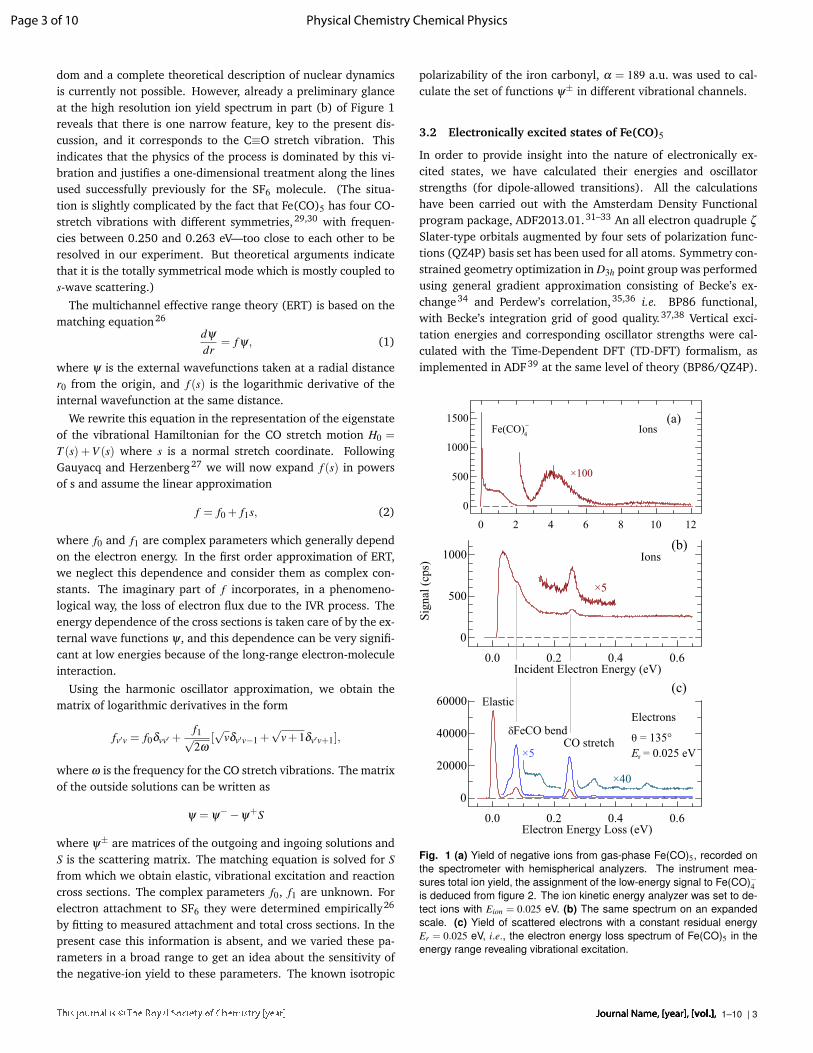
dom and a complete theoretical description of nuclear dynamicsis currently not possible. However, already a preliminary glanceat the high resolution ion yield spectrum in part (b) of Figure 1reveals that there is one narrow feature, key to the present dis-cussion, and it corresponds to the C≡O stretch vibration. Thisindicates that the physics of the process is dominated by this vi-bration and justifies a one-dimensional treatment along the linesused successfully previously for the SF6 molecule. (The situa-tion is slightly complicated by the fact that Fe(CO)5 has four CO-stretch vibrations with different symmetries,29,30 with frequen-cies between 0.250 and 0.263 eV—too close to each other to beresolved in our experiment. But theoretical arguments indicatethat it is the totally symmetrical mode which is mostly coupled tos-wave scattering.)
The multichannel effective range theory (ERT) is based on thematching equation26
dψ
dr= f ψ, (1)
where ψ is the external wavefunctions taken at a radial distancer0 from the origin, and f (s) is the logarithmic derivative of theinternal wavefunction at the same distance.
We rewrite this equation in the representation of the eigenstateof the vibrational Hamiltonian for the CO stretch motion H0 =
T (s) +V (s) where s is a normal stretch coordinate. FollowingGauyacq and Herzenberg27 we will now expand f (s) in powersof s and assume the linear approximation
f = f0 + f1s, (2)
where f0 and f1 are complex parameters which generally dependon the electron energy. In the first order approximation of ERT,we neglect this dependence and consider them as complex con-stants. The imaginary part of f incorporates, in a phenomeno-logical way, the loss of electron flux due to the IVR process. Theenergy dependence of the cross sections is taken care of by the ex-ternal wave functions ψ, and this dependence can be very signifi-cant at low energies because of the long-range electron-moleculeinteraction.
Using the harmonic oscillator approximation, we obtain thematrix of logarithmic derivatives in the form
fv′v = f0δvv′ +f1√2ω
[√
vδv′v−1 +√
v+1δv′v+1],
where ω is the frequency for the CO stretch vibrations. The matrixof the outside solutions can be written as
ψ = ψ−−ψ
+S
where ψ± are matrices of the outgoing and ingoing solutions andS is the scattering matrix. The matching equation is solved for Sfrom which we obtain elastic, vibrational excitation and reactioncross sections. The complex parameters f0, f1 are unknown. Forelectron attachment to SF6 they were determined empirically26
by fitting to measured attachment and total cross sections. In thepresent case this information is absent, and we varied these pa-rameters in a broad range to get an idea about the sensitivity ofthe negative-ion yield to these parameters. The known isotropic
polarizability of the iron carbonyl, α = 189 a.u. was used to cal-culate the set of functions ψ± in different vibrational channels.
3.2 Electronically excited states of Fe(CO)5
In order to provide insight into the nature of electronically ex-cited states, we have calculated their energies and oscillatorstrengths (for dipole-allowed transitions). All the calculationshave been carried out with the Amsterdam Density Functionalprogram package, ADF2013.01.31–33 An all electron quadruple ζ
Slater-type orbitals augmented by four sets of polarization func-tions (QZ4P) basis set has been used for all atoms. Symmetry con-strained geometry optimization in D3h point group was performedusing general gradient approximation consisting of Becke’s ex-change34 and Perdew’s correlation,35,36 i.e. BP86 functional,with Becke’s integration grid of good quality.37,38 Vertical exci-tation energies and corresponding oscillator strengths were cal-culated with the Time-Dependent DFT (TD-DFT) formalism, asimplemented in ADF39 at the same level of theory (BP86/QZ4P).
0.0 0.2 0.4 0.6
0
20000
40000
60000
Electron Energy Loss (eV)
Electrons
q = 135°
Er = 0.025 eV
Elastic
×5
×40
dFeCO bendCO stretch
(c)
0.0 0.2 0.4 0.6
0
500
1000
Incident Electron Energy (eV)
Sig
nal
(cp
s)
×5
Ions(b)
0 2 4 6 8 10 12
0
500
1000
1500Ions
×100
(a)Fe(CO)
4
-
Fig. 1 (a) Yield of negative ions from gas-phase Fe(CO)5, recorded onthe spectrometer with hemispherical analyzers. The instrument mea-sures total ion yield, the assignment of the low-energy signal to Fe(CO)−4is deduced from figure 2. The ion kinetic energy analyzer was set to de-tect ions with Eion = 0.025 eV. (b) The same spectrum on an expandedscale. (c) Yield of scattered electrons with a constant residual energyEr = 0.025 eV, i.e., the electron energy loss spectrum of Fe(CO)5 in theenergy range revealing vibrational excitation.
Journal Name, [year], [vol.],1–10 | 3
Page 3 of 10 Physical Chemistry Chemical Physics

0 1 2 3 4 5 6 7 8
F e ( C O ) -2
2 x 2 5 x
F e -
F e ( C O ) -3
02 0 0 04 0 0 06 0 0 08 0 0 0
1 0 0 0 01 2 0 0 0
E n e r g y ( e V )
Intens
ity (c
.p.s.)
F e ( C O ) -4
0 1 2 3 4 5 6 7 8 9 1 0 1 1 1 205
1 01 52 02 5
F e ( C O ) -
Fig. 2 Negative ion yield as a function of electron energy recorded on theDEA spectrometer with trochoidal monochromator and quadrupole massfilter.
4 Results and discussion4.1 DEA: experiment below 0.5 eV
Figure 1 shows the negative ion yield from Fe(CO)5 recorded onthe electron spectrometer with hemispherical analyzers and Fig-ure 2 the mass-resolved ion yields for individual anions recordedon the setup with trochoidal monochromator and quadrupolemass filter. The spectra from the two instruments are in very goodagreement. The ion yield shows an intense narrow peak at lowenergies. The results from the quadrupole instrument in Figure 2show that it is entirely due to the Fe(CO)−4 fragment. Figure 1bshows that the peak is only about 70 meV wide. It thus appearsto be less high in the spectra from the quadrupole instrument inFigure 2, where it is convoluted with the 200 meV wide energyprofile of the electron beam.
Essential features of the spectra agree with the early measure-ments of Compton and Stockdale,8 except that their spectra didnot show the 0 eV peak, but only a broad Fe(CO)−4 band with amaximum around 0.8 eV. The absence of the low energy peak intheir spectrum can presumably be attributed to the failure of theirinstrument to generate sufficiently slow electrons. The presentstrong DEA signal close to 0 eV is consistent with the high elec-tron attachment rates measured in an ICR cell10 and by the flow-ing afterglow technique.11
The high resolution spectrum in figure 1b reveals previouslyunreported fine features: a cusp at 0.08 eV and a small peak at0.26 eV. These structures closely resemble the structures closeto thresholds for vibrational excitation that were observed, forexample, in the DEA spectra of hydrogen halides or methylhalides.25 Such structures are due to interchannel coupling—opening of the vibrational excitation channel reduces the flux intothe DEA channel. Which vibrational levels of Fe(CO)5 are excitedat threshold is revealed by the electron-energy loss spectrum inFigure 1c. Comparison of the parts (b) and (c) of figure 1 revealthat the structures in the DEA cross section are very close to thethresholds for vibrational excitation. The two most prominent in-elastic peaks in this spectrum correspond to excitation of δFeCObending (overlap of v7,a′′2 and v11,e′ modes) and CO stretch (over-
0.0 0.1 0.2 0.3
0
200
400
Ion Kinetic Energy (eV)
Ion
Co
un
t R
ate
(c/s
)
0
100
200
Ei = 0.02 eV
Ei = 1.0 eV
Fig. 3 Ion kinetic energy distributions recorded at the two incident elec-tron energies indicated.
lap of four normal modes involving CO stretch). (The mode num-bering and assignment are identical to those of Refs.29,30.) Thistype of structures has been successfully reproduced either by thenonlocal resonance model or the effective range theory and the-ory has always provided a very valuable insight into the mech-anism of process.25 We have applied the latter theory here, asdetailed in the next subsection.
Revealing information about energy partitioning in the frag-mentation process is provided by the ion kinetic energy distribu-tions and we therefore measured ion kinetic energy spectra withthe electrostatic instrument as shown in Fig. 3. The spectra arecorrected for the analyzer response function, using the responsefunction determined for electrons. Two distributions were mea-sured. One, discussed in this section, at essentially zero incidentelectron energy, at the zero electronvolt DEA peak, the other,discussed at a later section, at Ei = 1 eV, within the 1 eV reso-nance. Both are narrow, the widths at half height are 60 meV atEi = 0.02 eV and 50 meV at Ei = 1.0 eV. Both distributions peakat the very low energy of 0.025 eV, whereby the instrumental ioncollection efficiency drops below about 25 meV, so that the truedistribution may peak at an even lower energy.
The maximum Fe(CO)−4 kinetic energy is given by the avail-able excess energy Ee = EA− BDE + Ei, where EA is the elec-tron affinity of the product negative ion Fe(CO)−4 , BDE theFe(CO)4−CO bond dissociation energy, and Ei the incident elec-tron energy. EA and BDE are, unfortunately, not known withhigh precision as discussed by Lacko et al.12, Shuman et al.11
and Buathong et al.14 Excess energy Ee = 0.6± 0.3 eV is ob-tained with EA = 2.4± 0.3 eV20 and the experimental value ofBDE = 1.8± 0.09 eV.40 Taking the calculated value of BDE =
1.43 eV12 yields Ee = 1.03±0.3. Ei = 0.025 eV can be neglected inview of the large error bar of EA.
Only 14% of the total kinetic energy release is given to theFe(CO)−4 fragment, so that the Fe(CO)−4 maximum kinetic energyis 0.09± 0.04 eV or 0.15± 0.04 eV for the two choices of BDE,respectively.
These numbers are higher than the measured peak position of0.025 eV (Fig. 3). This indicates that a major fraction of the avail-able excess energy is left as vibrational energy of the Fe(CO)−4
4 | 1–10Journal Name, [year], [vol.],
Page 4 of 10Physical Chemistry Chemical Physics

0.1 1.0
010
110
210
Electron Energy (eV)
Ela
stic
Cro
ss S
ecti
on (
Å/s
r)2
135°
Fig. 4 Differential elastic cross section measured at θ = 135◦.
fragment and thus supports the hypothesis of substantial IVR inthe Fe(CO)−5 attachment complex. On the other hand the factthat the tail of the distribution extends up to about 0.15 eV forEi = 0.025 eV in Fig. 3 indicates that the IVR process is not com-plete, the Fe(CO)−4 fragment is not fully thermalized. This resultagrees with the conclusion of Buathong et al.,14 based on study ofelectron attachment in Rydberg atom collisions, that partial butnot complete statistical redistribution of the excess energy priorto dissociation occurs, indicating dissociation of Fe(CO)−5 on timescales of a few vibrational periods.
Finally, since virtual states, implied in the theoretical treatmentbelow, are manifested by a sharp rise of the elastic cross section atlow energies, we report the elastic cross section in Figure 4. Thecross section does rise very sharply at low energy (observe that itis shown on a log-log scale), providing an experimental evidencefor a virtual state. A pronounced Ramsauer-Townsend minimumoccurs at 0.28 eV.
4.2 DEA: theory below 0.5 eVOur first choice of the ERT parameters was motivated by our pre-vious calculations of electron attachment to SF6.26 Specifically,we have chosen r0 = 3.23, f0 = 0.989+ 0.108i, f1 = −0.00991+0.0025i. (All parameters are in a.u.) Although, what can be calledthe “size" of Fe(CO)5, is greater than r0, the ERT radius cannotbe taken too large as this leads to the energy dependence of theparameters f0 and f1. Therefore we consider the extension ofthe polarization potential into the region r0 < r < R (where R isthe effective size of the molecule) as an empirical way to incor-porate the electron-molecule interaction in this region. Since theFe-C distance is 1.81 Å, and C-O distance 1.15 Å,29,30 R shouldbe about 6 a.u.
The listed set of parameters leads to a virtual-state scatteringat low energies, similar to e−SF6 scattering. Variation of f0 andf1 resulted in the following observations: The increase of Re f0leads to less pronounced virtual-state effect. The Im f0 parametermostly controls coupling between the scattering and attachmentchannels, and therefore influences only the magnitude of the at-tachment cross section, but not its shape. The parameter Re f1influences less the attachment cross section as it is mostly respon-sible for vibrational excitation. Finally, the attachment cross sec-tion has very little sensitivity to Im f1.
Fig. 5 Electron attachment to iron pentacarbonyl calculated with two setsof parameters as described in text. Solid (black curve), Re f1 =−0.0991;dashed (red) curve, Re f1 =−0.143.
Fig. 6 Electron attachment to iron pentacarbonyl calculated with polar-izabilities α = 220 a.u. (solid black curve) and α = 230 a.u. (dashed redcurve).
Journal Name, [year], [vol.],1–10 | 5
Page 5 of 10 Physical Chemistry Chemical Physics

In figure 5 we present two curves for attachment cross sec-tions corresponding to the original choice of parameters and withRe f1 replaced by −0.143. The cusp at the CO stretch threshold iscaused by the virtual state due to the e−Fe(CO)5 polarization at-traction. It is well known26,41 that by increasing e−M attraction,one can convert the virtual-state cusp into vibrational Feshbachresonance. In figure 6 we show the result of this numerical ex-periment performed by increasing the polarizability α. At α = 220a.u. the cusp becomes very pronounced meaning that the virtualstate is on the brink of conversion to the bound state. Then atα = 230 a.u. a below-threshold resonance appears meaning thatthe virtual state has been converted into a bound state.
The magnitude of the cross section can be checked by calcula-tion of the attachment rate coefficient k and comparison with themeasurements of Shuman et al.11 who obtained k = (7.9±1.4)×10−8 cm3/s at T = 300 K and k = (8.8±2)×10−8 cm3/s at T = 400K. Our first choice of the parameter Re f1 (solid curve in Fig. 5)gives k = 5.92×10⁻⁸ cm3/s and the second choice (dashed curvein Fig. 5) k = 6.26×10⁻⁸ cm3/s at T = 300 K. As was mentioned,the absolute value of the cross section is more sensitive to theparameter Re f0. In particular, the choice Re f0 = 0.7 a.u. leadsto k = 8.14×10⁻⁸ cm3/s at T = 300 K, closer to the experimentalvalue. With regard to the temperature dependence, since the the-ory incorporates explicitly only C-O stretch vibrations, the crosssection is almost independent of vibrational temperature at ther-mal energies, and all temperature dependence is determined bythe electron energy dependence of the cross section. In particu-lar, with the choice Re f0 = 0.7 a.u., the rate coefficient drops from8.14×10−8 to 7.45×10⁻⁸ cm3/s. Although this drop is within theexperimental uncertainty, it could be possible that the actual ratecoefficient grows with the temperature because of the growth ofpopulation of excited states corresponding to other modes withlower frequencies not included in our model.
Finally we add that the attachment rate, although rather high,is small as compared to the prediction of the Vogt-Wanniermodel42 describing quantum capture by the polarization poten-tial. The Vogt-Wannier thermal rate coefficient is given by43
kVW = 7.755×10⁻⁸α
1/2 cm3/s
where α is taken in a.u. For iron pentacarbonyl this estimate ex-ceeds the actual value by a factor 13.5. This makes this moleculerather inefficient attacher11 as compared, for example, with SF6
and CCl4.
4.3 DEA: experiment above 0.5 eV
This section discusses the DEA bands above 0.5 eV, shown in Figs.1 and 2. Our spectrum is in an excellent agreement with thatof Schukin et al.9 A number of resonant bands appear and weattempt their assignment to shape and Feshbach resonances. In-dependent information about shape resonances is obtained fromthe cross sections for vibrational excitation (VE) shown in Fig.7. All VE cross sections have very intense threshold peaks whichare due to the virtual state discussed in section 4.2. A numberof broad bands can be discerned at higher energies, assigned asoverlapping shape resonances with temporary occupation of CO-
0 1 2 3 4
0.00
0.04
0.08
0.12
Incident Electron Energy (eV)
Cro
ss S
ecti
on
(Å
)2/s
r
÷20
DE = 0.057 eV
0.00
0.04
0.08
0.12
÷50
DE = 0.079 eV
0.00
0.04
0.08
÷50
DE = 0.252 eV
Fe(CO)5
CO (÷12)
p*CO 135°
Fig. 7 Cross sections for vibrational excitation, indicative of resonances.
located virtual orbitals in Fe(CO)5 resulting from overlapping π∗COorbitals.
The v = 0 → 1 VE cross section of carbon monoxide is alsoshown in Fig. 7 for comparison and shows that the Fe(CO)5 π∗
bands are in the right energy range.There is only a limited correspondence between the DEA and
the VE spectra. The 1.2 eV Fe(CO)−3 band in Fig. 2 corresponds tothe 1.3 eV band in the CO stretch excitation cross section (∆E =
0.252 eV) in Fig. 7. The 0.7 eV Fe(CO)−4 band in Fig. 2 does nothave any clear corresponding band in the VE spectra. It could bethat there is a π∗ resonance at 0.7 eV but is obscured by the tailof the threshold peaks in the VE spectra. It could also be that the0.7 eV Fe(CO)−4 band in the DEA spectra is caused by the same π∗
resonance as the 1.3 eV band in the VE spectra, but the DEA bandis lowered by the “kinetic shift”, i.e., the resonance width beingnarrower at lower energies.
Electronic Feshbach resonances are generally located 0-0.4 eVbelow their parent triplet electronically excited state and shouldthus follow a pattern similar to that of triplet bands in an electronenergy loss (EEL) spectrum (shown in sec. 4.5 below). Compari-son of the EELS spectrum with the DEA spectrum in Fig. 2 revealssuch a similarity, in particular the shapes of the 5.9 and 8.8 eVFe(CO)− bands in Fig. 2 are reminiscent of the 5.76 eV and the9.2 eV triplet bands in in the EEL spectrum, permitting the assign-ment of these DEA bands to Feshbach resonances.
Finally we address the question of the decay dynamics of thehigher-lying resonances, in particular the one which gives rise tothe 1 eV shoulder in the ion yield in Fig. 1. We do this by recordingthe spectrum of electrons detached following a capture of a 1 eVelectron, as shown by the center trace in Fig. 8. The excitation ofthe CO stretch vibration, and that an overtone of it is excited, areindications of a temporary occupation of a π∗CO orbital. The inter-esting feature is the group of electrons around the energy-loss of1 eV in the center spectrum of Fig. 8, i.e., electrons detached withnearly zero energy. Such electrons are a manifestation of an ex-tremely fast radiationless decay, fast enough to compete with the
6 | 1–10Journal Name, [year], [vol.],
Page 6 of 10Physical Chemistry Chemical Physics

0.0 0.2 0.4 0.6 0.8 1.0
0
5
Electron Energy Loss (eV)
Cro
ss S
ecti
on (
Å/s
r)2
×10 Ei = 0.28 eV
0.0
0.5
1.0
1.5 Ei = 1.0 eV
×10
0
1
2
3
×50
Ei = 3.0 eV
0.252
0.252
0.252
CO str
2 CO str
0.08
0.08
0.08
0.058
0.058
0.058 Fe(CO)5
Fig. 8 Distributions of scattered electron energies at the incident ener-gies of 0.28, 1.0 and 3.0 eV.
ns-ps fast autodetachment of the resonance. This process, pre-sumably mediated by a conical intersection between the potentialsurfaces of the 1 eV π∗CO shape resonance and the ground stateFe(CO)−5 , leads to a rapid conversion of electronic to vibrationalenergy followed by detachment of thermal electrons.
Note the unusual situation in the Ei = 0.28 eV spectrum at thebottom of Fig. 8, where, as a consequence of the threshold peakin the CO stretch excitation (see top trace in Fig. 7) and of theRamsauer-Townsend minimum in the elastic cross section (Fig.4), the elastic peak is nearly 20× lower than the inelastic peak at∆E = 0.252 eV!
4.4 Two-dimensional EEL spectrum
Two-dimensional (2D) spectra provide insight into the dynamicsof resonances by mapping their decay channels and their capac-ity to thermalize electrons.44–46 The 2D spectrum of Fe(CO)5 isshown in Fig. 9.
Features already discussed above can be recognized: (i) TheRamsauer-Townsend minimum can be discerned at incident en-ergy Ei =0.28 eV on the ‘elastic ridge’ (situated vertically at anenergy-loss ∆E = 0), (ii) the threshold peaks in the excitation ofindividual vibrational modes, and (iii) enhancement of the exci-tation of the δFeCO bend and CO stretch vibrations in the 0.8-1.6 eV incident energy range, indicative of π∗ resonances. Notethat ejection of thermal electrons is mapped along the diagonal“threshold line” where ∆E = Ei, i.e., Er = 0. Interesting is thusthe “threshold ridge” signal (green diagonal line in Fig. 9) - it re-veals efficient ejection of thermal electrons in the incident energyrange zero to ∼1.4 eV. It is indicative of the fast dynamics, rapidthermalization of the electrons by IVR followed by thermal de-tachment. Interesting is further that the DEA signal, also shownfor comparison in the same figure, mimics the shape of the yieldof thermal electrons. This indicates that the two processes areclosely related—attachment of an electron into a π∗ resonance is
0.0
0.2
0.4
0.6
0.8
1.0
1.2
1.4
1.6
1.8
0.0 0.2 0.4 0.6 0.8 1.0 1.2 1.4 1.6 1.8 2.0
Electron Energy Loss (eV)
Inci
den
t E
lect
ron
En
erg
y (
eV)
Fe(CO)5
DEA
0.0
0.2
0.4
0.6
0.8
1.0
threshold lineelastic ridge
´4
Fig. 9 Two-dimensional electron energy loss spectrum and DEA spec-trum. The Incident Electron Energy scale (the ordinate) refers to both the2D energy loss spectrum and the DEA spectrum. The Energy Loss scaleapplies to the 2D spectrum only; it does not apply to the DEA spectrum -the horizontal scale shows the ion kinetic energy there.
followed by very rapid IVR leading to a hot Fe(CO)−5 anion whichdecays by one of the two competing decay processes, detachmentof a thermal electron or a thermal loss of a CO ligand.
There is a subtle interesting feature in the 2D spectrum: a faintdiagonal line parallel to the ‘threshold ridge’, but shifted left. Itindicates enhanced ejection of electrons with a discrete energyEr = 0.250 eV, independent of the incident energy, and over thesame range of incident energies where the zero eV electrons arealso ejected. With a certain overstatement one could say thatthe collision complex has become an electron monochromator—electrons with a range of energies are attached and monoener-getic electrons are ejected. A plausible explanation is that thethermalized Fe(CO)−5 can, apart of ejecting a thermal, nearly 0 eVelectron, also eject a 0.250 eV electron by simultaneously los-ing one quantum of the CO stretch vibration. The CO stretchmode promotes detachment. The process where a specific vi-brational mode promotes electron detachment has also been ob-served in acrylonitrile.44,46 Autodetachment mediated by specificvibrational modes has also been reported by Verlet and coworkersin a time-resolved photodetachment study of para-toluquinonetrimer cluster anion.47
4.5 Electronic excitationFigure 10 shows the electron-energy loss spectra in the energyrange 2 - 15 eV, measured on the spectrometer with hemispher-ical analyzers. The spectra were recorded at 0◦ and 180◦ scat-tering angles. In order to keep the analyzer response functionconstant, the spectra were recorded at constant residual energyof an electron Er and the incident energy was scanned (the x-axiscorresponds to the energy loss, a difference between the incidentand residual energy).
It is well established48 that if the incident electron has highenergy and undergoes little deflection (large impact parameters),
Journal Name, [year], [vol.],1–10 | 7
Page 7 of 10 Physical Chemistry Chemical Physics

2 3 4 5 6 7 8 9 10 11 12 13 14 150
30
60
90 = 0o, E
r = 20 eV
(a)
E'A
2''
2 3 4 5 6 7 8 9 10 11 12 13 14 150
4
8(b) = 180o, E
r = 1 eVSi
gnal
(cps
)
Electron energy loss (eV)
Fig. 10 Electron-energy loss spectra of gas phase Fe(CO)5 recorded attwo different scattering conditions. Vertical bars are at the calculated (TD-DFT) positions of the excited states, in panel (a) their heights correspondto oscillator strengths. Panel (a) shows the singlet states of E ′ and A′′2symmetries, panel (b) the triplet states.
the long-range interaction with the molecule leads to selectionrules identical to those for optical transitions. On the other hand,spin-forbidden transitions, due to spin-exchange scattering, arepreferred at low electron energies (incident electron wavelengthcomparable with the wavelength of valence electrons) and havenearly isotropic angular distribution. The excitation of dipole-allowed singlet states thus dominates in the forward direction athigher energies and the excitation of triplet states is favored atlow electron energies and large scattering angles. In figure 10awe thus show present TD-DFT excitation energies and oscillatorstrengths for dipole-allowed transitions. In figure 10b we com-pare the backward EELS spectrum with calculated excitation en-ergies of triplet states. Since the individual bars at the calculatedpositions of triplets states are not discernible, we have convolutedthe calculated spectrum with a Gaussian of 1 eV FWHM. The ta-bles with energies, configurations and (for allowed transitions)oscillator strengths are presented in the Supplementary Informa-tion.
Fe(CO)5 is a low-spin d8 complex with the trigonal bipyrami-dal structure. In D3h point group iron d orbitals split into e′ (dxy,dx2−y2 ), e′′ (dxz, dyz) and a′1 (dz2 ) that combine with the suitableMOs of CO ligands. As a consequence, the five highest occu-pied and the five lowest unoccupied MOs of Fe(CO)5 involve irond orbitals.49 MOs with dominant metal d-orbital character arestrongly σ -antibonding, empty 14a′1, and highest occupied 10e′
and 3e′′. The latter two sets are result of the π-back-bondingof the iron orbitals with the π∗ carbonyl orbitals. The ground
electronic state of Fe(CO)5 is 1A′1 and dipole-allowed transitionsare to the excited E ′ and A′′2 states, shown in Figure 10 as olive-green and blue bars, respectively. Calculated excitation energiesand oscillator strengths are in excellent agreement with the EELSspectrum (Figure 10a), and with near-UV gas-phase50 and solu-tion spectra.51 In particular, the most prominent bands at 5.0 and6.3 eV can be clearly assigned as metal-to-ligand charge transfer(MLCT) transitions to the E ′ and A′′2 excited states, respectively.Excitation energies by TDDFT are overestimated by∼ 0.25 eV. Re-cent high-level ab-initio studies52,53 reported significant overesti-mation of the first band (by 0.6-1.5 eV). The maximum in TDDFTspectrum lies between two groups of E ′ states with considerableoscillator strengths. The first group is of mixed MLCT and Ry-dberg 3d → 4s character, while the second one is of MLCT andRydberg 3d→ 4p type. In addition, our TDDFT results explain allthe other experimental features. The gradual ascent of the signalin the range 4-5 eV is dominated by MLCT transitions (one A′′2and two E ′ states). A dipole-allowed E ′ d− d transition is alsopredicted to be in this range, although it carries little oscillatorstrength. Finally, the broad band at energies higher than 8 eV isseen to be a consequence of a group of high-lying ligand-to-metalcharge transfer and intra-ligand transitions.
Very good agreement is also obtained for singlet-triplet transi-tions (Figure 2b). Obviously, peaks at EELS spectrum correspondto the regions where high density of triplet states is calculatedin TDDFT. The lowest triplet, 3E ′ state, due to the d−d spin-fliptransition (10e′ → 14a′1) is calculated to be 0.56 eV lower thancorresponding singlet state. It is noteworthy to mention that onecomponent of this, degenerate state, becomes the ground elec-tronic state upon dissociation of one CO ligand.54,55
5 ConclusionsWe provide new insight into elementary electron-induced decom-position processes in gas phase Fe(CO)5.
The dominant feature in DEA is a high and narrow (70 meV)peak in the Fe(CO)−4 formation (cleavage of one metal-ligandbond) at near-zero incident electron energy. Fine structures areobserved on the tail of this peak, at vibrational excitation thresh-olds.
A model based on the effective range theory with complexboundary conditions reproduces these structures and interpretsthem as evidence of virtual state scattering, with an importantrole being played by intramolecular vibrational redistribution(IVR).
The hypothesis of a virtual state dominating low energy(<0.5 eV) processes receives further support from the observa-tion of threshold peaks in vibrational excitation cross sections andfrom a dramatic rise of the elastic cross section at very low ener-gies.
The second highest DEA feature is a Fe(CO)−4 peak around0.8 eV which we interpret as due to a π∗ resonance. A two-dimensional (2D) EEL spectrum reveals efficient detachment ofnearly zero eV electrons over the same range of incident electronenergies as this DEA band, i.e., the capacity of Fe(CO)5 to ther-malize electrons within this π∗ resonance. This is taken as an ex-perimental evidence for a rapid IVR process being important also
8 | 1–10Journal Name, [year], [vol.],
Page 8 of 10Physical Chemistry Chemical Physics

for this π∗ resonance—it converts the anion formed by the initialattachment into a hot Fe(CO)−5 anion that then decays either byloss of one CO ligand or by detachment of a thermal electron. Thelow measured kinetic energies of the Fe(CO)−4 fragment providean additional evidence for IVR taking place prior to dissociation.
The 2D spectrum also reveals a somewhat exotic capacity ofthe CO stretch vibrational mode to promote detachment in thehot Fe(CO)−5 anion, leading to a small yield of superthermal elec-trons with a discrete energy equal to the CO stretch vibrationalquantum. These electrons are observed over a range of incidentelectron energies covering the entire width of the π∗ resonance.
Combination of electron-energy loss spectra and TD-DFT calcu-lations characterizes the electronically excited states of Fe(CO)5.The calculations agree with the experiment very well and repro-duce both the spin-allowed and spin-forbidden transitions. Thesignificance of these results is that (i) these excited states rep-resent a path to neutral dissociation and (ii) the energies ofthe triplet states provide indication of energies of Feshbach res-onances and permit conclusions about assignments of the higher-lying DEA bands.
The importance of the present findings is that they reveal mech-anisms via which iron pentacarbonyl is dissociated at various en-ergy ranges. The electrons with energies below 1 eV lead to veryefficient DEA. The DEA cross section is strongly enhanced by thelong-range forces: the high polarizability of Fe(CO)5 leads to avirtual state scattering. The presence of this virtual state leads tohigh DEA cross section. The long-range forces are thus crucial forthe low-energy DEA. On the other hand the electronic excitation,the first step in the neutral dissociation pathway, can be viewed asa direct excitation and thus a short range process. This differenceopens a major question: when the iron pentacarbonyl reacts withelectrons in an environment (e.g., adsorbed at a surface at realis-tic FEBID conditions), the typical distances between molecules (orFe(CO)5 molecules and ’bulk’) are smaller than the distances onwhich the electron-induced-dipole interaction is operative. Howdoes this fact influence the low-energy DEA effectivity? This ques-tion has been addressed in our cluster beam study.56
AcknowledgementsThis work has been supported by the the Czech Science Founda-tion project Nr. 17-04844S (J.F.), Swiss National Science Founda-tion Project No. 200020-144367/1 (M.A.), US National ScienceFoundation Grant No. PHY-1401788 (I.I.F), Serbian Ministry ofScience Project 172035 (M.Z.), and by several short-term scien-tific missions withing the COST Action CM1301 CELINA.
References1 I. Utke, P. Hoffmann and J. Melngailis, J. Vac. Sci. Technol. B,
2008, 26, 1197.2 M. Takeguchi, M. Shimojo and K. Furuya, Nanotechnology,
2005, 16, 1321–1325.3 T. Lukasczyk, M. Schirmer, H.-P. Steinrück and H. Marbach,
Small, 2008, 4, 841–846.4 J. M. De Teresa and A. Fernández-Pacheco, Appl. Phys. A,
2014, 117, 1645.
5 R. M. Thorman, T. P. R. Kumar, D. H. Fairbrother and O. In-gólfsson, Beilstein J. Nanotechnol., 2015, 6, 1904–1926.
6 H. Miyazoe, I. Utke, H. Kikuchi, S. Kiriu, V. Friedli, J. Michlerand K. Terashima, J. Vac. Sci. Tech. B, 2010, 28, 744.
7 B. Geier, C. Gspan, R. Winkler, R. Schmied, J. D. Fowlkes,H. Fitzek, S. Rauch, J. Rattenberger, P. D. Rack and H. Plank,J. Phys. Chem. C, 2014, 118, 14009.
8 R. N. Compton and J. A. D. Stockdale, Int. J. Mass. Spectrom.Ion Phys., 1976, 22, 47–55.
9 P. V. Shchukin, M. V. Muftakhov and R. V. Khatymov, Issled.Russ., 2005.
10 P. M. George and J. L. Beauchamp, J. Chem. Phys., 1982, 76,2959.
11 N. S. Shuman, T. M. Miller, J. F. Friedman and A. A. Viggiano,J. Phys. Chem. A, 2012, 117, 1102.
12 M. Lacko, P. Papp, K. Wnorowski and Š. Matejcík, Eur. Phys.J. D., 2015, 69, 84.
13 M. Lacko, P. Papp, Š. Matejcík and K. Wnorowski, WDS’14Proceedings of Contributed Papers - Physics, 2014, 292–297.
14 S. Buathong, M. Kelley and F. B. Dunning, J. Chem. Phys.,2016, 145, 134309.
15 S. Massey, A. D. Bass, E. Alizadeh and L. Sanche, J. Phys.:Conf. Ser., 2015, 635, 062012.
16 S. Massey, A. D. Bass and L. Sanche, J. Phys. Chem. C, 2015,119, 12708.
17 C. Hauchard and P. A. Rowntree, Can. J. Chem., 2011, 89,1163–1173.
18 J. Lengyel, J. Kocišek, , M. Fárník and J. Fedor, J. Phys. Chem.C, 2016, 120, 7397–7402.
19 J. Lengyel, J. Fedor and M. Fárník, J. Phys. Chem. C, 2016,120, 17810–17816.
20 P. C. Engelking and W. C. Lineberger, J. Am. Chem. Soc., 1979,101, 5569.
21 M. Zlatar, M. Allan and J. Fedor, J. Phys. Chem. C, 2016, 120,10667–10674.
22 M. Allan, J. Phys. B: Atom. Molec. Phys., 1992, 25, 1559.23 M. Allan, J. Phys. B: Atom. Molec. Phys., 2005, 38, 3655.24 M. Stano, Š. Matejcík, J. D. Skalny and T. D. Märk, J. Phys. B:
Atom. Molec. Phys., 2003, 36, 261.25 H. Hotop, M.-W. Ruf, M. Allan and I. I. Fabrikant, Adv. At. Mol.
Opt. Phys., 2003, 49, 85.26 I. I. Fabrikant, H. Hotop and M. Allan, Phys. Rev. A, 2005, 71,
022712.27 J. P. Gauyacq and A. Herzenberg, J. Phys. B: Atom. Molec.
Phys., 1984, 17, 1155.28 L. D. Landau and E. M. Lifshitz, Quantum Mechanics (Nonrel-
ativistic Theory), Butterworth-Heinenann, 1981.29 V. Jonas and W. Thiel, J. Chem. Phys., 1995, 102, 8474–8484.30 J. H. Jang, J. G. Lee, H. Lee, Y. Xie and H. F. S. III, J. Phys.
Chem. A, 1998, 102, 5298–5304.31 C. Fonseca Guerra, J. G. Snijders, G. te Velde and E. J.
Baerends, Theor. Chem. Acc., 1998, 99, 391–403.32 G. te Velde, F. M. Bickelhaupt, E. J. Baerends, C. Fonseca
Journal Name, [year], [vol.],1–10 | 9
Page 9 of 10 Physical Chemistry Chemical Physics

Guerra, S. J. A. van Gisbergen, J. G. Snijders and T. Ziegler, J.Comput. Chem., 2001, 22, 931–967.
33 ADF: Density Functional Theory (DFT) software for chemists,version 2013.01, http://www.scm.com/, 2013.
34 A. D. Becke, Phys. Rev. A, 1988, 38, 3098–3100.35 J. P. Perdew, Phys. Rev. B, 1986, 33, 8822–8824.36 J. P. Perdew, Phys. Rev. B, 1986, 34, 7406–7406.37 A. D. Becke, J. Chem. Phys., 1988, 88, 2547–2553.38 M. Franchini, P. H. T. Philipsen and L. Visscher, J. Comput.
Chem., 2013, 34, 1819–1827.39 F. Kootstra, P. L. de Boeij and J. G. Snijders, J. Chem. Phys.,
2000, 112, 6517–6531.40 K. E. Lewis, D. M. Golden and G. P. Smith, J. Am. Chem. Soc.,
1984, 106, 3905–3912.41 E. Leber, I. I. Fabrikant, J. M. Weber, M.-W. Ruf and H. Hotop,
in Dissociative Recombination: Theory, experiment and applica-tions IV, ed. A. Mitchell and I. F. Schneider, World Scientific,Singapore, 2000, p. 69U76.
42 E. Vogt and G. H. Wannier, Phys. Rev., 1954, 95, 1190U1198.43 I. I. Fabrikant and H. Hotop, Phys. Rev. A, 2001, 63, 022706.44 K. Regeta and M. Allan, Phys. Rev. Lett., 2013, 110, 203201.
45 K. Regeta and M. Allan, J. Chem. Phys., 2015, 142, 184307.46 M. Allan, K. Regeta, J. D. Gorfinkiel, Z. Mašín, S. Grimme and
C. Bannwarth, Europ. Phys. J. D, 2016, 70, 123.47 J. N. Bull and J. R. R. Verlet, Science Advances, 2017, 3, year.48 M. Allan, J. Electron. Spectrosc. Relat. Phenom., 1989, 48,
219–351.49 A. J. Atkins, M. Bauer and C. R. Jacob, Phys. Chem. Chem.
Phys., 2015, 17, 13937–13948.50 M. Kotzian, N. Roesch, H. Schroeder and M. C. Zerner, J. Am.
Chem. Soc., 1989, 111, 7687–7696.51 M. Dartiguenave, Y. Dartiguenave and H. B. Gray, Bull. Soc.
Chim. Fr., 1969, 12, 4223.52 L. M. J. Huntington and M. Nooijen, J. Chem. Phys., 2015,
142, 194111.53 L. M. J. Huntington, O. Demel and M. Nooijen, J. Chem. The-
ory Comput., 2016, 12, 114–132.54 M. Poliakoff and E. Weitz, Acc. Chem. Res., 1987, 20, 408–414.55 M. Besora, J.-L. Carreón-Macedo, Álvaro Cimas and J. N. Har-
vey, Adv. Inorg. Chem., 2009, 61, 573 – 623.56 J. Lengyel, P. Papp, Š. Matejcík, J. Kocišek, M. Fárník and
J. Fedor, Beilstein J. Nanotechnol., 2017, 8, 2200.
10 | 1–10Journal Name, [year], [vol.],
Page 10 of 10Physical Chemistry Chemical Physics