distributed neural systems for face perceptionhaxbylab.dartmouth.edu/publications/hg11.pdfand for...
TRANSCRIPT

Chapter 6
Distributed Neural Systems for Face Perception James V. Haxby and Maria Ida Gobbini
Introduction Face perception plays a central role in social communication and is, arguably, one of the most sophisticated visual perceptual skills in humans. Consequently, face perception has been the sub-ject of intensive investigation and theorizing in both visual and social neuroscience.
The organization of neural systems for face perception has stimulated intense debate. Much of this debate has focused on models that posit the existence of a module that is specialized for face perception (Kanwisher et al., 1997 ; Kanwisher and Yovel, 2006 ) versus models that propose that face perception is mediated by distributed processing (Haxby et al., 2000 , 2001 ; Ishai et al., 2005 ; Ishai 2008 ). These two perspectives on the neural systems that underlie face perception are not necessarily incompatible (see Kanwisher and Barton, Chapter 7, this volume). In our work, we have proposed that face perception is mediated by distributed systems, both in terms of the involvement of multiple brain areas and in terms of locally distributed population codes within these areas (Haxby et al., 2000 , 2001 ). Specifically, we proposed a model for the distributed neural system for face perception that has a Core System of visual extrastriate areas for visual analysis of faces and an Extended System that consists of additional neural systems that work in concert with the Core System to extract various types of information from faces (Haxby et al., 2000 ). We also have shown that in visual extrastriate cortices, information that distinguishes faces from other categories of animate and inanimate objects is not restricted to regions that respond maximally to faces, i.e. the fusiform and occipital face areas (Haxby et al., 2001 ; Hanson et al., 2004 ).
In this chapter we present an updated model of distributed human neural systems for face per-ception. We will begin with a discussion of the Core System for visual analysis of faces with an emphasis on the distinction between perception of invariant features for identity recognition and changeable features for recognition of facial gestures such as expression and eye gaze. The bulk of the chapter will be a selective discussion of neural systems in the Extended System for familiar face recognition and for extracting the meaning of facial gestures, in particular facial expression and eye gaze. We will discuss the roles of systems for the representation of emotion, for person knowledge, and for action understanding in face recognition and perception of expression and gaze. The neu-ral systems that are recruited for extracting socially-relevant information from faces are an unbounded set whose membership accrues with further investigations of face perception. Rather than attempt an exhaustive review, our intention is to present systems that we believe are of par-ticular relevance for social communication and that are illustrative of how distributed systems are engaged by face perception. Many of the chapters in this volume provide a more detailed account for areas that we touch on in this chapter. Our review also is biased towards work that we have been directly involved in, with selective references to closely related work by other investigators. We will finish with a discussion of modularity and distributed processing in neural representation.
C6.S1
06-Calder-06.indd 9306-Calder-06.indd 93 12/23/2010 10:03:40 AM12/23/2010 10:03:40 AM
OUP UNCORRECTED PROOF-FPP, 12/23/2010, GLYPH

DISTRIBUTED NEURAL SYSTEMS FOR FACE PERCEPTION 94
The path that has led to our current model has been neither direct nor predictable. Our path was guided by empirical results that forced us repeatedly to question the ideas that motivated our experiments and to redirect our subsequent efforts. The work began in the late 1980s in the early days of imaging brain blood flow with positron emission tomography. One of us (JVH) was working at the National Institutes of Health with Cheryl Grady, Barry Horwitz, and Leslie Ungerleider, with critical support from Mortimer Mishkin and Stanley Rapoport. We set out to investigate whether the human visual system was organized into ventral object vision and dorsal spatial vision pathways. We used a face perception task to activate the ventral pathway (Haxby et al., 1991 , 1994 ), reasoning that face processing, as compared to visual processing of other objects, is more restricted to the ventral pathway. It was soon obvious, however, that faces had a distinct representation within the ventral pathway and were, therefore, a poor choice for revealing the full functional architecture of the ventral visual pathway. We began to conduct our own stud-ies of differential responses to faces and other objects in the ventral temporal cortex (Chao et al., 1999 ; Haxby et al., 1999 ; Ishai et al., 1999 ). When we examined the data from these experiments, it was apparent that the functional architecture of the ventral pathway was not a simple division into regions that responded preferentially to faces and regions that responded preferentially to other object categories. The two of us began working together at this point. We explored how the distinct responses to faces and other object categories were, in fact, organized, leading to the development of multivariate pattern (MVP) analysis and understanding the response to faces in ventral temporal cortex as a distributed population code (Haxby et al., 2001 ). At the same time, we became interested in the role of face perception in social communication, leading notably to studies of familiar face recognition (Gobbini et al., 2004 , 2006 ; Leibenluft et al., 2004 ; Gobbini and Haxby, 2007 ) and the perception of gaze direction and facial expressions (Hoffman and Haxby, 2000 ; Engell and Haxby, 2007 ; Montgomery and Haxby, 2008 ; Montgomery et al., 2009 ; Said et al., 2010 ). It soon became apparent that the functional architecture for these face percep-tion operations was not restricted to the ventral temporal cortex, and we were forced, again by the data, to look at the role played by other areas in visual extrastriate cortex and by areas in non-visual neural systems. The result of these repeated reformulations is the model that we present in this chapter. The broad outlines of our model have been relatively stable for the past 9 to 10 years, but we know from experience that future empirical research may well lead to further, possibly radical, revisions. We cannot predict what form the next revision may take.
The core system for visual analysis of faces Three extrastriate visual regions that respond maximally to faces Functional neuroimaging consistently has shown that three bilateral regions in occipital-temporal extrastriate visual cortex respond more strongly when viewing faces than when viewing other vis-ual images. These areas are in the inferior occipital gyrus — the occipital face area (OFA), the lateral fusiform gyrus — the fusiform face area (FFA), and the posterior superior temporal sulcus (pSTS) (Figure 6.1 ). Although these regions can usually be identified in both the right and left hemi-spheres, they tend to be larger and more reliably found in the right hemisphere. In addition to these areas that respond maximally to faces, nearby cortices also respond significantly during face recog-nition, and these non-maximal responses carry information about the distinction between faces and other stimuli (Haxby et al., 2001 ; Hanson et al., 2004 ) and are related to successful face identi-fication (Grill-Spector et al., 2004 ).
We proposed that these three areas — along with adjacent cortex that responds significantly, albeit not maximally, to faces — constitute a “Core System” for the visual analysis of face images
C6.S2.1
C6.S2
06-Calder-06.indd 9406-Calder-06.indd 94 12/23/2010 10:03:40 AM12/23/2010 10:03:40 AM
OUP UNCORRECTED PROOF-FPP, 12/23/2010, GLYPH

THE CORE SYSTEM FOR VISUAL ANALYSIS OF FACES 95
(Haxby et al., 2000 ). We proposed further that different regions within the Core System have representations that emphasize different types of visual information about faces.
Two classes of face perception operations In their classic paper on cognitive systems for face perception, Bruce and Young ( 1986 ) distin-guished between processes for recognizing facial identity and processes for analyzing facial expression and speech-related facial movements. Their model focused primarily on face identity recognition. We have emphasized how these complementary processes are dissociated in the Core System.
Identity recognition is dependent on perceiving facial features that are invariant across facial movements, such as facial expression and speech-related movements, and across variable view-ing conditions, such as angle of profile and lighting. Face recognition has a virtually unlimited capacity for representing distinct identities. The representations of these identities must be gen-erative and robust in that they allow recognition of a familiar face from a novel image — a particular image that one has never seen before — with a wide range of variations in expression, gaze, and mouth movements. Furthermore, the face recognition system is capable of perceiving that a previously unfamiliar face is unique and distinct from all faces that one has seen previ-ously. By contrast, analysis of facial expression is targeted at nuanced perception of changes that are associated with facial movement. For facial expression perception, precisely the variations that are irrelevant for identity recognition are of paramount importance. Moreover, categories of facial expressions — such as smiling, scowling, disgust, disdain, boredom — are perceived as the same expression even though they are produced by different familiar or unfamiliar individuals. Thus, these two classes of face perception operations are distinct and have the potential to inter-fere with each other. A change in expression should not be perceived as a change in identity. A particular expression should be interpreted as carrying similar information if produced by different individuals.
We have proposed that these two classes of face perception operations are kept distinct within the Core System by anatomic segregation of representational spaces that emphasize invariant features for identification versus changeable features such as expression and eye gaze changes
Fig. 6.1 Three visual extrastriate regions in occipitotemporal cortex that respond strongly to face stimuli. This figure shows the areas that responded more strongly to images of faces than to images of houses in a single subject. The activations are on a cortical surface that has been inflated to reveal sulcal cortex and tipped to show both lateral and ventral surfaces. Adapted from Haxby et al. ( 2000 ).
C6.S2.2
C6.F1
06-Calder-06.indd 9506-Calder-06.indd 95 12/23/2010 10:03:40 AM12/23/2010 10:03:40 AM
OUP UNCORRECTED PROOF-FPP, 12/23/2010, GLYPH

DISTRIBUTED NEURAL SYSTEMS FOR FACE PERCEPTION 96
(Haxby et al., 2000 ). We formulated this hypothesis based on findings from single-unit record-ings in macaque cortex (Hasselmo et al., 1989 ) that showed that neurons that were tuned to vari-ations in identity were located more ventrally in the convexity of inferior temporal (IT) cortex whereas neurons that were tuned to variations in expression were found in greater concentrations in the STS. We hypothesized that a similar dissociation exists between face-responsive cortices in the human fusiform and STS cortices.
Perception of both eye and mouth movements selectively evoke responses in the pSTS but not the FFA (Puce et al., 1998 ). Subsequent work has supported our hypothesis that the pSTS is involved both in the perception of facial movements and in the perception of features, such as eye gaze and expression, that are changed by movements. We found that selective attention to identity increased the neural response in the FFA but not in pSTS, whereas selective attention to eye gaze direction increased the neural response in pSTS but not in the FFA (Hoffman and Haxby, 2000 ), supporting our hypothesis that the FFA is involved more in the representation of identity whereas the pSTS is involved more in eye gaze perception. In another study, we compared the FFA and pSTS responses to dynamic changes in the eye gaze direction and identity (Engell et al., 2006 ) and found that the FFA responded more strongly to identity than gaze changes, whereas the pSTS responded more to gaze than identity changes. The response to faces shows adaptation to repeated expression in the pSTS as well as in a more anterior STS region but not in the fusiform cortex (Winston et al., 2004 ). In addition the STS responses to faces are significantly stronger for dynamic facial expressions than for still images of expressions, but no such enhancement is seen in the FFA (Figure 6.2 ). A functional localizer that uses dynamic videos of faces reveals a much larger face responsive STS region than does a localizer that uses static images of faces, whereas the addition of facial motion had little effect on the size of the fusiform face-responsive area (Hasson et al., 2004 ).
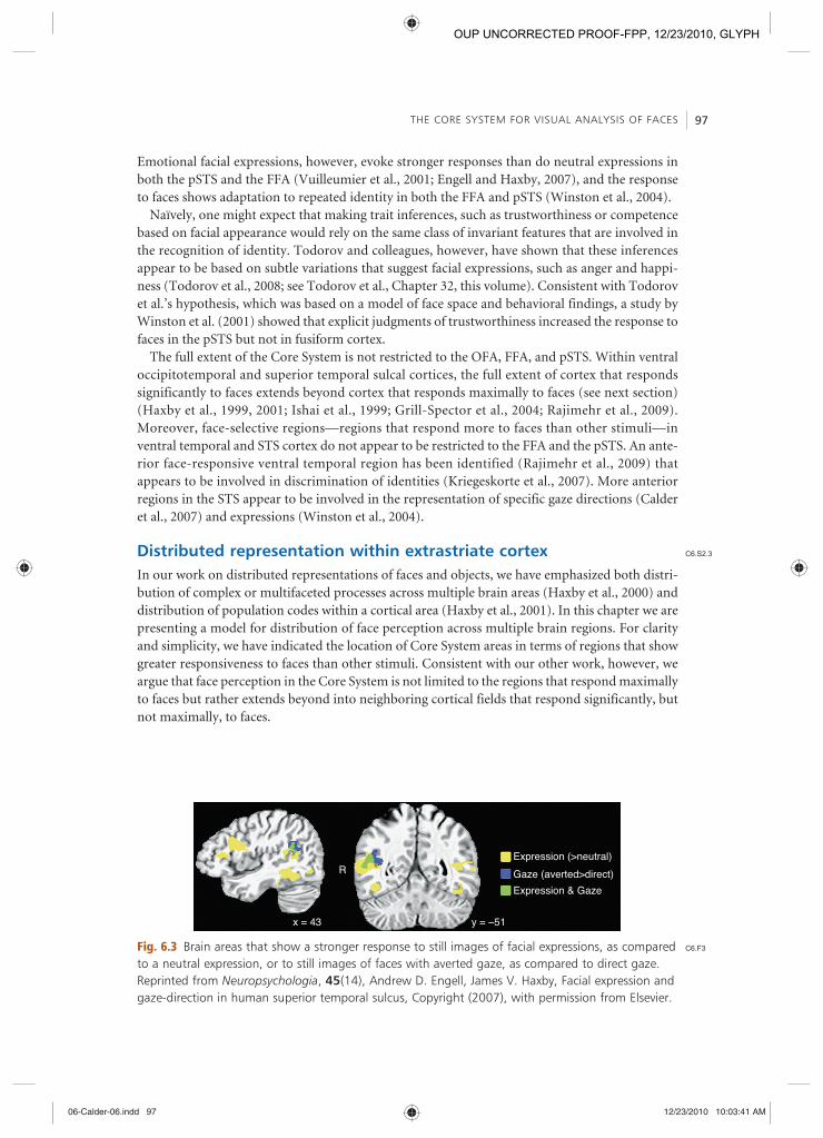
Although there is abundant evidence that the representation of faces in the FFA plays a greater role in recognizing identity and the representation of faces in the pSTS plays a greater role in perception of changeable features, such as expression and gaze, the distinction is not black and white. Consequently, the dominant roles of the FFA in face identity perception and the pSTS in expression perception have been challenged (Calder and Young, 2005 ; see Calder, Chapter 22, this volume). Perception of gaze changes appears to be more restricted to the pSTS than is percep-tion of expression, and within the STS expression perception appears to involve more rostral locations than does gaze perception (Winston et al., 2004 ; Engell and Haxby, 2007 ) (Figure 6.3 ).
Fig. 6.2 Brain areas that showed a significantly stronger response while viewing dynamic videos of actors producing facial expressions than while viewing still images of the same expressions. (Kirkland and Haxby, previously unpublished.)
C6.F2
06-Calder-06.indd 9606-Calder-06.indd 96 12/23/2010 10:03:40 AM12/23/2010 10:03:40 AM
OUP UNCORRECTED PROOF-FPP, 12/23/2010, GLYPH
THE CORE SYSTEM FOR VISUAL ANALYSIS OF FACES 97
Emotional facial expressions, however, evoke stronger responses than do neutral expressions in both the pSTS and the FFA (Vuilleumier et al., 2001 ; Engell and Haxby, 2007 ), and the response to faces shows adaptation to repeated identity in both the FFA and pSTS (Winston et al., 2004 ).
Naïvely, one might expect that making trait inferences, such as trustworthiness or competence based on facial appearance would rely on the same class of invariant features that are involved in the recognition of identity. Todorov and colleagues, however, have shown that these inferences appear to be based on subtle variations that suggest facial expressions, such as anger and happi-ness (Todorov et al., 2008 ; see Todorov et al., Chapter 32, this volume). Consistent with Todorov et al.’s hypothesis, which was based on a model of face space and behavioral findings, a study by Winston et al. (2001) showed that explicit judgments of trustworthiness increased the response to faces in the pSTS but not in fusiform cortex.
The full extent of the Core System is not restricted to the OFA, FFA, and pSTS. Within ventral occipitotemporal and superior temporal sulcal cortices, the full extent of cortex that responds significantly to faces extends beyond cortex that responds maximally to faces (see next section) (Haxby et al., 1999 , 2001 ; Ishai et al., 1999 ; Grill-Spector et al., 2004 ; Rajimehr et al., 2009 ). Moreover, face-selective regions — regions that respond more to faces than other stimuli — in ventral temporal and STS cortex do not appear to be restricted to the FFA and the pSTS. An ante-rior face-responsive ventral temporal region has been identified (Rajimehr et al., 2009 ) that appears to be involved in discrimination of identities (Kriegeskorte et al., 2007 ). More anterior regions in the STS appear to be involved in the representation of specific gaze directions (Calder et al., 2007 ) and expressions (Winston et al., 2004 ).
Distributed representation within extrastriate cortex In our work on distributed representations of faces and objects, we have emphasized both distri-bution of complex or multifaceted processes across multiple brain areas (Haxby et al., 2000 ) and distribution of population codes within a cortical area (Haxby et al., 2001 ). In this chapter we are presenting a model for distribution of face perception across multiple brain regions. For clarity and simplicity, we have indicated the location of Core System areas in terms of regions that show greater responsiveness to faces than other stimuli. Consistent with our other work, however, we argue that face perception in the Core System is not limited to the regions that respond maximally to faces but rather extends beyond into neighboring cortical fields that respond significantly, but not maximally, to faces.
R
y = –51
Expression & Gaze
Expression (>neutral)Gaze (averted>direct)
x = 43
Fig. 6.3 Brain areas that show a stronger response to still images of facial expressions, as compared to a neutral expression, or to still images of faces with averted gaze, as compared to direct gaze. Reprinted from Neuropsychologia , 45 (14), Andrew D. Engell, James V. Haxby, Facial expression and gaze-direction in human superior temporal sulcus, Copyright (2007), with permission from Elsevier.
C6.S2.3
C6.F3
06-Calder-06.indd 9706-Calder-06.indd 97 12/23/2010 10:03:41 AM12/23/2010 10:03:41 AM
OUP UNCORRECTED PROOF-FPP, 12/23/2010, GLYPH

DISTRIBUTED NEURAL SYSTEMS FOR FACE PERCEPTION 98
We have shown that the pattern of neural response to viewed faces in cortex that responds maximally to other visual stimuli distinguishes faces from other objects and different types of faces from each other (Figure 6.4 ) (Haxby et al., 2001 ; Connolly et al., in press). These distinctions were detected using multivariate pattern analysis (MVPA), which is more sensitive than conven-tional univariate analysis methods (Haynes and Rees, 2006 ; Norman et al., 2006 ; O’Toole et al., 2007 ). Whereas the mean response in the FFA does not distinguish between human faces and nonhuman mammalian faces (Tong et al., 1999), MVPA of responses, both within the FFA and in ventral temporal cortex that responds maximally to other visual stimuli, does detect such dis-tinctions (Connolly et al., in press). Grill-Spector et al. ( 2004 ) showed that half of the ventral temporal cortex that correlated with successful face identification, relative to face detection, was outside of the FFA. In macaque IT cortex, high concentrations of face-selective neurons are found in face-selective patches (Tsao et al., 2006; see Freiwald and Tsao, Chapter 36, this volume), but face-selective cells also are found outside of these patches, albeit in lower concentrations. Moreover, many face-responsive IT cells show coarse tuning profiles, with significant responses to both faces and other stimuli (Kiani et al., 2007 ). Thus, the full extent of cortex that participate in face perception in extrastriate visual areas is not restricted to the face-selective OFA, FFA, and pSTS and includes neurons that respond significantly to both faces and other stimuli.
The extended system for extracting information from faces Recognition of familiar faces Although recognition of a familiar face is accompanied by modulation of activity in the Core System FFA, this modulation can be positive or negative (for a review, see Gobbini and Haxby, 2007 ). Consequently, the strength of the FFA response may be more a reflection of top-down modulation than a signal of familiarity. Although facial identity may be encoded in the popula-tion response in the FFA (Rolls, 2000 ; Tsao et al., 2006; Freiwald et al., 2009 ; see Rolls, Chapter 4,
Fig. 6.4 Patterns of response in ventral temporal cortex to faces and three categories of objects — houses, chairs, and shoes. Data shown are in one axial section from one subject. The face-responsive fusiform areas are outlined in yellow. Note that the distinctive pattern of response to faces extends into areas outside of the face-selective areas. Data are from Haxby et al. ( 2001 ).
C6.S3
C6.S3.1
C6.F4
06-Calder-06.indd 9806-Calder-06.indd 98 12/23/2010 10:03:41 AM12/23/2010 10:03:41 AM
OUP UNCORRECTED PROOF-FPP, 12/23/2010, GLYPH

THE EXTENDED SYSTEM FOR EXTRACTING INFORMATION FROM FACES 99
and Freiwald and Tsao, Chapter 36, this volume) and other ventral temporal areas (e.g. anterior temporal cortex, Kriegeskorte et al., 2007 ), the contribution of other systems may modulate the overall strength of Core System responses. We have proposed that these other systems, which are outside of visual extrastriate cortex, play a central role in familiar face recognition — specifically in the automatic retrieval of person knowledge and in the emotional response to familiar individu-als. These systems are key parts of the Extended System for face perception.
Immediate retrieval of information about familiar individuals — such as their personality traits, their relationships with oneself and others, and their probable intentions — is a fundamental process that lays the foundation for appropriate social interactions. Therefore, recognition of the visual features of a familiar face is only the initial and relatively minor stage in the processes involved in familiar face recognition. A key component of the neural representation of familiar individuals is the retrieval of person knowledge. A second fundamental component of the neural representation of familiar individuals concerns one’s emotional response to that individual. Person knowledge refers to a broad class of information that encompasses personal traits, intentions, attitudes, transient mental states, biographical information and episodic memories related to specific individuals.
Research in social psychology has shown evidence for the spontaneous activation of traits and attitudes associated with the perceived individuals (Greenwald and Banaji, 1995 ; Bargh et al., 1996 ; Todorov and Uleman, 2002 ; Todorov et al., 2007 ). Furthermore, there is evidence that the representation of significant others is richer in thoughts, feelings and emotions as compared to non-significant others (Andersen and Cole, 1990; Andersen et al., 1998). As someone becomes more familiar the type of inferences made about these personally familiar individuals relates more to “psychological mediating variables” (such as goals and beliefs) and less to broad uncontextual-ized traits (e.g. “aggressive” or “friendly”) (Idson and Mischel, 2001 ).
The pattern of neural activity evoked by viewing a familiar face is modulated by the knowledge one has about that individual and by one’s emotional response to that person. We found that comparing faces with different levels of familiarity modulated the neural response in a distributed set of areas (Figure 6.5 ). We recorded a stronger response in areas associated with mentalizing or
Fig. 6.5 Brain areas that showed a stronger response to personally familiar faces, as compared to famous faces (upper panel, Gobbini et al., 2004 ), and to pictures of one’s own child, as compared to familiar, but unrelated, other children (lower panel, Leibenluft et al., 2004 ).
C6.F5
06-Calder-06.indd 9906-Calder-06.indd 99 12/23/2010 10:03:42 AM12/23/2010 10:03:42 AM
OUP UNCORRECTED PROOF-FPP, 12/23/2010, GLYPH

DISTRIBUTED NEURAL SYSTEMS FOR FACE PERCEPTION 100
theory of mind (ToM), such as the medial prefrontal cortex (MPFC) and the temporoparietal junction (TPJ), for faces of relatives and friends as compared to faces of famous individuals (politicians, actors, singers, athletes) and as compared to faces of strangers (Gobbini et al., 2004 ). The TPJ region that is involved in familiar face recognition and mentalizing can be dissociated from the nearby face-responsive pSTS (Gobbini et al., 2007 ). In a study of mothers of young chil-dren, the face of one’s own child as compared to faces of familiar but unrelated children (Leibenluft et al., 2004 ) also evoked stronger responses in these areas. Faces that are only visually familiar (through a behavioral training session in a laboratory prior to the functional magnetic resonance imaging (fMRI) experiment) but not associated with any semantic information, did not modu-late the MPFC and TPJ (Gobbini and Haxby, 2006 ). We have hypothesized that the MPFC and the TPJ encode those aspects of person knowledge (Mitchell et al., 2002 ) that are related to the representation of personal traits and mental states characteristic of a familiar individual.
The more familiar faces also evoked a stronger response in the posterior cingulate cortex and precuneus (PCC/PC) (Gobbini et al., 2004 ; Leibenluft et al., 2004 ). Faces that are visually familiar but with no associated person knowledge also evoked an increased response in the PCC/PC, sug-gesting that this region plays a role in the acquisition of simple visual familiarity (Kosaka et al., 2003 ; Gobbini and Haxby, 2006 ). The stronger response for the more familiar faces recorded in PCC/PC found in our studies and the involvement of the anterior temporal regions reported by others (Gorno-Tempini et al., 1998 ; Leveroni et al., 2000 ; Nakamura et al., 2000 ; Douville et al., 2005 ; Rotshtein et al., 2005 ) might indicate the involvement of these areas in retrieval of episodic memories and biographical information associated with familiar individuals.
Viewing faces of familiar individuals also modulated neural activity in regions usually involved in emotional responses, such as the amygdala and the insula. Faces of strangers evoked a stronger response in the amygdala as compared to the faces of relatives and friends. The stronger response of the amygdala to faces of stranger could reflect the wary attitude we experience when seeing someone new. On the other hand, mothers viewing the face of their own child evoked a stronger response in the amygdala as compared to faces of familiar unrelated children. Seeing the face of one’s own child also evoked a stronger response in the anterior insula. The insula responds to negatively valenced stimuli, such as expressions of disgust (Calder et al., 2001 ; Phillips et al., 2003 ), to being treated unfairly during negotiation games (Sanfey et al., 2003 ), but also might play a role also in mediating empathic reactions (Bartels and Zeki, 2000 ; Carr et al., 2003 ; Singer et al., 2004 ), and strong, positive emotions such as romantic love (Bartels and Zeki, 2000 ). The increased activity in the amygdala and in the insula elicited by viewing the face of one’s child might reflect the intense attachment and vigilant protectiveness that characterize the maternal relationship.
Emotional response plays an essential role in familiar face recognition but is dissociable from conscious recognition. The dissociation of these complementary processes is demonstrated by physiological data from prosopagnosic patients and patients with Capgras syndrome (see Langdon, Chapter 45, this volume). Prosopagnosia is characterized by the inability to consciously recognize familiar faces, but when prosopagnosics see pictures of familiar faces, their skin con-ductance response differs significantly from their skin conductance response to strangers (Tranel et al., 1995 ; Ellis and Lewis, 2001 ). This response must be elicited by sufficient processing to iden-tify the face for an emotional response but these processes are insufficient to evoke the conscious experience of recognition. By contrast, patients with Capgras syndrome can consciously recognize the visual features of a specific face as belonging to a familiar individual but believe that an impos-tor took the place of that familiar individual. Unlike prosopagnosics, patients with Capgras syn-drome do not show a differential skin conductance response to familiar faces (Ellis and Lewis, 2001 ). These findings indicate that when the emotional response to a familiar face is altered or missing, as it is in Capgras syndrome, familiar face recognition can be disrupted. The absence of
06-Calder-06.indd 10006-Calder-06.indd 100 12/23/2010 10:03:43 AM12/23/2010 10:03:43 AM
OUP UNCORRECTED PROOF-FPP, 12/23/2010, GLYPH

THE EXTENDED SYSTEM FOR EXTRACTING INFORMATION FROM FACES 101
the emotional “glow” that accompanies familiar face recognition can lead to the failure of con-scious recognition, which is complete only after one is certain of another’s identity.
Extracting the meaning of facial expressions The visual analysis of facial expression involves primarily extrastriate cortex in the pSTS, but extracting the significance of facial expression involves a distributed set of brain areas that are involved in action understanding and emotion. Perception of facial expression engages the puta-tive human mirror neuron system (hMNS), in particular the frontal operculum, supposedly reflecting the role of engaging motor representations for producing facial expressions in under-standing the meaning of facial expressions (Carr et al., 2004; Montgomery and Haxby, 2008 ; Montgomery et al., 2009 ). The similarity structure of patterns of response to particular facial expressions in the frontal operculum correlates highly with the similarity structure in the pSTS (Said et al., 2010 ), showing that the representational spaces for facial expressions in these two areas are closely related. Perception of facial expressions that convey particular emotions also engages areas that are associated with emotion processing (Breiter et al., 1996 ; Morris et al., 1996 ; Phillips et al., 1997; Whalen, 1998 ; Whalen et al., 1998 , 2004 ; Wicker et al., 2003 ).
Mirror neurons were discovered in single-unit recording studies in monkeys and are character-ized by their responses during both the execution of specific actions and the perception of others performing the same actions (Di Pellegrino et al., 1992 ; Gallese et al., 1996 ; Rizzolatti et al., 2001 ; Grafton, 2009 ). In humans the location of putative mirror neurons is established with fMRI by finding areas that respond both during the execution and perception of actions. FMRI adaptation and MVPA have provided ambiguous evidence for the specificity of these common responses to particular actions in humans (Lingnau et al., 2007; Dinstein et al., 2007, 2008; Kilner et al., 2009 ); nonetheless, the common responses to a range of actions are taken as a sufficient demonstration of mirror neuron properties. The areas that show these properties for the execution and percep-tion of facial expressions are in the inferior parietal lobe, the frontal operculum, and premotor cortex (Figure 6.6 ).
The frontal opercular area that responds during the perception of facial expression has a more anterior and inferior location than a nearby area that responds during the perception of hand movements. This result shows that the representation of facial expression in the frontal hMNS can be distinguished from the representation of other types of actions (Montgomery and Haxby, 2008 ).
Fig. 6.6 Brain areas that responded during viewing, imitation, and production of facial expressions. From Montgomery et al., 2009 , reprinted with permission from Sage Publications Ltd.
C6.S3.2
C6.F6
06-Calder-06.indd 10106-Calder-06.indd 101 12/23/2010 10:03:43 AM12/23/2010 10:03:43 AM
OUP UNCORRECTED PROOF-FPP, 12/23/2010, GLYPH
DISTRIBUTED NEURAL SYSTEMS FOR FACE PERCEPTION 102
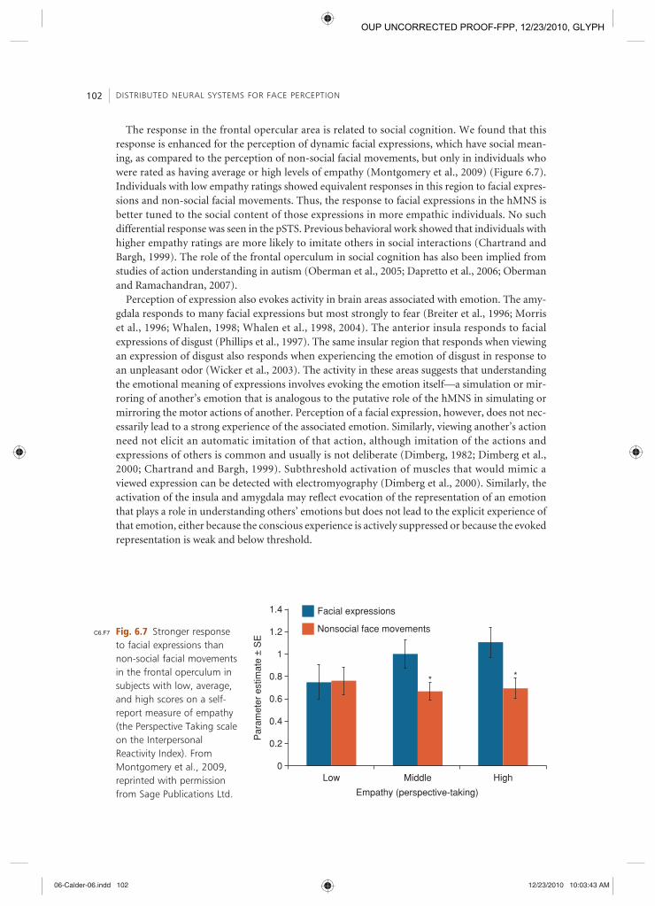
The response in the frontal opercular area is related to social cognition. We found that this response is enhanced for the perception of dynamic facial expressions, which have social mean-ing, as compared to the perception of non-social facial movements, but only in individuals who were rated as having average or high levels of empathy (Montgomery et al., 2009 ) (Figure 6.7 ). Individuals with low empathy ratings showed equivalent responses in this region to facial expres-sions and non-social facial movements. Thus, the response to facial expressions in the hMNS is better tuned to the social content of those expressions in more empathic individuals. No such differential response was seen in the pSTS. Previous behavioral work showed that individuals with higher empathy ratings are more likely to imitate others in social interactions (Chartrand and Bargh, 1999 ). The role of the frontal operculum in social cognition has also been implied from studies of action understanding in autism (Oberman et al., 2005 ; Dapretto et al., 2006 ; Oberman and Ramachandran, 2007 ).
Perception of expression also evokes activity in brain areas associated with emotion. The amy-gdala responds to many facial expressions but most strongly to fear (Breiter et al., 1996 ; Morris et al., 1996 ; Whalen, 1998 ; Whalen et al., 1998 , 2004 ). The anterior insula responds to facial expressions of disgust (Phillips et al., 1997). The same insular region that responds when viewing an expression of disgust also responds when experiencing the emotion of disgust in response to an unpleasant odor (Wicker et al., 2003 ). The activity in these areas suggests that understanding the emotional meaning of expressions involves evoking the emotion itself — a simulation or mir-roring of another’s emotion that is analogous to the putative role of the hMNS in simulating or mirroring the motor actions of another. Perception of a facial expression, however, does not nec-essarily lead to a strong experience of the associated emotion. Similarly, viewing another’s action need not elicit an automatic imitation of that action, although imitation of the actions and expressions of others is common and usually is not deliberate (Dimberg, 1982 ; Dimberg et al., 2000 ; Chartrand and Bargh, 1999 ). Subthreshold activation of muscles that would mimic a viewed expression can be detected with electromyography (Dimberg et al., 2000 ). Similarly, the activation of the insula and amygdala may reflect evocation of the representation of an emotion that plays a role in understanding others’ emotions but does not lead to the explicit experience of that emotion, either because the conscious experience is actively suppressed or because the evoked representation is weak and below threshold.
Low Middle
Empathy (perspective-taking)
0
0.2
Par
amet
er e
stim
ate
± S
E
0.4
0.6
0.8
1
1.2 Nonsocial face movements
* *
Facial expressions1.4
High
Fig. 6.7 Stronger response to facial expressions than non-social facial movements in the frontal operculum in subjects with low, average, and high scores on a self-report measure of empathy (the Perspective Taking scale on the Interpersonal Reactivity Index). From Montgomery et al., 2009 , reprinted with permission from Sage Publications Ltd.
C6.F7
06-Calder-06.indd 10206-Calder-06.indd 102 12/23/2010 10:03:43 AM12/23/2010 10:03:43 AM
OUP UNCORRECTED PROOF-FPP, 12/23/2010, GLYPH

MODULARITY AND DISTRIBUTED PROCESSING IN NEURAL REPRESENTATION 103
The role of the amygdala in facial expression perception appears multifaceted. The amygdala is critical for fear conditioning, but it appears to involve more than simply mirroring the emotion of fear. The amygdala can also be engaged by positive emotions and positive experiences. Whalen ( 1998 ) has proposed that the amygdala is part of a vigilance system that is activated in ambiguous situations with biological relevance. In this formulation, the ambiguity, not the emotion of fear, is the critical factor and can be either positive or negative.
Action understanding: eye gaze perception Eye gaze is another changeable feature of faces that plays an important role in social communica-tion. The pSTS plays a central role in the perception of others’ eye gaze direction, but the modula-tion of pSTS activity by direct and averted eye gaze is variable (Nummenmaa and Calder, 2009 ). Analogous to the variable modulation of fusiform responses by familiarity, the variable response of the pSTS may be due to the ambiguous meaning of direct and averted eye gaze. Direct eye gaze — eye contact — can be a sign of interest, an attempt to catch one’s attention, a sign of engage-ment in a social interaction, or a sign of threat. Averted gaze can signify lack of interest, lack of threat, or, conversely, can indicate another’s attention to an event or presence elsewhere in the environment that one should also be aware of oneself. Consequently, small differences in psycho-logical experiments can lend different meanings to direct and averted eye gaze and, thus, have variable effects on responses. The extraction of these different meanings is mediated by other neural systems that we include in the Extended System for face perception. Similar to the top-down effect of Extended System areas for person knowledge and emotion on fusiform responses to familiar faces, Extended System areas for spatial attention and mentalizing can exert a modu-lating influence on the pSTS response to eye gaze.
Perception of another’s averted eye gaze evokes a spontaneous shift of one’s own attention in the same direction (Friesen and Kingstone, 1998; Driver et al., 1999; Hietanen, 1999; Langton et al., 1999). We found that viewing faces with eye gaze averted unpredictably to the right and left evoked a stronger response in the intraparietal sulcus than did viewing faces with direct eye gaze (Hoffman and Haxby, 2000 ). The intraparietal sulcus plays a central role in shifting the direction of attention and in oculomotor control, along with the frontal eye fields (Beauchamp et al., 2001 ). The frontal eye fields were not included in the imaging volume in our study of gaze perception, which was restricted to coronal sections through posterior cortices (Figure 6.8a ). Subsequent studies by others, however, have shown that the frontal eye fields also are engaged by eye gaze perception (Nummenmaa and Calder, 2009 ) (Figure 6.8b ). The role of the neural system for shifting attention and eye movements in gaze perception is analogous to the role of the hMNS in the perception of facial expression.
Eye gaze perception additionally activates several other brain areas that play other roles in extracting information from the gaze of others. In a meta-analysis of imaging studies, Nummenmaa and Calder ( 2009 ) found consistent evidence for the engagement of the amygdala and MPFC, in addition to other areas. They proposed that the amygdala involvement reflects a role for this area in eye contact and the emotional response to being looked at. The role of the MPFC may relate to making inferences about the mental states that are implied by gaze, such as the focus of attention and intentions to act.
Modularity and distributed processing in neural representation We present here a model of face perception that involves multiple brain regions that work in concert in various configurations to extract different types of information from faces. Our per-spective contrasts with the emphasis others have placed on a dominant role for a single region
C6.S3.3
C6.S4
06-Calder-06.indd 10306-Calder-06.indd 103 12/23/2010 10:03:43 AM12/23/2010 10:03:43 AM
OUP UNCORRECTED PROOF-FPP, 12/23/2010, GLYPH

DISTRIBUTED NEURAL SYSTEMS FOR FACE PERCEPTION 104
(a)
Superior parietallobule
Postcentralsulcus
Frontaleye field
Inferior frontalcortex
aSTSSTS region,MT/V5
Medial prefrontalcortex
Amygdala/Hippocampus
(b)
Fig. 6.8 (a) A region in the intraparietal sulcus that responded during an eye gaze perception task. Frontal cortices are darkened to indicate that the imaging volume in this study (Hoffman and Haxby, 2000 ) did not include those areas. (b) Right hemisphere areas that showed involvement in gaze perception from a meta-analysis of PET and fMRI studies. Reprinted from Trends in Cognitive Sciences , 13 (3), Lauri Nummenmaa and Andrew J. Calder, Neural mechanisms of social attention, Copyright (2009), with permission from Elsevier.
in fusiform cortex, the FFA (Kanwisher et al., 1997 ; Kanwisher and Yovel, 2006 ). The modular and distributed system perspectives on the neural architecture that underlies face perception, however, are not necessarily incompatible (see Kanwisher and Barton, Chapter 7, this volume). We are not casting doubt on the existence of the FFA as an area that responds more during perception of faces than during perception of other objects, or even denying that the FFA plays a major role in many face perception operations. Our position also is not that all parts of the distributed systems that we describe play an equipotential role in face perception. We propose, instead, that an adequate account of the neural architecture that underlies face perception,
C6.F8
06-Calder-06.indd 10406-Calder-06.indd 104 12/23/2010 10:03:43 AM12/23/2010 10:03:43 AM
OUP UNCORRECTED PROOF-FPP, 12/23/2010, GLYPH

MODULARITY AND DISTRIBUTED PROCESSING IN NEURAL REPRESENTATION 105
which accounts both for the data from neuroimaging experiments on face perception and for the full scope of cognitive operations that fall within the domain of face perception, cannot be achieved with an exclusive focus on a single area. Studies of face perception consistently find that the occipitotemporal extrastriate visual cortices that respond significantly to faces includes inferior occipital and STS cortices, in addition to fusiform cortex, and extends into regions that do not respond maximally to faces. Similarly, multiple face-selective regions have been found in monkey IT cortex (see Freiwald and Tsao, Chapter 36, this volume). Studies of familiar face recognition, facial expression perception, and gaze perception consistently find activations in numerous, widely-distributed brain areas, such as the MPFC, TPJ, frontal operculum, IPS, FEF, amygdala, and anterior insula. Cognitively, face perception involves more than passive viewing of still face images or matching face images. Face perception also includes recognition of famil-iar faces, interpretation of facial expressions, and processing eye gaze, among other functions. These face perception operations are dependent upon the activation of person knowledge, the activation of motor programs for producing facial expressions and for oculomotor control, and emotional responses. The contribution of a single region, such as the FFA, to face perception must take into account the context in which that region functions, both in terms of the distrib-uted system of cortical areas and in terms of the full range of cognitive operations that fall within the domain of face perception.
Extended system
Core system
Motor simulation
Visual appearance
Posterior STSDynamic features of facial gesture
Inferior occipital & fusiform gyriInvariant features for identification
Inferior parietal & frontal operculumFacial expression
IPS and frontal eye fieldsEye gaze & spatial attention
Emotion
Amygdala.InsulaStriatum/reward system
Person knowledge
Medial prefrontal cortex (MPFC)
Temporoparietal junction (TPJ)Intentions, mental states
Anterior temporal cortexBiographical knowledge
Episodic memoriesPrecuneus/posterior cingulate
Personal traits, attitudes, mental statesTop-down modulatory feedback
Fig. 6.9 Updated model of distributed neural systems for face perception. C6.F9
06-Calder-06.indd 10506-Calder-06.indd 105 12/23/2010 10:03:44 AM12/23/2010 10:03:44 AM
OUP UNCORRECTED PROOF-FPP, 12/23/2010, GLYPH

DISTRIBUTED NEURAL SYSTEMS FOR FACE PERCEPTION 106
Summary and conclusion Face perception plays a multifaceted role in social communication, conveying many different types of information about the identity and mental states of others. Here we present an update of our model of the distributed system for face perception, which we first proposed in 2000 (Figure 6.9 ). This model divides brain areas that are involved in face perception into a Core System — occipitotemporal visual extrastriate areas that play a central role in the visual analysis of faces — and an Extended System — neural systems whose functions are not primarily visual but play critical roles in extracting information from faces. In the Core System, we emphasize a distinction between representation of invariant features that are critical for recognizing facial identity and representation of changeable features that are critical for facial gestures, such as expressions and eye gaze. We emphasize three sets of brain areas in the Extended System that are involved, respectively, in the representation of person knowledge, in action understanding (including gaze and attention), and in emotion. Familiar face recognition involves visual codes for familiar individuals in Core System areas in the fusiform, and possibly anterior temporal, cortex, along with the automatic activation of person knowledge and emotional responses. Facial expression involves visual codes in the STS, along with activation of representations of emotion and motor programs for producing expressions. Perception of eye gaze similarly involves visual codes in the STS, along with activation of brain areas for shifting attention and oculomotor control.
References Bargh , J.A. , Chen , M. , and Burrows , L. ( 1996 ). Automaticity of social behavior: direct effects of trait
construct and stereotype-activation on action . Journal of Personality and Social Psychology , 71 , 230 – 244.
Bartels , A. and Zeki , S. ( 2000 ). The neural basis of romantic love . Neuroreport , 11 , 3829 – 3834.
Beauchamp , M.S. , Petit , L. , Ellmore , T.M. , Ingeholm , J. , and Haxby J.V. ( 2001 ). A parametric fMRI study of overt and covert shifts of visuospatial attention . Neuroimage , 14 , 310 – 321.
Breiter , H.C. , Etcoff , N.L. , Whalen , P.J. , et al . ( 1996 ). Response and habituation of the human amygdala during visual processing of facial expression . Neuron , 17 , 875 – 887.
Bruce , V. and Young , A. ( 1986 ). Understanding face recognition . British Journal of Psychology , 77 , 305 – 327.
Calder , A.J. , Burton , A.M. , Miller , P. , Young , A.W. , and Akamatsu , S. ( 2001 ). A principal component analysis of facial expressions . Vision Research , 41 , 1179 – 1208.
Calder , A.J. and Young , A.W. ( 2005 ). Understanding the recognition of facial identity and facial expression . Nature Reviews Neuroscience , 6 , 641 – 651.
Calder , A.J. , Beaver , J.D. , Davis , M.H. , van Ditzhuijzen , J. , Keane , J. , and Lawrence , A.D. ( 2007 ). Disgust sensitivity predicts the insula and pallidal response to pictures of disgusting foods . European Journal of Neuroscience , 25 , 3422 – 3428.
Carr , L. , Iacoboni , M. , Dubeau , M.C. , Mazziotta , J.C. , and Lenzi GL. ( 2003 ). Neural mechanisms of empathy in humans: a relay from neural systems for imitation to limbic areas . Proceedings of the National Academy of Science USA , 100 , 5497 – 5502.
Chao , L.L. , Martin , A. , and Haxby , J.V. ( 1999 ). Are face-responsive regions selective only for faces? Neuroreport , 10 , 2945 – 2950.
Chartrand , T.L. and Bargh , J.A. ( 1999 ). The chameleon effect: the perception-behavior link and social interaction . Journal of Personality and Social Psychology , 76 , 893 – 910.
Connolly , A.C. , Gobbini , M.I. , and Haxby , J.V. (in press). Three virtues of similarity-based multi-voxel pattern analysis . In N. Kriegeskorte and G. Kreiman (eds.) Understanding visual population codes (UVPC)–toward a common multivariate framework for cell recording and functional imaging . Boston , MA MIT Press .
C6.S5
06-Calder-06.indd 10606-Calder-06.indd 106 12/23/2010 10:03:45 AM12/23/2010 10:03:45 AM
OUP UNCORRECTED PROOF-FPP, 12/23/2010, GLYPH

REFERENCES 107
Dapretto , M. , Davies , M.S. , Pfeifer , J.H. , et al . ( 2006 ). Understanding emotions in others: mirror neuron dysfunction in children with autism spectrum disorders . Nature Neuroscience , 9 , 28 – 30.
Dimberg , U. ( 1982 ). Facial reactions to facial expressions . Psychophysiology , 19 , 643 – 647.
Dimberg , U. , Thunberg , M. , and Elmehed K. ( 2000 ). Unconscious facial reactions to emotional facial expressions . Psychological Science , 11 , 86 – 89.
Dinstein , I. , Hasson , U. , Rubin , N. , and Heeger , D.J. ( 2007 ). Brain areas selective for both observed and executed movements . Journal of Neurophysiology , 98 , 1415 – 1427.
Dinstein , I. , Gardner , J.L. , Jazayeri , M. , and Heeger , D.J. ( 2009 ). Executed and observed movements have different distributed representations in human aIPS . Journal of Neuroscience , 28 , 11231 – 11239.
di Pellegrino , G. , Fadiga , L. , Fogassi , L. , Gallese , V. , and Rizzolatti , G. ( 1992 ). Understanding motor events: a neuropsychological study . Experimental Brain Research , 91 , 176 – 180.
Douville , K. , Woodard , J.L. , Seidenberg , M. , et al . ( 2005 ). Medial temporal lobe activity for recognition of recent and remote famous names: an event-related fMRI study . Neuropsychologia , 43 , 693 – 703.
Ellis , H.D. and Lewis , M.B. ( 2001 ). Capgras delusion: A window on face recognition . Trends in Cognitive Sciences , 5 , 149 – 156.
Engell , A.D. , Gobbini , M.I. , and Haxby , J.V. ( 2006 ). Gaze-change perception in the early visual cortex . Society for Neuroscience Abstracts , 438 . 12.
Engell , A.D. and Haxby , J.V. ( 2007 ). Facial expression and gaze-direction in human superior temporal sulcus . Neuropsychologia , 45 , 3234 – 3241.
Freiwald , W.A. , Tsao , D.Y. , and Livingstone , M.S. ( 2009 ). A face feature space in the macaque temporal lobe . Nature Neuroscience , 12 , 1187 – 1196.
Gallese , V. , Fadiga , L. , Fogassi , L. , and Rizzolatti , G. ( 1996 ). Action recognition in the premotor cortex . Brain , 119 , 593 – 609.
Greenwald , A.G. and Banaji , M.R. ( 1995 ). Implicit social cognition: attitudes, self-esteem, and stereotypes . Psychological Review , 102 , 4 – 27.
Gobbini , M. I. , Leibenluft , E. , Santiago , N. , and Haxby , J. V. ( 2004 ). Social and emotional attachment in the neural representation of faces . NeuroImage , 22 , 1628 – 1635.
Gobbini , M.I. and Haxby , J.V. ( 2006 ). Neural response to the visual familiarity of faces . Brain Research Bulletin , 71 , 76 – 82.
Gobbini , M. I. and Haxby , J. V. ( 2007 ). Neural systems for recognition of familiar faces . Neuropsychologia , 45 , 32 – 41.
Gobbini , M.I. , Koralek , A.C. , Bryan , R.E. , Montgomery , K.J. , and Haxby , J.V. Two takes on the social brain: A comparison of theory of mind tasks . Journal of Cognitive Neuroscience , 19 , 1803 – 1814 .
Gorno-Tempini , M.L. , Price , C.J. , Josephs , O. , et al . ( 1998 ). The neural systems sustaining face and proper-name processing . Brain , 121 , 2103 – 2118.
Grafton , S.T. ( 2009 ). Embodied cognition and the simulation of action to understand others . Annals of the New York Academy of Sciences , 1156 , 97 – 117.
Grill-Spector , K. , Knouf , N. , and Kanwisher , N. ( 2004 ). The fusiform face area subserves face perception, not generic within-category identification . Nature Neuroscience , 7 , 555 – 562.
Hanson , S.J. , Matsuka , T. , and Haxby , J.V. ( 2004 ). Combinatorial codes in ventral temporal lobe for object recognition: Haxby (2001) revisited: is there a “face” area? Neuroimage , 23 , 156 – 166.
Hasselmo , M.E. , Rolls , E.T. , and Baylis , G.C. ( 1989 ). The role of expression and identity in the face-selective responses of neurons in the temporal visual cortex of the monkey . Behavioral Brain Research , 32 , 203 – 218.
Hasson , U. , Nir , Y. , Levy , I. , Fuhrmann , G. , and Malach , R. ( 2004 ). Intersubject synchronization of cortical activity during natural vision . Science , 303 , 1634 – 1640.
Haxby , J.V. Grady , C.L. , Horwitz , B. , et al . ( 1991 ). Dissociation of object and spatial visual processing pathways in human extrastriate cortex . Proceedings of the National Academy of Science, U S A , 88 , 1621 – 1625.
06-Calder-06.indd 10706-Calder-06.indd 107 12/23/2010 10:03:45 AM12/23/2010 10:03:45 AM
OUP UNCORRECTED PROOF-FPP, 12/23/2010, GLYPH

DISTRIBUTED NEURAL SYSTEMS FOR FACE PERCEPTION 108
Haxby , J.V. , Horwitz , B. , Ungerleider , L.G. , Maisog , J.M. , Pietrini , P. , and Grady , C.L. ( 1994 ) The functional organization of human extrastriate cortex: A PET-rCBF study of selective attention to faces and locations . Journal of Neuroscience , 14 , 6336 – 6353.
Haxby , J.V. , Ungerleider , L.G. , Clark , V.P. , Schouten , J.L. , Hoffman , E.A. , and Martin , A. ( 1999 ). The effect of face inversion on activity in human neural systems for face and object perception . Neuron , 22 , 189 – 199.
Haxby , J.V. , Hoffman , E.A. , and Gobbini , M.I. ( 2000 ). The distributed human neural system for face perception . Trends in Cognitive Sciences , 4 , 223 – 233.
Haxby , J.V. , Gobbini , M.I. , Furey , M.L. , Ishai , A. , Schouten , J.L. , and Pietrini , P. ( 2001 ). Distributed and overlapping representations of faces and objects in ventral temporal cortex . Science , 293 , 2425 – 2430.
Haynes , J.D. and Rees , G. ( 2006 ). Decoding mental states from brain activity in humans . Nature Reviews Neuroscience , 7 , 523 – 534.
Hoffman , E.A. and Haxby , J.V. ( 2000 ). Distinct representations of eye gaze and identity in the distributed human neural system for face perception . Nature Neuroscience , 3 , 80 – 84.
Idson , L.C. and Mischel , W. ( 2001 ). The personality of familiar and significant people: the lay perceiver as a social-cognitive theorist . Journal of Personality and Social Psychology , 80 , 585 – 96.
Ishai , A. ( 2008 ). Let’s face it: It’s a cortical network . NeuroImage , 40 , 415 – 419.
Ishai , A. , Ungerleider , L.G. , Martin , A. , Schouten , J.L. , and Haxby , J.V. ( 1999 ). Distributed representation of objects in the human ventral visual pathway . Proceedings of the National Academy of Sciences, USA , 96 , 9379 – 9384.
Ishai , A. , Schmidt , C.F. , and Boesiger , P. ( 2005 ). Face perception is mediated by a distributed cortical network . Brain Research Bulletin , 67 , 87 – 93.
Kanwisher , N. and Yovel , G. ( 2006 ). The fusiform face area: a cortical region specialized for the perception of faces . Philosophical Transactions of the Royal Society B , 361 , 2109 – 2128.
Kanwisher , N. , McDermott , J. , and Chun , M.M. ( 1997 ). The fusiform face area: a module in human extrastriate cortex specialized for face perception . Journal of Neuroscience , 17 , 4302 – 4311.
Kiani , R. , Esteky , H. , Mirpour , K. , and Tanaka , K. ( 2007 ). Object category structure in response patterns of neuronal population in monkey inferior temporal cortex . Journal of Neurophysiology . 97 , 4296 – 4309.
Kilner , J.M. , Neal , A. , Weiskopf , N. , Friston , K.J. , and Frith C.D. ( 2009 ). Evidence of mirror neurons in human inferior frontal gyrus . Journal of Neuroscience , 29 , 10153 – 10159.
Kosaka , H. Omori , M. , Iidaka , T. et al . ( 2003 ). Neural substrates participating in acquisition of facial familiarity: an fMRI study . Neuroimage , 20 , 1734 – 1742.
Kriegeskorte , N. , Formisano , E. , Sorger , B. , and Goebel , R. ( 2007 ). Individual faces elicit distinct response patterns in human anterior temporal cortex . Proceedings of the National Academy of Sciences , USA, 104 , 20600 – 20605.
Leibenluft , E. , Gobbini , M.I. , Harrison , T. , and Haxby J.V. ( 2004 ). Mothers’ neural activation in response to pictures of their children and other children . Biological Psychiatry , 56 , 225 – 32.
Leveroni , C.L. , Seidenberg , M. , Mayer , A.R. , Mead , L.A. , Binder , J.R. , and Rao SM. ( 2000 ). Neural systems underlying the recognition of familiar and newly learned faces . Journal of Neuroscience , 20 , 878 – 86.
Lingnau , A. , Gesierich , B. , and Caramazza , A. ( 2009 ). Asymmetric fMRI adaptation reveals no evidence for mirror neurons in humans . Proceedings of the National Academy of Sciences, USA , 106 , 9925 – 9930.
Mitchell , J.P. , Heatherton , T.F. , and Macrae , C.N. ( 2002 ). Distinct neural systems subserve person and object knowledge . Proceedings of the National Academy of Sciences, USA , 99 , 15238 – 15234.
Montgomery , K.J. and Haxby J.V. ( 2008 ). Mirror neuron system differentially activated by facial expressions and social hand gestures: a functional magnetic resonance imaging study . Journal of Cognitive Neuroscience , 20 , 1866 – 1877.
Montgomery , K.J. , Seeherman , K.R. , and Haxby , J.V. ( 2009 ). The well-tempered social brain . Psychological Science , 20 , 1211 – 1213.
06-Calder-06.indd 10806-Calder-06.indd 108 12/23/2010 10:03:45 AM12/23/2010 10:03:45 AM
OUP UNCORRECTED PROOF-FPP, 12/23/2010, GLYPH

REFERENCES 109
Morris , J.S. , Frith , CD. , Perrett , D.I. , et al . ( 1996 ). A differential neural response in the human amygdala to fearful and happy facial expressions . Nature , 383 , 812 – 815.
Nakamura , K. , Kawashima , R. , Sato , N. , et al . ( 2000 ). Functional delineation of the human occipito-temporal areas related to face and scene processing . A PET study. Brain , 123 , 1903 – 1912.
Norman , K.A. , Polyn , S.M. , Detre , G.J. , and Haxby , J.V. ( 2006 ). Beyond mind-reading: multi-voxel pattern analysis of fMRI data . Trends in Cognitive Science , 10 , 424 – 430.
Nummenmaa , L. and Calder , A.J. ( 2009 ). Neural mechanisms of social attention . Trends in Cognitive Sciences. 13 , 135 – 143.
Oberman , L.M. , Hubbard , E.M. , McCleery , J.P. , Altschuler , E.L. , Ramachandran , V.S. , and Pineda , J.A. ( 2005 ). EEG evidence for mirror neuron dysfunction in autism spectrum disorders . Brain Research, Cognitive Brain Research , 24 , 190 – 198.
Oberman , L.M. and Ramachandran , V.S. ( 2007 ). The simulating social mind: the role of the mirror neurons system and simulation in the social and communicative deficits of autism spectrum disorders . Psychological Bulletin , 133 , 310 – 327.
O’Toole , A.J. , Jiang , F. , Abdi , H. , Penard , N. , Dunlop , J.P. , and Parent , M.A. ( 2007 ) Theoretical, statistical, and practical perspectives on pattern-based classification approaches to the analysis of functional neuroimaging data . Journal of Cognitive Neuroscience , 18 , 1735 – 1752.
Phillips , M.L. , Young , A.W. , Senior , C. , et al . ( 1996 ). A specific neural substrate for perceiving facial expressions of disgust . Nature , 389 , 495 – 498.
Phillips , M.L. , Drevets , W.C. , Rauch , S.L. , and Lane , R. ( 2003 ). Neurobiology of emotion perception . I: the neural basis of normal emotion perception. Biological Psychiatry , 54 , 504 – 514.
Puce , A. , Allison , R. , Bentin , S. , Gore , J.C. , and McCarthy , G. ( 1998 ). Temporal cortex activation in humans viewing eye and mouth movements . Journal of Neuroscience , 18 , 2188 – 2199.
Rajimehr , R. , Young , J.C. , and Tootell , R.B.H. ( 2009 ). An anterior temporal face patch in human cortex, predicted by macaque maps . Proceedings of the National Academy of Sciences, USA , 106 , 1995 – 2000.
Rizzolatti , G. , Fogassi , L. , and Gallese , V. ( 2001 ). Neurophysiological mechanisms underlying the understanding and imitation of action . Nature Reviews Neuroscience , 2 , 661 – 670.
Rolls , E.T. ( 2000 ). Functions of the primate temporal lobe visual cortices in invariant visual object and face recognition . Neuron , 27 , 205 – 218.
Rotshtein , P. , Henson , R. N. , Treves , A. , Driver , J. , and Dolan , R. J. ( 2005 ). Morphing Marilyn into Maggie dissociates physical and identity face representations in the brain . Nature Neuroscience , 8 , 107 – 113.
Said , C.P. , Moore , C.D. , Engell , A.D. , Todorov , A. , and Haxby J.V. ( 2010 ). Distributed representations of dynamic facial expression in the superior temporal sulcus . Journal of Vision, 10 : 11 , 1 – 12 .
Sanfey , A.G. , Rilling , J.K. , Aronson , J.A. , Nystrom , L.E. , and Cohen , J.D. ( 2003 ). The neural basis of economic decision-making in the ultimatum game . Science , 300 , 1755 – 1758.
Singer , T. , Seymou , B. , O’Doherty , J. , Kaube , H. , Dolan , R.J. , and Frith , C.D. ( 2004 ). Empathy for pain involves the affective but not sensory components of pain . Science , 303 , 1157 – 1162.
Todorov , A. and Uleman , J.S. ( 2002 ). Spontaneous trait inferences are bound to actors’ faces: evidence from a false recognition paradigm . Journal of Personality and Social Psychology , 83 , 1051 – 1065.
Todorov , A. , Gobbini , M.I. , Evans , K.K. , and Haxby , J.V. ( 2007 ). Spontaneous retrieval of affective person knowledge in face perception . Neuropsychologia , 45 , 163 – 173.
Todorov , A. , Said , C.P. , Engell , A.D. , and Oosterhof , N.N. ( 2008 ). Understanding evaluation of faces on social dimensions . Trends in Cognitive Sciences , 12 , 455 – 460.
Tranel , D. , Damasio , A.R. , and Damasio , H. ( 1995 ). Double dissociation between overt and covert recognition . Journal of Cognitive Neurosciences , 7 , 425 – 432.
Vuilleumier , P. , Armony , J.L. , Driver , J. , and Dolan , R.J. ( 2001 ). Effects of attention and emotion on face processing in the human brain: An event-related fMRI study . Neuron , 30 , 829 – 841.
06-Calder-06.indd 10906-Calder-06.indd 109 12/23/2010 10:03:45 AM12/23/2010 10:03:45 AM
OUP UNCORRECTED PROOF-FPP, 12/23/2010, GLYPH

DISTRIBUTED NEURAL SYSTEMS FOR FACE PERCEPTION 110
Whalen , P.J. ( 1998 ). Fear, vigilance, and ambiguity: initial neuroimaging studies of the human amygdala . Current Directions in Psychological Science , 7 , 177 – 188.
Whalen , P.J. , Rauch , S.L. , Etcoff , N.L. , McInerney , S.C. , Lee , M.B. , and Jenike , M.A. ( 1998 ). Masked presentations of emotional face expressions modulate amygdala activity without explicit knowledge . Journal of Neuroscience , 18 , 411 – 418.
Whalen , P.J. , Kagan , J. , Cook , R.G. , et al . ( 2004 ). Human amygdala responsivity to masked fearful eye whites . Science , 306 , 2061 .
Wicker , B. , Keysers , C. , Plailly , J. , Royet , J.-P. , Gallese , V. , and Rizzolatti , G. ( 2003 ). Both of us disgusted in my insula: The common neural basis of seeing and feeling disgust . Neuron , 40 , 655 – 664.
Winston , J.S. , Henson , R.N.A. , Fine-Goulden , M.R. , and Dolan , R.J. ( 2004 ). fMRI-adaptation reveals dissociable neural representations of identity and expression in face perception . Journal of Neurophysiology , 92 , 1830 – 1839.
06-Calder-06.indd 11006-Calder-06.indd 110 12/23/2010 10:03:45 AM12/23/2010 10:03:45 AM
OUP UNCORRECTED PROOF-FPP, 12/23/2010, GLYPH