disulfide bond formation of thiols by carbon nanotubes · 2017-04-05 · disulfide bond formation...
TRANSCRIPT

S1
Supplementary Information
Disulfide Bond Formation of Thiols by Carbon Nanotubes
A. Hirano,*a T. Kameda,b S. Sakuraba,c M. Wada,a T. Tanaka,a H. Katauraa
aNanomaterials Research Institute, National Institute of Advanced Industrial Science and
Technology (AIST), Tsukuba, Ibaraki 305-8565 Japan
bComputational Biology Research Center National Institute of Advanced Industrial Science and
Technology (AIST) Koto, Tokyo 135-0064 Japan
cGraduate School of Frontier Sciences, The University of Tokyo, Kashiwa, Chiba 277-8561
Japan
* To whom correspondence should be addressed: Nanomaterials Research Institute, National
Institute of Advanced Industrial Science and Technology, Tsukuba, Ibaraki 305-8565, Japan.
Tel.: +81-29-849-1064, Fax: +81-29-861-2786, E-mail: [email protected]
Electronic Supplementary Material (ESI) for Nanoscale.This journal is © The Royal Society of Chemistry 2017

S2
1. Materials and Methods
1.1. Chemicals. CNTs synthesized by the high-pressure catalytic CO (HiPco) decomposition
method were purchased from Nano-Integris. CNTs synthesized by the arc plasma jet (APJ)
method and by the enhanced direct injection pyrolytic synthesis (eDIPS) method (MEIJO eDIPS
EC2.0) were purchased from Meijo Nano Carbon Co., Ltd. Sodium dodecyl sulfate (SDS),
sodium deoxycholate, ammonia solution, acetic acid, L-cysteine (Cys), L(−)-cystine (Cys-Cys),
(+/−)-dithiothreitol (red-DTT), glutathione (GSH and GSSG), formic acid, methanol, guanidine
hydrochloride, urea and lysozyme from egg white were obtained from Wako Pure Chemical
Industries, Ltd. The oxidized form of dithiothreitol, i.e., trans-4,5-dihydroxy-1,2-dithiane,
designated ox-DTT, was purchased from Sigma-Aldrich Co. LLC.
Ethylenediamine-N,N,N',N'-tetraacetic acid, disodium salt, dehydrate (EDTA) was obtained from
Dojindo Laboratories.
1.2. Redox Reaction of Dispersant-Free HiPco CNTs with Thiols Using Ultrasonication.
The HiPco CNTs were used to examine the reaction of CNTs with thiols. First, the raw HiPco
CNTs were dried under vacuum to remove coexisting volatile impurities such as organic solvents.
Subsequently, 1 mg of the CNTs was mixed with 1 mL of 0.1% v/v ammonium acetate buffer
solution (pH 4.5) and 10 L of 100 mM thiols dissolved in the buffer solution using an ultrasonic
processor (Nanoruptor NR-350, Cosmo Bio Co., Ltd.) for 15 min at a power of 350 W to create
1-mL suspensions containing 1 mg/mL CNTs and 1 mM thiols. After mixing, these samples
were centrifuged at 3,000 rpm for 10 min (H-103N, Kokusan Co. Ltd.). The upper 400-L
aqueous fractions obtained from the supernatants were then filtered using a
polytetrafluoroethylene (PTFE) syringe filter membrane (As One Corp.) with a 0.22-m pore
S3
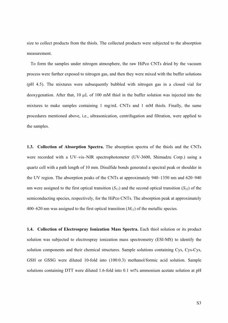
size to collect products from the thiols. The collected products were subjected to the absorption
measurement.
To form the samples under nitrogen atmosphere, the raw HiPco CNTs dried by the vacuum
process were further exposed to nitrogen gas, and then they were mixed with the buffer solutions
(pH 4.5). The mixtures were subsequently bubbled with nitrogen gas in a closed vial for
deoxygenation. After that, 10 L of 100 mM thiol in the buffer solution was injected into the
mixtures to make samples containing 1 mg/mL CNTs and 1 mM thiols. Finally, the same
procedures mentioned above, i.e., ultrasonication, centrifugation and filtration, were applied to
the samples.
1.3. Collection of Absorption Spectra. The absorption spectra of the thiols and the CNTs
were recorded with a UV–vis–NIR spectrophotometer (UV-3600, Shimadzu Corp.) using a
quartz cell with a path length of 10 mm. Disulfide bonds generated a spectral peak or shoulder in
the UV region. The absorption peaks of the CNTs at approximately 940–1350 nm and 620–940
nm were assigned to the first optical transition (S11) and the second optical transition (S22) of the
semiconducting species, respectively, for the HiPco CNTs. The absorption peak at approximately
400–620 nm was assigned to the first optical transition (M11) of the metallic species.
1.4. Collection of Electrospray Ionization Mass Spectra. Each thiol solution or its product
solution was subjected to electrospray ionization mass spectrometry (ESI-MS) to identify the
solution components and their chemical structures. Sample solutions containing Cys, Cys-Cys,
GSH or GSSG were diluted 10-fold into (100:0.3) methanol/formic acid solution. Sample
solutions containing DTT were diluted 1.6-fold into 0.1 wt% ammonium acetate solution at pH

S4
9.5 to adjust their pH to approximately 7; they were then diluted 10-fold into (100:0.2)
methanol/formic acid solution. Finally, they were filtered through a 0.22-m pore size PTFE
membrane and subjected to ESI-MS. The ESI mass spectrometer (ZQ2000, Nihon Waters K.K.)
was operated in positive-ion mode for Cys, Cys-Cys, GSH and GSSG and in negative-ion mode
for DTT.
1.5. On-Column Redox Reaction of Dispersant-Free HiPco CNTs with Thiols Using a
Liquid Chromatography System. The redox reaction of the CNTs with the thiols was
examined using a high-performance liquid chromatographic column, which was filled with
CNT-immobilized silica matrices. The matrices (Chemcobond NH2, Chemco Plus Scientific Co.,
Ltd.) have amino groups, enabling physical adsorption of the CNTs onto their surfaces. The
chromatographic system (Shimadzu Corp.) comprised a degasser (DGU-20A), a pump
(LC-20AT), a photodiode array detector (SPD-M20A) and an autosampler (SIL-20AC). The
on-column redox reaction using the CNTs on the matrices was conducted at a flow rate of 0.05
mL/min at room temperature using a 0.1% v/v ammonium acetate buffer solution (pH 4.5) as a
running buffer, where 20 L of 500 M thiol solutions dissolved in the same buffer solutions
were loaded into the columns.
1.6. Collection of Absorption Spectra of the SDS-Dispersed CNTs in the Presence of
Thiols. The HiPco CNTs dispersed in 1 wt% sodium dodecyl sulfate (SDS) solution (pH 4.5)
were reduced by the addition of different concentrations of the thiol solutions containing 1 wt%
SDS. An aliquot of 30 mg of HiPco CNTs was suspended in 30 mL of 1 wt% SDS (1 mg/mL)
using an ultrasonic processor (Nanoruptor NR-350) for 1 min at a power of 350 W as a

S5
preliminary dispersion treatment. The suspension was further dispersed for 1 h at a power
density of ca. 20 W cm-2 using an ultrasonic homogenizer (Sonifire 250D, Branson) equipped
with a 0.5-in flat tip. The glass vial containing this dispersion was immersed in a water bath at
18°C to prevent an increase in the temperature of the dispersions during the ultrasonication
treatment. The dispersed sample was subsequently centrifuged at 210,000 × g for 1 h using an
ultracentrifuge (himac CS100GXII and S80AT3 rotor, Hitachi Koki Co., Ltd.) to remove the
residue of catalytic metal particles, CNT bundles, and other impurities. The upper 70% of the
supernatant was collected as a purified CNT dispersion containing 1 wt% SDS. The CNT sample
was then diluted into 1 wt% SDS solution to adjust the absorbance of the dispersion at 600 nm to
0.28, which corresponds to 0.13 mg/mL; it was then further diluted 15-fold into a 1 wt% SDS
solution. The pH value of the dispersion was subsequently adjusted to 4.5 using 0.1 M HCl.
Additionally, 0–20 mM thiol solutions (pH 4.5) containing 1 wt% SDS were prepared by
dissolving the thiols into SDS solutions and by adding HCl or NaOH for pH adjustment. Finally,
aliquots of the CNT dispersions (375 L), which had been incubated for several days for
equilibration, were mixed with the thiol solutions (125 L) to prepare samples containing 0.0065
mg/mL CNTs, 0–5 mM thiols and 1 wt% SDS. The absorbances of these samples at 1250 nm at
various time periods are shown in Figure S7.

S6
2. Results
2.1. Absorption Spectra of Cys, Cys-Cys, GSH and GSSG.
Figure S1. Absorption spectra of the thiols in 0.1% v/v ammonium acetate buffer (pH 4.5).
S7
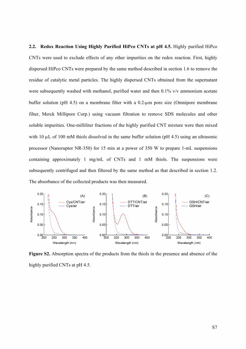
2.2. Redox Reaction Using Highly Purified HiPco CNTs at pH 4.5. Highly purified HiPco
CNTs were used to exclude effects of any other impurities on the redox reaction. First, highly
dispersed HiPco CNTs were prepared by the same method described in section 1.6 to remove the
residue of catalytic metal particles. The highly dispersed CNTs obtained from the supernatant
were subsequently washed with methanol, purified water and then 0.1% v/v ammonium acetate
buffer solution (pH 4.5) on a membrane filter with a 0.2-m pore size (Omnipore membrane
filter, Merck Millipore Corp.) using vacuum filtration to remove SDS molecules and other
soluble impurities. One-milliliter fractions of the highly purified CNT mixture were then mixed
with 10 L of 100 mM thiols dissolved in the same buffer solution (pH 4.5) using an ultrasonic
processor (Nanoruptor NR-350) for 15 min at a power of 350 W to prepare 1-mL suspensions
containing approximately 1 mg/mL of CNTs and 1 mM thiols. The suspensions were
subsequently centrifuged and then filtered by the same method as that described in section 1.2.
The absorbance of the collected products was then measured.
Figure S2. Absorption spectra of the products from the thiols in the presence and absence of the
highly purified CNTs at pH 4.5.
S8
2.3. Redox Reaction Using Highly Purified HiPco CNTs at Neutral pH Values. A
procedure similar to that mentioned in section 2.2 was performed in the absence of the buffer
solution to examine pH changes in the mixture of the CNTs and thiols; that is, the washing
process described in section 2.2 was completed using a sufficiently large amount of purified
water, resulting in a sample pH of approximately 6. The highly purified buffer-free CNT sample
was mixed with buffer-free thiol solution at approximately pH 6 (adjusted to that level using
NaOH) using an ultrasonic processor (Nanoruptor NR-350) for 15 min at a power of 350 W to
prepare 800-L suspensions containing approximately 0 or 1 mg/mL of CNTs and 1 mM thiols.
The suspensions were subsequently centrifuged at 3000 rpm for 10 min using a centrifuge
(H103n). pH values of the supernatants were measured to examine pH changes in the presence
and absence of the CNTs. Finally, the supernatants were filtered through a PTFE membrane with
a 0.22-m pore size and then subjected to the absorption measurement.
The pH values were found to be 6.0 ± 0.2 and 7.1 ± 0.2 in the presence and absence of the
CNTs, respectively. The absorption spectra of the filtered supernatants are shown in Figure S3.
The spectrum of the sample collected from the mixture in the presence of CNTs exhibited a
shoulder at approximately 250 nm, indicating the presence of the disulfide bond. By contrast, the
spectrum of the sample collected from the mixture in the absence of CNTs showed no such
spectral shoulder. This result indicates that formation of the disulfide bond can be induced by the
CNTs even at neutral pH. Incidentally, the sample in the absence of CNTs showed a weak
absorption at approximately 225–300 nm, which was different from that of Cys-Cys. This weak
absorption is ascribable to deprotonation of the thiol group of Cys, which is available at neutral
pH values.1 Thus, the deprotonation can be prevented by the electron transfer from the thiols into
the CNTs in the presence of the CNTs.

S9
Figure S3. Absorption spectra of the products from cysteine in the presence and absence of the
highly purified CNTs in buffer-free solution.

S10
2.4. Redox Reaction of Dispersant-Free HiPco CNTs with Thiols Using Ultrasonication
Figure S4. Mass spectra of the products from DTT (A-C) or GSH (D-F) under different
conditions.
S11
2.5. Comparison between Different CNT Types in the Redox Reaction. Different types of
CNTs were used to examine their redox reactions with thiols. CNTs synthesized by the APJ
method and by the eDIPS method as well as by the HiPco method were used as model carbon
nanotubes. The APJ CNTs have diameters of 1.4 ± 0.1 nm, and the eDIPS CNT have diameters
of 2.0 ± 0.8 nm. Each 5-mg CNT sample was mixed with 6 mL of 0.1% v/v ammonium acetate
buffer solution (pH 4.5) using an ultrasonic homogenizer (Sonifire 250D) equipped with a
0.25-in flat tip for 10 min at a power density of ca. 20 W cm−2 as a preliminary dispersion
treatment. Subsequently, each 6-mL mixture was centrifuged at 10,000 × g for 10 min using an
ultracentrifuge (himac CS100GXII and S80AT3 rotor). The upper 2-mL fractions of the
centrifuged 6-mL samples were then removed to obtain 4 mL of CNT mixtures as the samples
used for the redox reaction with the thiols. Additionally, the upper 4-mL fractions of the other
centrifuged 6-mL samples were collected to obtain 4 mL of CNT-free solutions as the control
samples; note that these control samples were used to examine effects of coexisting soluble
impurities on the redox reaction. Each 4-mL sample was mixed with 1 mL of 10 mM thiols in
0.1% v/v ammonium acetate buffer solution (pH 4.5) using the ultrasonic homogenizer for 1 h at
a power density of ca. 20 W cm−2 to prepare 5-mL suspensions containing 0 or 1 mg/mL of
CNTs and 2 mM thiols. After mixing, each sample was centrifuged at 10,000 × g for 10 min
using the ultracentrifuge. The upper 1-mL aqueous fractions were obtained from the supernatants
to collect products from the thiols and were then subjected to an absorption measurement (Figure
S5). Table S1 listed the product yields from the thiols under the respective conditions; note that
the mixing treatment in this experiment were performed for 2 mM thiols using the ultrasonic
homogenizer equipped with the flat tip, which is different from the method shown in Section 1.2.

S12
Figure S5. Representative absorption spectra of the products from 2 mM thiol solutions mixed
with or without different types of CNTs, i.e., HiPco, eDIPS, or APJ, under ambient air: (A-C)
Cys; (D-F) DTT; (G-I) GSH.
Table S1. Product yields of the oxidized forms of the thiols in the presence of the CNTs.
CNT Yield (%)
Cys DTT GSH
HiPco 75 44 24
APJ 27 39 16
eDIPS 84 21 4

S13
2.6. Absorption spectra of the different types of CNTs
The absorption spectra of the CNTs used in this study were shown in Figure S6; the CNTs were
dispersed in 1 wt% sodium deoxycholate solution for 1 h at a power density of ca. 20 W cm-2
using an ultrasonic homogenizer (Sonifire 250D) equipped with a 0.25-in flat tip.
Figure S6. Absorption spectra of the different types of CNTs, i.e., HiPco, eDIPS, or APJ,
dispersed in 1 wt% sodium deoxycholate.

S14
2.7. Reduction Kinetics of the SDS-Dispersed CNTs by the Addition of Thiols.
Figure S7. Time-dependent absorbance changes of the SDS-dispersed CNTs at 1250 nm in the
presence of 250 M thiols.

S15
2.8. Molecular Dynamics Simulations of Thiol Molecules around an Oxidized CNT.
All-atom molecular dynamics (MD) simulations of Cys and GSH with a charged CNT in an
explicit water box were performed. The charged (6,6) CNT was used as a model of an oxidized
CNT, as described in our previous study.2 The CNT has a total charge of +5, which corresponds
to one positive charge per 92 carbon atoms of the CNT. The net charge was evenly divided
among all carbon atoms of the CNT. The system contained a CNT, five chloride ions and 3444
water molecules for Cys or 3420 water molecules for GSH in addition to the thiol
molecules—specifically, three Cys molecules, two Cys-Cys molecules, three GSH molecules or
two GSSG molecules. As an initial state, the thiol molecules were placed in the vicinity of the
CNT. The structure of the (6,6) CNT with 48-Å length was generated using TubeGen 3.4.3 The
CNT was modeled using the general AMBER force field (GAFF),4 and the thiols were modeled
using the Amber99SB force field.5 The water molecules were modeled as TIP3P water.6
Chloride ions were modeled using the monovalent anion parameters by Joung and Cheatham.7
Simulations were conducted with the NPT ensemble at a temperature of 298 K and a pressure of
1.0 bar in a box (approximately 48 × 48 × 48 Å3). To prevent the CNT from protruding from the
periodic boundary box, the CNT was fixed along the z-axis at the center of the box by a
harmonic restraint with a spring constant of k = 2.390 kcal mol−1 Å−2 (= 1000 kJ mol−1 nm−2),
and scaling of the box size in the z-axis direction was omitted. The temperature was controlled
by a Langevin thermostat with an inverse friction constant of 0.5 ps−1. The pressure was
controlled by a Parrinello–Rahman barostat8 with a relaxation time of 2.0 ps. The electrostatics
were treated by using the particle mesh Ewald (PME) method9 with a 10.0 Å cut-off distance.
The van der Waals interactions were expressed by the cut-off method with 10.0 Å cut-off
distances. The covalent bonds for the hydrogen atoms in the thiol molecules were constrained by
S16
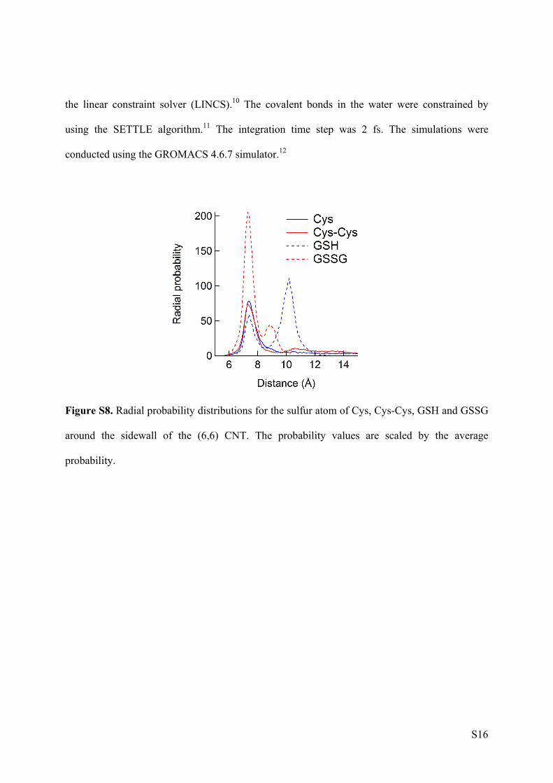
the linear constraint solver (LINCS).10 The covalent bonds in the water were constrained by
using the SETTLE algorithm.11 The integration time step was 2 fs. The simulations were
conducted using the GROMACS 4.6.7 simulator.12
Figure S8. Radial probability distributions for the sulfur atom of Cys, Cys-Cys, GSH and GSSG
around the sidewall of the (6,6) CNT. The probability values are scaled by the average
probability.

S17
2.9. Reduction of the Surfactant-Dispersed CNTs by the Addition of Lysozyme. Highly
dispersed HiPco CNTs were prepared by the same method described in section 1.6 and then
diluted into 1 wt% SDS solution to adjust the absorbance of the dispersion at 600 nm to 0.28,
which corresponds to 0.13 mg/mL. Reduced lysozyme from egg white was prepared as follows.
Native lysozyme was dissolved into a solution containing guanidine hydrochloride, DTT and
EDTA to make a 50 mg/mL lysozyme solution in the presence of 6 M guanidine hydrochloride
30 mM DTT and 2 mM EDTA. Subsequently, the lysozyme solution was dialyzed against 2 mM
EDTA solution using dialysis tubing (Visking tubing, MWCO 12,000–14,000). The resulting
precipitate of lysozyme was dissolved into a solution containing 6 M urea and 2 mM EDTA. The
dissolved lysozyme solution was filtered through a 3,000 NMWL membrane (Amicon Ultra-15
Centrifugal Filter Units with Ultracel-3 membrane, EMD Millipore Corp.) by the centrifugation
at 3,000 rpm (H-103N, Kokusan Co. Ltd.) to remove DTT and then the concentrated sample was
diluted five-fold into a solution containing 6 M urea and 2 mM EDTA; this process was repeated
five times. Finally, the reduced lysozyme concentration was adjusted to 100 M using a solution
containing 6 M urea and 2 mM EDTA. Native lysozyme was also dissolved into a solution
containing 6 M urea and 2 mM EDTA and used as a control sample. The CNT and lysozyme
samples prepared above were diluted into a solution containing urea, EDTA and SDS to prepare
samples containing 0.0065 mg/mL CNTs, 1 M lysozyme, 0.6 M urea, 2 mM EDTA and 0.05
wt% SDS. The absorption spectra of these samples are shown in Figure S9.

S18
Figure S9. Absorption spectra of HiPco CNTs in the presence and absence of the reduced and
native lysozyme (Lyz).

S19
References
(1) Edsall, J. T. Biochemistry 1965, 4, 28. (2) Hirano, A.; Kameda, T.; Yomogida, Y.; Wada, M.; Tanaka, T.; Kataura, H. ChemNanoMat 2016, 2, 911. (3) Frey, J. T.; Doren, D. J. TubeGen 3.4, (web-interface, http://turin.nss. udel.edu/research/tubegenonline.html), University of Delaware, Newark DE, 2011. (4) Wang, J.; Wolf, R. M.; Caldwell, J. W.; Kollman, P. A.; Case, D. A. J. Comput. Chem. 2004, 25, 1157. (5) Hornak, V.; Abel, R.; Okur, A.; Strockbine, B.; Roitberg, A.; Simmerling, C. Proteins 2006, 65, 712. (6) Jorgensen, W. L.; Chandrasekhar, J.; Madura, J. D.; Impey, R. W.; Klein, M. L. J. Chem. Phys. 1983, 79, 926. (7) Joung, I. S.; Cheatham, T. E. 3rd J. Phys. Chem. B 2008, 112, 9020. (8) Parrinello, M.; Rahman, A. J. Appl. Phys. 1981, 52, 7182–7190. (9) Essmann, U.; Perera, L.; Berkowitz, M. L.; Darden, T.; Lee, H.; Pedersen, L. G. J. Chem. Phys. 1995, 103, 8577. (10) Hess, B.; Bekker, H.; Berendsen, H. J. C.; Fraaije, J. G. E. M. J. Comput. Chem. 1997, 18, 1463. (11) Miyamoto, S.; Kollman, P. A. J. Comput. Chem. 1992, 13, 952. (12) Hess, B.; Kutzner, C.; van der Spoel, D.; Lindahl, E. J. Chem. Theory Comput. 2008, 4, 435.