divalent ion trapping inside potassium channels of …crt.biomol.uci.edu/pub/pdf/1989 grissmer...
TRANSCRIPT
Divalent Ion Trapping Inside Potassium Channels of Human T Lymphocytes
S. GRISSMER a n d M. D. CAHALAN
From the Department of Physiology and Biophysics, University of California, Irvine, Califor- nia 92717
ABSTRACT Using the patch-clamp whole-cell recording technique, we investi- gated the influence o f external Ca 2+, Ba ~+, K +, Rb +, and internal Ca 2+ on the rate o f K + channel inactivation in the human T lymphocyte-derived cell line, Jurkat E6-1. Raising external Ca 2+ or Ba ~+, or reducing external K +, accelerated the rate o f the K + current decay during a depolarizing voltage pulse. External Ba ~+ also produced a use-dependent block of the K + channels by entering the open channel and becoming trapped inside. Raising internal Ca 2+ accelerated inactivation at lower concentrations than external Ca ~+, but increasing the Ca z+ buffering with BAPTA did not affect inactivation. Raising [K+]o or adding Rb + slowed inactiva- tion by competing with divalent ions. External Rb + also produced a use-dependent removal of block of K + channels loaded with Ba 2+ or Ca 2+. From the removal o f this block we found that under normal conditions ~25% of the channels were loaded with Ca ~+, whereas under conditions with 10 #M internal Ca ~+ the propor- tion o f channels loaded with Ca ~+ increased to ~50%. Removing all the divalent cations from the external and internal solution resulted in the induction o f a non- selective, voltage-independent conductance. We conclude that Ca 2+ ions from the outside or the inside can bind to a site at the K § channel and thereby block the channel or accelerate inactivation.
I N T R O D U C T I O N
Vol tage-dependent K § channels in h u m a n T lymphocytes inactivate almost com- pletely over a per iod o f hundreds o f milliseconds dur ing depolar izing voltage steps (DeCoursey et al., 1984; Matteson and Deutsch, 1984). The inactivation rate is sen- sitive to the presence o f divalent ions in the bathing solution; elevated external Ca ~+ o r added lan thanum increase, whereas manganese and cobalt decrease, the rate o f inactivation (DeCoursey et al., 1985). O n e possible explanat ion for these results is that Ca 2+ or l an thanum can gain access to a site that inactivates the channel, whereas manganese and cobalt may prevent Ca ~+ f rom gaining access to this site. Increas ing [Ca~+]i also reduces the K § conduc tance o f T cells (Bregestovski et al., 1986) and accelerates inactivation o f K § channels o f B cells (Choquet et al., 1987). Thus, bo th external and in temal Ca 2+ regulate the effective n u m b e r o f K + chan- nels.
Address reprint requests to Dr. M. D. Cahalan, Department of Physiology and Biophysics, Univer- sity of California, Irvine, CA 92717.
j . GEN. PHYSIOL. ~) The Rockefeller University Press �9 0022-1295/89/04/0609/22 $2.00 Volume 93 April 1989 609-630
609
610 T H E J O U R N A L O F G E N E R A L P H Y S I O L O G Y �9 V O L U M E 93 �9 1 9 8 9
The purpose o f the experiments repor ted here is to define mechanisms o f ion interaction with K + channels. The similarity o f effects when raising external o r inter- nal [Ca 2+] suggests a c o m m o n site o f action. In some nerve preparat ions, inactiva- t ion o f voltage-gated Ca z+ channels depends on Ca 2+ ent ry th rough o p e n Ca 2+ channels and intracellular accumulat ion, ra ther than on membrane potential alone, a p h e n o m e n o n known as Ca~+-dependent inactivation o f the Ca t+ channel (for review see Eckert and Chad, 1984). An analogous mechanism in which sparingly permeant Ca ~+ ions pass th rough the K + channel and lead to inactivation might account fo r the effects o f external di- and polyvalent cations on the rate o f K + chan- nel inactivation described above. We therefore investigated whether the model o f Cai+-dependent inactivation o f Ca ~+ channels could be also applied to K § channel inactivation o f h u m a n T lymphocytes. O u r exper iments suggest that ra ther than modifying a site accessible to intracellular Ca *+ buffers, divalent ions can become t rapped inside K + channels. Since [Ca~+]i is known to rise shortly after mi togen addition, the interaction o f ions within the K + channel may be relevant to signal t ransduct ion mechanisms. Some o f the results have been repor ted in preliminary communica t ions (Grissmer and Cahalan, 1987, 1988).
ME T HODS
The experiments were carried out on single cells of a human T lymphocyte-derived cell line, Jurkat E6-1, using the whole-cell recording mode of the patch<lamp technique (Hamill et al., 1981). All experiments were done at room temperature (22-26~
Solutions
The cells under investigation were bathed in normal Ringer solution. External solutions con- taining varying concentrations of Ca ~§ Ba ~+, K +, and Rb + are listed in Table I. The chamber volume contained ~250 ~1 of solution which could be totally exchanged during the record- ings by bath perfnsion within 15-20 s. The patch-pipette usually contained 160 mM K-aspar- tate with [Ca 2+] buffered to 10 -8 M (see Table I). In some experiments the pipette solution contained higher [Ca~+].
Data Acquisition
The holding potential was adjusted in all experiments to E = - 8 0 mV. The patch-clamp amplifier (either List L/M-EPC 7, Adams and List Associates, Ltd., Great Neck, NY; or Axon Instruments, Inc., Axopatch, Burlingame, CA) was used in the voltage-clamp mode without series resistance compensation. Electrodes were pulled from Accufill 90 Micropets (Becton, Dickinson & Co., Parsippany, NJ) in three stages, coated with Sylgard (Dow Coming Corp., Midland, MI) and fire polished to resistances, measured in the bath, of 2-7 Mfl. K + channel inactivation was not different if hard glass was used as pipette glass (compare Cota and ArTn- strong, 1988; Furman and Tanaka, 1988).
In all experiments, the command input of the patch-clamp amplifier was controlled by a computer (PDP 11/73) via a digital-analog converter, and membrane currents were recorded at a bandwidth of 2 kHz. Correction for linear leakage and capacitative currents was achieved by analog subtraction and by digital subtraction of an appropriately scaled mean current asso- ciated with eight pulses delivered from a hyperpolarized potential. All potentials were correc- ted for the liquid junction potential that develops at the tip of the pipette if the pipette solu- tion is different from that of the bath. The liquid junction potential between the normal internal (pipette) and external (bath) solution was - 1 3 mV.
G R I S S M E R A N D CAHALAN Lymphocyte K + Channel Inactivation
T A B L E I
Solutions
6 1 1
External
CI- an ion
(pH 7.4)
Na § K + Ca ~+ M g 2+ H E P E S EGTA Ba ~+ Rb +
Norma l r i nge r C a 2 +
" 0 "
0.5
4.6
12
54
106
K +
0
2.2
10
20
40
8O
133
140
160
Rb+-Ringer Ba 2+
0.3
2
10
54
106
In te rna l
(pH 7.2)
160
160
160
156
144
8O
164.5
162.2
154.5
144.5
124.5
84.5
31.5
24.5
4.5
4.5
160
160
144
8O
4.5 2 1 5
4 .5 - - 3 5 1
4.5 0.5 2.5 5 - -
4 .5 4.6 1 5 - -
4 .5 12.4 1 5 - -
4 .5 53 .8 1 5 - -
4 .5 105.7 1 5 - -
D - -
h _ _
B - -
- - 2 1 5
2.2 2 1 5 - -
10 2 1 5 - -
20 2 1 5 - - - -
40 2 1 5 - - - -
80 2 1 5 - - - -
133 2 1 5 - - - -
140 2 1 5 - - - -
160 2 1 5 - - - -
- - 2 1 5 - -
4.5 - - 2.7 5 - - 0 .3
4.5 - - 1 5 - - 2.0
4.5 2 1 5 - - 10.4
4.5 2 1 5 - - 53.8
4.5 2 1 5 - - 105 .7
m _ _
160
K + Ca ~+ Mg ~+ H E P E S EGTA CI- A s p -
[Ca2+]~ - 10 -s M 160 0.1 2 5 1.1 4.2 160
[Ca~+]~ - 10 -5 M 160 1.1 2 5 1.1 4.2 160
[Ca2+]i - 2 x 1 0 - s 160 2.0 1 5 - - 1 6 6 - -
The measured osmolar i ty o f the solutions was 2 9 0 - 3 2 0 mosmol. Asp- , aspartate- ;
HEPES, N-2-hydroxyethylpiperazine-N'-2-ethanesulfonic acid; EGTA, ethylene glycol-
bis(B-aminoethylether)N,N,N' ,N'- te tmacet ic acid. [Ca~+]i was calcula ted assuming a disso- ciat ion cons tan t for EGTA a n d Ca ~+ a t p H 7.2 o f 10 -7 M acco rd ing to Portzehl et al.,
1964; in some exper iments the intracel lular Ca 2§ buf fe r ing was increased by including 55
mM 1,2-bis(2-aminophenoxy)-ethane-N,N,N' ,N'- te t raacet ic acid (BAPTA) and 5 mM
CaCI2 (free [Ca2+]t - 10 -s M) in the pipet te solution. All concen t ra t ions are in millimo-
lar.
Analysis
E x p o n e n t i a l s w e r e f i t t e d t o t h e d e c a y o f t h e K + c u r r e n t a s d e s c r i b e d i n m o r e d e t a i l b y
C a h a l a n e t al . ( 1 9 8 5 ) b y i n p u t t i n g v a l u e s t h r o u g h p o t e n t i o m e t e r s . A t t e m p t s t o f i t t h e d e c a y o f
t h e K + c u r r e n t i n h i g h [ K + ] o w i t h a s i n g l e e x p o n e n t i a l f a i l e d t o d e s c r i b e t h e t i m e c o u r s e o f
6 1 2 THE JOURNAL OF GENERAL PHYSIOLOGY �9 VOLUME 93 �9 1 9 8 9
the K + channel inactivation sufficiently. The fit, however, was improved by including a certain amount of baseline offset. This is consistent with the idea that there are K + channels that do not inactivate during a depolarizing pulse, or that there is always a certain number of open K + channels during depolarizations due to an equilibrium between the rate of activation and inactivation. Even with this fitting procedure, including an offset, the decay of the K + current could not be adequately characterized, especially during the first 300-500 ms after the onset of the depolarizing pulse. Therefore, the decay of the K + current in high [K+]o was described with a double exponential function.
R E S U L T S
External Ca 2+ and Ba 2+ Enhance Inactivation
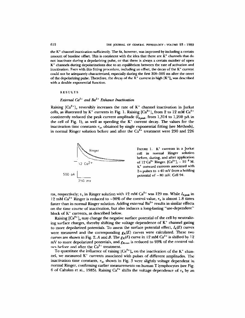
Raising [Ca~+]o reversibly increases the rate o f K § channel inactivation in Jurka t cells, as illustrated by K § currents in Fig. 1. Raising [Ca2+]o f rom 2 to 12 mM Ca 2+ consistently reduced the peak cur ren t ampli tude ( IK~ , f rom 1,314 to 1,200 pA in the cell o f Fig. 1), as well as speeding the K + cur ren t decay. The values for the inactivation time constants ~'h, obta ined by single exponent ia l fitting (see Methods), in normal Ringer solution before and after the Ca 2+ t rea tment were 230 and 226
12 Ca 2+
500 pA
250 ms
FIGURE 1. K § currents in a Jurkat cell in normal Ringer solution before, during, and after application of I2 Ca ~+ Ringer. [Ca~+]i = 10 -8 M. K + outward currents associated with 2-s pulses to +40 mV from a holding potential of - 8 0 inV. Cell 94.
ms, respectively; rh in Ringer solution with 12 mM Ca ~+ was 129 ms. While I ~ in 12 mM Ca 2+ Ringer is r educed to ~90% o f the control value, 7h is almost 1.8 times faster than in normal Ringer solution. Adding external Ba 2+ results in similar effects on the time course o f inactivation, but also induces a long-lasting "use -dependen t " block o f K + currents , as described below.
Raising [Ca2+]o may change the negative surface potential o f the cell by neutraliz- ing surface charges, thereby shifting the voltage dependence o f K + channel gating to more depolar ized potentials. To assess the surface potential effect, IK(E) curves were measured and the co r respond ing gK(E) curves were calculated. These two curves are shown in Fig. 2, A and B. The gK(E) curve in 12 mM Ca 2+ is shifted by 12 mV to more depolar ized potentials, and g K ~ is reduced to 93% o f the control val- l~e~ b e f o r e and after the Ca 2+ treatment.
To quanti tate the influence o f raising [Ca2+]o on the inactivation o f the K + chan- nel, we measured K + currents associated with pulses o f different amplitudes. The inactivation time constants, rh, shown in Fig. 3 were slightly voltage dependen t in normal Ringer, conf i rming earlier measurements on h u m a n T lymphocytes (see Fig. 6 o f Cahalan et al., 1985). Raising Ca *§ shifts the voltage dependence o f rh by an
G R I $ S M E R A N D C A H A L A N
A
I K
2 .0 "
1.0
. / -40 o
( h A )
i i ) i
4O 80
Lymphocyte K + Channel Inactivation
B
gK (nS)
~2[ .
Ringer 4 12 Ca 2+
- 4 0 0 4 0 8 0
E (rnV) E (mY)
618
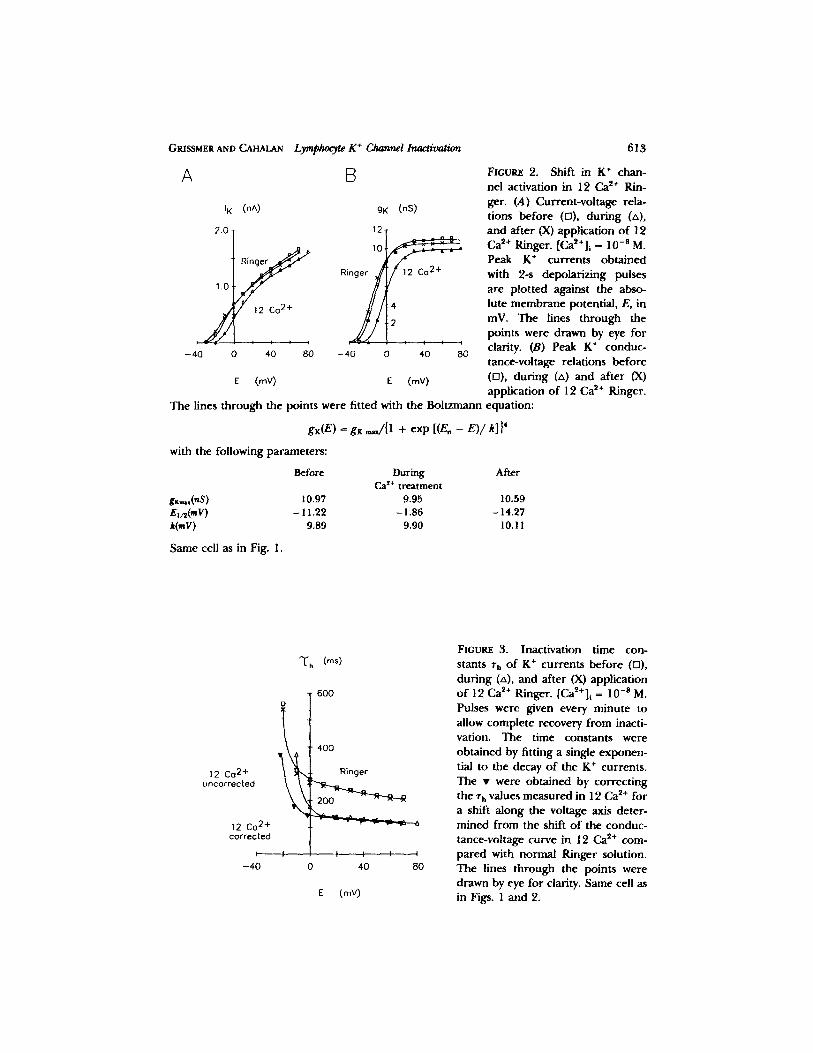
FIOUnE 2. Shift in K + chan- nel activation in 12 Ca 2+ Rin- ger. (A) Current-voltage rela- tions before (ra), dur ing (zx), and after (X) application o f 12 Ca ~+ Ringer. [Ca~+] i ffi 10 -8 M. Peak K + currents obtained with 2-s depolarizing pulses are plotted against the abso- lute membrane potential, E, in mV. The lines through the points were drawn by eye for clarity. (B) Peak K + conduc- tance-vohage relations before (D), during (zx) and after (X) application o f 12 Ca ~+ Ringer.
The lines through the points were fitted with the Boltzmann equation:
gK(E) = gK ,~/{1 + exp [(E, - E)/k]} 4
with the following parameters:
Before During After Ca ~+ treatment
gxm,,(nS ) 10.97 9.95 10.59 E,/~(mV) - 11.22 - 1.86 - 14.27 k(mV) 9.89 9.90 10. ! 1
Same cell as in Fig. 1.
12 Co 2+ uncorrected
12 Co 2+ corrected
I-, -40
" ( h (m@
600
400
R i n g e r
�9 ,A .
I l I , I I
0 4o 8o
E (my)
FIGURE 3. Inactivation time con- stants ~', o f K + currents before (F1), dur ing (zx), and after (X) application o f 12 Ca ~+ Ringer. [Ca2+]i = 10 - s M. Pulses were given every minute to allow complete recovery f rom inacti- vation. The time constants were obtained by fitting a single exponen- tial to the decay of the K + currents. The �9 were obtained by correct ing the T h values measured in 12 Ca ~+ for a shift along the voltage axis deter- mined from the shift o f the conduc- tance-voltage curve in 12 Ca 2+ com- pared with normal Ringer solution. The lines through the points were drawn by eye for clarity. Same cell as in Figs. 1 and 2.
614 THE JOURNAL OF GENERAL PHYSIOLOGY �9 VOLUME 9 3 �9 1 9 8 9
a m o u n t c o m p a r a b l e to the shift o f act ivat ion shown in Fig. 2. In add i t i on to this shift, rh is r e d u c e d in 12 mM Ca~+-Ringer. The combina t i on o f b o t h effects causes c rossover o f the con t ro l curve and the curve o b t a i n e d in 12 mM Ca 2+. To co r rec t for surface po ten t ia l effects, the ~'h values o b t a i n e d in 12 mM Ca ~+ ( labeled "12 Ca u n c o r r e c t e d " ) were shif ted to m o r e hype rpo l a r i zed poten t ia l s by the same a m o u n t that the gz(E) curve was shif ted to m o r e depo l a r i zed poten t ia l s (12 mV) when [Ca2+]o was ra ised f rom 2 to 12 mM ("12 Ca co r r ec t ed" ) . The r educ t i on o f rh was then d e t e r m i n e d by ca lcula t ing the rat ios o f the con t ro l Zh and the c o r r e c t e d ~'h in 12 mM Ca ~+. At E = 10 mV the r educ t i on was to 52% o f the con t ro l value, whereas at E = 50 rnV the r e d u c t i o n was to 57% o f the con t ro l value. This means that , aside f rom a shift, the Ca ~+ effect on the inact ivat ion t ime cons tan t shows very little, if any, vol tage d e p e n d e n c e .
p
1.o
0.0 �9 .
o.1 1.o l o l o o l o o o
[X2+] o (mM)
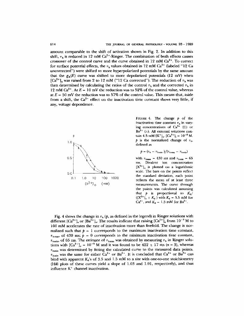
FIGURE 4. The change p of the inactivation time constant rh in vary- ing concentrations of Ca ~+ (n) or Ba ~+ (zx). All external solutions con- tain 4.5 mM [K+]o. [Ca2+]i = 10 -s M. p is the normalized change of rh, defined as
p = (rh - T h m i n ) / / ( r h m a x - - rhmin)
with rh,,~ = 430 ms and ~',mi~ = 65 ms. Divalent ion concentration [X2+]o is plotted on a logarithmic scale. The bars on the points reflect the standard deviation, each point reflects the mean of at least three measurements. The curve through the points was calculated assuming that p is proportional to Kd/ ([X2+]o + Kd ) with Kd = 3.5 mM for Ca 2+, and Kd = 1.5 mM for Ba 2+.
Fig. 4 shows the change in ~rh (p, as de f ined in the legend) in Ringer solut ions with d i f fe ren t [Ca~+]o o r [Ba2+]o. The results indicate tha t ra is ing [Ca2+] o f rom 10 -9 M to 100 mM accelera tes the ra te o f inact ivat ion m o r e than fivefold. The change is nor - mal ized such that p = 1 c o r r e s p o n d s to the m a x i m u m inact ivat ion t ime cons tant , rhm~,, o f 430 ms; p = 0 c o r r e s p o n d s to the m i n i m u m inact ivat ion t ime cons tant , rhmln, o f 65 ms. The es t imate o f rhea, was o b t a i n e d by measu r ing rh in Ringer solu- t ions with [Ca~+]o = 10 -9 M and it was f o u n d to be 422 -+ 17 ms (n = 3), whereas rhm~ was d e t e r m i n e d by f i t t ing the ca lcu la ted curve to the m e a s u r e d da ta points . Zhmm was the same for e i ther Ca 2+ o r Ba 2+. I t is conc luded that Ca ~+ o r Ba 2+ can b ind with a p p a r e n t Kd'S o f 3.5 and 1.5 mM to a site with one - to -one s to ich iomet ry (Hill plots o f these curves yield a s lope o f 1.03 and 1.01, respectively), and thus inf luence K + channel inact ivat ion.
GRISSMER AND CAHALAN Lymphocyte K + Channel Inactivation 615
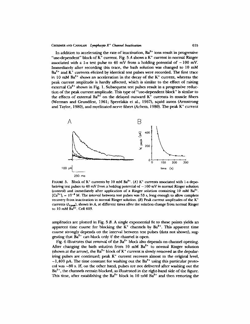
In addition to accelerating the rate of inactivation, Ba ~+ ions result in progressive "use-dependent" block of K + current. Fig. 5 A shows a K + current in normal Ringer associated with a 1-s test pulse to 40 mV from a holding potential o f - 1 0 0 inV. Immediately after recording this trace, the bath solution was changed to 10 mM Ba ~+ and K + currents elicited by identical test pulses were recorded. The first trace in 10 m M B a ~+ shows an acceleration in the decay o f the K + current, whereas the peak current amplitude is hardly affected, which is similar to the effect of raising external Ca ~+ shown in Fig. 1. Subsequent test pulses result in a progressive reduc- tion o f the peak current amplitude. This type of "use-dependent block" is similar to the effects o f external Ba ~+ on the delayed outward K + currents in muscle fibers (Werman and Grundfest, 1961; Sperelakis et al., 1967), squid axons (Armstrong and Taylor, 1980), and myelinated nerve fibers (Arhem, 1980). The peak K + current
A B
~- 400
200
~: - 0
I O0 pA~
250 ms
1 oo 200 3O0
time (s)
FtGUltE 5. Block of K § currents by 10 mM Ba ~+. (A) K + currents associated with 1-s depo- larizing test pulses to 40 mV from a holding potential of - 100 mV in normal Ringer solution (control) and immediately after application of a Ringer solution containing 10 mM Ba *+. [Ca~+]~ = 10 -s M. The interval between test pulses was 53 s, long enough to allow complete recovery from inactivation in normal Ringer solution. (B) Peak current amplitudes of the K § currents (I~0,~), shown in A, at different times after the solution change from normal Ringer to 10 mMBa 2+. Cell 603.
amplitudes are plotted in Fig. 5 B. A single exponential fit to these points yields an apparent time course for blocking the K § channels by Ba 2+. This apparent time course strongly depends on the interval between test pulses (data not shown), sug- gesting that Ba 2+ can block only if the channel is open.
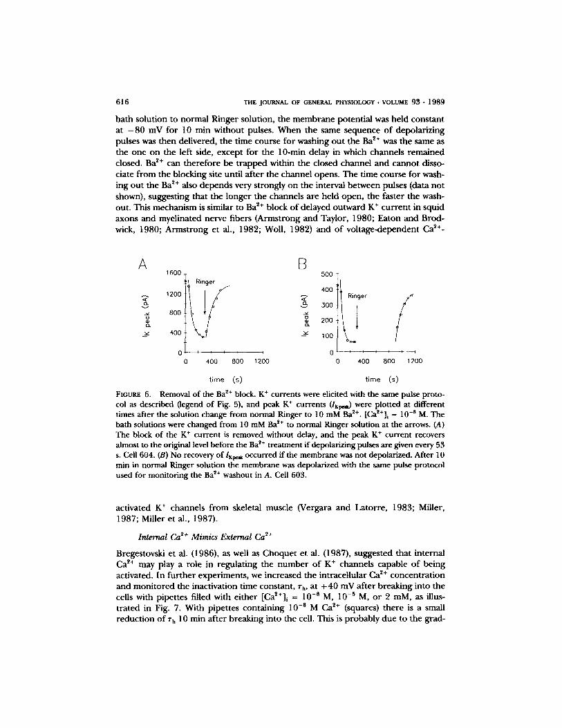
Fig. 6 illustrates that removal o f the Ba *§ block also depends on channel opening. After changing the bath solution f rom 10 mM Ba ~+ to normal Ringer solution (shown at the arrow), the Ba ~+ block of K + current is slowly removed as the depolar- izing pulses are continued; peak K + current recovers almost to the original level,
1,400 pA. The time constant for washing out the Ba ~+ using this particular proto- col was ~80 s. If, on the other hand, pulses are not delivered after washing out the Ba 2+, the channels remain blocked, as illustrated in the fight-hand side of the figure. This time, after establishing the Ba 2+ block in 10 m M B a *+ and then restoring the
616 THE JOURNAL OF GENERAL PHYSIOLOGY �9 VOLUME 93 �9 1989
ba th so lu t ion to no rma l R inger solut ion, the m e m b r a n e po ten t ia l was he ld cons tan t at - 8 0 mV for 10 min wi thou t pulses. W h e n the same sequence o f depo la r i z ing pulses was then del ivered, the t ime course for washing o u t the Ba ~+ was the same as the one on the left side, excep t for the 10-min delay in which channels r e m a i n e d closed. Ba z+ can t h e r e f o r e be t r a p p e d within the c losed channe l and canno t disso- ciate f rom the b lock ing site unt i l a f te r the channe l opens . The t ime course for wash- ing ou t the Ba 2+ also d e p e n d s very s t rongly on the interval be tween pulses (data no t shown), sugges t ing that the longe r the channels a re he ld open , the fas ter the wash- out. This mechan i sm is s imilar to Ba ~+ b lock o f de layed ou tw a rd K § c u r r e n t in squid axons and myel ina ted nerve f ibers (Arms t rong and Taylor , 1980; Ea ton a n d Brod- wick, 1980; A r m s t r o n g et al., 1982; Woll , 1982) and o f v o l t a g e - d e p e n d e n t Ca ~+-
A B
v
1600
1200
800
400
0
500 Ringer
400
300
200
-~ 1oo
I I I I I * 0
0 400 800 1200
Ringer
- - I t I
o 400
/ I I - - - - - 4
800 1200
time (s) t ime (s)
FIGURE 6. Removal of the Ba 2+ block. K + currents were elicited with the same pulse proto- col as described (legend of Fig. 5), and peak K + currents (IK~.~) were plotted at different times after the solution change from normal Ringer to 10 mM Ba 2+. [CaZ+]i = 10 -s M. The bath solutions were changed from 10 mM Ba 2+ to normal Ringer solution at the arrows. (A) The block of the K + current is removed without delay, and the peak K + current recovers almost to the original level before the Ba i+ treatment if depolarizing pulses are given every 53 s. Cell 604. (B) No recovery of IK~. i occurred if the membrane was not depolarized. After 10 min in normal Ringer solution the membrane was depolarized with the same pulse protocol used for monitoring the Ba ~+ washout in A. Cell 603.
ac t iva ted K + channels f rom skeletal muscle (Vergara and La to r r e , 1983; Miller, 1987; Mil ler e t al., 1987).
Internal Ca 2+ Mimics External Ca 2+
Bregestovski e t al. (1986), as well as C h o q u e t et al. (1987), sugges ted that in te rna l Ca ~+ may play a role in r egu la t ing the n u m b e r o f K + channels capab le o f be ing activated. In fu r the r exper imen t s , we increased the in t race l lu la r Ca ~+ concen t r a t i on and m o n i t o r e d the inact ivat ion t ime cons tant , rh, at + 40 mV af te r b r eak ing into the cells with p ipe t tes filled with e i ther [Ca2+] i = 10 -s M, 10 -5 M, o r 2 raM, as illus- t r a t ed in Fig. 7. Wi th p ipe t tes con ta in ing 10 -8 M Ca ~+ (squares) the re is a small r educ t i on o f rh 10 rain a f te r b reak ing in to the cell. This is p r o b a b l y due to the grad-
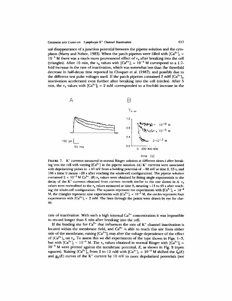
GRISSMER AND CAHALAN Lymphocyte K + Channel Inactivation 617
ual d i s a p p e a r a n c e o f a j u n c t i o n po ten t i a l be tween the p ipe t t e so lu t ion and the cyto- p lasm (Marty a n d Neher , 1983). W h e n the pa tch p ipe t tes were fi l led with [Ca~+]i = 10-5 M the re was a much m o r e p r o n o u n c e d effect o f rh a f te r b r eak ing in to the cell (triangles). Af t e r 10 min, the ~'h values with [Ca~+]i = 10 -s M c o r r e s p o n d to a 1.7- fold increase in the ra te o f inact ivat ion, which was somewhat less than the t h ree fo ld dec rease in hal f -decay t ime r e p o r t e d by C h o q u e t et al. (1987), a n d possibly due to the d i f fe ren t test pulse vol tages used. I f the pa tch p ipe t tes con t a ined 2 m M [Ca2+]i, inact ivat ion acce le ra ted even fu r t he r a f te r b reak ing into the cell (circles). A f t e r 5 min, the rh values with [Ca~+]i = 2 mM c o r r e s p o n d e d to a f ivefold increase in the
A [3
" ~ ' h r e l
1.2
0.8
0.4 l
lOO pA[
50 ms 0.0
~ ~ lO - 8 M
~ ~ 10-5 M
~ 2.1o - 3 M
r i q i
0 200 400 600
time (s)
FIGURE 7. K + currents measured in normal Ringer solution at different times t after break- ing into the cell with varying [Ca z+] in the pipette solution. (A) K + currents were associated with depolarizing pulses to +40 mV from a holding potential of - 8 0 mV at time 0, 53 s, and 106 s (time 0 means ~20 s after reaching the whole-ceU configuration). The pipette solution contained 2 x 10 -s M Ca ~+. (B) rh values were obtained by fitting single exponentials to the decay of the K § currents obtained from current records similar to the one shown in A. rh values were normalized to the zh values measured at time 0, meaning ~ 15 to 45 s after reach- ing the whole-cell configuration. The squares represent ten experiments with [Ca~+]i = 10 -8 M, the triangles represent nine experiments with [Ca~t+]i ~ 10 -s M, the circles represent four experiments with [Ca~§ i = 2 mM. The lines through the points were drawn by eye for clar- ity.
ra te o f inactivat ion. With such a high in te rna l Ca 2+ c o n c e n t r a t i o n it was imposs ib le to r e c o r d longe r than 6 min a f t e r b reak ing into the cell.
I f the b ind ing site for Ca ~+ that inf luences the ra te o f K § channe l inact ivat ion is loca ted within the m e m b r a n e field, and Ca ~+ is able to reach this site f rom e i the r side o f the m e m b r a n e , ra is ing [Ca2+]i may a l te r the vol tage d e p e n d e n c e o f the effect o f [Ca2+]o on r h. To assess this we d id e x p e r i m e n t s o f the type shown in Figs. 1 -3 , b u t with [Ca~+]i = 10 -s M. The rh values o b t a i n e d in no rma l R inger with [Cag+]i = 10 -s M were p lo t t ed agains t the m e m b r a n e potent ia l , E, as shown in Fig. 8 (open squares). Rais ing [Ca2+]o f rom 2 to 12 mM with [Ca~+]l = 10 -s M shif ted the IK(E )
a n d gK(E) curves o f the K + c u r r e n t by 10 mV to m o r e d e p o l a r i z e d poten t ia l s (not
618 THE JOURNAL OF GENERAL PHYSIOLOGY �9 VOLUME 93 �9 1 9 8 9
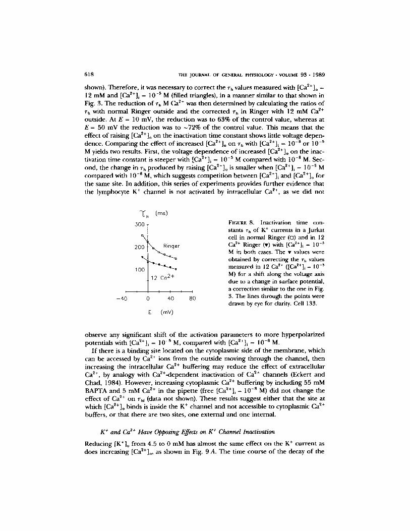
shown). Therefore , it was necessary to correc t the 7h values measured with [Ca~+]o = 12 mM and [Ca~+]i = 10 -s M (filled triangles), in a manne r similar to that shown in Fig. 3. The reduct ion o f zh M Ca ~+ was then de te rmined by calculating the ratios o f r h with normal Ringer outside and the cor rec ted T h in Ringer with 12 mM Ca ~+ outside. At E = 10 mV, the reduct ion was to 63% o f the control value, whereas at E = 50 tnV the reduct ion was to ~72% o f the control value. This means that the effect o f raising [Ca~+]o on the inactivation time constant shows little voltage depen- dence. Compar ing the effect o f increased [Ca2+]o on ~'h with [Ca2+]i = 10 -s o r 10 -s M yields two results. First, the voltage dependence o f increased [Ca2+]o on the inac- tivation time constant is s teeper with [Ca~+]i = 10 -s M c o m p a r e d with 10 -s M. Sec- ond, the change in zh p r o d u c e d by raising [Ca~+]o is smaller when [Ca2+]i = 10 -5 M compared with 10 -s M, which suggests compet i t ion between [Ca~+]i and [Ca~+]o for the same site. In addition, this series o f experiments provides fur ther evidence that the lymphocyte K + channel is not activated by intracellular Ca ~+, as we did not
'Tf h (ms)
2 0 ~ r
100 t , , I 12 Ca 2 , , ,+ , ,
- 4 0 0 40 80
E (mV)
F I G U R E 8 . Inactivation time con- stants ~'h of K § currents in a Jurkat cell in normal Ringer (D) and in 12 Ca ~+ Ringer (v) with [Ca~+]i = 10 -5 M in both cases. The �9 values were obtained by correcting the r h values measured in 12 Ca 2+ ( [ C a 2 + ] i = l 0 - s
M) for a shift along the voltage axis due to a change in surface potential, a correction similar to the one in Fig. 3. The lines through the points were drawn by eye for clarity. Cell 133.
observe any significant shift o f the activation parameters to more hyperpolar ized potentials with [Ca~+]i = 10 -~ M, c o m p a r e d with [Ca2+]i = l 0 -a M.
I f there is a binding site located on the cytoplasmic side o f the membrane , which can be accessed by Ca 2+ ions f rom the outside moving th rough the channel, then increasing the intracellular Ca ~+ buffer ing may reduce the effect o f extracellular Ca 2§ by analogy with Ca~+-dependent inactivation o f Ca ~+ channels (Eckert and Chad, 1984). However , increasing cytoplasmic Ca ~+ buffer ing by including 55 mM BAPTA and 5 mM Ca 2+ in the pipet te (free [Ca2+]i = 10 -s M) did no t change the effect o f Ca 2+ on Thf (data not shown). These results suggest ei ther that the site at which [Ca~+] o binds is inside the K § channel and not accessible to cytoplasmic Ca 2+ buffers, o r that there are two sites, one external and one internal.
K + and Ca 2+ Have Opposing Effects on K + Channel Inactivation
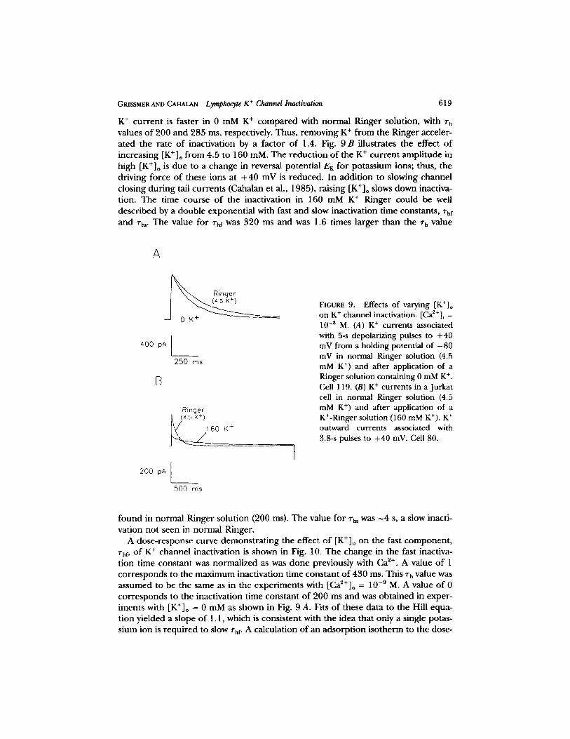
Reducing [K+]o f rom 4.5 to 0 mM has almost the same effect on the K + cur ren t as does increasing [Ca2+]o, as shown in Fig. 9 A. The time course o f the decay o f the
GRISSMER AND CAHALAN Lymphocyte K + Channel Inactivation 619
K + c u r r e n t is fas ter in 0 m M K + c o m p a r e d with n o r m a l R inger solut ion, with rh values o f 200 and 285 ms, respectively. Thus, r emov ing K + f rom the Ringer acceler- a t ed the ra te o f inact ivat ion by a fac tor o f 1.4. Fig. 9 B i l lustrates the effect o f increas ing [K + ]o f rom 4.5 to 160 mM. The r educ t i on o f the K + c u r r e n t a m p l i t u d e in high [K+]o is d u e to a change in reversal po ten t ia l EK for po tass ium ions; thus, the dr iv ing force o f these ions at + 40 mV is r educed . In add i t i on to s lowing channe l c los ing d u r i n g tail cu r r en t s (Cahalan et al., 1985), ra is ing [K+]o slows down inactiva- t ion. The t ime course o f the inact ivat ion in 160 mM K + Ringer cou ld be well de sc r ibed by a doub le exponen t i a l with fast a n d slow inact ivat ion t ime constants , rhf and Vh,. The value for l"hf was 320 ms and was 1.6 t imes l a rge r than the % value
A
Ringer
400 pA
B
200 pA [ _ _
250 ms
Ringer j ~ 6 (4-5 K+)
:0 K +
500 ms
"-].
FIGURE 9. Effects of varying [K+]o on K § channel inactivation. [Ca2+]i = 10 -8 M. (A) K + currents associated with 5-s depolarizing pulses to +40 mV from a holding potential of - 8 0 mV in normal Ringer solution (4.5 mM K +) and after application of a Ringer solution containing 0 mM K § Cell 119. (B) K + currents in a Jurkat cell in normal Ringer solution (4.5 mM K § and after application of a K+-Ringer solution (160 mM K*). K + outward currents associated with 3.8-s pulses to +40 mV. Cell 80.
f o u n d in no rma l R inger so lu t ion (200 ms). The value for "rhs was ~4 s, a slow inacti- va t ion no t seen in no rma l Ringer .
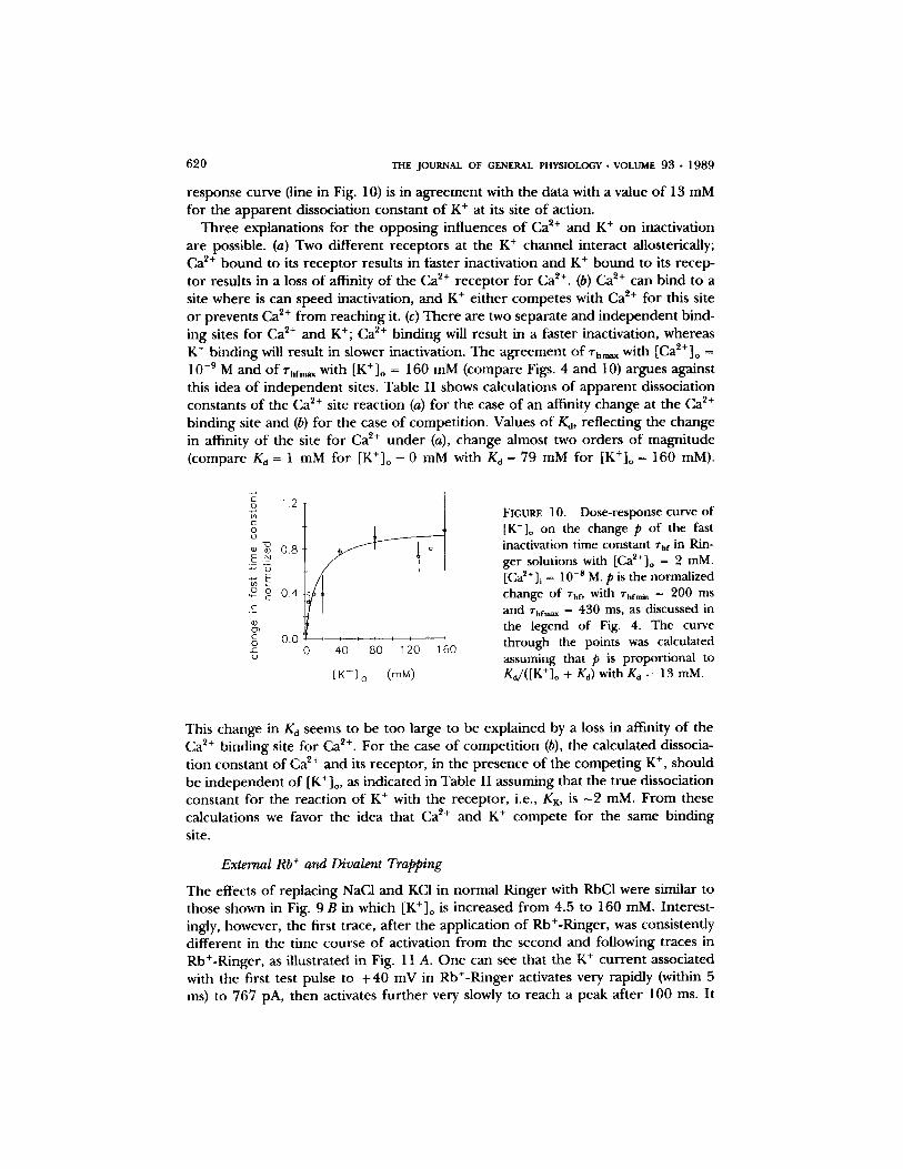
A dose - r e sponse curve d e m o n s t r a t i n g the effect o f [K+]o on the fast c o m p o n e n t , rhr, o f K + channel inact ivat ion is shown in Fig. 10. The change in the fast inactiva- t ion t ime cons tan t was no rma l i zed as was d o n e previously with Ca 2+. A value o f 1 c o r r e s p o n d s to the m a x i m u m inact ivat ion t ime cons tan t o f 430 ms. This rh value was a s sumed to be the same as in the expe r imen t s with [Ca2+]o = 10 -9 M. A value o f 0 c o r r e s p o n d s to the inact ivat ion t ime cons tan t o f 200 ms and was o b t a i n e d in expe r - iments with [K+]o = 0 mM as shown in Fig. 9 A. Fits o f these da ta to the Hill equa- t ion y ie lded a s lope o f 1.1, which is cons is ten t with the idea that only a single potas- s ium ion is r e q u i r e d to slow the. A calcula t ion o f an a d s o r p t i o n i so the rm to the dose-
6 2 0 THE JOURNAL OF GENERAL PHYSIOLOGY �9 VOLUME 9 3 �9 1 9 8 9
response curve (line in Fig. 10) is in ag reement with the data with a value o f 13 mM for the apparen t dissociation constant o f K + at its site o f action.
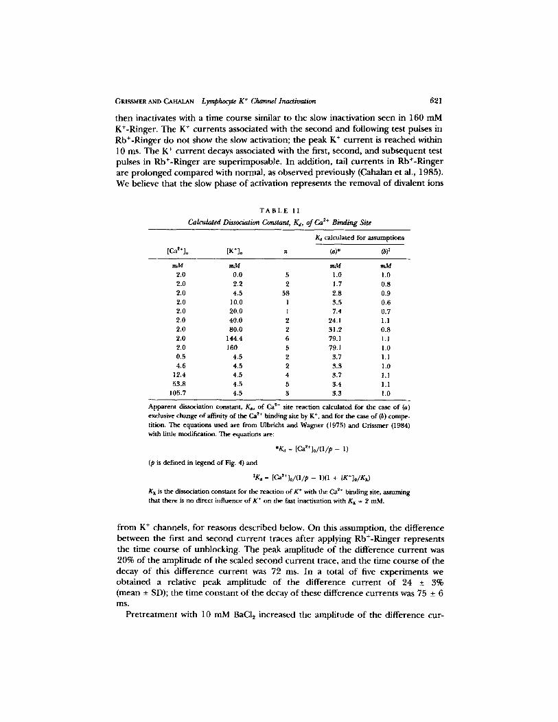
Three explanations for the oppos ing influences o f Ca ~§ and K + on inactivation are possible. (a) Two different receptors at the K + channel interact allosterically; Ca e+ b o u n d to its r ecep tor results in faster inactivation and K + b o u n d to its recep- tor results in a loss o f affinity o f the Ca 2+ receptor for Ca ~§ (b) Ca ~+ can bind to a site where is can speed inactivation, and K + ei ther competes with Ca ~+ for this site o r prevents Ca ~+ f rom reaching it. (c) There are two separate and independent bind- ing sites for Ca ~+ and K§ Ca 2+ binding will result in a faster inactivation, whereas K + binding will result in slower inactivation. The agreement o f "rhm~ with [Ca2+]o = 10 -9 M and o f %fm~ with [K+]o = 160 mM (compare Figs. 4 and 10) argues against this idea o f independent sites. Table I I shows calculations o f apparen t dissociation constants o f the Ca 2+ site react ion (a) for the case o f an affinity change at the Ca ~+ binding site and (b) for the case o f competi t ion. Values o f Ka, reflecting the change in affinity o f the site for Ca ~+ u n d e r (a), change almost two orders o f magni tude (compare Kd = 1 mM for [K+]o = 0 mM with Kd = 79 mM for [K+]o = 160 mM).
.gq 1.2
0 U
' ~ 0.8
2 ~ 0.4 .C_
c 0.0 C~
i i I I i i I i
0 40 80 120 160
[ K + ] o (raM)
FIGURE 10 . D o s e - r e s p o n s e c u r v e o f
[K+]o on the change p of the fast inactivation time constant %f in Rin- ger solutions with [Ca2+]o = 2 mM. [Ca2+]i = 10 -s M. p is the normalized change of %f, with rhfmn, = 200 ms and rhf,,~, = 430 ms, as discussed in the legend of Fig. 4. The curve through the points was calculated assuming that p is proportional to Ka/([K+]o + Kd) with Kd = 13 mM.
This change in Kd seems to be too large to be explained by a loss in affinity o f the C a 2+ binding site for Ca ~+. For the case o f compet i t ion (b), the calculated dissocia- tion constant o f Ca ~+ and its receptor , in the presence o f the compet ing K § should be independent o f [K+]o, as indicated in Table I I assuming that the t rue dissociation constant for the reaction o f K + with the receptor , i.e., KK, is - 2 raM. F rom these calculations we favor the idea that Ca 2+ and K + compete for the same binding site.
External Rb + and Divalent Trapping
The effects o f replacing NaC1 and KCI in normal Ringer with RbCI were similar to those shown in Fig. 9 B in which [K+]o is increased f rom 4.5 to 160 mM. Interest- ingly, however, the first trace, after the application o f Rb+-Ringer, was consistently different in the time course o f activation f rom the second and following traces in Rb+-Ringer, as illustrated in Fig. 11 A. O n e can see that the K + cur ren t associated with the first test pulse to + 4 0 mV in Rb§ activates very rapidly (within 5 ins) to 767 pA, then activates fur ther very slowly to reach a peak after 100 ms. It
GRISSMER AND CAHALAN Lymphocyte K § Channel Inactivation 621
t h e n inac t iva tes wi th a t i m e c o u r s e s imi la r to t he s low inac t iva t ion s e e n in 160 m M
K+-Ringe r . T h e K + c u r r e n t s a s soc ia t ed wi th t h e s e c o n d a n d f o l l o w i n g test pu lses in
R b + - R i n g e r d o n o t s h o w the s low ac t iva t ion ; t he p e a k K + c u r r e n t is r e a c h e d wi th in
10 ms. T h e K + c u r r e n t decays a s soc ia t ed wi th t he first, s e cond , a n d s u b s e q u e n t test
pu l ses in R b + - R i n g e r a r e s u p e r i m p o s a b l e . I n add i t i on , tail c u r r e n t s in R b + - R i n g e r
a r e p r o l o n g e d c o m p a r e d wi th n o r m a l , as o b s e r v e d p r ev ious ly (Caha lan e t al,, 1985).
W e be l i eve tha t t he s low p h a s e o f ac t iva t ion r e p r e s e n t s t he r e m o v a l o f d iva l en t ions
T A B L E II
Calculated Dissociation Constant, K~, of Ca 2+ Binding Site
/~ calculated for assumptions
[Ca~+]o [K*]o n (a)* (~):
mM mM mM ram 2.0 0.0 5 1.0 1.0 2.0 2.2 2 1.7 0.8 2.0 4.5 58 2.8 0.9 2.0 10.0 1 3.5 0.6 2.0 20.0 t 7.4 0.7 2.0 40.0 2 24.1 1.1 2.0 80.0 2 31.2 0.8 2.0 144.4 6 79.1 1.1 2.0 160 5 79.1 1.0 0.5 4.5 2 3.7 1.1 4.6 4.5 2 3.3 1.0
12.4 4.5 4 3.7 1.1 53.8 4.5 5 3.4 1.1
105.7 4.5 3 3.3 1.0
Apparent dissociation constant, /Ca, of Ca s+ site reaction calculated for the case of (a) exclusive change of affinity of the Ca ~+ binding site by K +, and for the case of (b) compe- tition. The equations used are from Ulbricht and Wagner (1975) and Grissmer 0984) with little modification. The equations are:
*Kd- [CaS+]o/(1/p- 1)
(p is defined in legend of Fig. 4) and
tKa = [ C a ~ + ] o / ( 1 / p - 1)(1 + [K+]o/Kx)
K x is the dissociation constant for the reaction ofK + with the Ca :~+ binding site, assuming that there is no direct influence of K + on the fast inactivation with Kx - 2 raM.
f r o m K + c h a n n e l s , f o r r e a s o n s d e s c r i b e d be low. O n this a s s u m p t i o n , t he d i f f e r e n c e
b e t w e e n the first a n d s e c o n d c u r r e n t t races a f t e r a p p l y i n g R b + - R i n g e r r e p r e s e n t s
t he t i m e c o u r s e o f u n b l o c k i n g . T h e p e a k a m p l i t u d e o f t he d i f f e r e n c e c u r r e n t was
20% o f t he a m p l i t u d e o f t h e sca led s e c o n d c u r r e n t t race , a n d the t i m e c o u r s e o f t he
decay o f this d i f f e r e n c e c u r r e n t was 72 ms. I n a to ta l o f f ive e x p e r i m e n t s we
o b t a i n e d a re la t ive p e a k a m p l i t u d e o f t he d i f f e r e n c e c u r r e n t o f 24 _+ 3%
( m e a n _+ SD); t h e t ime c o n s t a n t o f t he decay o f t he se d i f f e r e n c e c u r r e n t s was 75 _+ 6 ms.
P r e t r e a t m e n t wi th 10 m M BaCl 2 i n c r e a s e d the a m p l i t u d e o f t he d i f f e r e n c e cu r -
622 THE JOURNAL OF GENERAL PHYSIOLOGY �9 VOLUME 93 �9 1989
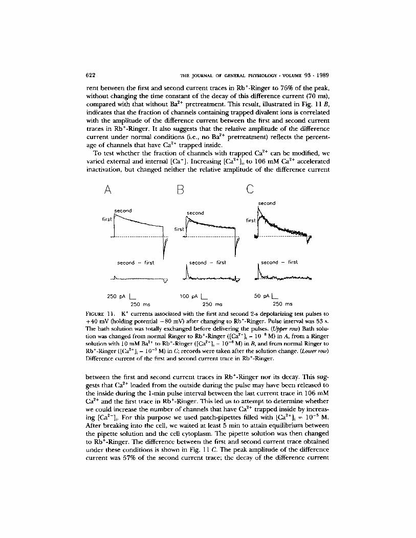
rent between the first and second cur ren t traces in Rb+-Ringer to 76% o f the peak, without changing the time constant o f the decay o f this difference cur ren t (70 ms), compared with that without Ba ~+ pret reatment . This result, illustrated in Fig. 11 B, indicates that the fraction o f channels conta ining t rapped divalent ions is correla ted with the ampli tude o f the difference cur ren t between the first and second cur ren t traces in Rb+-Ringer. It also suggests that the relative ampli tude o f the difference cur ren t unde r normal condit ions (i.e., no Ba 2+ pret reatment) reflects the percent- age o f channels that have Ca ~+ t rapped inside.
To test whether the fraction o f channels with t rapped Ca z+ can be modified, we varied external and internal [Ca+]. Increasing [Ca2+] o to 106 mM Ca 2+ accelerated inactivation, but changed nei ther the relative ampli tude o f the difference cur rent
/x B
second second
second - first second - first
C second
f i r s ~
250 pA L_ 100 pA [ ~ 50 pAL__ 250 ms 250 ms 250 ms
FIGURE l 1. K + currents associated with the first and second 2-s depolarizing test pulses to +40 mV (holding potential - 8 0 mV) after changing to Rb+-Ringer. Pulse interval was 53 s. The bath solution was totally exchanged before delivering the pulses. (Upper row) Bath solu- tion was changed from normal Ringer to Rb+-Ringer ([Ca2+] i = 10 -8 M) in A, from a Ringer solution with 10 mM Ba 2+ to Rb+-Ringer ([Ca~+]~ = 10 -s M) in B, and from normal Ringer to Rb+-Ringer ([Ca2+]i = 10 -5 M) in C; records were taken after the solution change. (Lower row) Difference current of the first and second current trace in Rb+-Ringer.
between the first and second cur ren t traces in Rb+-Ringer no r its decay. This sug- gests that Ca ~+ loaded f rom the outside dur ing the pulse may have been released to the inside dur ing the 1-min pulse interval between the last cur ren t trace in 106 mM Ca ~+ and the first trace in Rb+-Ringer. This led us to a t tempt to determine whether we could increase the n u m b e r o f channels that have Ca 2+ t rapped inside by increas- ing [Ca2+]i. For this purpose we used patch-pipettes filled with [Ca~+]i = 10 -5 M. After breaking into the cell, we waited at least 5 rain to attain equilibrium between the pipette solution and the cell cytoplasm. The pipette solution was then changed to Rb+-Ringer. The difference between the first and second cur ren t trace obtained unde r these condit ions is shown in Fig. 11 C. The peak ampli tude o f the difference cur ren t was 57% o f the second cur ren t trace; the decay o f the difference cur rent
GR1SSMER AND C A ~ Lymphocyte K + Channel Inactivation 623
was 71 ms. In a total o f five experiments, we obtained 52 _ 8% for the relative amplitude of the difference current; the time course for the decay of these differ- ence currents was 76 _+ 4 ms. We conclude that it is possible to load the channels with Ca ~+ f rom the inside, and that this Ca ~+ can be released to the outside.
Effects of Removing All Divalent Cations
To see whether the residual inactivation rate of K + channels (which corresponds to rhm~, in low external and internal [Ca ~+ ] is due to block by Mg ~+ ions, which would be analogous to the Mg ~+ block of inward rectifier K + channels (Matsuda et al., 1987; Vandenberg, 1987), experiments were per formed using internal solutions with no added Mg 2+ ([Ca~+]i = 10 -8 M). After breaking into a cell we recorded nor- real inactivating K + currents even after 15 min with normal Ringer outside. I f we changed to a Ringer solution with no added Mg ~+ and [Ca~+]o = 10 -9 M, changes in the membrane current that were similar to those described by Armstrong and Lopez-Barneo (1987) occurred. We observed the following results. (a) The inward holding current increased. (b) When the voltage was stepped to E = + 40 mV from a holding potential o f - 8 0 mV, there was an instantaneous j u m p in current f rom inward to outward. (c) The K + current became smaller, with slower decay of the current (comparable with the experiments with [Ca~+]o = 10 -9 M), and disappeared at last. Extending these results, which are similar to those found previously on squid neurons (Armstrong and Lopez-Barneo, 1987), we found that: (d) the induction of the new "leakage" conductance could be prevented by 2 mM Mg 2+ inside. (e) This conductance could be blocked by either 1 mM Ca ~+ or 1 mM Mg ~+ outside. ( f ) Ba ~+ could not only prevent the induction of this conductance but could also block it af ter its development with a Kd around 3 raM. (g) The induction of this conductance could not be prevented by totally blocking the K + channels with 10 nM charybdo- toxin, a potent blocker of the K + channel (Sands et al., 1988), ei ther before or dur- ing treatment with zero-divalent external and internal solutions. Although raising [Mg2+]o to 12 mM has little effect on the K + channel inactivation rate (data not shown), even 1 mM Mg ~§ can "pro tec t" the K § channels while also preventing the increased leakage current seen in zero divalent. We are uncertain whether this con- ductance is related to the K § channel protein, or, alternatively, represents a nonspe- cific conductance induced by zero divalent.
D I S C U S S I O N
It is well established that ions can alter the gating of voltage-dependent channels. One mechanism involves electrostatic attraction and binding of ions to negative sur- face charges at each side of the membrane (for review, see Hille, 1984), thereby influencing the voltage dependence o f channel gating. Several observations, how- ever, cannot be explained by surface potential theory alone. One example is that externally applied Zn ~§ ions slow activation without altering the time course of deac- tivation in squid axons (Gilly and Armstrong, 1982). State-dependent binding mech- anisms, in which the ion binds more strongly to a particular channel conformation, provide an explanation for effects of divalent ions on channel gating. A special case of state-dependent mechanisms involves ion binding to open channels. For example,
6 2 4 THE JOURNAL OF GENERAL PHYSIOLOGY �9 VOLUME 93 �9 1 9 8 9
occupancy of an open channel by either a permeant or blocking ion has been hypothesized to hinder channel closing for both acetylcholine receptors and K § channels (Marchais and Marty, 1979; Swenson and Armstrong, 1981; Cahalan and Pappone, 1983; Matteson and Swenson, 1986; Armstrong and Matteson, 1986). The effects of permeant monovalent, divalent, and blocking ions on T cell K § chan- nels are further examples of state-dependent channel mechanisms. A novel aspect o f the results repor ted here is that we have focused on the effects o f ions on channel inactivation gating.
Divalent Effects on Inactivation: Block or Modulat ion
We have shown that the lymphocyte K + channel inactivation rate is strongly influenced by external Ca 2+ (or Ba2+), as well as internal Ca 2+. There are two possi- ble mechanisms that should be considered. Ca P+ binding to a site in the channel may result in (or modulate the rate of) a conformational change that inactivates the channel. A second possibility is that block of the K + channel by a Ca P+ ions is the inactivation mechanism. In this case, depolarization to open the channel would also enable entry and block by divalent ions; the slow recovery f rom inactivation would represent the unbinding of t rapped Ca P+ . In either mechanism, if Ca P+ entry were decreased (a) by reducing [CAP+] o, (b) by competit ion with an external permeant ion, or (c) by a K + channel blocker, then inactivation should become slower. We now consider experimental evidence on each of these points. (a) As [CaP+] o is reduced, the time constant o f inactivation approaches a maximum value, thm~,, o f --430 ms. This residual inactivation observed in essentially CaP+-free internal and external solutions may be due to an intrinsic inactivation mechanism, or due to block by another ion present, for example Mg ~+. The induction of "leak" conductance and disappearance of K + conductance upon the removal o f all divalents prevents a clear test o f this hypothesis. (b) When [K+]o is elevated, inactivation becomes slower and incomplete, even during pulses lasting 4 s (Fig. 9 B). Table I I describes the interac- tion between [K+]o and [CaP+]o on the fast component of inactivation; permeant monovalent ions may compete with Ca P+ for entry into the channel. A slow compo- nent of inactivation with a time constant greater than thr~ is revealed when [K+]o is raised. This may indicate that thm~, is not due to an intrinsic inactivation mechanism. (c) External tetraethylammonium ([TEA+]o) slows the time constant o f inactivation as it reduces the current, resulting in crossover of the K § currents in the presence and absence of TEA + and a constant current integral during long depolarizing pulses as [TEA+]o is elevated (Grissmer and Cahalan, 1989). Similar crossover of K + current due to channel block, combined with slowing of inactivation, was also observed with external Mn P+ or Co P+ ions (DeCoursey et al., 1985). The kinetic effects o f TEA + on inactivation were modeled by a scheme in which channels blocked by TEA + cannot inactivate (Grissmer and Cahalan, 1989). TEA + (or Mn P+ or Co P+) blocking the K + channel may prevent inactivation by preventing Ca P+ entry. I f this is true, then the fact that TEA + results in th values far greater than thm~ might argue against an intrinsic th of 430 ms, as discussed in (a) above, and in favor of divalent entry being a prerequisite for inactivation. Either divalent block or a conformational change induced by a divalent ion within the channel would be con- sistent with the evidence described above.
GRISSMER AND CAHALAN Lymphocyte K § Channel Inactivation 625
The concentration dependence of the Ca ~+ (or Ba 2+) effect on inactivation is apparently inconsistent with a simple divalent-block hypothesis; thm~ saturates at a minimum value of 65 ms as [Ca~+] o or [Ba2+]o is elevated. According to a simple divalent-block mechanism for inactivation, one would expect the time constant o f inactivation to decrease monotonically as the concentration of divalent is raised, as is the case for internal Ba 9+ block of squid axon K + channels (Armstrong and Tay- lor, 1980; Eaton and Brodwick, 1980; Armstrong et al., 1982). The effects of both [Ca~+] o and [Ba2+] o on lymphocyte K + channel inactivation can be described by a simple 1:1 binding isotherm, with saturation at high [Ca2+]o or [Ba~+]o at the same value of the, (Fig. 4). This result suggests that divalent binding to a site inside the channel induces a conformational change that inactivates the channel with a limiting rate corresponding to thmi,. A more complicated blocking model involving extremely slow single filing of Ca ~+ (or Ba ~§ between an outermost saturable site and a block- ing site within the channel might also account for thrum- Regardless of the exact mechanism, the acceleration of inactivation by divalent ions parallels the idea that channel closing (deactivation) is more rapid when channels contain Ca 2+ than when they contain monovalent ions, as proposed for squid axon K + channels (Armstrong and Matteson, 1986).
Blocking Model for [CaZ + ]o and [Ca2 + ]i
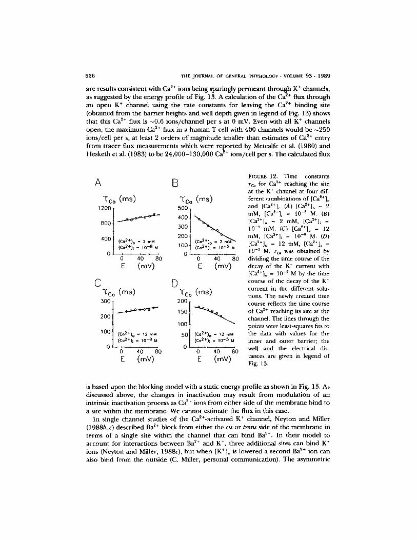
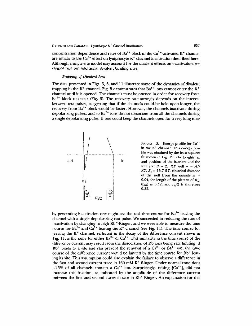
Raising either internal or external [Ca 2+ ] accelerates the rate of inactivation. Com- petition between internal and external Ca 2+ ions for the same binding site within the channel may result in the diminished effect o f [Ca2+]o on Zh when [Ca~+]i is raised (Figs. 3 and 8). We modeled the change in rh by assuming that Ca ~+ can block the open channel nearly irreversibly, and that Ca 2+ may reach the blocking site f rom either side of the membrane. Under this assumption one can separate rh due to an intrinsic inactivation process (measured in [Ca~+]o = 10 -9 M and [Ca2+]i = 10 -8 M) f rom rc~ due to Ca ~+ reaching the site. These ~'c~ values measured in different [Ca2+]o and different [Ca2+]i were plotted against the absolute membrane potential E as shown in Fig. 12 (open squares). The voltage dependent of rc~ changes f rom a small increase of Zc~ with more depolarization in [Ca2+] i = 10 -8 M, to a marked reduction of rc~ with depolarization in [Ca~+]i = 10 -~ M. The lines through the points are least-squares fits to the data considering that the K + channel has one local free energy minimum (site) that can either be empty or occupied by Ca 2+. Ca 2+ moves f rom either side of the membrane to the site over an intervening energy max- imum (barrier). The data could be reasonably described by a free energy profile shown in Fig. 13, which illustrates a two-barrier one-site model. In this model the binding site for Ca ~+ is almost at the extracellular side of the membrane, perhaps near the channel mouth. The reason for the very broad plateau of the inner barr ier is the weak voltage dependence ofzc~ in all the solutions with different Ca 2+ concen- trations. Alternatively, a second two-barrier one-site model, with the plateau at the outer barrier, is indistinguishable f rom the one shown in Fig. 13. The Ca ~+ binding site would then be about two thirds of the way across the electric field f rom the outside.
The similar effects raising internal and external [Ca 2+ ] on K + channel inactivation (Figs. 3 and 8), and the unblocking of divalent cations f rom K + channels (Fig. 11),
626 T H E J O U R N A L O F GENERAL P H Y S I O L O G Y �9 V O L U M E 93 �9 1 9 8 9
are results cons is ten t with Ca 2+ ions be ing spar ingly p e r m e a n t t h r o u g h K + channels , as sugges ted by the energy prof i le o f Fig. 13. A calcula t ion o f the Ca 2+ flux t h r o u g h an o p e n K + channel us ing the ra te cons tan ts for leaving the Ca 2+ b ind ing site (ob ta ined f rom the b a r r i e r heights and well d e p t h given in l egend o f Fig. 13) shows that this Ca 2+ flux is ~0 .6 i ons / c ha nne l p e r s at 0 mV. Even with all K + channels open , the m a x i m u m Ca 2+ flux in a h u m a n T cell with 400 channels would be ~250 ions /ce l l p e r s, at least 2 o r d e r s o f m a g n i t u d e smal ler than es t imates o f Ca ~+ en t ry f rom t r ace r f lux measu remen t s which were r e p o r t e d by Metcalfe et al. (1980) and Heske th et al. (1983) to be 2 4 , 0 0 0 - 1 3 0 , 0 0 0 Ca ~+ ions /ce l l p e r s. The ca lcu la ted flux
A 13
"~Co (ms) ~'Co (ms) 500 .
1200 t 4oot 3~176 2001
( C o 2 + ) o = 2 m M . I O0 ( C o 2 + ) i = 10 - 8 M 0 / . . . . . 0 1 ,10 ,5 I (Co2+), M
0 40 80 0 40 80 E (mY) E (mY)
C D 'co (ms) "t'co (ms)
200.
,ool 100 I (C~176 = 12 mM 50,t CccC:22:))o = 112
o ! ' .,o.o o o/ 0 40 80 0 40 80 E (mV) E (mV)
FIGURE 12. Time constants Zc~ for Ca 2+ reaching the site at the K + channel at four dif- ferent combinations of [Ca~+]o and [Ca2+]i. (A) [Ca~+]o = 2 raM, [Ca~+]i = 10 -8 M. (B) [Ca2+]o = 2 raM, [Ca2+] i =
10 -5 mM. (C) [Ca~*]o = 12 mM, [Ca~+]i = 10 -8 M. (D) [Ca~+]o = 12 raM, [Ca2§ i = 10 -~ M. rc~ was obtained by dividing the time course of the decay of the K + current with [Ca2+]o = 10 -9 M by the time course of the decay of the K + current in the different solu- tions. The newly created time course reflects the time course of Ca 2+ reaching its site at the channel. The lines through the points were least-squares fits to the data with values for the inner and outer barrier; the well and the electrical dis- tances are given in legend of Fig. 13.
is based u p o n the b locking mode l with a static energy prof i le as shown in Fig. 13. As discussed above, the changes in inact ivat ion may resul t f rom m o d u l a t i o n o f an intr insic inact ivat ion process as Ca 2+ ions f rom e i ther side o f the m e m b r a n e b ind to a site within the m e m b r a n e . We canno t es t imate the flux in this case.
In single channe l s tudies o f the Ca2+-activated K + channel , Ney ton a n d Mil ler (1988b, c) desc r ibed Ba 2+ b lock f rom e i ther the c/s o r trans side o f the m e m b r a n e in te rms o f a single site within the channe l that can b ind Ba 2+. In the i r mode l to accoun t for in terac t ions be tween Ba 2+ and K +, th ree addi t iona l sites can b ind K + ions (Neyton and Miller, 1988c), b u t when [K+]o is lowered a second Ba 2+ ion can also b ind f rom the ou ts ide (C. Miller, pe rsona l communica t ion) . The asymmetr ic
GRXSSMEIt AND ~ Lymphocyte K + Channel Inactivation 627
concent ra t ion dependence and rates o f Ba 2+ block in the Ca2+-activated K + channel are similar to the Ca ~+ effect on lymphocyte K § channel inactivation described here. Al though a single-site model may account for the divalent effects on inactivation, we cannot rule out additional divalent binding sites.
Trapping o f Divalent Ions
The data presented in Figs. 5, 6, and 11 illustrate some o f the dynamics o f divalent t rapping in the K + channel. Fig. 5 demonst ra tes that Ba 2+ ions cannot enter the K + channel until it is opened. The channels must be opened in o rde r for recovery f rom Ba 2+ block to occur (Fig. 6). The recovery rate strongly depends on the interval between test pulses, suggesting that if the channels could be held o p e n longer, the recovery f rom Ba 2+ block would be faster. However , the channels inactivate dur ing depolar izing pulses, and so Ba 2+ ions do not dissociate f rom all the channels dur ing a single depolar izing pulse. I f one could keep the channels open for a very long time
out
Sl
in
FIGURE 13. Energy profile for Ca 2§ in the K § channel. This energy pro- file was obtained by the least-squares fit shown in Fig. 12. The heights, B, and position of the barriers and the well are: Bi = 21 RT, well = -14 .7 RT, Bo ~ 15.7 RT, electrical distance of the well from the outside s~ 0.04, the length of the plateau of Bo~t (PB2) is 0.52, and s2/2 is therefore 0.22.
by prevent ing inactivation one might see the real time course for Ba 2+ leaving the channel with a single depolar izing test pulse. We succeeded in reduc ing the rate o f inactivation by changing to high Rb+-Ringer, and we were able to measure the time course for Ba ~+ and Ca ~+ leaving the K + channel (see Fig. 11). The time course for leaving the K + channel, reflected in the decay o f the difference cur ren t shown in Fig. 11, is the same for ei ther Ba ~+ or Ca 2+. This similarity in the time course o f the difference cur ren t may result f rom the dissociation o f Rb ions being rate limiting; if Rb + binds to a site and can prevent the removal o f a Ca ~+ or Ba ~+ ion, the time course o f the difference cur ren t would be limited by the time course for Rb + leav- ing its site. This assumption could also explain the failure to observe a difference in the first and second cur ren t trace in 160 mM K + Ringer. Unde r normal condit ions - 2 5 % o f all channels contain a Ca 2+ ion. Surprisingly, raising [Ca~+]o did no t increase this fraction, as indicated by the ampli tude o f the difference cur ren t between the first and second cur ren t trace in Rb+-Ringer. An explanat ion for this
628 T H E J O U R N A L O F GENERAL P H Y S I O L O G Y �9 V O L U M E 93 �9 1989
behav ior cou ld be that Ca 2+ is able to leave its b ind ing site even i f the channe l is closed. The la t te r possibil i ty is s u p p o r t e d by the expe r imen t s with an increase in [Ca2+]i to 10 -5 M, which accelera tes inact ivat ion and increases the d i f fe rence cur- ren t to >50%.
Several l ines o f ev idence are p r e s e n t e d in this p a p e r which indicate that Ca ~+ and Ba 2+ are able to e n t e r o p e n K + channels f rom the outs ide . I f the channel closes, Ca ~+ a n d Ba 2+ b e c o m e t r a p p e d inside the channel . Ba l+ remains t r a p p e d , whereas Ca ~+ is though t to cross the m e m b r a n e to the inside d u r i n g the interval be tw e e n test pulses. Ba 2+ t r a p p i n g has been desc r ibed previously for the high conduc t ance Ca z+- ac t iva ted K + channel f rom rabb i t o r ra t muscle i n c o r p o r a t e d into p l ana r l ipid bi layers (Vergara and La to r r e , 1983; Miller, 1987; Mil ler et al., 1987; Ney ton and Miller, 1988a). The exis tence o f a Ba~+-trapping mechanism, as well as the previ- ously desc r ibed sensitivity to cha rybdo tox in o f the Ca~§ " m a x i " - K + chan- nel and the lymphocyte K + channel (Miller et al., 1985; Sands et al., 1988), implies s t ruc tura l similarit ies be tween these two channel types and raises the ques t ion o f whe the r the K + channel in T lymphocytes might be Ca 2+ activated. O u r expe r imen t s with high in terna l Ca 2+ o f u p to 2 mM show no a p p a r e n t act ivat ion o f this channel ; instead, raising [Ca2+]i speeds up inactivat ion. O n e conc lus ion f rom o u r exper i - men t s o n Ba 2+ t r a p p i n g is that d ivalent ions in the in ternal o r ex te rna l so lu t ion must wait for the channel to o p e n be fo re en t e r ing the pore . I f the channe l closes, Ba 2+ is t r a p p e d inside the channe l and remains t r a p p e d even in the absence o f ex t race l lu la r and in t race l lu lar Ba ~+. F r o m the s idedness o f the Ba 2+ act ion, one has to conc lude that bo th the Ca~+-activated K + channel and the vol tage-ga ted K + channel in T lym- phocytes have a ga t ing mechan i sm at the ex te rna l side that can shield the inside o f the channe l f rom externa l ly app l i ed divalent ions, as well as a ga t ing mechan i sm at the cytoplasmic side o f the m e m b r a n e that shields the inside o f the channe l f rom in terna l b lockers (cf. Arms t rong , 1971).
The work was supported by National Institutes of Health grants NS-14609 and GM-14514, by a grant from the Office of Naval Research, and by a Research Fellowship to S. Grissmer from the Deutsche Forschungsgemeinschaft (Gr 848/2-I and Gr 848/2-2).
Original version received 16 May 1988 and accepted version received 19 October 1988.
R E F E R E N C E S
Arhem, P. 1980. Effects of rubidium, caesium, strontium, barium and lanthanum on ionic currents in myelinated nerve fibres of Xenopus laevis. Acta Physiologica Scandinavica. 108:7-16.
Armstrong, C. M., and J. Lopez-Barueo. 1987. External calcium ions are required for potassium channel gating in squid neurons. Sc/ence. 236:712-714.
Armstrong, C. M., and D. R. Matteson. 1986. The role of calcium ions in the closing of K channels. Journal of General Physiology. 87:817-832.
Armstrong, C. M., R. P. Swenson, and S. R. Taylor. 1982. Block of squid axon K channels by internally and externally applied barium ions.Journal ofC, eneral Physiology. 80:663-682.
Armstrong, C. M., and S. R. Taylor. 1971. Interaction of tetraethylammonium ion derivatives with the potassium channels of giant axons. Journal of General Physiology. 58:413-437.
Armstrong, C. M., and S. R. Taylor. 1980. Interaction of barium ions with potassium channels in squid giant axons. Biophysical Journal. 30:473-488.
GRISSMER AND CAHALAN Lymphocyte K + Channel Inactivation 629
Bregestovski, P., A. Redkozubov, and A. Alexeev. 1986. Elevation of intracellular calcium reduces voltage-dependent potassium conductance in human T cells. Nature. 319:776-778.
Cahalan, M. D., K. G. Chandy, T. E. DeCoursey, and S. Gupta. 1985. A voltage-gated potassium channel in human T lymphocytes. Journal of Physiology. 358:197-237.
Cahalan, M. D., and P. A. Pappone. 1983. Chemical modification of potassium channel gating in frog myelinated nerve by trinitrobenzene sulphonic acid. Journal of Physiology. 342:119-143.
Choquet, D., P. Sarthou, D. Primi, P. A. Cazenave, and H. Korn. 1987. Cyclic AMP-modulated potassium channels in murine B cells and their precursors. Sc/ence. 235:1211-1214.
Cota, G., and C. M. Armstrong. 1988. Potassium channel "inactivation" induced by soft-glass pi-
pettes. Biophysical Journal. 53:107-109. DeCoursey, T. E., K. G. Chandy, S. Gupta, and M. D. Cahalan. 1984. Voltage-gated K § channels in
human T lymphocytes: a role in mitogenesis? Nature. 307:465-468. DeCoursey, T. E., K. G. Chandy, S. Gupta, and M. D. Cahalan. 1985. Voltage-dependent ion chan-
nels in T lymphocytes. Journal of Neuroimmunology. 10:71-95. Eaton, D. C., and M. S. Brodwick. 1980. Effects of barium on the potassium conductance of squid
axon. Journal of General Physiology. 75:727-750. Eckert, R., andJ . E. Chad. 1984. Inactivation of Ca channels. Progress in Biophysics and Molecular
Biology. 44:215-267. Furman, R. E., and J. c . Tanaka. 1988. Patch electrode glass composition affects ion channel cur-
rents. Biophysical Journal. 53:287-292. Gilly, W. F., and C. M. Armstrong. 1982. Divalent cations and the kinetics of potassium channels in
squid giant axons. Journal of General Physiology. 79:965-996. Grissmer, S. 1984. Effect of various cations and anions on the action of tetrodotoxin and saxitoxin
on frog myelinated nerve fibres. PfliJgers Archly. 402:353-359. Grissmer, S., and M. D. Cahalan. 1987. Calcium and potassium ions alter the rate of potassium
channel inactivation in T lymphocytes. Biophysical Journal. 51:365a. (Abstr.) Grissmer, S., and M. D. Cahalan. 1988. Ba ~+ and Ca ~+ trapped inside the K § channels of human T
lymphocytes. Biophysical Journal. 53:260a. (Abstr.) Grissmer, S., and M. D. Cahalan. 1989. Open K § channels of human T lymphocytes that are
blocked by TEA + cannot inactive. Biophysical Journal. 55:203-206. Hamill, O. P., A. Mart),, E. Neher, B. Sakmann, and F. J. Sigworth. 1981. Improved patch-clamp
techniques for high-resolution current recording from cells and cell-free membrane patches.
Pfliigers Archiv. 391:85-100. Hesketh, T. R., G. A. Smith, J. P. Moore, M. V. Taylor, andJ. C. Metcalfe. 1983. Free cytoplasmic
calcium concentration and the mitogenic stimulation of lymphocytes. Journal of Biological Chem- istry. 258:4876-4882.
Hille, B. 1984. Ionic Channels of Excitable Membranes. Sinauer Associates Inc., MA. 426 pp. Marchais, D., and A. Marty. 1979. Interaction of permeant ions with channels activated by acetyl-
choline in Aplysia neurones. Journal of Physiology. 297:9-45. Marty, A., and E. Neher. 1983. Tight-seal whole-cell recording. In Single Channel Recording. B.
Sakmann and E. Neher, editors. Plenum Publishing Corp., New York. 107-122.
Matsuda, H., A. Saigusa, and H. Irisawa. 1987. Ohmic conductance through inwardly rectifying K channel and blocking by internal Mg e+. Nature. 325:156-159.
Matteson, D. R., and C. Deutsch. 1984. K channels in T lymphocytes: a patch clamp study using monoctonal antibody adhesion. Nature. 307:468-471.
Matteson, D. R., and R. P. Swenson, Jr. 1986. External monovalent cations that impede the closing of K channels. Journal of General Physiology. 87:795-816.
Metcalfe, J. C., T. Pozzan, G. A. Smith, and T. R. Hesketh. 1980. A calcium hypothesis for the control of cell growth. Biochemical Society Symposia. 45:1-26.
650 THE JOURNAL OF GENERAL PHYSIOLOGY �9 VOLUME 93 �9 1989
Miller, C. 1987. Trapping single ions inside single ion channels. Biophysical Journal. 52:123-126. Miller, C., R. Latorre, and I. Reisin. 1987. Coupling of voltage-dependent gating and Ba 2+ block in
the high-conductance, Ca2+-activated K + channel. Journal of General Physiology. 90:427-449. Miller, C., E. Moczydlowski, R. Latorre, and M. Phillips. 1985. Charybdotoxin, a protein inhibitor
of single Ca2+-activated K + channels from mammalian skeletal muscle. Nature. 313:316-318.
Neyton, J., and C. Miller. 1988a. External permeant cations lock Ba ~+ ions into the large Ca 2+-
activated K + channel. Biophysical Journal. 53:259a. (Abstr.) Neyton, J., and C. Miller. 1988b. Potassium blocks barium permeation through a calcium-activated
potassium channel. Journal of General Physiology. 92:548-567.
Neyton, J., and C. Miller. 1988c. Discrete Ba ~+ block as a probe of ion occupancy and pore struc- ture in the high-conductance Ca2+-activated K + channel. Journal of General Physiology. 92:569-
586.
Portzehl, H., P. C. Caldwell, and J. C. Ruegg. 1964. The dependence of contraction and relaxation of muscle fibers from the crab Maia squinado on the internal concentration of free calcium ions.
Biochimica et Biophysica Acta. 79:581-591.
Sands, S. B., R. S. Lewis, and M. D. Cahalan. 1988. Charybdotoxin blocks voltage-gated K + chan-
nels in T lymphocytes. Biophysical Journal. 53:260a. (Abstr.)
Sperelakis, N., M. Schneider, and E.J . Harris. 1967. Decreased K conductance produced by Ba ++
in frog sartorius fibers. Journal of General Physiology. 50:1565-1583. Swenson, R. P., and C. M. Armstrong. 1981. K + channels close more slowly in the presence of
external K + and Rb +. Nature. 291:427-429.
Vandenberg, C. A. 1987. Inward rectification of heart potassium channel (Ki) depends on internal
magnesium. Biophysical Journal. 51:66a. (Abstr.) Vergara, C., and R. Latorre. 1983. Kinetics of Ca~+-activated K + channels from rabbit muscle
incorporated into planar bilayers. Evidence for a Ca ~+ and Ba ~+ blockade. Journal of General Physiology. 82:543-568.
Werman, R., and H. Grundfest. 1961. Graded and all-or-none electrogenesis in the ar thropod
muscle. II. The effects of alkali-earth and onium ions on lobster muscle fibers. Journal of C, en~al Physiology. 44:997-1027.
Woll, K. H. 1982. The effects of internal barium on the K current of the node of Ranvier. Pfl~gers Archly. 393:318-321.