diversification of a protein kinase cascade: ime-2 is involved in
TRANSCRIPT

INVESTIGATION
Diversification of a Protein Kinase Cascade: IME-2Is Involved in Nonself Recognition and Programmed
Cell Death in Neurospora crassaElizabeth A. Hutchison,1 Joanna A. Bueche, and N. Louise Glass2
Department of Plant and Microbial Biology, University of California, Berkeley, California 94720
ABSTRACT Kinase cascades and the modification of proteins by phosphorylation are major mechanisms for cell signaling andcommunication, and evolution of these signaling pathways can contribute to new developmental or environmental response pathways.The Saccharomyces cerevisiae kinase Ime2 has been well characterized for its role in meiosis. However, recent studies have revealedalternative functions for Ime2 in both S. cerevisiae and other fungi. In the filamentous fungus Neurospora crassa, the IME2 homolog(ime-2) is not required for meiosis. Here we determine that ime-2 interacts genetically with a transcription factor vib-1 during nonselfrecognition and programmed cell death (PCD). Mutations in vib-1 (Dvib-1) suppress PCD due to nonself recognition events; however,a Dvib-1 Dime-2 mutant restored wild-type levels of cell death. A role for ime-2 in the post-translational processing and localization ofa mitochondrial matrix protein was identified, which may implicate mitochondria in N. crassa nonself recognition and PCD. Further,Dvib-1 strains do not produce extracellular proteases, but protease secretion reverted to near wild-type levels in a Dvib-1 Dime-2 strain.Mass spectrometry analysis revealed that the VIB-1 protein is phosphorylated at several sites, including a site that matches the IME-2consensus. The genetic and biochemical data for ime-2 and vib-1 indicate that IME-2 is a negative regulator of VIB-1 and suggestparallel negative regulation by IME-2 of a cell death pathway in N. crassa that functions in concert with the VIB-1 cell death pathway.Thus, IME2 kinase function has evolved following the divergence of S. cerevisiae and N. crassa and provides insight into the evolutionof kinases and their regulatory targets.
CELL–CELL signaling via kinase cascades is an essentialmechanism for communication within and between or-
ganisms. Protein kinases are one of the largest protein familiesin eukaryotes and as much as 30% of any given eukaryoticproteome is phosphorylated (Deshmukh et al. 2010; Mosesand Landry 2010). Although kinases have constrained targetspecificities, these proteins are often structured in a modularway, such that they can evolve new functions via interactionwith scaffolds, adapters, or docking proteins (Bhattacharyyaet al. 2006). In addition, duplication of kinase targets can
result in reciprocal loss of phosphorylation sites and sub-functionalization of these targets and/or a gain of new phos-phorylation sites, resulting in neo-functionalization (Amoutziaset al. 2010). Thus, changes in kinase structure, along withtarget duplication and divergence, can affect the structureand signaling output of kinase pathways. Although the majorclasses of kinases are conserved across fungal species, thereis evidence for duplication, family expansion, and differencesin domain organization, suggesting that fungi can changetheir kinase signaling pathways to accommodate changes inenvironment or developmental processes (Kosti et al. 2010).
In Saccharomyces cerevisiae, Ime2 is a serine/threonineprotein kinase involved in the induction of meiosis and spor-ulation (Smith and Mitchell 1989). Ime2 has both early andlate roles in meiosis, including the initiation of meiosis,meiotic DNA replication, meiotic divisions I and II, and sporeformation (Benjamin et al. 2003; Honigberg 2004; Brush et al.2012). Nutritional signals for meiosis converge at Ime1, a tran-scriptional regulator of Ime2, as well as at Ime2 itself, tocoordinate meiotic initiation (Honigberg and Purnapatre
Copyright © 2012 by the Genetics Society of Americadoi: 10.1534/genetics.112.142612Manuscript received June 7, 2012; accepted for publication July 12, 2012Available freely online through the author-supported open access option.Supporting information is available online at http://www.genetics.org/content/early/2012/07/16/genetics.112.142612/suppl/DC1.Microarray data from this article have been deposited at the GEO database at NCBIas series GSE35905.1Present address: Cornell University Microbiology Department, 156 Wing Hall,Ithaca, NY 14853.
2Corresponding author: Department of Plant and Microbial Biology, 111 Koshland Hall,University of California, Berkeley, CA 94720. E-mail: [email protected]
Genetics, Vol. 192, 467–482 October 2012 467

2003; Kassir et al. 2003). One of the major roles of Ime2 isto contribute to activation of the major middle meiotic tran-scription factor NDT80 (Pak and Segall 2002a; Sopko et al.2002; Shin et al. 2010), in part through phosphorylationof the repressor Sum1 (Pak and Segall 2002b; Ahmed et al.2009;Winter 2012). In addition, Ime2 directly phosphorylatesNdt80, which is associated with an increased ability to acti-vate transcription of Ndt80 target genes (Sopko et al. 2002;Shubassi et al. 2003). Ndt80 binds to the middle sporulationelement (MSE) and activates expression of middle meioticgenes (Chu and Herskowitz 1998; Chu et al. 1998); cellslacking Ndt80 arrest at pachytene, prior to nuclear divisionin meiosis I (Xu et al. 1995).
In the filamentous fungus Neurospora crassa, there is oneIME2 homolog (ime-2), but three NDT80 homologs (femalesexual development, fsd-1; vegetative incompatibility blocked,vib-1; and NCU04729) (Borkovich et al. 2004; Hutchisonand Glass 2010). Homologs to IME1 and SUM1 are lackingin the N. crassa genome. Recently, we showed neither ime-2nor the NDT80 homologs fsd-1, vib-1, or NCU04729 are in-volved in meiotic functions in N. crassa (Hutchison andGlass 2010). Mutations in ime-2 did not affect the transcrip-tion or activity of fsd-1, the homolog most closely relatedto S. cerevisiae NDT80. However, ime-2, vib-1, and fsd-1mutants were affected in the production of female reproduc-tive structures, termed protoperithecia. The development ofprotoperithecia in N. crassa is induced under conditions ofnitrogen starvation (Westergaard and Mitchell 1947; Hirsh1954). The Dfsd-1 and Dvib-1 mutants formed few protoper-ithecia under nitrogen starvation and an Dfsd-1 Dvib-1mutantwas female sterile. In contrast, a Dime-2 strain produced pro-toperithecia under conditions where development of thesestructures is normally suppressed (nitrogen sufficiency) andsignificantly more protoperithecia are under nitrogen star-vation conditions (Hutchison and Glass 2010). A deletion ofime-2 restored protoperithecial development in a Dfsd-1mu-tant, while a Dime-2 Dvib-1 mutant showed a Dvib-1 pheno-type (few protoperithecia). These observations indicate anetwork of regulatory interactions between ime-2 and the
NDT80 homologs fsd-1 and vib-1 during development offemale reproductive structures in N. crassa.
The vib-1 mutant was first identified in a search formutations that alleviate heterokaryon incompatibility (HI)in N. crassa (Xiang and Glass 2002). Heterokaryon incom-patibility mediates nonself recognition and is a ubiquitousphenomenon in filamentous fungi (Aanen et al. 2010; Choiet al. 2012). Within a single filamentous fungal colony, in-dividual hyphae can fuse and form interconnected networks(Fleissner et al. 2008; Read et al. 2010). However, if fusionoccurs between strains that contain alternative specificitiesat heterokaryon incompatibility (het) loci, either the fusioncell is walled off and rapidly killed or the growth of theheterokaryon is inhibited (Glass and Kaneko 2003; Aanenet al. 2010) (Figure 1). In N. crassa, there are 11 het loci andgenetic differences at any one of these 11 loci are sufficientto restrict heterokaryon formation (Glass and Dementhon2006). Strains carrying genetic differences at het loci, butalso loss-of-function mutations in vib-1, will form vigorousheterokaryons and do not show HI-associated programmedcell death (PCD) (Xiang and Glass 2002, 2004; Lafontaineand Smith 2012). vib-1 is also necessary for the expressionof several genes known to be involved in cell death due toHI (Figure 1) (Dementhon et al. 2006). In Aspergillus nidu-lans, deletion of a vib-1 homolog, xprG, prevents secretion ofextracellular proteases upon nitrogen or carbon starvation(Katz et al. 2006) and vib-1 mutants exhibit this phenotypeas well.
In this study, we show that in N. crassa, a deletion ofime-2 restores HI-induced PCD in a Dvib-1 strain and alsorestores the production of extracellular proteases. We fur-ther investigated the ime-2 phenotype using transcriptionalprofiling to assess physiological differences between ime-2mutants and a wild-type strain and identified a possible rolefor IME-2 in mitochondrial homeostasis. Our data suggestthat IME-2 is a negative regulator of a cell death pathwaythat functions in parallel to the VIB-1 HI pathway to specifi-cally regulate nonself recognition and cell death when strainscarry incompatible specificities at het loci.
Figure 1 Schematic for VIB-1 regulation of HI and celldeath. VIB-1 is required for HI mediated by genetic differ-ences at mating type, het-6, and het-c pin-c (Xiang andGlass 2002, 2004; Lafontaine and Smith 2012) in additionto activating PCD through additional unknown downstreameffectors (Dementhon et al. 2006). het-6 incompatibility ismediated by un-24 (ribonucleotide reductase) and het-6 inter-actions (Micali and Smith 2006; Lafontaine and Smith 2012);mating-type incompatibility is mediated by mating type A-1,mating type a-1, and tol (Pittenger 1957; Newmeyer1970; Glass et al. 1990; Shiu and Glass 1999); while het-cincompatibility is mediated by het-c pin-c interactions (Glassand Kaneko 2003; Kaneko et al. 2006); alternative HET-Cpolypeptides have been shown to physically interact (Sarkaret al. 2002). Phenotypic consequences of HI in N. crassainclude growth inhibition and suppression of conidiation(Perkins 1988). Hyphal compartments within heterokaryons
that carry alternate het haplotypes (such as het-c/pin-c, rnr/het-6, or mat-a/A/tol) undergo compartmentation and rapid cell death (Glass and Kaneko2003) and thus stain positive for vital dyes such as methylene blue (arrows).
468 E. A. Hutchison, J. A. Bueche, and N. L. Glass
Materials and Methods
Strains and growth conditions
All strains used in this study are listed in Table 1. Deletionstrains (FGSC 11308, FGSC 11309, FGSC 17936, and FGSC17937) were constructed by the Neurospora program projectgrant (Colot et al. 2006) and obtained from the FungalGenetics Stock Center (FGSC) (McCluskey 2003). Strainswere grown on Vogel’s minimal media (MM) (Vogel 1956)unless otherwise specified, and crosses were performed onWestergaard’s media (Westergaard and Mitchell 1947). Trans-formations were performed as previously described (Margolinet al. 1997). To obtain forced heterokaryons, conidial sus-
pensions from strains of complementary auxotrophic markerswere mixed and plated on minimal media. Growth rates weredetermined by growing strains in race tubes. Protoperithecialdevelopment was assessed over a 9-day time period of growthon water agar (Hutchison and Glass 2010).
Protease and cell death assays
For extracellular protease assays, strains were grown (intriplicate) in MM (Vogel 1956) overnight. Protease assayswere performed as described previously (Dementhon et al.2006). Cell death frequency was measured by staining withmethylene blue (Hutchison et al. 2009). Heterokaryons wereinoculated onto MM overlaid with cellophane, grown for
Table 1 List of strains used in this study
Strain name Genotypea Origin or referenceb
FGSC 2489 A FGSCFGSC 11308, FGSC
11309Dvib-1::hph a; Dvib-1::hph A FGSC
FGSC 17936, FGSC17937
Dime-2::hph a; Dime-2::hph A FGSC
FGSC 4564 ad-3B cyh-1 am1 FGSCR15-7 his-3; a Dementhon et al. (2006)C9-15 het-c2 pin-c2 thr-2 A Smith et al. (2000)C9-2 het-c2 pin-c2 thr-2 a Smith et al. (2000)Xa-3 het-c2 pin-c2 arg-5; pan-2 A Xiang and Glass (2002)JH3 het-c2 pin-c2 arg-5; a C9-2 · Xa-3
Dementhon et al. (2006)R14-42 his-3 rid-1 Dsad-1::hph A Gift from P. K. T. Shiu; Rasmussen et al. (2008)KD02-10 his-3; pyr-4; pan-2 a Dementhon et al. (2006)DVI.4 Dime-2::hph; Dvib-1::hph A Hutchison and Glass (2010)D49.10 his-3; DNCU09915::hph; DNCU04729::hph A Hutchison and Glass (2010)KD13-21 his-3; Dvib-1::hph A Dementhon et al. (2006)KD13-51 Dvib-1::hph; pan-2 A Dementhon et al. (2006)KD13-33 Dvib-1::hph; pan-2 a Dementhon et al. (2006)KD13-01 het-c2 pin-c2 thr-2; Dvib-1::hph a Dementhon et al. (2006)KD13-23 his-3; het-c2 pin-c2; Dvib-1::hph; pan-2 A Dementhon et al. (2006)DVI.HIS.40 his-3; Dime-2; Dvib-1 a DVI.4 · KD02-10SV1 his-3::pccg1-vib-1-gfp; pyr-4; Dvib-1; pan-2 A Gift from J. Sun, Glass laboratoryBH13c his-3::pccg1-vib-1-gfp; Dvib-1::hph SV1 · DVI.HIS.40DI.PYR.4 Dime-2::hph pyr-4 a DVI.4 · KD02-10DI.HIS.10 his-3; Dime-2::hph a DVI.4 · KD02-10DI.A.22 Dime-2::hph arg-5 het-c2 pin-c2 a DVI.A.78 · D49.10DVI.PYR.63 Dime-2::hph pyr-4; Dvib-1::hph a DVI.4 · KD02-10DVI.HIS.48 his-3; Dime-2::hph; Dvib-1::hph a DVI.4 · KD02-10DVI.A.101 Dime-2::hph arg-5; Dvib-1::hph a DVI.4 · JH3DV.80 het-c2 pin-c2 arg-5; Dvib-1 A KD02-10 · DVI.4R14-42arg4gfp his-3::pccg1-arg-4-gfp rid-1 Dsad-1::hph A R14-42 [pccg1-arg-4-gfp]Dvib-1arg4gfp his-3::pccg1-arg4-gfp; Dvib-1::hph A KD13-21 [pccg1-arg-4-gfp]Dime2arg4gfp his-3::pccg1-arg4-gfp; Dime-2::hph a DI.HIS.10 [pccg1-arg-4-gfp]2XKOarg4gfp his-3::pccg1-arg4-gfp; Dime-2; Dvib-1::hph DVI.HIS.48 [pccg1-arg-4-gfp]DVI.A.78 Dime-2::hph arg-5; Dvib-1::hph a DVI.4 · JH3D49VI.HIS.1 his-3; Dime-2::hph; DNCU09915::hph; Dvib-1::hph;
DNCU04729::hph aD49.10 · DVI.A.78
1XA his-3::pccg1-vib-1(S60A)-gfp; het-c2 pin-c2 Dvib-1::hph; pan-2 A KD13-23 [pccg1-vib-11 · PMA-gfp]5XA his-3::pccg1-vib-1(S60A, S413A, S537A, S542A, S545A)-gfp;
het-c2 pin-c2 Dvib-1::hph; pan-2 AKD13-23 [pccg1-vib-14 · PMA-gfp]
1XD his-3::pccg1-vib-1(S60D)-gfp; het-c2 pin-c2 Dvib-1::hph; pan-2 A KD13-23 [pccg1-vib-11 · PMD-gfp]5XD his-3::pccg1-vib-1(S60D, S413D, S537D, S542D, S545D)-gfp;
het-c2 pin-c2 Dvib-1::hph; pan-2 AKD13-23 [pccg1-vib-14 · PMD-gfp]
a Strains are of het-c1 pin-c1 genotype unless otherwise indicated.b“x”, strains derived from crosses.
N. crassa ime-2 Regulates Cell Death 469

2–3 days, and stained for 1–2 min with 0.003% methyleneblue. Approximately 20 random images were taken andthe percentage of dead (blue) hyphal compartments wasdetermined.
RNA extraction and quantitative RT-PCR
RNA extraction was performed on mycelia ground in liquidnitrogen or on sections of mycelia grown on cellophane.Mycelia were mixed with 0.3 g of 0.5-mm silica beads and1 mL of TRIzol (Invitrogen, Carlsbad, CA) and disruptedusing a bead beater (Mini-BeadBeater-8; Biospec Products).RNA was extracted according to the manufacturer’s protocolfor TRIzol (Invitrogen). Samples were purified using anRNAeasy kit (QIAGEN, Valencia, CA) and DNAwas removedwith QIAGEN DNase (no. 79254) or Ambion Turbo DNase(no. AM2238). RNA concentration and quality were assessedusing a Nanodrop (Thermo Scientific) and gel electrophore-sis. Quantitative RT-PCR (Q-RT-PCR) was performed using anEXPRESS One-Step SYBR GreenER kit (Invitrogen) accordingto the manufacturer’s protocol, run on an ABI 7300 machine,and analyzed with ABI 7300 system software. Actin mRNAwas used as the endogenous control, and reactions wereperformed in triplicate.
Microarray analysis
Microarray slide production, hybridization, and analysis wereperformed as described in Tian et al. (2007). Neurosporamicroarray slides are available from the FGSC (http://www.fgsc.net/). Approximately 10 mg of DNase-treated RNA wasused as a template for cDNA synthesis (ChipShot IndirectcDNA Synthesis kit; Promega, Madison, WI), and hybrid-izations were performed using ProntoPlus kits (Promega),according to manufacturer instructions. Slides were scannedusing an Axon GenePix 4000B scanner and analyzed usingGenePix Pro 6 software (Molecular Devices). Three indepen-dent hybridizations pooled from three biological replicateswere performed. Data were analyzed using Bayesian analy-sis of gene expression levels (BAGEL) (Townsend and Hartl2002). Microarray data were verified by Q-RT-PCR, usingtemplate RNA from an independent experiment. All micro-array data were deposited at the Filamentous Fungal GeneExpression Database (Zhang and Townsend 2010) and theGene Expression Omnibus (GEO) database (ID GSE35905).Functional category enrichment analysis was carried outthrough the Munich Information Center for Protein Sequen-ces (MIPS) database (http://www.helmholtz-muenchen.de/en/mips/projects/funcat) (Ruepp et al. 2004), which uses ahypergeometric distribution to calculate P-values.
Mitochondrial staining
Mitochondria were visualized using 10 mM MitoTracker RedFM (Invitrogen; no. M22425) (Hickey et al. 2004). Approx-imately 105 conidia were inoculated into a 30-ml flask ofMM and shaken at 30� for �6 hr. MitoTracker Red FM wasadded to 1-ml aliquots of the conidia, followed by shakingat 30� for an additional 15–20 min. Conidia were pelleted by
centrifugation and washed once with MM. Conidia were thenspread on a MM plate and incubated at 30� for 5–10 min.Mitochondria were imaged using a Deltavision Spectris DV4deconvolution microscope (Applied Precision Instruments).A stack of �20 images was taken 0.2 mm apart, deconvolvedusing SVI Huygens, and visualized using Bitplane Imarissoftware.
Protein extraction, immunoprecipitation, andWestern blotting
Protein was extracted from mycelia for immunoprecipitation(IP), using a method adapted from the FGSC Neurosporaprotocol page (http://www.fgsc.net/neurosporaprotocols/Immunoprecipitation%20final.pdf). Briefly, 20–30 g of my-celia was ground in liquid nitrogen and homogenized usinga 6770 Freezer/Mill from the SPEX CertiPrep Group, usingthree cycles of 1 min precool, 1 min run time, and 1 min cooltime, at a speed of 15 cycles per second (CPS). Homogenizedmycelia were added to HEPES IP extraction buffer [50 mMHEPES (pH 7.4), 137 mM NaCl, 10% glycerol] containingcomplete mini-EDTA–free protease inhibitor and PhosSTOPphosphatase inhibitor (Roche) and vortexed to homogeniza-tion. Samples were centrifuged at 3400 rpm for 10 min. Super-natants were concentrated via centrifugation, using Vivaspin15R protein concentrators [10,000 molecular weight cutoff (MWCO); Sartorius Stedium Biotech]. Four millilitersof each sample was immunoprecipitated using Protein GDynabeads (Invitrogen), according to manufacturer’s instruc-tions, with the following exceptions: mouse anti-GFP anti-body (Roche) was incubated with the beads for 1 hr, and thesample was immunoprecipitated for 2 hr at 4�. Protein wasremoved from the beads by boiling for 5 min, and sampleswere run on a 4–15% Criterion Tris-HCl gel (Bio-Rad,Hercules, CA). Protein gels were either subjected to West-ern blot analysis using standard methods or stained withSimplyBlue SafeStain Coomassie G-250 stain (Invitrogen) tovisualize protein, and gel bands of interest were extractedusing a razor blade.
Mass spectrometry
Gel bands of interest from Coomassie-stained gels were cutout and minced into ,1-mm2 pieces. Protein bands wereextracted following a protocol adapted from the Universityof California (Berkeley) QB3 Proteomics/Mass SpectrometryLaboratory (http://qb3.berkeley.edu/pmsl/protocols.htm).Briefly, gel pieces were washed in 500 ml NH4HCO3 for20 min. After discarding the NH4HCO3 wash, 150 ml ofNH4HCO3 (100 mM) and 10 ml of DTT (45 mM) were addedand samples were incubated for 15 min at 50�. After coolingto room temperature, 10 ml of iodoacetamide (100 mM) wasadded and samples were incubated at room temperature for15 min in the dark. The solvent was discarded and gel pieceswere washed in 500 ml of a 1:1 mixture of acetonitrile andNH4HCO3 (100 mM) for 20 min. Then, gel pieces were in-cubated for 10–15 min in 50 ml of acetonitrile and dried ina speedvac. Gel pieces were rehydrated in 10 ml of NH4HCO3
470 E. A. Hutchison, J. A. Bueche, and N. L. Glass

(25 mM) and digested overnight with either trypsin or endo-proteinase C, according to the manufacturer’s instructions.Proteins were extracted twice with a mix of 60% acetonitrileand 0.1% formic acid and once with 100% acetonitrile.Extracted proteins were dried using a speedvac and two-dimensional mass spectrometry analysis was performed atthe QB3 Proteomics/Mass Spectrometry facility.
Results
A deletion of ime-2 restores HI-mediated cell death ina Dvib-1 mutant
In N. crassa, if two individuals are genetically identical at allnonself recognition loci (termed het), they can undergo cellfusion to form a “compatible” heterokaryon that looks iden-tical to a homokaryotic wild-type strain. However, if individ-uals are genetically different at any 1 of 11 het loci, hyphalfusion results in compartmentalization of the fusion celland subsequent death; these individuals are referred to as“incompatible” (Glass and Kaneko 2003) (Figure 1). In ad-dition to cell death, incompatible heterokaryons have a re-duced growth rate and do not conidiate. Strains that containloss-of-function mutations in vib-1 form vigorous hetero-karyons even if they differ in het allelic specificity (Xiangand Glass 2002, 2004) (Figure 2, C and D). Whereas com-patible wild-type heterokaryons conidiate upon contact withplate edges, Dvib-1 heterokaryons of identical or alternatehet-c pin-c haplotype display deregulated conidiation (Figure 2,C and D).
Due to genetic interactions identified between ime-2 andvib-1 with regard to formation of female reproductive struc-tures (Hutchison and Glass 2010), we evaluated whetherIME-2 plays a role in HI by assessing the incompatibilityphenotype of Dime-2 and Dvib-1 Dime-2 mutants comparedto wild-type and Dvib-1 mutants. To evaluate HI, we useda forced heterokaryon approach, using strains with the samehet-c pin-c specificity (het-c1 pin-c1 and thus compatible) vs.using strains containing alternate het-c pin-c haplotypes(het-c1 pin-c1 + het-c2 pin-c2 and thus incompatible) (Kanekoet al. 2006; Hall et al. 2010) (Figure 1 and Table 1). Further,these strains have different, complementary auxotrophicmarkers; when isolates carrying different auxotrophic markersare placed on minimal media, only the heterokaryotic strainis able to grow.
Compatible heterokaryons of identical het-c pin-c haplo-type of wild-type, Dvib-1, Dime-2, or Dvib-1 Dime-2 strainswere phenotypically identical to a homokaryotic wild-typeor mutant strain by itself (Figure 2, A, C, E, and G). Wild-type heterokaryons with incompatible combinations of het-cpin-c haplotypes exhibited a severely decreased growth rate
Figure 2 A deletion of ime-2 partially restores HI in Dvib-1 mutants.Heterokaryons of identical het-c pin-c haplotype (C, compatible) show agrowth and conidiation pattern indistinguishable from that of homokaryoticstrains, including WT (A) (FGSC 4564 + R15-7), Dvib-1 (C) (KD13-21 +KD13-51), Dime-2 (E) (DI.PYR.4 + DI.HIS.10), and Dvib-1 Dime-2 (G) (DVI.HIS.48 + DVI.PYR.63) heterokaryons (Table 1). Strains carrying incompat-ible het-c pin-c haplotypes (Inc, incompatible) in a WT background showgrowth inhibition and suppression of conidiation (B) (FGSC 4564 + C9-15).Heterokaryons of incompatible het-c pin-c haplotypes, but carrying homo-zygous Dvib-1 mutations are similar in phenotype to compatible Dvib-1heterokaryons (D) (KD13-33 + KD13-1). Heterokaryons carrying incom-patible het-c pin-c haplotypes and homozygous Dime-2 mutations show
typical HI (F) (DI.HIS.10 + DI.A.22), while heterokaryons carrying in-compatible het-c pin-c haplotypes and homozygous for Dvib-1 Dime-2mutations show partial restoration of HI (decreased growth rate andsuppression of conidiation) (H) (DVI.PYR.63 + DVI.A.101).
N. crassa ime-2 Regulates Cell Death 471

and lack of conidiation (compare Figure 2B with 2A). Het-erokaryons carrying incompatible het-c pin-c haplotype andhomozygous deletions of ime-2 also showed an identical in-compatible phenotype to wild type (compare Figure 2F with2B), indicating that ime-2 is not required for HI. In contrast,homozygous Dvib-1 heterokaryons of incompatible het-c pin-chaplotype showed a phenotype more similar to compatibleDvib-1 heterokaryons (compare Figure 2D to 2C); a deletionof vib-1 suppresses HI. However, a heterokaryon carrying in-compatible het-c pin-c haplotypes, plus homozygous Dvib-1Dime-2 mutations showed significantly poorer growth andconidiation than a Dvib-1 suppressed incompatible hetero-karyon (compare Figure 2H to 2D). The Dvib-1 Dime-2 het-cpin-c incompatible heterokaryon had a growth rate of 2.80 60.55 cm/day (Figure 2H), compared to a wild-type incom-patible heterokaryon (1.16 6 0.53 cm/day; Figure 2B) anda Dvib-1 incompatible heterokaryon (6.7 6 0.40 cm/day;Figure 2D). In addition, the Dvib-1 Dime-2 het-c pin-c incom-patible heterokaryon produced fewer conidia comparedto a Dvib-1 suppressed incompatible heterokaryon (com-pare Figure 2H to 2D) and exhibited an altered, densegrowth pattern that did not resemble either single mutants(Dvib-1 or Dime-2; Figure 2, C and E) or the Dvib-1 Dime-2compatible strain (Figure 2G). Thus, in a Dvib-1 mutant,a deletion of ime-2 partially restored the inhibited growth
and aconidial phenotype associated with nonself recogni-tion and HI.
In addition to growth inhibition and absence of con-idiation, wild-type heterokaryons carrying incompatiblehet-c pin-c haplotypes show substantial PCD, with �30% ofhyphal segments showing compartmentalization and death,which is assessed by staining with vital dyes such as meth-ylene blue (Figure 1) (Glass and Kaneko 2003). The Dvib-1,Dime-2, and Dvib-1 Dime-2 heterokaryons of identical het-cpin-c haplotype exhibited very little cell death, �2–5%, whichwas similar to that of a wild-type compatible heterokaryon(Figure 3A). Wild-type heterokaryons carrying incompatiblehet-c pin-c haplotypes showed �30% hyphal compartmenta-tion and death (Xiang and Glass 2002). Similar to wild-typeincompatible heterokaryons and consistent with the HI phe-notype (Figure 2F), the Dime-2 heterokaryon carrying in-compatible het-c pin-c haplotypes showed �27% hyphalcompartmentation and death (Figure 3A). As observed pre-viously (Xiang and Glass 2002), Dvib-1 heterokaryons carryingincompatible het-c pin-c haplotypes showed a substantiallyreduced cell death frequency that was similar to that of eitherDvib-1 or wild-type compatible heterokaryons (�2–5%). Incontrast, the Dvib-1 Dime-2 heterokaryon carrying incompat-ible het-c pin-c haplotypes showed a cell death frequency sim-ilar to that of wild-type incompatible heterokaryons (�25%)
Figure 3 A deletion of ime-2 restores wild-type levels ofcell death in Dvib-1 incompatible heterokaryons. (A) Het-erokaryons of compatible and incompatible het-c pin-chaplotype from Figure 2 were plated on minimal mediaoverlaid with cellophane and grown for 1–3 days. The per-centage of dead cell compartments was evaluated usingmicroscopy and methylene blue staining (Xiang and Glass2002; Hutchison et al. 2009). Heterokaryons of incompat-ible het-c pin-c haplotype but carrying homozygous vib-1deletions were suppressed for cell death (Dvib-1), whilethe addition of Dime-2 mutation in these strains restoredwild-type levels of cell death (Dvib-1 Dime-2). (B) Expressionof ime-2, vib-1, and two HET domain genes (pin-c, tol) wasassessed using quantitative RT-PCR. Deletion of vib-1 abol-ishes expression of HET domain genes during vegetativegrowth. Expression of pin-c and tol was not restored in aDvib-1 Dime-2 mutant.
472 E. A. Hutchison, J. A. Bueche, and N. L. Glass

(Figure 3A). These data indicate that loss-of-function muta-tions in ime-2 restored hyphal compartmentation and deathto Dvib-1 heterokaryons carrying incompatible het-c pin-chaplotypes.
Many proteins involved in HI, like PIN-C, contain a con-served protein domain of unknown function, termed HET(PF06985) (Espagne et al. 2002). HET domains are fila-mentous fungal-specific protein domains that can cause anHI-like cell death when overexpressed (Paoletti and Clave2007). Previously, it was shown that vib-1 is necessary forthe expression of HET domain genes pin-c, tol, and het-6,which are required for HI in N. crassa (Dementhon et al.2006) (Figure 1). To test whether restoration of cell deathin Dvib-1 Dime-2 strains correlated with HET domain geneexpression, we performed quantitative RT-PCR for pin-c andtol in wild-type, Dvib-1, Dime-2, and Dvib-1 Dime-2 strains(Figure 3B). As expected, expression of pin-c and tol was notdetected in a Dvib-1 mutant. Although expression of pin-cand tol in Dime-2 strains was not significantly different fromthat in wild type, expression of vib-1 was significantly in-creased, suggesting that IME-2 negatively regulates vib-1expression levels. In the Dvib-1 Dime-2 strain, however, ex-pression of pin-c and tol was similar to expression levelsobserved in the Dvib-1 strain (Figure 3B), indicating thatrestoration of cell death by Dime-2 mutations in a Dvib-1strain carrying incompatible het-c pin-c haplotypes is notdue to induced expression of pin-c.
Microarray analysis reveals that mutations in ime-2affect mitochondrial function
Because we identified a genetic interaction between vib-1and ime-2 during HI, we evaluated what physiological pro-cesses were affected in the Dime-2 mutant by performinggene expression profiling of wild type vs. a Dime-2 strainunder nitrogen starvation conditions. The initiation of femalereproductive structures (protoperithecia) in N. crassa is reg-ulated by the availability of nitrogen (Westergaard andMitchell 1947; Hirsh 1954), and we hypothesized that dif-ferences in gene expression between wild-type and Dime-2deletion strains may be more pronounced under nitrogenstarvation conditions (Hutchison and Glass 2010). The wild-type strain (FGSC 2489) and a Dime-2 strain (FGSC 17937)were grown overnight (�16 hr) in minimal media. The my-celia were then washed and subsequently transferred to a flaskcontaining minimal media without nitrogen and grown foran additional 4 hr (see Materials and Methods). Myceliafrom both strains were harvested for RNA extraction andmicroarray analysis. Three replicate microarrays, includingdye swaps, were performed.
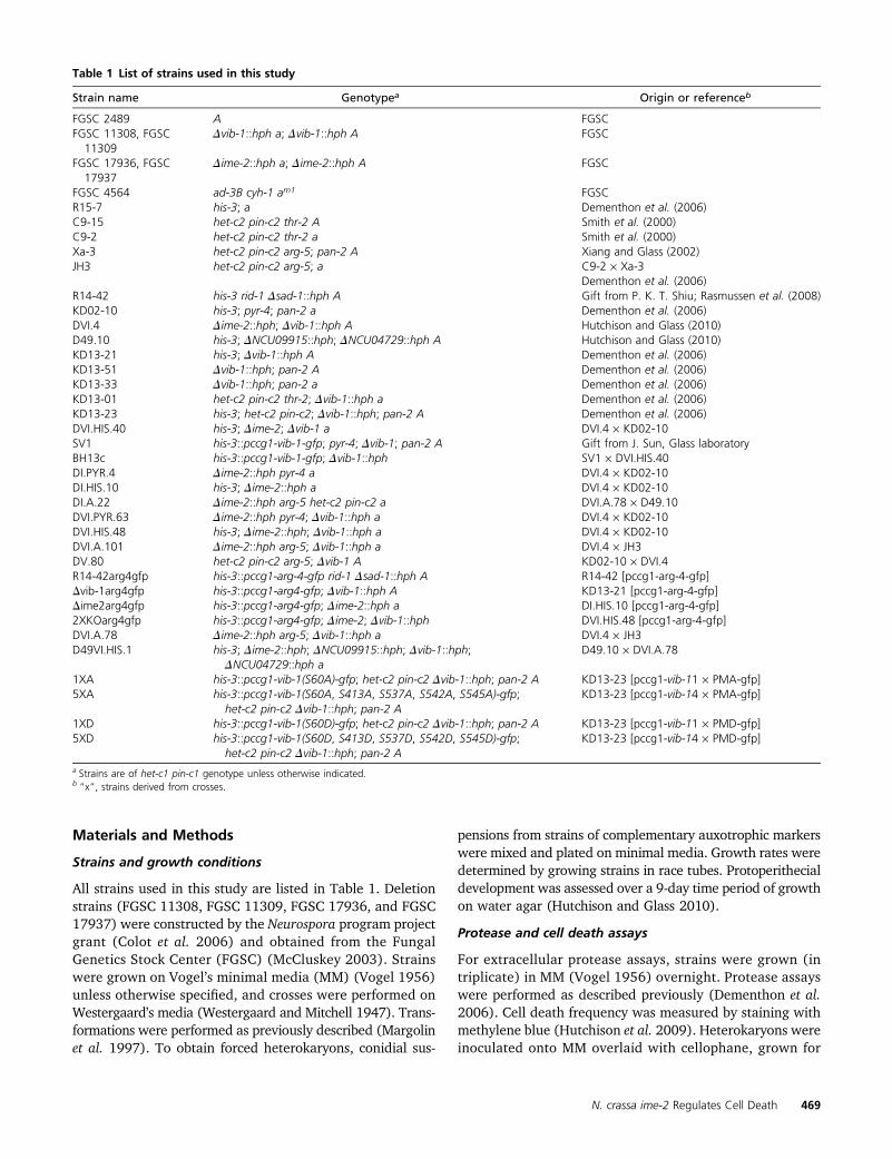
In the Dime-2 strain, a total of 187 genes showed a sig-nificant decrease in expression level of at least 1.5-fold.Functional category analysis (Ruepp et al. 2004) of this geneset showed enrichment for energy (P, 0.0001); transcription(P , 0.05); cellular transport (P , 0.005); and transposableelements, viral and plasmid proteins (P , 1e-11) (Figure 4).Many of the genes in the energy functional category belonged
to pathways involving electron transport and respiration.Furthermore, the enrichment of genes belonging to the “trans-posable elements, viral and plasmid proteins” functional cate-gory was due almost exclusively to genes belonging to themitochondrial genome. In fact, 13 of the 29 genes compos-ing the mitochondrial genome showed decreased expressionlevels in the Dime-2 strain compared to wild type (SupportingInformation, Table S1), a significant enrichment (P, 0.006).These observations suggest mitochondrial impairment in theDime-2 mutant and are consistent with the fact that Dime-2strains exhibit a slower growth rate (3.5–4 cm/day) com-pared to wild-type strains (�7 cm/day).
A total of 506 genes showed increased expression levelsin the Dime-2 strain compared to wild type (Figure 4). Thesegenes were enriched in a variety of functional categories(Ruepp et al. 2004), including metabolism (P , 1e-11);energy (P , 1e-17); cell-cycle and DNA processing (P ,0.0001); transcription (P, 0.005); protein synthesis (P ,1e-24); protein fate (P , 1e-5); protein with binding func-tion or cofactor requirement (P , 1e-44); regulation ofmetabolism and protein function (P , 0.0005); cellulartransport (P , 1e-14); cellular communication and signaltransduction (P , 1e-4); cell rescue, defense, and virulence(P , 0.0005); interaction with the environment (P , 1e-6);cell fate (P , 1e-8); development (P , 0.005); diogenesisof cellular components (P , 1e-12); and cell-type differentia-tion (P , 1e-6). Although we hypothesized that the constitu-tive production of protoperithecia in Dime-2 strains (Hutchisonand Glass 2010) may be due to a defect in nitrogen sensing,we did not observe significant differences in gene expressionwith respect to nitrogen metabolism genes or genes involvedin the metabolism of amino acids. Thus, the array data in-stead suggest that Dime-2 mutants are not deficient in nitro-gen sensing specifically, but that these strains may havea more general nutrient-sensing defect.
Interestingly, genes involved in mitochondrial biogene-sis (within the biogenesis of cellular components category)were also significantly enriched among genes that showed in-creased expression levels in the Dime-2mutant (P, 0.0001).A recent study by Keeping et al. (2010) used mass spectrom-etry as well as computational methods to compile a com-prehensive list of 738 genes that compose the N. crassamitochondrial proteome. Using this data set, we askedwhether nuclear-encoded mitochondrial genes were ex-pressed differently between wild-type and Dime-2 strains.In fact, these genes were significantly enriched (P , 1e-18)in the set of 506 genes that showed increased expressionlevels in the Dime-2 mutant. As previously mentioned, 13 ofthe 29 genes composing the mitochondrial genome showedlower expression levels in the Dime-2 strain (Table S1).These data suggest that the Dime-2 mutant may have im-paired mitochondria (evidenced by decreased expression ofgenes within the mitochondrial genome), resulting in a reg-ulatory feedback loop such that Dime-2 strains increaseexpression of nuclear-encoded mitochondrial genes to com-pensate for this defect. Overall, the microarray data suggest
N. crassa ime-2 Regulates Cell Death 473
that IME-2 plays a role in mitochondrial function in N. crassa.We therefore assessed, via microscopy, whether mitochondrialmorphology or protein localization was altered in Dime-2mutants compared to wild type and the Dvib-1 mutants.
ime-2 mutants affect post-translational processing ofthe mitochondrial protein ARG-4
To evaluate the mitochondrial phenotype in wild-type, Dvib-1,Dime-2, and Dvib-1 Dime-2 strains, we stained conidial germ-lings with MitoTracker Red FM and also transformed eachstrain with the nuclear-encoded GFP-tagged mitochondrialmarker gene encoding ARG-4 (Bowman et al. 2009). Thenuclear arg-4 locus encodes acetylornithine-glutamate ace-tyltransferase (arginine biosynthetic pathway), which isimported into the mitochondrial matrix (Cybis and Davis1975). In wild-type hyphae, mitochondria appear as longtubules in the apical regions of hyphae and as more punctatestructures farther back from the hyphal tip (Bowman et al.2009). When stained with MitoTracker Red FM, all strains(Dvib-1, Dime-2, and Dvib-1 Dime-2) looked identical to wildtype (Figure 5A) and had brightly stained, long, tubularmitochondrial networks. Similarly, when mitochondria werevisualized via ARG-4-GFP localization, wild type and theDvib-1 mutant exhibited long, tubular mitochondria thatlooked identical to the MitoTracker Red FM-stained mito-chondria (Figure 5B). However, Dime-2 strains exhibited analtered localization pattern. Instead of long, tubular struc-
tures, ARG-4-GFP localized either to vesicles or to punctatestructures or was diffuse in the cytoplasm (Figure 5B). Mito-Tracker Red FM can permeate the cell membrane and accu-mulates in mitochondria based on membrane potential(Macho et al. 1996; Poot et al. 1996), and thus it is likelythat the mitochondria observed in all strains, including theDime-2 strains, were active and have a functioning mem-brane potential. However, the lack of ARG-4-GFP localiza-tion in the Dime-2 strain suggested that IME-2 may playa role in protein targeting to the mitochondria. Interestingly,the Dvib-1 Dime-2 strain restored localization of ARG-4-GFPto tubular mitochondria (Figure 5B).
In yeast, the ARG-4 homolog Arg7 undergoes a post-translational autoproteolytic processing step that results inthe formation of two smaller subunits, each of which localizesto the mitochondrial matrix, where they associate in a com-plex (Abadjieva et al. 2000). The autoproteolytic activity ofyeast Arg7 is dependent on a threonine residue, and thisresidue is conserved in N. crassa ARG-4. Therefore, we de-termined whether the defect in ARG-4-GFP localization inDime-2 strains was due to a defect in protein processing.Two distinct bands for N. crassa ARG-4-GFP (�75 kDa and50 kDa) have previously been reported (Bowman et al.2009), which is expected if ARG-4 is proteolytically cleavedat the conserved threonine residue. In the wild-type andDvib-1 strains, two ARG-4-GFP bands at �75 kDa and 50 kDawere detected (Figure 6, A and B). However, in Dime-2 strains
Figure 4 Functional category analysis of gene expressiondifferences in wild type vs. the Dime-2 mutant. Shown isthe distribution of significantly enriched MIPS functionalcategories (http://www.helmholtz-muenchen.de/en/mips/projects/funcat) (Ruepp et al. 2004) for the microarray dataset of wild type compared to a Dime-2 deletion strain. Atotal of 187 genes showed a reduction in expression inDime-2 relative to wild type, while 506 genes showed anincrease in expression level in the Dime-2 strain relative towild type.
474 E. A. Hutchison, J. A. Bueche, and N. L. Glass

much less ARG-4-GFP protein was present and only the 50-kDaARG-4-GFP band was detected. Despite the lack of detectablefull-length ARG-4 in the Dime-2mutants, these mutants werenot arginine auxotrophs. In S. cerevisiae, arg7 mutants ex-hibit a leaky Arg phenotype (Crabeel et al. 1997). Consistentwith microscopy results, the Dvib-1 Dime-2 strain showed awild-type pattern for ARG-4 processing (Figure 6, A and B).In fact, Dvib-1 Dime-2 strains appeared to produce moreARG-4-GFP than wild-type or Dvib-1 deletion strains.
The arg-4-gfp construct used to visualize ARG-4-GFP lo-calization is under the regulation of the ccg-1 promoter(Bowman et al. 2009), which is commonly used for consti-tutive gene expression in N. crassa. The wild-type and theDvib-1, Dime-2, and Dvib-1 Dime-2 strains transformed withthe pccg-arg-4-gfp construct also contain a native copy ofarg-4. Thus, we quantified the transcription of arg-4 in com-parison to ccg-1, using Q-RT-PCR in all strains to assessexpression levels of arg-4 (Figure 6D). Expression levelsfor arg-4 (a readout for both the ccg-1–regulated and theresident arg-4 genes) were very similar between wild typeand Dvib-1 and Dime-2mutants. These data indicate that thedifferences observed in ARG-4 protein levels between wild-type and Dime-2 strains were not due to decreased expres-sion levels of arg-4 in the Dime-2 mutant (Figure 6, A, B,and D). However, the expression level of arg-4 was signifi-cantly elevated in the Dvib-1 Dime-2 strain, consistent withincreased ARG-4 protein levels in this strain detected viaWestern blot (Figure 6B). Wild type and the Dime-2 mutantalso showed similar levels of ccg-1 expression, while theDvib-1and Dvib-1 Dime-2 strains showed slightly lower levels of ccg-1expression (Figure 6D). Because the ccg-1 promoter was notdownregulated in the Dime-2 strain or upregulated in theDvib-1 Dime-2 strain, it is likely that differences in proteins
levels of ARG-4 are not due to the regulation of the arg-4transgene. Either the increased arg-4 transcription in theDvib-1 Dime-2 strain originated from the native arg-4 locusor arg-4 transcripts were stabilized in this strain. Thus, ime-2and vib-1 affect the post-translational modification of ARG-4and, to some degree in the Dvib-1 Dime-2 strain, transcrip-tional regulation of arg-4.
Mutations in ime-2 revert the protease secretionphenotype of vib-1 mutants
Mutations in the transcription factor vib-1 cause a visiblephenotype during vegetative growth, such that Dvib-1mutants show pinkish (rather than orange) conidial pigmen-tation, deregulated conidiation, decreased aerial hyphaeformation (Figures 2C and 7A), and a slight decrease ingrowth rate compared to wild type (Xiang and Glass 2002;Dementhon et al. 2006; Hutchison et al. 2009). In contrast,wild type and the Dime-2 mutant show robust aerial hyphaeformation and conidiation at the top of the tube or edge of theplate and were nearly indistinguishable, with the exception ofthe slightly yellow pigmentation observed in the Dime-2 mu-tant (Figure 7A). In the Dvib-1 Dime-2 mutant, aerial hyphaeformation was restored and conidiation occurred only at thetop of the slants, a phenotype similar to wild type and theDime-2 mutant (Figure 7A). However, conidia of the Dime-2Dvib-1 mutant are pinkish in color, like those of the Dvib-1single mutants.
In addition to the vegetative conidiation phenotype, Dvib-1mutants do not secrete extracellular proteases in response tonitrogen or carbon starvation (Dementhon et al. 2006), a phe-notype similar to the vib-1 homolog in A. nidulans, xprG (Katzet al. 2006). We therefore evaluated whether mutations inime-2 restored protease activity in the Dvib-1 mutant. When
Figure 5 Mutations in ime-2 affect lo-calization of the mitochondrial proteinARG-4. (A) Mitochondria stained withMitoTracker Red FM in FGSC 2489(WT), FGSC 11308 (Dvib-1), FGSC17937 (Dime-2), and DVI.4 (Dvib-1Dime-2) (Table 1). The right column isan enlargement of the region high-lighted by a white box in the center col-umn. (B) Localization of ARG-4-GFP tomitochondria in wild-type and deletionstrains transformed with pccg1-arg-4-gfp (Table 1). Localization of ARG-4-GFPto mitochondria in wild type is identi-cal to mitochondria stained by MitoTrackerRed FM and to that previously reported(Bowman et al. 2009). ARG-4-GFP lo-calization in the Dvib-1 and the Dvib-1Dime-2 mutants was identical to that inWT. However, in the Dime-2 strain, mi-tochondrial tubule structures were notobserved and instead ARG-4-GFP local-ized to either vesicles or punctae or wasdiffuse in the cytoplasm. Bar, 5 mm.
N. crassa ime-2 Regulates Cell Death 475

nitrogen was provided, extracellular proteases were not in-duced in the wild-type, Dvib-1, Dime-2, or Dvib-1 Dime-2strains (Figure 7B). When the wild-type strain was trans-ferred to nitrogen starvation medium, extracellular proteaseactivity was induced, while no activity was detected in theDvib-1 mutant. A strain carrying a deletion of Dime-2 pro-duced slightly elevated levels of proteases in response tonitrogen starvation (Figure 7, B and C). However, unlikethe Dvib-1 mutant, the Dvib-1 Dime-2 mutant showed wild-type protease activity in response to nitrogen starvation(Figure 7, B and C), indicating that loss-of-function muta-tions in ime-2 suppressed the defect in protease secretion inDvib-1 mutants.
Based on the Dvib-1 Dime-2 phenotype, we hypothesizedthat additional regulators may be functioning redundantly tovib-1 to restore protease production. Two obvious candidategenes that may have redundant functions with vib-1 are thevib-1 paralogs fsd-1 and NCU04729. Previously, we deter-mined that strains containing a deletion of NCU04729 wereindistinguishable from wild type under all conditions, whileDfsd-1 mutants shows defects in protoperithecial formationand ascospore maturation (Hutchison and Glass 2010); nei-ther fsd-1 nor NCU04729 affect cell death due to HI. Wetherefore tested the ability of a Dvib-1 Dime-2 Dfsd-1DNCU04729 deletion strain (Table 1, strain D49VI.HIS.1)to produce extracellular proteases. As shown in Figure 7,the quadruple mutant (Dvib-1 Dime-2 Dfsd-1 DNCU04729)produced near wild-type levels of extracellular proteases(Figure 7B), indicating that neither fsd-1 or NCU04729were responsible for the restoration of protease secretionin the Dvib-1 Dime-2 mutant.
VIB-1 is phosphorylated at a predicted IME-2consensus site
In S. cerevisiae, the consensus sequence for Ime2 phosphory-lation (R-P-x-S/T-A/R-G) has been well characterized (Holtet al. 2007; Moore et al. 2007). We analyzed the N. crassagenome for matches to the yeast Ime2 phosphorylation con-sensus sequence, using the Scansite program (http://scansite.mit.edu/) (Obenauer et al. 2003) with a slightly modifiedphosphorylation consensus identified for N. crassa (R-P-x-S/T-P/A/R-G) (L. Holt Laboratory, unpublished data). Thereare 30 total predicted phosphorylation targets of IME-2 inthe N. crassa genome (Table S2). Consistent with the role ofime-2 in protoperithecial formation, one of the predictedIME-2 phosphorylation targets present in the Scansite dataset is NIT-2 (NCU09068), a major regulator of nitrogenutilization. In addition, both AL-1 (albino-1; NCU00552),a phytoene dehydrogenase involved in carotenoid biosyn-thesis (Schmidhauser et al. 1990), and NRC-2 (NCU01797),a serine-threonine kinase involved in regulation of entry intothe conidiation pathway and conidial development (Kothe andFree 1998), were present in the Scansite data set. Dime-2strains appear to have a slightly different conidiation pheno-type from that of wild type (WT), including less pigmentationand fewer conidia (Figures 2E and 7A). The N. crassa homo-log of the yeast protein kinase Ste20 (NCU03894) is alsoa predicted IME-2 phosphorylation target. Ste20 and itshomologs in mammals (Mst1 and Mst2) have been previouslyshown to have a role in the apoptotic signaling cascade(Madeo et al. 2009; Radu and Chernoff 2009). Additionally,the VIB-1 protein contains a match for the Ime2 consensus,while neither of the other two NDT80 homologs in N. crassa(fsd-1 or NCU04729) have an Ime2 consensus site. From aphosphoproteomics study (A. Leeder and N. L. Glass, unpub-lished results), we identified a phosphopeptide for VIB-1 atthe predicted IME-2 consensus site (RPRS*60), as well as fouradditional phosphorylation sites (MPQS*413, PSKS*537, andRHGS*542HGS*545) (Figure 8A).
To test whether the predicted IME-2 consensus site wasnecessary for VIB-1 function, we constructed mutant vib-1alleles such that the IME-2 site was mutated to alanine (pre-dicted to be phospho-null; S60-to-A mutation) (1XA, Table 1)or mutated to aspartate (phospho-mimetic; S60-to-D muta-tion) (1XD, Table 1). The growth rates of strains carrying thevib-1S60A and vib-1S60D alleles were identical to those of wildtype as was nuclear localization of VIB-1S60A-GFP and VIB-1S60D -GFP (Figure S1 and Figure S2). Although identical inphenotype to wild type, both the vib-1S60A-gfp and the vib-1S60D -gfp strains showed slightly lower protease levels, sug-gesting that phosphorylation of the S60 site contributes toVIB-1 activity (Figure 7C). Similarly, mutations of the IME-2consensus sequence in VIB-1 significantly reduced the numbersof protoperithecia produced in vib-1S60A-gfp and vib-1S60D -gfpstrains under conditions of nitrogen starvation (Figure S3).These data indicate that mutations in the Ime2 consensussite on VIB-1 negatively affect protoperithecial development.
Figure 6 Strains carrying a deletion in ime-2 affect the post-transcriptionalregulation of ARG-4. (A) Western blot for ARG-4-GFP in wild-type, Dvib-1,Dime-2, and Dvib-1 Dime-2 strains, with molecular weight ladder (kDa) onthe left. ARG-4-GFP is detected as two distinct bands, �75 kDa and 50 kDain WT, as previously reported (Bowman et al. 2009). (B) Longer exposure ofthe blot from A, more clearly showing the 50-kDa ARG-4-GFP in the Dime-2strain. (C) Western blot of b-tubulin showing that equal amounts of proteinwere loaded in each well. (D) Quantitative RT-PCR of arg-4 and ccg-1transcript levels in wild-type, Dvib-1, Dime-2, and Dvib-1 Dime-2 strains.
476 E. A. Hutchison, J. A. Bueche, and N. L. Glass

To assess the role of the Ime2 phosphorylation sites on HI,the vib-1S60A-gfp and vib-1S60D-gfp alleles were transformedinto Dvib-1 strains of het-c2 pin-c2 haplotype (Table 1). Eachstrain was then forced in a heterokaryon with a Dvib-1 strainof het-c1 pin-c1 haplotype. The Dvib-1 mutation is recessive,
such that a single functional copy of vib-1 in a heterokaryon issufficient to trigger HI (Xiang and Glass 2002; Dementhonet al. 2006) (Figure 8B, compare panels 3 and 4). Whenstrains containing vib-1S60A-gfp or vib-1S60D-gfp were forcedin a heterokaryon with a Dvib-1 strain of incompatible het-c
Figure 7 An ime-2 deletion restores wild-type conidiationpatterns and protease production to a Dvib-1 mutant. (A)Wild-type (FGSC 2489), Dvib-1 (FGSC 11308), Dime-2(FGSC 17936), and Dvib-1 Dime-2 (DVI.4) strains grownon minimal media slants. (B) Extracellular protease activityof wild-type and deletion strains (from A), as well as thatof the Dvib-1 Dime-2 Dfsd-1 DNCU04729 strain (D49VI.HIS.1), was assessed on media with and without nitrogen.Only the vib-1 deletion strain showed inability to secreteproteases. Protease activity units are normalized to WT innitrogen starvation (2nitrogen) media. (C) Extracellularprotease activity of vib-1 phospho-mutants (vib-1S60A
1XA, vib-1S60D 1XD, vib-1S60A;S413A;S537A;S542A;S545A 5XA,and vib-1S60D;S413D;S537D;S542D;S545D 5XD) (Table 1). Dataare shown as the fold increase of protease secretion com-pared to that in the strains grown on media containingnitrogen. Asterisks (*) in B and C indicate strains with pro-tease production significantly different from that in WT(P , 0.05).
N. crassa ime-2 Regulates Cell Death 477

pin-c haplotype, a HI response was triggered that was in-distinguishable from a wild-type HI phenotype (Figure 8B,panels 5 and 6). In addition, the vib-1S60A-gfp and vib-1S60D-
gfp incompatible heterokaryons displayed wild-type levels ofcell death (Figure 8C).
We also constructed strains where all five identified phos-phorylation sites were mutated to alanine (S60A, S413A,S537A, S542A, S545A, and 5XA) or aspartate (S60D, S413D,S537D, S542D, S545D, and 5XD) (Table 1). Both the 5XAand 5XD strains grew significantly slower thanWT (Figure S2)and were also impaired in cell death during HI (Figure 8C).The 5XA and 5XD mutants also showed a significant decreasein protoperithecial formation compared to WT (Figure S2),with the 5XD strain showing the most significant reduction,with values similar to those of the Dvib-1 and Dvib-1D ime-2mutants. Consistent with our data that five amino acid sub-stitutions in VIB-1 negatively affect its function, the 5XA and5XD strains showed less protease activity than WT (P =0.05), with values similar to those of the 1XA and 1XDstrains.
Discussion
Diversification of kinase cascades may provide a mechanismfor eukaryotes to evolve new developmental pathways oradapt to new environments (Bhattacharyya et al. 2006). Inthis study, we showed that N. crassa ime-2 regulates celldeath due to HI in the absence of vib-1 and also regulatespost-translational processing of the mitochondrial matrixprotein ARG-4. IME2 homologs have not been previouslyimplicated in programmed cell death, but recent studies inyeast and other fungi have provided evidence that Ime2 andits homologs can function in cellular processes other thanmeiosis. For instance, Strudwick et al. (2010) described arole for Ime2 in yeast pseudohyphal formation, and studiesin other fungal species showed that although Ime2 homo-logs often function in sexual differentiation or nutrient sens-ing, they are not generally meiotic regulators (Irniger 2011).Our results provide additional evidence that the functionof the Ime2 pathway differs among fungal species and im-plicate N. crassa ime-2 in nonself recognition and pro-grammed cell death.
Data from this study as well as from a previous study(Hutchison and Glass 2010) indicate that ime-2 interactsgenetically with the transcription factor vib-1. For some phe-notypes, such as HI, protease production, and conidiation,ime-2 is epistatic to vib-1. However, for other phenotypes,including protoperithecial formation, HET domain gene ex-pression, and ARG-4 localization and protein processing, vib-1is epistatic to ime-2. These data suggest that the ime-2/vib-1signaling pathway is not a simple, linear interaction, but thatthere are other genetic interactors present depending on thecellular process. We propose that the overall structure of thepathway (with the IME2 homolog functioning upstream ofthe NDT80 homolog) is likely conserved, that IME-2 nega-tively regulates VIB-1 (likely at the protein level), and thatIME-2 regulates a parallel pathway that functions redun-dantly with VIB-1 (Figure 9) to regulate HI and proteaseproduction. In this scenario, protease production and cell
Figure 8 Phosphorylation of VIB-1 and phenotype of phospho-sitemutants. (A) VIB-1 is phosphorylated on multiple sites (determined bymass spectrometry), including a predicted IME-2 consensus site. The as-terisk indicates the phosphorylation site that matches the consensus foryeast Ime2. (B) Mutations of the IME-2 consensus phosphorylation site inVIB-1 do not affect growth inhibition and suppression of conidiationassociated with HI. Dvib-1 compatible (1, KD13-21 + KD13-51) and in-compatible (2, KD13-21 + DV.80) heterokaryons (suppressed for HI) areshown. A heterokaryon with only one functional copy of vib-1 and car-rying incompatible het-c pin-c haplotypes shows typical HI (Dvib-1 muta-tions are recessive) (4, KD13-51 + JH1) compared to a heterokaryoncarrying one functional copy of vib-1, but of compatible het-c pin-c hap-lotype (3, KD13-21 + FGSC 6103). Heterokaryons with a phospho-nullmutation (S60 to A) (5, 1XA + KD13-21) or a phospho-mimetic mutation(S60 to D) (6, 1XD + KD13-21) at the predicted IME-2 consensus site inVIB-1 exhibited a typical wild-type HI phenotype. (C) Mutations of theIME-2 consensus phosphorylation site in VIB-1 do not affect cell-deathpercentages associated with HI. However, mutations at all five VIB-1phosphorylation sites reduce cell-death percentages, although growthinhibition is still observed. All incompatible (Inc) heterokaryons exhibitsignificantly higher cell death than compatible (C) heterokaryons. Celldeath percentages for incompatible heterokaryons labeled with an aster-isk (*) were significantly different (P, 0.05) from those for heterokaryonslabeled with an arrowhead (;).
478 E. A. Hutchison, J. A. Bueche, and N. L. Glass
death in a Dvib-1 Dime-2 strain is restored. Mutation ofcomponents in the parallel cell-death induction pathway inaddition to vib-1 mutations would ameliorate cell death andHI completely, regardless of the presence or absence of ime-2.Further experiments will be needed to identify IME-2 targetsand additional members of this pathway.
Loss-of-function mutations in vib-1 result in an inability tosecrete proteases in response to nitrogen starvation and pres-ence of extracellular protein, a phenotype that is suppressed inDime-2 Dvib-1 mutants. In eukaryotes, including S. cerevisiae,caspase proteases (metacaspases in S. cerevisiae) are integralin the activation of the apoptotic cell-death cascade (Madeoet al. 2004; Tait and Green 2010; Abdelwahid et al. 2011).In N. crassa, metacaspases are not required for HI-inducedcell death (Hutchison et al. 2009). However, it is possiblethat a link between HI-induced cell death and VIB-1/IME-2–regulated proteases occurs in N. crassa by the utilization ofnonmetacaspase proteases to induce cell death.
Mitochondria are key players in apoptosis and cell-deathpathways (Tait and Green 2010). During apoptotic cell death,the pro-apoptotic Bcl-2 proteins BAX and BAK can cause themitochondrial outer membrane to permeabilize, disruptingmitochondrial function, energy production, and redox poten-tial and promoting the release of pro-apoptotic factors suchas cytochrome c (Degterev and Yuan 2008; Tait and Green2010; Abdelwahid et al. 2011). In fungi, mitochondria havealso been implicated in apoptotic cell death and have impor-tant roles in life span and senescence (Madeo et al. 2004;Maheshwari and Navaraj 2008; Sharon et al. 2009). Muta-tions in ime-2 affected localization and post-transcriptionalprocessing of ARG-4, a phenotype that was restored to awild-type pattern in the Dvib-1 Dime-2 mutant. The obser-vation that IME-2 affects post-translational processing ofa mitochondrial matrix protein suggests that the parallelcell-death pathway could be acting through the mitochon-dria or a mitochondria-related pathway. S. cerevisiae IME2and its homolog in Schizosaccharomyces pombe, mde3/pit1,have a role in meiosis (Abe and Shimoda 2000; Kassir et al.2003; Honigberg 2004) as well as in pseudohyphal growth
(Strudwick et al. 2010). Both of these developmental processesare associated with nitrogen starvation, suggesting an addi-tional role for Ime2 in nutrient sensing, similar to that pro-posed for N. crassa (Hutchison and Glass 2010). It will be ofinterest to assess whether ime2D mutants in S. cerevisiae alsohave in common a defect in the localization and processing ofArg7 (ortholog of N. crassa arg-4), as observed in N. crassaDime-2 mutants. Further experiments on the relationship be-tween ime-2/IME2 and mitochondrial function are warranted.
We identified five phosphorylation sites on VIB-1, in-cluding a site that matches the predicted consensus site forIme2. However, mutations in the Ime2 consensus site in VIB-1that are predicted to result in phospho-null mutations (S to A)or activating mutations (S to D) resulted in no to only subtlephenotypic differences from WT. In particular, no role for theIme2 phosphorylation site was observable for HI. Strainscontaining vib-1 alleles containing five S-to-A or five S-to-Dmutations showed more severe defects, particularly reducedvegetative growth and protoperithecial development, as wellas a reduced percentage of cell death during HI. These obser-vations suggest that altering these five sites results in a VIB-1protein that is not fully functional (hypomorphic allele). Ourgenetic and phenotypic data show a genetic interaction be-tween ime-2 and vib-1 during protoperithecial developmentand during HI and protease secretion in response to nitrogenstarvation and for the localization and processing of a mito-chondrial matrix protein. These observations suggest a com-plex regulatory interaction between these two proteins in anumber of cellular functions. Further experiments will un-ravel the interconnection of these two proteins and the sig-naling and transcriptional regulatory pathways they regulatein different cellular contexts.
Acknowledgments
We thank the Glass laboratory for helpful comments anddiscussions. Additionally, we thank Abby Leeder for help withconfocal microscopy and for sharing unpublished results onVIB-1 phosphorylation and Jianping Sun for her expertise
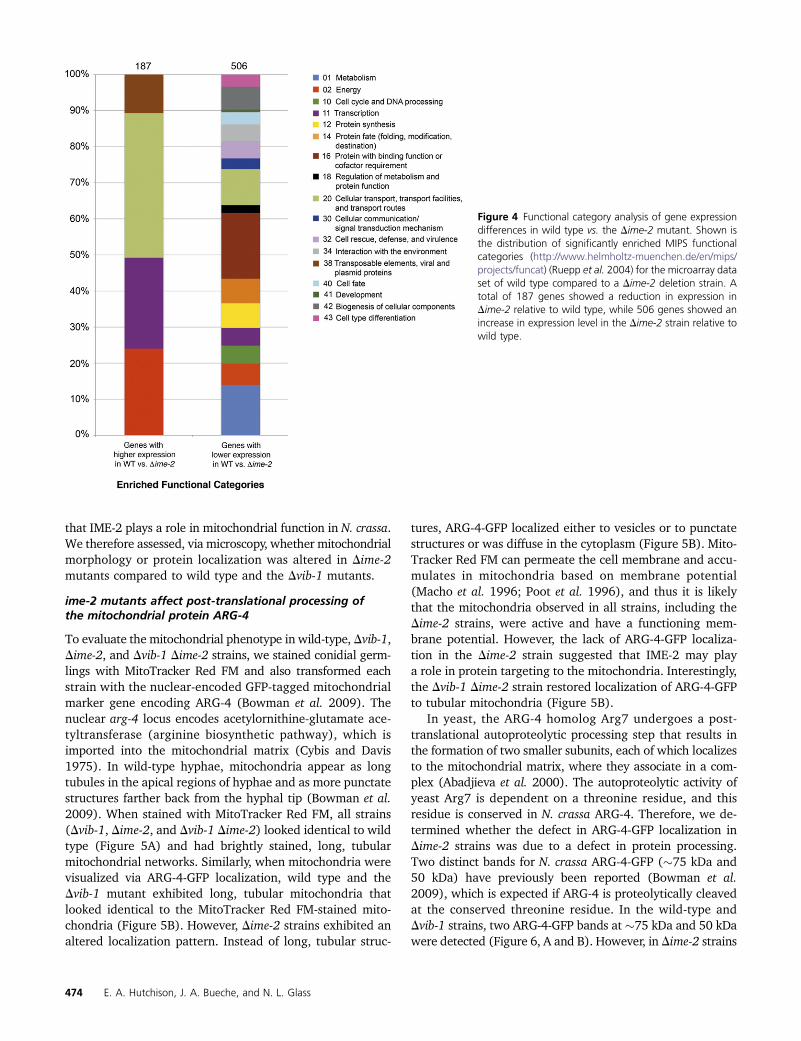
Figure 9 Model for the ime-2 and vib-1 genetic pathway. (A)VIB-1 positively regulates HI and cell death, as well as HETdomain gene expression (via a separate mechanism). IME-2negatively regulates additional HI cell deathmediators that func-tion in parallel to the VIB-1 pathway. Due to the role of IME-2 inthe processing of a mitochondrial protein, we reasoned that thealternate cell-death effectors and/or pathway could be actingthrough the mitochondria. Further, since deletions in ime-2 donot exhibit increased cell death during HI, we suggest that VIB-1negatively regulates these alternate death effectors. (B) VIB-1also positively regulates secreted proteases and, similar to themechanism for HI cell-death regulation, IME-2 negatively regu-lates a parallel pathway for secreted proteases. Deletions in ime-2 cause a significant increase in secreted proteases, and thus inthis pathway, VIB-1 does not regulate the parallel pathway forsecreted proteases. Further, IME-2 appears to negatively regu-late VIB-1 with respect to protoperithecial development viaa separate pathway. For both pathways, it is likely that IME-2is regulated by cellular signals of nitrogen availability.
N. crassa ime-2 Regulates Cell Death 479

with protein and mass spectrometry experiments. We alsothank Lori Kohlstaedt at the University of California (Berkeley)Mass Spectrometry facility. Finally, we thank Liam Holt forhelpful discussions regarding Ime2, yeast meiosis, and phos-phorylation site prediction methods. This work was supportedby a National Institutes of Health (NIH) grant to N.L.G.(GM60468). We acknowledge use of materials generated byNIH grant P01 GM068087, “Functional analysis of a modelfilamentous fungus.”
Literature Cited
Aanen, D. K., A. J. Debets, N. L. Glass, and S. J. Saupe,2010 Biology and genetics of vegetative incompability infungi, pp. 274–288 in Cellular and Molecular Biology of Filamen-tous Fungi, edited by K. A. Borkovich, and D. J. Ebbole. ASMPress, Washington, DC.
Abadjieva, A., P. Hilven, K. Pauwels, and M. Crabeel, 2000 Theyeast ARG7 gene product is autoproteolyzed to two subunitpeptides, yielding active ornithine acetyltransferase. J. Biol.Chem. 275: 11361–11367.
Abdelwahid, E., S. Rolland, X. Teng, B. Conradt, J. M. Hardwicket al., 2011 Mitochondrial involvement in cell death of non-mammalian eukaryotes. Biochim. Biophys. Acta 1813: 597–607.
Abe, H., and C. Shimoda, 2000 Autoregulated expression of Schiz-osaccharomyces pombe meiosis-specific transcription factor Mei4and a genome-wide search for its target genes. Genetics 154:1497–1508.
Ahmed, N. T., D. Bungard, M. E. Shin, M. Moore, and E. Winter,2009 The Ime2 protein kinase enhances the disassociation of theSum1 repressor from middle meiotic promoters. Mol. Cell. Biol.29: 4352–4362.
Amoutzias, G. D., Y. He, J. Gordon, D. Mossialos, S. G. Oliver et al.,2010 Posttranslational regulation impacts the fate of dupli-cated genes. Proc. Natl. Acad. Sci. USA 107: 2967–2971.
Benjamin, K. R., C. Zhang, K. M. Shokat, and I. Herskowitz,2003 Control of landmark events in meiosis by the CDKCdc28 and the meiosis-specific kinase Ime2. Genes Dev. 17:1524–1539.
Bhattacharyya, R. P., A. Remenyi, B. J. Yeh, and W. A. Lim,2006 Domains, motifs, and scaffolds: the role of modular in-teractions in the evolution and wiring of cell signaling circuits.Annu. Rev. Biochem. 75: 655–680.
Borkovich, K. A., L. A. Alex, O. Yarden, M. Freitag, G. E. Turneret al., 2004 Lessons from the genome sequence of Neurosporacrassa: tracing the path from genomic blueprint to multicellularorganism. Microbiol. Mol. Biol. Rev. 68: 1–108.
Bowman, B. J., M. Draskovic, M. Freitag, and E. J. Bowman,2009 Structure and distribution of organelles and cellular lo-cation of calcium transporters in Neurospora crassa. Eukaryot.Cell 8: 1845–1855.
Brush, G. S., N. A. Najor, A. A. Dombkowski, D. Cukovic, and K. E.Sawarynski, 2012 Yeast IME2 functions early in meiosis up-stream of cell cycle-regulated SBF and MBF targets. PLoS ONE7: e31575.
Choi, G. H., A. L. Dawe, A. Churbanov, M. L. Smith, M. G. Milgroomet al., 2012 Molecular characterization of vegetative incom-patibility genes that restrict hypovirus transmission in thechestnut blight fungus Cryphonectria parasitica. Genetics 190:113–127.
Chu, S., and I. Herskowitz, 1998 Gametogenesis in yeast is regu-lated by a transcriptional cascade dependent on Ndt80. Mol.Cell 1: 685–696.
Chu, S., J. DeRisi, M. Eisen, J. Mulholland, D. Botstein et al.,1998 The transcriptional program of sporulation in buddingyeast. Science 282: 699–705.
Colot, H. V., G. Park, G. E. Turner, C. Ringelberg, C. M. Crew et al.,2006 A high-throughput gene knockout procedure for Neuros-pora reveals functions for multiple transcription factors. Proc.Natl. Acad. Sci. USA 103: 10352–10357.
Crabeel, M., A. Abadjieva, P. Hilven, J. Desimpelaere, and O. Soet-ens, 1997 Characterization of the Saccharomyces cerevisiaeARG7 gene encoding ornithine acetyltransferase, an enzymealso endowed with acetylglutamate synthase activity. Eur. J.Biochem. 250: 232–241.
Cybis, J., and R. H. Davis, 1975 Organization and control in thearginine biosynthetic pathway of Neurospora. J. Bacteriol. 123:196–202.
Degterev, A., and J. Yuan, 2008 Expansion and evolution of celldeath programmes. Nat. Rev. Mol. Cell Biol. 9: 378–390.
Dementhon, K., G. Iyer, and N. L. Glass, 2006 VIB-1 is required forexpression of genes necessary for programmed cell death inNeurospora crassa. Eukaryot. Cell 5: 2161–2173.
Deshmukh, K., K. Anamika, and N. Srinivasan, 2010 Evolution ofdomain combinations in protein kinases and its implications forfunctional diversity. Prog. Biophys. Mol. Biol. 102: 1–15.
Espagne, E., P. Balhadere, M. L. Penin, C. Barreau, and B. Turcq,2002 HET-E and HET-D belong to a new subfamily of WD40proteins involved in vegetative incompatibility specificity in thefungus Podospora anserina. Genetics 161: 71–81.
Fleissner, A., A. R. Simonin, and N. L. Glass, 2008 Cell fusion inthe filamentous fungus, Neurospora crassa. Methods Mol. Biol.475: 21–38.
Glass, N. L., and K. Dementhon, 2006 Non-self recognition andprogrammed cell death in filamentous fungi. Curr. Opin. Micro-biol. 9: 553–558.
Glass, N. L., and I. Kaneko, 2003 Fatal attraction: nonself recog-nition and heterokaryon incompatibility in filamentous fungi.Eukaryot. Cell 2: 1–8.
Glass, N. L., J. Grotelueschen, and R. L.Metzenberg, 1990 Neurosporacrassa A mating-type region. Proc. Natl. Acad. Sci. USA 87:4912–4916.
Hall, C., J. Welch, D. J. Kowbel, and N. L. Glass, 2010 Evolutionand diversity of a fungal self/nonself recognition locus. PLoSONE 5: e14055.
Hickey, P. C., S. R. Swift, M. G. Roca, and N. D. Read, 2004 Live-cell imaging of filamentous fungi using vital flourescent dyesand confocal microscopy. Methods Microbiol. 34: 63–87.
Hirsh, H. M., 1954 Environmental factors influencing the differ-entiation of protoperithecia and their relation to tyrosinaseand melanin formation in Neurospora crassa. Physiol. Plant.7: 72–92.
Holt, L. J., J. E. Hutti, L. C. Cantley, andD.O.Morgan, 2007 Evolutionof Ime2 phosphorylation sites on Cdk1 substrates provides amechanism to limit the effects of the phosphatase Cdc14 inmeiosis. Mol. Cell 25: 689–702.
Honigberg, S. M., 2004 Ime2p and Cdc28p: co-pilots driving mei-otic development. J. Cell. Biochem. 92: 1025–1033.
Honigberg, S. M., and K. Purnapatre, 2003 Signal pathway inte-gration in the switch from the mitotic cell cycle to meiosis inyeast. J. Cell Sci. 116: 2137–2147.
Hutchison, E., S. Brown, C. Tian, and N. L. Glass, 2009 Tran-scriptional profiling and functional analysis of heterokaryon in-compatibility in Neurospora crassa reveals that reactive oxygenspecies, but not metacaspases, are associated with programmedcell death. Microbiology 155: 3957–3970.
Hutchison, E. A., and N. L. Glass, 2010 Meiotic regulators Ndt80and Ime2 have different roles in Saccharomyces and Neurospora.Genetics 185: 1271–1282.
480 E. A. Hutchison, J. A. Bueche, and N. L. Glass

Irniger, S., 2011 The Ime2 protein kinase family in fungi: moreduties than just meiosis. Mol. Microbiol. 80: 1–13.
Kaneko, I., K. Dementhon, Q. Xiang, and N. L. Glass, 2006 Nonallelicinteractions between het-c and a polymorphic locus, pin-c, areessential for nonself recognition and programmed cell death inNeurospora crassa. Genetics 172: 1545–1555.
Kassir, Y., N. Adir, E. Boger-Nadjar, N. G. Raviv, I. Rubin-Bejeranoet al., 2003 Transcriptional regulation of meiosis in buddingyeast. Int. Rev. Cytol. 224: 111–171.
Katz, M. E., K. A. Gray, and B. F. Cheetham, 2006 The Aspergillusnidulans xprG (phoG) gene encodes a putative transcriptionalactivator involved in the response to nutrient limitation. FungalGenet. Biol. 43: 190–199.
Keeping, A., D. Deabreu, M. Dibernardo, and R. A. Collins,2010 Gel-based mass spectrometric and computational ap-proaches to the mitochondrial proteome of Neurospora. FungalGenet. Biol. 48: 526–536.
Kosti, I., Y. Mandel-Gutfreund, F. Glaser, and B. A. Horwitz,2010 Comparative analysis of fungal protein kinases and as-sociated domains. BMC Genomics 11: 133.
Kothe, G. O., and S. J. Free, 1998 The isolation and characteriza-tion of nrc-1 and nrc-2, two genes encoding protein kinases thatcontrol growth and development in Neurospora crassa. Genetics149: 117–130.
Lafontaine, D. L., and M. L. Smith, 2012 Diverse interactions me-diate asymmetric incompatibility by the het-6 supergene com-plex in Neurospora crassa. Fungal Genet. Biol. 49: 65–73.
Macho, A., D. Decaudin, M. Castedo, T. Hirsh, S. A. Susin et al.,1996 Chloromethyl-X-Rosamine is an aldehyde-fixable poten-tial-sensitive fluorochrome for the detection of early apoptosis.Cytometry 25: 333–340.
Madeo, F., E. Herker, S. Wissing, H. Jungwirth, T. Eisenberg et al.,2004 Apoptosis in yeast. Curr. Opin. Microbiol. 7: 655–660.
Madeo, F., D. Carmona-Gutierrez, J. Ring, S. Buettner, T. Eisenberget al., 2009 Caspase-dependent and caspase-independent celldeath pathways in yeast. Biochem. Biophys. Res. Commun. 382:227–231.
Maheshwari, R., and A. Navaraj, 2008 Senescence in fungi: theview from Neurospora. FEMS Microbiol. Lett. 280: 135–143.
Margolin, B. S., M. Freitag, and E. U. Selker, 1997 Improvedplasmids for gene targeting at the his-3 locus of Neurosporacrassa by electroporation. Fungal Genet. Newsl. 44: 34–36.
McCluskey, K., 2003 The Fungal Genetics Stock Center: frommolds to molecules. Adv. Appl. Microbiol. 52: 246–262.
Micali, C. O., and M. L. Smith, 2006 A nonself recognition genecomplex in Neurospora crassa. Genetics 173: 1991–2004.
Moore, M., M. E. Shin, A. Bruning, K. Schindler, A. Vershon et al.,2007 Arg-Pro-X-Ser/Thr is a consensus phosphoacceptor se-quence for the meiosis-specific Ime2 protein kinase in Saccharo-myces cerevisiae. Biochemistry 46: 271–278.
Moses, A. M., and C. R. Landry, 2010 Moving from transcriptionalto phospho-evolution: generalizing regulatory evolution?Trends Genet. 26: 462–467.
Newmeyer, D., 1970 A suppressor of the heterokaryon-incompat-ibility associated with mating type in Neurospora crassa. Can. J.Genet. Cytol. 12: 914–926.
Obenauer, J. C., L. C. Cantley, and M. B. Yaffe, 2003 Scansite 2.0:proteome-wide prediction of cell signaling interactions usingshort sequence motifs. Nucleic Acids Res. 31: 3635–3641.
Pak, J., and J. Segall, 2002a Regulation of the premiddle andmiddle phases of expression of the NDT80 gene during sporu-lation of Saccharomyces cerevisiae. Mol. Cell. Biol. 22: 6417–6429.
Pak, J., and J. Segall, 2002b Role of Ndt80, Sum1, and Swe1 astargets of the meiotic recombination checkpoint that control exitfrom pachytene and spore formation in Saccharomyces cerevi-siae. Mol. Cell. Biol. 22: 6430–6440.
Paoletti, M., and C. Clave, 2007 The fungus-specific HET domainmediates programmed cell death in Podospora anserina. Eukar-yot. Cell 6: 2001–2008.
Perkins, D. D., 1988 Main features of vegetative incompatibility inNeurospora crassa. Fungal Genet. Newsl. 35: 44–46.
Pittenger, T. H., 1957 The mating type alleles and heterokaryonformation in Neurospora crassa. Microbiol. Genet. Bull. 15: 21–22.
Poot, M., Y. Z. Zhang, J. A. Kramer, K. S. Wells, L. J. Jones et al.,1996 Analysis of mitochondrial morphology and function withnovel fixable fluorescent stains. J. Histochem. Cytochem. 44:1363–1372.
Read, N. D., A. Fleissner, M. G. Roca, and N. L. Glass,2010 Hyphal fusion, pp. 260–273 in Cellular and MolecularBiology of Filamentous Fungi, edited by K. A. Borkovich, andD. J. Ebbole. ASM Press, Washington, DC.
Radu, M., and J. Chernoff, 2009 The DeMSTification of mamma-lian Ste20 kinases. Curr. Biol. 19: R421–R425.
Ruepp, A., A. Zollner, D. Maier, K. Albermann, J. Hani et al.,2004 The FunCat, a functional annotation scheme for system-atic classification of proteins from whole genomes. Nucleic AcidsRes. 32: 5539–5545.
Sarkar, S., G. Iyer, J. Wu, and N. L. Glass, 2002 Nonself recogni-tion is mediated by HET-C heterocomplex formation during veg-etative incompatibility. EMBO J. 21: 4841–4850.
Schmidhauser, T. J., F. R. Lauter, V. E. Russo, and C. Yanofsky,1990 Cloning, sequence, and photoregulation of al-1, a carot-enoid biosynthetic gene of Neurospora crassa. Mol. Cell. Biol. 10:5064–5070.
Sharon, A., A. Finkelstein, N. Shlezinger, and I. Hatam, 2009 Fungalapoptosis: function, genes and gene function. FEMS Microbiol.Rev. 33: 833–854.
Shin, M. E., A. Skokotas, and E. Winter, 2010 The Cdk1 and Ime2protein kinases trigger exit from meiotic prophase in Saccharo-myces cerevisiae by inhibiting the Sum1 transcriptional repressor.Mol. Cell. Biol. 30: 2996–3003.
Shiu, P. K., and N. L. Glass, 1999 Molecular characterization oftol, a mediator of mating-type-associated vegetative incompati-bility in Neurospora crassa. Genetics 151: 545–555.
Shubassi, G., N. Luca, J. Pak, and J. Segall, 2003 Activity of phos-phoforms and truncated versions of Ndt80, a checkpoint-regulatedsporulation-specific transcription factor of Saccharomyces cerevi-siae. Mol. Genet. Genomics 270: 324–336.
Smith, H. E., and A. P. Mitchell, 1989 A transcriptional cascadegoverns entry into meiosis in Saccharomyces cerevisiae. Mol. Cell.Biol. 9: 2142–2152.
Sopko, R., S. Raithatha, and D. Stuart, 2002 Phosphorylation andmaximal activity of Saccharomyces cerevisiae meiosis-specifictranscription factor Ndt80 is dependent on Ime2. Mol. Cell. Biol.22: 7024–7040.
Strudwick, N., M. Brown, V. M. Parmar, andM. Schroder, 2010 Ime1and Ime2 are required for pseudohyphal growth of Saccharomycescerevisiae on nonfermentable carbon sources. Mol. Cell. Biol. 30:5514–5530.
Tait, S. W., and D. R. Green, 2010 Mitochondria and cell death:outer membrane permeabilization and beyond. Nat. Rev. Mol.Cell Biol. 11: 621–632.
Tian, C., T. Kasuga, M. S. Sachs, and N. L. Glass, 2007 Transcriptionalprofiling of cross pathway control in Neurospora crassa and compar-ative analysis of the Gcn4 and CPC1 regulons. Eukaryot. Cell 6:1018–1029.
Townsend, J. P., and D. L. Hartl, 2002 Bayesian analysis of geneexpression levels: statistical quantification of relative mRNAlevel across multiple strains or treatments. Genome Biol. 3:RESEARCH0071.
Vogel, H. J., 1956 A convenient growth medium for Neurospora.Microbiol. Genet. Bull. 13: 42–46.
N. crassa ime-2 Regulates Cell Death 481

Westergaard, M., and H. K. Mitchell, 1947 A synthetic mediumfavoring sexual reproduction. Am. J. Bot. 34: 573–577.
Winter, E., 2012 The Sum1/Ndt80 transcriptional switch andcommitment to meiosis in Saccharomyces cerevisiae. Microbiol.Mol. Biol. Rev. 76: 1–15.
Xiang, Q., and N. L. Glass, 2002 Identification of vib-1, a locusinvolved in vegetative incompatibility mediated by het-c in Neu-rospora crassa. Genetics 162: 89–101.
Xiang, Q., and N. L. Glass, 2004 The control of mating type het-erokaryon incompatibility by vib-1, a locus involved in het-c
heterokaryon incompatibility in Neurospora crassa. FungalGenet. Biol. 41: 1063.
Xu, L., M. Ajimura, R. Padmore, C. Klein, and N. Kleckner,1995 NDT80, a meiosis-specific gene required for exit frompachytene in Saccharomyces cerevisiae. Mol. Cell. Biol. 15:6572–6581.
Zhang, Z., and J. P. Townsend, 2010 The filamentous fungal geneexpression database (FFGED). Fungal Genet. Biol. 47: 199–204.
Communicating editor: A. P. Mitchell
482 E. A. Hutchison, J. A. Bueche, and N. L. Glass

GENETICSSupporting Information
http://www.genetics.org/content/early/2012/07/16/genetics.112.142612/suppl/DC1
Diversification of a Protein Kinase Cascade: IME-2Is Involved in Nonself Recognition and Programmed
Cell Death in Neurospora crassaElizabeth A. Hutchison, Joanna A. Bueche, and N. Louise Glass
Copyright © 2012 by the Genetics Society of AmericaDOI: 10.1534/genetics.112.142612

E. A. Hutchinson, J. A. Bueche, and N. L. Glass 2 SI
Figure S1 Phenotype of vib-‐1 phosphorylation site mutants. (A) wild type (FGSC 2489), (B) Δvib-‐1 (FGSC 11309), (C) transformant with vib-‐1S60A-‐gfp mutant allele (strain 1XA) and (D) transformant with vib-‐1S60D-‐gfp mutant allele (strain 1XD). Strains expressing VIB-‐1S60A-‐GFP (E) and VIB-‐1S60D-‐GFP (F) exhibit normal nuclear localization (shown in panels at right). Brightfield images of hyphae are shown in corresponding panels to the left.

E. A. Hutchinson, J. A. Bueche, and N. L. Glass 3 SI
Figure S2 Average growth rate (mm/day) of strains grown on minimal media in race tubes. Asterisks (*) indicate strains with growth rates significantly different (p<0.05) than the WT strain. Strains used are listed in table 1 and are as follows: WT (FGSC 2489), Δvib-‐1 (FGSC 11309), Δime-‐2 (FGSC 17937), Δvib-‐1 Δime-‐2 (DVI.4).
*
* *
* *
*

E. A. Hutchinson, J. A. Bueche, and N. L. Glass 4 SI
Figure S3 Formation of N. crassa protoperithecia after 7 days of growth on water agar. Numbers of protoperithecia for each strain are shown as a percentage of wild type. Strains used are listed in table 1 and are as follows: WT (FGSC 2489), Δvib-‐1 (FGSC 11309), Δime-‐2 (FGSC 17937), Δvib-‐1 Δime-‐2 (DVI.4).

E. A. Hutchinson, J. A. Bueche, and N. L. Glass 5 SI
Table S1 Mitochondrial genes
Gene Annotationa Decreased expression levels in Δime-‐
2
NCU_16001 NADH dehydrogenase subunit 2
NCU_16002 conserved hypothetical yes
NCU_16003 cytochrome c oxidase subunit 3 yes
NCU_16004 NADH dehydrogenase subunit 6
NCU_16005 mitochondrial ribosomal protein S5 yes
NCU_16006 NADH dehydrogenase subunit 2
NCU_16007 NADH dehydrogenase subunit 3 yes
NCU_16008 NADH dehydrogenase subunit 4L yes
NCU_16009 hypothetical protein
NCU_16010 laglidadg endonuclease
NCU_16011 hypothetical protein
NCU_16012 NADH dehydrogenase subunit 5
NCU_16013 cytochrome b
NCU_16014 group I intron endonuclease yes
NCU_16015 laglidadg endonuclease yes
NCU_16016 cytochrome c oxidase subunit 1 yes
NCU_16017 hypothecial protein yes
NCU_16018 NADH dehydrogenase subunit 1 yes
NCU_16019 group I intron endonuclease
NCU_16020 NADH dehydrogenase subunit 4 yes
NCU_16021 laglidadg endonuclease
NCU_16022 hypothetical protein
NCU_16023 hypothetical protein
NCU_16024 ATPase subunit 8 yes
NCU_16025 ATPase subunit 6
NCU_16026 laglidadg endonuclease
NCU_16027 ATPase subunit 9 yes
NCU_16028 cytochrome c oxidase subunit 2 aBroad Institute

E. A. Hutchinson, J. A. Bueche, and N. L. Glass 6 SI
Table S2 Potential N. crassa IME-‐2 targets for phosphorylation, predicted by Scansitea, b
NCU # Annotation
NCU00101 PBN-‐1
NCU00258 40S ribosomal protein S7
NCU00269 Histone-‐lysine N-‐methyltransferase, H3 lysine-‐36 specific
NCU00276 DNA polymerase gamma, mitochondrial
NCU00311 Protein VTS1
NCU00552 Phytoene dehydrogenase; albino 1 protein
NCU00675 Protein EFR-‐3
NCU01157 Glutamate-‐cysteine ligase
NCU01206 Histone-‐lysine N-‐methyltransferase, H3 lysine-‐4 specific
NCU01312 Myb-‐like DNA-‐binding protein MYB-‐1
NCU01797 Serine/threonine-‐protein kinase NRC-‐2 (Non-‐repressible conidiation protein 2)
NCU01954 Pre-‐mRNA-‐splicing factor CWC-‐24
NCU02466 SVP1-‐like protein 2
NCU03678 Protein SSH-‐4
NCU03725 Transcription factor VIB-‐1
NCU03819 COPII coat assembly protein SEC-‐16
NCU03894 Serine/threonine-‐protein kinase STE-‐20
NCU04190 Probable exocyst complex component SEC-‐8
NCU04191 Protein RAI-‐1
NCU05589 Protein SIP-‐5
NCU05876 pH-‐response regulator protein PAL-‐A/RIM-‐20
NCU06276 Increased rDNA silencing protein 4
NCU06498 Pre-‐mRNA-‐splicing factor CWC-‐21
NCU07693 Protein STU-‐1
NCU07863 Chromatin modification-‐related protein VID-‐21
NCU08566 Nuclear distribution protein nudE homolog 1 (RO-‐11)
NCU08699 Putative oxidoreductase BLI-‐4, mitochondrial
NCU09068 Nitrogen catabolic enzyme regulatory protein
NCU09730 Kinesin heavy chain
NCU09993 Helicase SWR-‐1 a http://scansite.mit.edu/; b one mismatch was allowed at the final consensus position
GENETICSSupporting Information
http://www.genetics.org/content/early/2012/07/16/genetics.112.142612/suppl/DC1
Diversification of a Protein Kinase Cascade: IME-2Is Involved in Nonself Recognition and Programmed
Cell Death in Neurospora crassaElizabeth A. Hutchison, Joanna A. Bueche, and N. Louise Glass
Copyright © 2012 by the Genetics Society of AmericaDOI: 10.1534/genetics.112.142612

E. A. Hutchinson, J. A. Bueche, and N. L. Glass 2 SI
Figure S1 Phenotype of vib-‐1 phosphorylation site mutants. (A) wild type (FGSC 2489), (B) Δvib-‐1 (FGSC 11309), (C) transformant with vib-‐1S60A-‐gfp mutant allele (strain 1XA) and (D) transformant with vib-‐1S60D-‐gfp mutant allele (strain 1XD). Strains expressing VIB-‐1S60A-‐GFP (E) and VIB-‐1S60D-‐GFP (F) exhibit normal nuclear localization (shown in panels at right). Brightfield images of hyphae are shown in corresponding panels to the left.

E. A. Hutchinson, J. A. Bueche, and N. L. Glass 3 SI
Figure S2 Average growth rate (mm/day) of strains grown on minimal media in race tubes. Asterisks (*) indicate strains with growth rates significantly different (p<0.05) than the WT strain. Strains used are listed in table 1 and are as follows: WT (FGSC 2489), Δvib-‐1 (FGSC 11309), Δime-‐2 (FGSC 17937), Δvib-‐1 Δime-‐2 (DVI.4).
*
* *
* *
*

E. A. Hutchinson, J. A. Bueche, and N. L. Glass 4 SI
Figure S3 Formation of N. crassa protoperithecia after 7 days of growth on water agar. Numbers of protoperithecia for each strain are shown as a percentage of wild type. Strains used are listed in table 1 and are as follows: WT (FGSC 2489), Δvib-‐1 (FGSC 11309), Δime-‐2 (FGSC 17937), Δvib-‐1 Δime-‐2 (DVI.4).
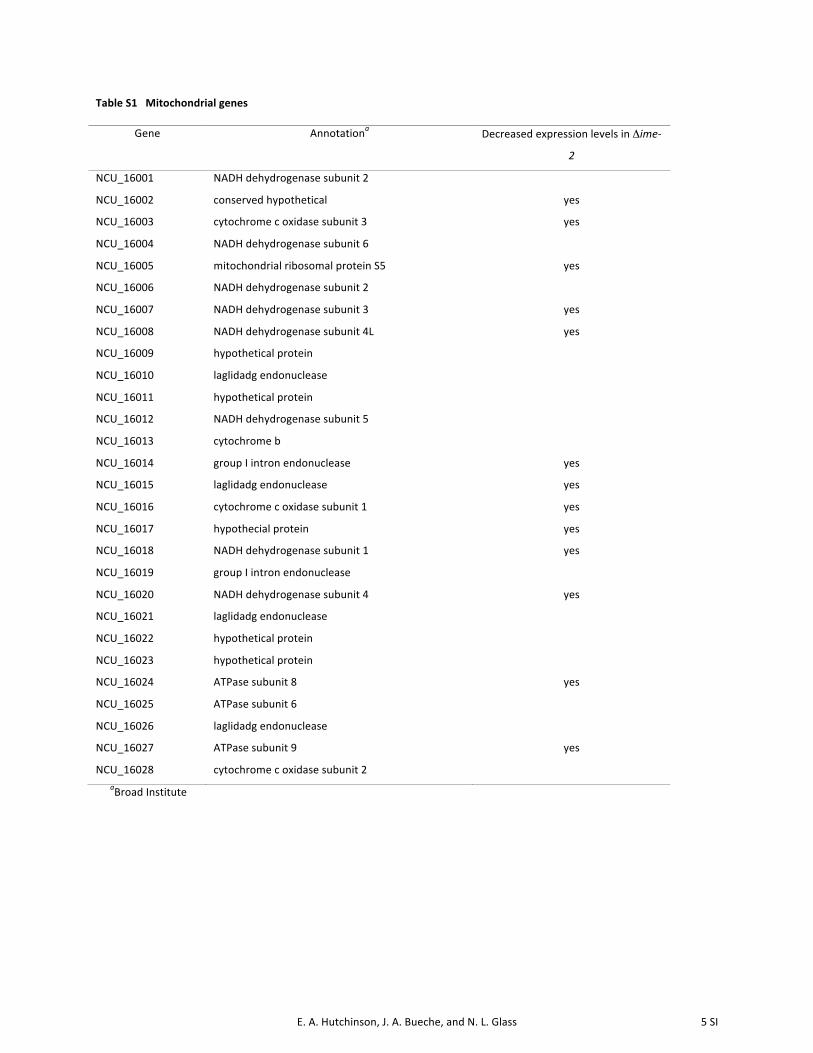
E. A. Hutchinson, J. A. Bueche, and N. L. Glass 5 SI
Table S1 Mitochondrial genes
Gene Annotationa Decreased expression levels in Δime-‐
2
NCU_16001 NADH dehydrogenase subunit 2
NCU_16002 conserved hypothetical yes
NCU_16003 cytochrome c oxidase subunit 3 yes
NCU_16004 NADH dehydrogenase subunit 6
NCU_16005 mitochondrial ribosomal protein S5 yes
NCU_16006 NADH dehydrogenase subunit 2
NCU_16007 NADH dehydrogenase subunit 3 yes
NCU_16008 NADH dehydrogenase subunit 4L yes
NCU_16009 hypothetical protein
NCU_16010 laglidadg endonuclease
NCU_16011 hypothetical protein
NCU_16012 NADH dehydrogenase subunit 5
NCU_16013 cytochrome b
NCU_16014 group I intron endonuclease yes
NCU_16015 laglidadg endonuclease yes
NCU_16016 cytochrome c oxidase subunit 1 yes
NCU_16017 hypothecial protein yes
NCU_16018 NADH dehydrogenase subunit 1 yes
NCU_16019 group I intron endonuclease
NCU_16020 NADH dehydrogenase subunit 4 yes
NCU_16021 laglidadg endonuclease
NCU_16022 hypothetical protein
NCU_16023 hypothetical protein
NCU_16024 ATPase subunit 8 yes
NCU_16025 ATPase subunit 6
NCU_16026 laglidadg endonuclease
NCU_16027 ATPase subunit 9 yes
NCU_16028 cytochrome c oxidase subunit 2 aBroad Institute

E. A. Hutchinson, J. A. Bueche, and N. L. Glass 6 SI
Table S2 Potential N. crassa IME-‐2 targets for phosphorylation, predicted by Scansitea, b
NCU # Annotation
NCU00101 PBN-‐1
NCU00258 40S ribosomal protein S7
NCU00269 Histone-‐lysine N-‐methyltransferase, H3 lysine-‐36 specific
NCU00276 DNA polymerase gamma, mitochondrial
NCU00311 Protein VTS1
NCU00552 Phytoene dehydrogenase; albino 1 protein
NCU00675 Protein EFR-‐3
NCU01157 Glutamate-‐cysteine ligase
NCU01206 Histone-‐lysine N-‐methyltransferase, H3 lysine-‐4 specific
NCU01312 Myb-‐like DNA-‐binding protein MYB-‐1
NCU01797 Serine/threonine-‐protein kinase NRC-‐2 (Non-‐repressible conidiation protein 2)
NCU01954 Pre-‐mRNA-‐splicing factor CWC-‐24
NCU02466 SVP1-‐like protein 2
NCU03678 Protein SSH-‐4
NCU03725 Transcription factor VIB-‐1
NCU03819 COPII coat assembly protein SEC-‐16
NCU03894 Serine/threonine-‐protein kinase STE-‐20
NCU04190 Probable exocyst complex component SEC-‐8
NCU04191 Protein RAI-‐1
NCU05589 Protein SIP-‐5
NCU05876 pH-‐response regulator protein PAL-‐A/RIM-‐20
NCU06276 Increased rDNA silencing protein 4
NCU06498 Pre-‐mRNA-‐splicing factor CWC-‐21
NCU07693 Protein STU-‐1
NCU07863 Chromatin modification-‐related protein VID-‐21
NCU08566 Nuclear distribution protein nudE homolog 1 (RO-‐11)
NCU08699 Putative oxidoreductase BLI-‐4, mitochondrial
NCU09068 Nitrogen catabolic enzyme regulatory protein
NCU09730 Kinesin heavy chain
NCU09993 Helicase SWR-‐1 a http://scansite.mit.edu/; b one mismatch was allowed at the final consensus position