diversity of fusarium species and mycotoxins contaminating pineapple
TRANSCRIPT
MICROBIAL GENETICS • ORIGINAL PAPER
Diversity of Fusarium species and mycotoxinscontaminating pineapple
Łukasz Stępień & Grzegorz Koczyk &
Agnieszka Waśkiewicz
Received: 21 January 2013 /Revised: 11 March 2013 /Accepted: 12 March 2013 /Published online: 10 April 2013# The Author(s) 2013. This article is published with open access at Springerlink.com
Abstract Pineapple (Ananas comosus var. comosus) is animportant perennial crop in tropical and subtropical areas. Itmay be infected by various Fusarium species, contaminat-ing the plant material with mycotoxins. The aim of thisstudy was to evaluate Fusarium species variability amongthe genotypes isolated from pineapple fruits displaying fun-gal infection symptoms and to evaluate their mycotoxigenicabilities. Forty-four isolates of ten Fusarium species wereobtained from pineapple fruit samples: F. ananatum, F.concentricum, F. fujikuroi, F. guttiforme, F. incarnatum, F.oxysporum, F. polyphialidicum, F. proliferatum, F.temperatum and F. verticillioides. Fumonisins B1–B3,beauvericin (BEA) and moniliformin (MON) contents werequantified by high-performance liquid chromatography(HPLC) in pineapple fruit tissue. Fumonisins are likely themost dangerous metabolites present in fruit samples (themaximum FB1 content was 250 μg g−1 in pineapple skinand 20 μg ml−1 in juice fraction). In both fractions, BEA andMON were of minor significance. FUM1 and FUM8 geneswere identified in F. fujikuroi, F. proliferatum, F.temperatum and F. verticillioides. Cyclic peptide synthase
gene (esyn1 homologue) from the BEA biosynthetic path-way was identified in 40 isolates of eight species. Based onthe gene-specific polymerase chain reaction (PCR) assays,none of the isolates tested were found to be able to producetrichothecenes or zearalenone.
Keywords Ananas comosus . FUM genes . Mycotoxins .
Phylogeny . Tropical fruit diseases
Introduction
Pineapple [Ananas comosus (L.) Merr. var. comosus, syn.Ananas ananas (L.) Voss] is a crop native to tropical andsubtropical areas of South America, where it holds a con-siderable economic importance (Ploetz 2001). This perenni-al plant is susceptible to a number of fungal diseases, ofwhich fusariosis is the most severe. The disease affectsalmost all parts of the plant, damaging particularly fruitand stem apices. However, the agent causing the disease isambiguous and other diseases caused by Fusarium fungi(like fruitlet core rot) have also been reported (Ploetz 2006).As a result of the systemic dispersal inside the host plant,many members of the Fusarium genus have the ability ofcolonising perennial crops with only scarce symptoms ofinfection while still being detectable (de Oliveira Rocha etal. 2011; Weber et al. 2006). Moreover, even in the case ofan asymptomatic infection, significant mycotoxin contami-nation of the plant tissues is possible (Desjardins 2006;Stankovic et al. 2007; Thiel et al. 1992; von Bargen et al.2009). Fusarium guttiforme has been frequently associatedwith pineapple fusariosis in Brazil (Ploetz 2006), while F.subglutinans emerged as the main cause associated withfruitlet core rot in Hawaii (Rohrbach and Pfeiffer 1976).Both species belong to the Gibberella fujikuroi speciescomplex (GFSC); however, F. subglutinans is currentlynot regarded as a pineapple pathogen (Leslie and Summerell
Electronic supplementary material The online version of this article(doi:10.1007/s13353-013-0146-0) contains supplementary material,which is available to authorized users.
Ł. Stępień (*)Department of Pathogen Genetics and Plant Resistance, Institute ofPlant Genetics, Polish Academy of Sciences, Strzeszyńska 34,60-479 Poznań, Polande-mail: [email protected]
G. KoczykDepartment of Biometry and Bioinformatics, Institute of PlantGenetics, Polish Academy of Sciences, Strzeszyńska 34,60-479 Poznań, Poland
A. WaśkiewiczDepartment of Chemistry, Poznań University of Life Sciences,Wojska Polskiego 75,60-625 Poznań, Poland
J Appl Genetics (2013) 54:367–380DOI 10.1007/s13353-013-0146-0
2006). More recently, a novel species infecting pineappleand originating from South Africa was characterized as F.ananatum. The new pathogenic species was found to beclosely related to both F. subglutinans and F. guttiforme(Jacobs et al. 2010). The geographical incidence of thosespecies, as well as their host species range, is still to bedetermined. It has been demonstrated that, for some patho-gens (like F. oxysporum and F. subglutinans), a considerablelevel of host–pathogen specificity was developed (Lievenset al. 2007; Steenkamp et al. 1999; Sutherland and Pegg1992). Recently, Boutigny et al. (2011) found some evi-dence that members of the Gibberella zeae species complexalso exhibit host preference to some extent. The analysis ofthe tef-1α sequence is widely used to infer phylogeneticrelationships between closely related genotypes (e.g. Juradoet al. 2010; Kristensen et al. 2005; Moretti et al. 2008; Punjaet al. 2008; Stępień et al. 2011a, 2011b, 2012). In addition tothe tef-1α sequence, the sequencing of other polymorphicregions like β-tubulin, MAT alleles, H3 histone,cellobiohydrolase-C (cbh-C) and topoisomerase II (topII)has become increasingly useful for the molecular identifica-tion of Fusaria (Hatsch et al. 2004; Jacobs et al. 2010;O’Donnell et al. 2004; Steenkamp et al. 1999).
To date, there is little information on secondary metabolitesproduced by Fusaria in infected pineapple plants. Based onthe in vitro and also in planta mycotoxin synthesis comparisonof the GFSC, one should consider moniliformin (MON),beauvericin (BEA), fumonisins, fusaproliferin, fusarins andfusaric acid as likely major metabolites (Kvas et al. 2009). Thefumonisin biosynthetic pathway has been well recognized anddescribed (Proctor et al. 2006) and FUM genes (particularlyFUM1) have often been used for studies of fumonisin-producing fungi (Khaldi and Wolfe 2011; Proctor et al.2008; Stępień et al. 2011a; Waalwijk et al. 2004). Recently,the structure of the BEA biosynthetic gene cluster in F.proliferatum has been revealed (Zhang et al. 2012). Moreover,two clusters responsible for the synthesis of fusarins andfusaric acid by F. verticillioides have been elucidated (Brownet al. 2012). The progress in the studies focused on theFusarium secondary metabolite biosynthetic pathways hasbeen recently summarized (Stępień 2013).
Fusarium proliferatum is able to synthesize many of themycotoxins produced by the members of the Fusariumgenus. Although, until now, the species was not regardedas a pineapple pathogen (Jacobs et al. 2010; Ploetz 2006),nevertheless, the worldwide distribution of this polyphagousand cosmopolitan species (Kenényi et al. 2002; Jurado et al.2010) may implicate that its occurrence on pineapple ispossible and some isolates have already been described(Stępień et al. 2011a; 2011b). In consequence, a broad rangeof Fusarium mycotoxins would likely occur in pineappletissues. This group includes fumonisins, which, togetherwith trichothecenes and zearalenone, are regarded as the
most dangerous to animal and human health (Desjardins2006).
The two main aims of this study were to evaluate thespecies variability of Fusarium isolates obtained from pine-apple fruit with fungal infection symptoms and to examinetheir mycotoxigenic potential. The latter was done by (i)quantitative analyses of fumonisins, BEA and MON presentin pineapple tissue samples and (ii) identifying essentialgenes of fumonisin, trichothecene, zearalenone andBEA/enniatin metabolic pathways using gene-specific poly-merase chain reaction (PCR) assays.
Materials and methods
Fusarium strains purification
Plant material consisted of commercially available pineap-ple fruit originating from Costa Rica, Ecuador, Hondurasand Hawaii. Additionally, some fruit samples from Indone-sia (IN) and Vietnam (VN) were collected directly from thelocal market in a pineapple breeding area. The pineapplefruits were examined for the presence of Fusarium fungiover the course of a 3-year survey. Pieces of fruit tissue werecut out of the pineapple fruit core and plated on potatodextrose agar (PDA) medium. The core is usually the partwhere mycelia of the contaminating fungi occur first. After afew days of incubation at room temperature, the tips ofsingle hyphae of all Fusarium fungi were transferred ontonew plates. The obtained fungal strains were passaged ontofresh PDA plates the same way at least three times to assuretheir purity. For most of the isolates, single spore cultureswere performed according to Leslie and Summerell (2006).All isolates are deposited in the KF Fusarium collection atthe Institute of Plant Genetics, Polish Academy of Sciences,Poznań, Poland.
Colony growth rate measurement
The growth speed of 31 Fusarium isolates was measured asthe diameter of fungal colonies on 90-mm Petri plates withPDA medium (Oxoid, Basingstoke, Hampshire, UK) fol-lowing 7 days of incubation in 24-h intervals, replicatedtwice. The temperature of 25 °C was chosen, as it appearedto be the optimum for differentiation between fast- andslow-growing Fusarium strains in previous studies (Stępieńet al. 2011b). The mean values were calculated andpresented.
DNA extraction, primers, PCR assays and DNA sequencing
Mycelia of the isolates studied were grown on solid PDAmedium to control and eliminate any bacterial or fungal
368 J Appl Genetics (2013) 54:367–380
contaminations. They were harvested after 7 days and storedat −20 °C. Genomic DNAs of all isolates were extractedusing a hexadecyltrimethylammonium bromide (CTAB)method, as described previously (Stępień et al. 2004), andthe DNA concentrations were adjusted to 10 ng μl−1. All theprimer details are presented in Table 1. Some of the primersused for PCR assays were designed with Primer3 andPrimerBlast software using sequences of biosynthetic genesdeposited in the NCBI GenBank, while others, like tef-1α,were previously published and validated. The 25-μl reactionvolume consisted of 1 unit of Platinum HotStart Taq DNApolymerase (Invitrogen, Carlsbad, CA, USA), 1× PCR buff-er, 12.5 pmol of each forward and reverse primers,2.5 mmol l−1 of each dNTPs and 10–20 ng of genomicDNA as template. The PCR conditions were as follows:15 min at 95 °C, 35 cycles of (30–60 s at 94 °C, 30–60 sat 58–63 °C, 1–2 min at 72 °C) and 10 min at 72 °C (seeTable 1 for annealing temperatures). Amplicons wereelectrophoresed in 1.5 % agarose gels (Invitrogen) withethidium bromide staining and visualised using UV light.
Fragments of tef-1α, FUM1 and FUM8 genesobtained with the Fum1F1/R2 and Antfum8F/R primerswere sequenced. Prior to the sequencing reaction, PCR-amplified DNA fragments were purified using exonucle-ase I (Epicentre, Madison, WI, USA) and shrimp alka-line phosphatase (Promega, Madison, WI, USA) using
the following program: 30 min at 37 °C, followed by15 min at 80 °C. Both strands were labelled using theBigDye Terminator v3.1 kit (Applied Biosystems, FosterCity, CA, USA) and purified by ethanol precipitationaccording to the procedure outlined by Błaszczyk et al.(2004). Sequence reading was performed using AppliedBiosystems 3130 equipment.
Sequence analysis and phylogeny reconstruction
In order to evaluate the phylogenetic relationships betweenthe isolates studied, multiple alignments of tef-1α, FUM1and FUM8 sequences were created with ClustalW (Larkin etal. 2007). Subsequently, they were realigned separately forintron and exon regions using MUSCLE (Edgar 2004) andedited in SEAVIEW (Gouy et al. 2010). Phylogenetic re-lationships were reconstructed using MEGA4 (Tamura et al.2007). All gene sequences were analyzed using the maxi-mum parsimony approach (closest neighbour interchangeheuristics), with complete deletion (i.e. no positionscontaining gaps were considered in the phylogeny analysis).All reconstructions were validated by bootstrapping with1,000 replicates.
Aligned sequences of the translation elongation factortef-1α (alignment length of 427 bp) from tested strains werecompared to the reference sequences deposited in the NCBI
Table 1 Target genes amplified by polymerase chain reaction (PCR), primer sequences, annealing temperatures (Tm), expected amplicon sizes,GenBank accessions numbers and references
Target gene Primers 5′>3′ sequence Tm (°C)/ exp. size GenBank acc. References
tef-1α Ef728M CATCGAGAAGTTCGAGAAGG 63 Multiple Stępień et al. (2011a)Tef1R GCCATCCTTGGAGATACCAGC ∼600
FUM1 Fum1F1 CACATCTGTGGGCGATCC 62.5 AF155773 Stępień et al. (2011a)Fum1R2 ATATGGCCCCAGCTGCATA 1,126
FUM8 Antfum8F ACGGCTCTCCCGTTGTCTGC 60 AY577456 Stępień et al. (2011a)Antfum8R GGCCAGCCGTCTCTCAAGCG 651
PKS4 PKS4_F AGACGGCGCAACAAGGGCTG 60 AY495638.1 Stępień et al. (2012)PKS4_R GCAGTTGCCCGTGTCGGACA 355
PKS13 PKS13_1 CCCAGCCAAGCCCAGTACGC 60 DQ019316.1 Stępień et al. (2012)PKS13_2 ACAGCGGCTGACCTGGGTCA 532
TRI5 TRI5_1 AGCGACTACAGGCTTCCCTC 62 EF661664.1 Stępień et al. (2011a)TRI5_2 AAACCATCCAGTTCTCCATCT 545
TRI13 TRI13_NIVF CCAAATCCGAAAACCGCAG 58 AY057841.1 Nicholson et al. (2004)TRI13_NIVR TTGAAAGCTCCAATGTCGTG 290
TRI13 TRI13_DONF CATCATGAGACTTGTKCRAGT 58 AF336366.2 Nicholson et al. (2004)TRI13_DONR GCTAGATCGATTGTTGCATTGAG 225
esyn1 Esyn_1 GCCGTTGGCGAGCTGGTCAT 60 Z18755.3 Stępień and Waśkiewicz(2013)Esyn_2 GCAAAGCACGCGTCAACGCA 997
bsyn1 beas_1 TKGARCAGCGBCAYGAGACM 58 Multiplea Stępień and Waśkiewicz(2013)beas_2 GGWCGRGGGAARTCRGTDGG 495
a Based on consensus sequence elucidated from three NCBI database sequences and three unpublished sequences obtained by the authors inpreliminary experiments
J Appl Genetics (2013) 54:367–380 369

GenBank using the BLASTN algorithm for species identi-fication. Fourteen reference strains were also included in thephylogenetic reconstruction: F. solani FGSC 9596(NECHADRAFT_59329), F. graminearum NRRL 31084(FGSG_08811), F. oxysporum f. sp. lycopersici NRRL34936 (FOXG_03515), F. verticillioides FGSC 7600(FVEG_02381), F. ananatum NRRL 53131 (HM347128),F. begoniae NRRL 25300 (AF160293), F. bulbicola NRRL13618 (AF160294), F. concentricum NRRL 26434(AF333933), F. guttiforme ITEM 7611 (JN092343), F.guttiforme NRRL 22945 (AF160297; re-identified as F.ananatum by Jacobs et al. 2010), F. guttiforme MRC 6782(DQ282170), F. guttiforme MRC 7539 (DQ282165), F.subglutinans MUCL 52468 (HM067691), F. succisaeNRRL 13613 (AF160291), F. temperatum MUCL 52462(HM067690). The reference sequences for F. graminearum,F. verticillioides and F. oxysporum f. sp. lycopersici weretaken from the Fungal Genome Initiative (Haas et al. 2011).The reference F. solani (Sayers et al. 2012) sequence wasobtained from NCBI/RefSeq. All other reference sequenceswere obtained from NCBI/GenBank. The F. solani sequencewas used as an outgroup, in accordance with previous re-constructions of Fusarium phylogeny demonstrating earlydivergence of this species (Watanabe et al. 2011).
In the case of the FUM1 gene, the analysed region wascoding (partial 2nd exon of the gene; ca. 920 bp) andcorresponds to the linker region between ketoacyl synthaseand acyltransferase domains, as well as ca. 73 N-terminalamino acids of the acyl transferase domain (domain bound-aries predicted based on NCBI/CDD matches—Marchler-Bauer et al. 2011). For FUM8, a shorter region of ca. 620 bpwas successfully amplified and sequenced. The region cov-ered a stretch of sequence including both coding and non-coding regions (full 4th and partial 5th exons, as well aspartial 3rd and full 4th intron sequences). Additional refer-ence sequences were included, in order to verify the mono-phyly of F. proliferatum biosynthetic genes. Theserepresented known fumonisin-producing strains: F.oxysporum FRC O-1890 (Proctor et al. 2008) and F.verticillioides FGSC 8961 (Proctor et al. 2006).
Preparation of plant tissue samples
Two fruit fractions, pineapple skin and pineapple juice,were prepared for each sample in order to determine thein planta mycotoxin content. The skin was freeze-dried,homogenized and subjected to the same extraction pro-cedure as used for rice cultures (see below). In juicepreparation, the ripe fruit flesh was frozen at −80 °C,thawed, blended and centrifuged at 11,000 × g and 6 °Cto recover clear juice, which, after filtering throughWhatman no. 4 filter paper, was used for the mycotoxinextraction protocol.
Preparation of rice cultures
Rice cultures (in three repetitions) were prepared for indi-vidual Fusarium isolates (Kostecki et al. 1999; Moretti et al.2008). Long-grain rice samples (50 g per flask) were leftovernight at room temperature with the addition of 12.5 mlof water and sterilized by autoclaving the next day. Ricesamples were subsequently inoculated with 4 cm2 of 7-day-old mycelium on PDA medium. The average culture humid-ity was kept at around 30 % and maintained for 14 days at25 °C. Afterwards, cultures were dried at room temperaturefor 48 h.
Mycotoxin quantification
Fumonisins B1, B2 and B3
Sample extracts were prepared from the dry 2-week-old ricecultures and pineapple skin fractions. Five grammes of eachsample were homogenized for 3 min in 10 ml of methanol–water (3:1, v/v) and filtered through Whatman no. 4 filterpaper. The detailed procedure of extraction and purificationof FB analogues was reported earlier (Waśkiewicz et al.2010). Purified fumonisins content of methanol extractsand pineapple juice was measured using the high-performance liquid chromatography (HPLC) method(Waśkiewicz et al. 2010). A Waters 2695 apparatus (WatersDivision of Millipore, Milford, MA, USA) with an X-Bridge column (3.9×100 mm) and a Waters 2475 fluores-cence detector (λEx=335 nm and λEm=440 nm) were used.The detection limits were 10 ng g−1 for FB1–FB3. Prelimi-nary, positive results (on the basis of retention time) wereconfirmed by thorough HPLC analysis and compared withthe relevant calibration curves (the correlation coefficientsfor FB1, FB2 and FB3 were 0.9967, 0.9981 and 0.9966,respectively). Recoveries for FB1, FB2 and FB3 were 93,97 and 87 %, respectively. Recovery coefficients were mea-sured in triplicate by extracting the mycotoxins from blanksamples spiked with 10–100 ng g−1 of the compound. Therelative standard deviations for all fumonisins were less than8 %.
Beauvericin and moniliformin
Samples (15 g) of rice cultures and pineapple skin werehomogenized with 75 ml of acetonitrile:methanol:water(16:3:1, v/v/v) and filtered (Whatman no. 4 filter paper).Pineapple juice samples were also filtered. The filtrate wasdefatted twice with 25 ml of heptane. The bottom layer wasevaporated to dryness and the resulting residue subsequentlydissolved in 50 ml of methanol:water (55:45, v/v) andextracted twice with 25 ml of dichloromethane. The aque-ous, bottom phase (containing MON) was concentrated to
370 J Appl Genetics (2013) 54:367–380
1 ml, while the CH2Cl2 phase (containing BEA) was evap-orated to dryness and purified according to the methoddescribed by Kostecki et al. (1999).
BEA and MON were quantified using a Waters 2695apparatus with a C18 Nova Pak column (3.9×150 mm forBEA and 3.9×300 mm for MON) and a Waters 2996 Photo-diode Array Detector (Waters, Milford, MA, USA) (λmax=205 nm for BEA and λmax=229 nm for MON). The HPLCconditions for BEA included a constant flow rate of0.6 ml min−1 and acetonitrile:water (85:15, v/v) was used asthe mobile phase. The detection limit for BEAwas 10 ng g−1.Acetonitrile:water (15:85, v/v) buffered with 10 ml 0.1 MK2HPO4 in 40 % t-butyl-ammonium hydroxide in 1 L ofsolvent was used as the mobile phase for MON analysis (flowrate 0.6 ml min−1), with the detection limit set at 25 ng g−1.Positive results (on the basis of retention time) were confirmedby HPLC analysis and comparison with the relevant calibra-tion curve (the correlation coefficients for BEA and MONwere 0.9970 and 0.9988, respectively). Recoveries for BEAand MON were 91 and 94 %, respectively, which were mea-sured in triplicate by extracting the mycotoxins from blanksamples spiked with 10–100 ng g−1 of the compounds. Therelative standard deviation values were less than 7 % for BEAand less than 5 % for MON.
Results
Strain isolation, molecular identification and phylogeny
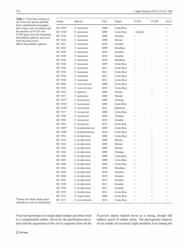
Forty-four Fusarium strains were isolated and purifiedduring this study. Some plant samples yielded morethan one individual, but only single strains of a specieswere subjected to subsequent analyses. To assure thatthe strains are pure and unique, a single-sporesubculturing was performed where possible using theprocedure described by Leslie and Summerell (2006).This was not possible for all strains, as some of themdid not develop spores under laboratory conditions (re-sults not shown).
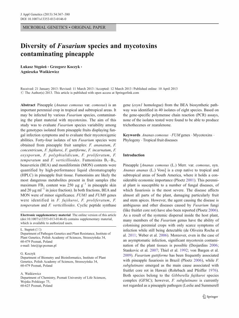
Partial sequences of the translation elongation factor gene(tef-1α) from the strains tested were compared to the refer-ence sequences deposited in the NCBI GenBank usingBLASTN. Ten Fusarium species were identified: F.ananatum, F. concentricum, F. fujikuroi, F. guttiforme, F.incarnatum, F. oxysporum, F. polyphialidicum , F.proliferatum, F. temperatum and F. verticillioides. F.proliferatum and F. ananatum were identified with thehighest frequencies (15 and 14 isolates, respectively). Onlysingle occurrences of F. fujikuroi, F. guttiforme, F.temperatum and F. verticillioides species were observed(Fig. 1 and Table 2). Most of the isolates originated fromCosta Rica and Ecuador, with only a few samples being
from Hawaii, Honduras, Indonesia and Vietnam. Phyloge-netic relationships between the isolates were reconstructed(Fig. 1), including the reference sequences of model strainsof F. graminearum, F. oxysporum, F. verticillioides and F.solani, as well as several other accessions of closely relatedspecies (see the Materials and Methods section).
The phylogeny reconstruction for the tef-1α fragmentsupports clear, early divergence of F. polyphialidicumstrains from the bulk of the considered species. Thereconstructed phylogeny shows moderate support for twoclades for the majority of all the analyzed F. proliferatumstrains, except for KF 3408 (two clades with supports of 66and 56 %, respectively).
Notably, F. guttiforme descent is unresolved in this phy-logeny. Some F. guttiforme strains (KF 3327, NRRL 22945,ITEM 7611) appear to share common descent with F.ananatum (61 % bootstrap support), while grouping withF. begoniae NRRL 25300 is implied for two other strains(MRC 6782 and MRC 7539). This suggests the possiblemisidentification of KF 3327 and ITEM 6711 strains in theirrespective collections, as some of the model F. guttiformestrains (e.g. NRRL 22945) have been previously re-identified as F. ananatum in the paper describing this re-cently characterized species (Jacobs et al. 2010).
Growth speed
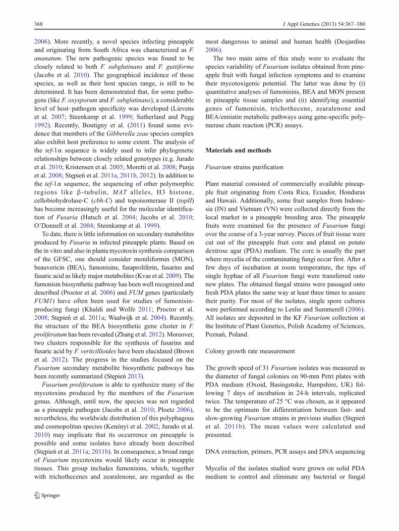
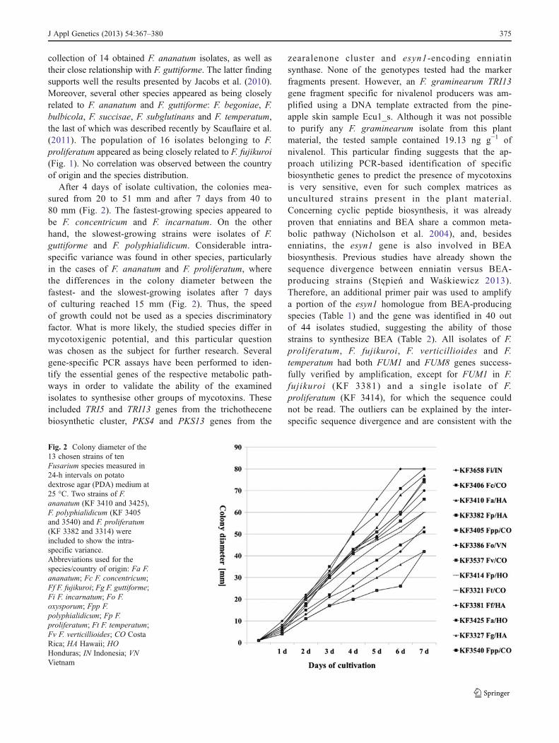
Thirty-one Fusarium isolates representing ten identifiedspecies were subjected to the analysis of growth speed at25 °C. F. incarnatum and F. concentricum strains formed thecolonies with the greatest speed and F. guttiforme and F.polyphialidicum were the species for which colony sizeincrease was observed with the lowest rates. To maximizethe clarity of the results obtained, only one isolate perspecies was included (Fig. 2), except for two isolates of F.ananatum, F. polyphialidicum and F. proliferatum, sampleswhich delineate the limits of intra-specific differences ob-served for growth speeds.
Identification of mycotoxin biosynthetic genes
The presence of FUM1 and FUM8 genes (encoding thepolyketide synthase and PLP-dependent aminotransferasefrom the fumonisin biosynthetic gene cluster, respectively)was confirmed in the F. fujikuroi, F. proliferatum, F.temperatum and F. verticillioides strains, though with minorexceptions, namely, the F. fujikuroi KF 3381 strain gave noamplification for FUM1 and a single strain of F. ananatum(KF 3316) amplified the FUM1 gene marker with lowefficiency. The markers for trichothecene (TRI5 encodingthe trichodiene synthase and TRI13 encoding the P450monooxygenase determining deoxynivalenol and nivalenolchemotypes), as well as two zearalenone biosynthetic genes
J Appl Genetics (2013) 54:367–380 371

372 J Appl Genetics (2013) 54:367–380

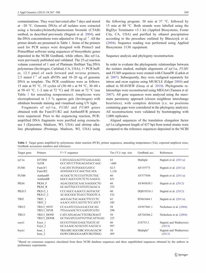
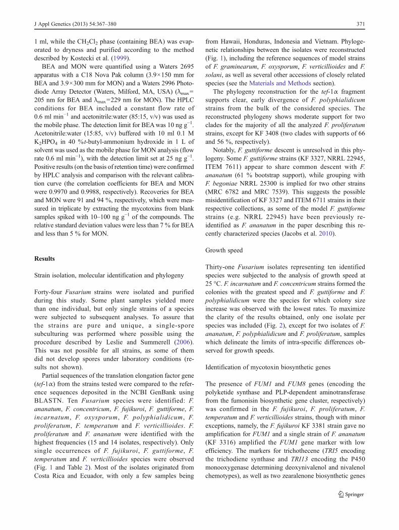
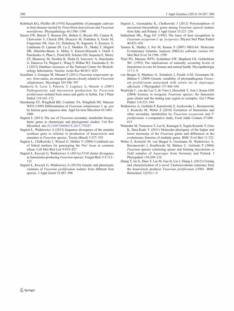
(PKS4 and PKS13 encoding two polyketide synthases),were not found in any of the analysed samples (data notshown). A weak amplification of TRI13 fragment specificfor nivalenol producers was observed in the case of DNAextracted from pineapple skin sample Ecu1_s. Sequencingof the fragment was confirmed to be the P450monooxygenasefrom the TRI cluster of the F. graminearum variant (253nucleotides with 99% identities). Although it was not possibleto purify any F. graminearum isolate from this plant material,still, the skin sample contained trace amounts of nivalenol(19.13 ng g−1), which suggests the presence of the pathogen.Using an esyn1 gene-specific marker, it was not possible toconfirm the presence of the target gene. However, an addi-tional pair of primers was used to amplify the sequence fromBEA-producing species (Table 1). All isolates except for threeF. incarnatum and one F. verticillioides had the gene present(Table 2). Partial sequences of FUM1 and FUM8 genes fromall isolates with the genes present were examined to verifytheir origin and cross-reference the toxigenic capability withinferred species (assignments based on the tef-1α sequence).The only exceptions were isolates KF 3316 (F. ananatum) andKF 3414 (F. proliferatum), for which neither FUM1 norFUM8 sequences could not be obtained, in spite of theirsuccessful amplification (Table 2). Phylogenetic trees wereconstructed, bootstrapped and topology visualized with notless than 50 % of support (Figs. 3 and 4).
Notably, the divergence of F. proliferatum and F.verticillioides/F. oxysporum (Waalwijk et al. 2004) is wellsupported by both FUM1 and FUM8 phylogenies. The re-spective reconstructions also demonstrate close relationshipsbetween singular F. temperatum (FUM1 and FUM8) and F.
fujikuroi (FUM8) genes with the F. proliferatum genes (>90%bootstrap support values for clades).
Mycotoxins synthesized in planta and in vitro
The mycotoxins of three groups, BEA, MON andfumonisins B1, B2 and B3, were quantified in five samplesof pineapple juice and skin fractions originating from Ecua-dor and Costa Rica. To investigate the mycotoxigenic abil-ities of individual isolates, the cultures of sterilized rice wereused. Only a small group of isolates could not be tested;being highly susceptible to the storage conditions (nine iso-lates out of 44 studied), the mycelia were not viable after afew months of cold storage at −20 °C.
The inter-specific variability of mycotoxigenic abilitiesdisplayed by the set of isolates studied is presented inTable 3. To facilitate the comparison of in planta versusin vitro mycotoxin contents, isolates purified from ana-lyzed pineapple fruit samples were separated from theothers and appended in Table 4.
Discussion
Recent studies reported mainly F. guttiforme as the causativeagent of pineapple fusariosis (Ploetz 2006). Nevertheless,Jacobs et al. (2010) demonstrated F. ananatum as the emerg-ing species involved in pineapple fruit lesions in South Africa.In the present study, numerous Fusarium species wereobtained from pineapple plant samples besides the twoabove-mentioned species, namely, F. concentricum, F.fujikuroi, F. incarnatum, F. oxysporum, F. polyphialidicum,F. proliferatum, F. temperatum and F. verticillioides. In severalcases, two different species were identified in the same plantsample (Table 4). The majority of the species occurred withlow frequencies (see Table 2) and have not been reported aspineapple pathogens, thus, inoculation experiments are need-ed in order to confirm the ability of those strains to infect andcolonize the pineapple tissue. Taking into account that theharvested pineapple fruit is usually protected from pathogeninfiltration and also from colonization by saprophytic agentsby spraying a fungicide (like Bayleton), this finding could behelpful in corroborating the supposition that the identifiedFusaria are not necessarily exogenous contaminants intro-duced during transport and storage.
The high incidence of F. proliferatum deserves signifi-cant attention. Recently, Stępień et al. (2011b) characterizedthe genetic diversity of several strains obtained from pine-apple in respect to genotypes obtained from different hosts,showing significant intra-specific diversity in relation to thehost of origin. Pineapple-derived strains grouped firmlytogether, apart from garlic, maize, rice and asparagus strains(Stępień et al. 2011b). The co-occurrence of multiple
Fig. 1 The most parsimonious tree created on the basis of trans-lation elongation factor 1α (tef-1α) sequences of 44 isolates often Fusarium species, as well as 14 additional sequences frompublicly available model strains. F. solani (H. haematococca MPVI) was used as an outgroup in the analysis. The consistencyindex for the first most parsimonious tree was 0.587, and theretention index was 0.803. A total of 277 positions were analysed(complete deletion of gapped positions from the initial alignmentof 427 base pairs), of which 69 were parsimony informative. Thetree was obtained using the maximum parsimony approach (clos-est neighbour interchange with search level 3 based on ten repli-cates for the initial tree construction) and tested by bootstrapping(1,000 replicates) with a cut-off value of 50 %. Abbreviationsused for the species/country of origin: Fa F. ananatum; , Ff F.fujikuroi; Fg F. guttiforme; Fi F. incarnatum; Fo F. oxysporum;Fpp F. polyphialidicum; Fp F. proliferatum; Ft F. temperatum; FvF. verticillioides; CR Costa Rica; EC Ecuador; HA Hawaii; HOHonduras; IN Indonesia; VN Vietnam. For 12 model strains, eithergene identifiers (from BROAD/JGI model sequences of F.graminearum, F. verticillioides, F. oxysporum, F. solani) orGenBank accession codes (all other cases) are given in parenthe-ses. *F. guttiforme NRRL 22945 subsequently re-identified as F.ananatum by Jacobs et al. (2010); see also the discussion of thetef-1α phylogeny in the Results section
R
J Appl Genetics (2013) 54:367–380 373
Fusarium genotypes in a single plant sample can often resultin a contaminated culture. However, the purification proce-dure and the acquisition of the tef-1α sequence from all the
Fusarium strains studied serves as a strong, though stillindirect, proof of culture purity. The phylogenetic analysisof our isolate set revealed a high similarity level among the
Table 2 Forty-four isolates often Fusarium species purifiedfrom contaminated pineapples,their origin, year of isolation andthe presence of FUM1 andFUM8 genes from the fumonisinbiosynthetic pathway and bsyn1from the beauvericin(BEA) biosynthetic pathway
aStrains for which single sporesubcultures were not performed
Isolate Species Year Origin FUM1 FUM8 bsyn1
KF 3302a F. ananatum 2008 Costa Rica − − +
KF 3316a F. ananatum 2008 Costa Rica + (weak) − +
KF 3330 F. ananatum 2008 Ecuador − − +
KF 3410 F. ananatum 2009 Hawaii − − +
KF 3417 F. ananatum 2010 Ecuador − − +
KF 3425 F. ananatum 2009 Honduras − − +
KF 3436 F. ananatum 2010 Ecuador − − +
KF 3438 F. ananatum 2010 Ecuador − − +
KF 3542 F. ananatum 2010 Honduras − − +
KF 3544 F. ananatum 2009 Costa Rica − − +
KF 3546 F. ananatum 2011 Costa Rica − − +
KF 3552 F. ananatum 2011 Costa Rica − − +
KF 3554 F. ananatum 2011 Costa Rica − − +
KF 3556 F. ananatum 2011 Costa Rica − − +
KF 3406 F. concentricum 2009 Costa Rica − − +
KF 3536 F. concentricum 2010 Costa Rica − − +
KF 3381 F. fujikuroi 2009 Hawaii − + +
KF 3327 F. guttiforme 2008 Hawaii − − +
KF 3387a F. incarnatum 2009 Vietnam − − −
KF 3543a F. incarnatum 2009 Costa Rica − − −
KF 3658a F. incarnatum 2011 Indonesia − − −
KF 3317 F. oxysporum 2008 Costa Rica − − +
KF 3386 F. oxysporum 2009 Vietnam − − +
KF 3504 F. oxysporum 2010 Ecuador − − +
KF 3555 F. oxysporum 2011 Costa Rica − − +
KF 3405a F. polyphialidicum 2009 Costa Rica − − +
KF 3540a F. polyphialidicum 2010 Costa Rica − − +
KF 3301 F. proliferatum 2008 Costa Rica + + +
KF 3315 F. proliferatum 2008 Hawaii + + +
KF 3382 F. proliferatum 2009 Hawaii + + +
KF 3383 F. proliferatum 2009 Hawaii + + +
KF 3385 F. proliferatum 2009 Vietnam + + +
KF 3404 F. proliferatum 2009 Costa Rica + + +
KF 3407 F. proliferatum 2009 Costa Rica + + +
KF 3408 F. proliferatum 2009 Costa Rica + + +
KF 3414 F. proliferatum 2010 Honduras + + +
KF 3439 F. proliferatum 2010 Ecuador + + +
KF 3444 F. proliferatum 2010 Ecuador + + +
KF 3547 F. proliferatum 2011 Ecuador + + +
KF 3548 F. proliferatum 2011 Ecuador + + +
KF 3550 F. proliferatum 2011 Ecuador + + +
KF 3553 F. proliferatum 2011 Costa Rica + + +
KF 3321 F. temperatum 2008 Costa Rica + + +
KF 3537 F. verticillioides 2010 Costa Rica + + −
374 J Appl Genetics (2013) 54:367–380
collection of 14 obtained F. ananatum isolates, as well astheir close relationship with F. guttiforme. The latter findingsupports well the results presented by Jacobs et al. (2010).Moreover, several other species appeared as being closelyrelated to F. ananatum and F. guttiforme: F. begoniae, F.bulbicola, F. succisae, F. subglutinans and F. temperatum,the last of which was described recently by Scauflaire et al.(2011). The population of 16 isolates belonging to F.proliferatum appeared as being closely related to F. fujikuroi(Fig. 1). No correlation was observed between the countryof origin and the species distribution.
After 4 days of isolate cultivation, the colonies mea-sured from 20 to 51 mm and after 7 days from 40 to80 mm (Fig. 2). The fastest-growing species appeared tobe F. concentricum and F. incarnatum. On the otherhand, the slowest-growing strains were isolates of F.guttiforme and F. polyphialidicum. Considerable intra-specific variance was found in other species, particularlyin the cases of F. ananatum and F. proliferatum, wherethe differences in the colony diameter between thefastest- and the slowest-growing isolates after 7 daysof culturing reached 15 mm (Fig. 2). Thus, the speedof growth could not be used as a species discriminatoryfactor. What is more likely, the studied species differ inmycotoxigenic potential, and this particular questionwas chosen as the subject for further research. Severalgene-specific PCR assays have been performed to iden-tify the essential genes of the respective metabolic path-ways in order to validate the ability of the examinedisolates to synthesise other groups of mycotoxins. Theseincluded TRI5 and TRI13 genes from the trichothecenebiosynthetic cluster, PKS4 and PKS13 genes from the
zearalenone cluster and esyn1-encoding enniatinsynthase. None of the genotypes tested had the markerfragments present. However, an F. graminearum TRI13gene fragment specific for nivalenol producers was am-plified using a DNA template extracted from the pine-apple skin sample Ecu1_s. Although it was not possibleto purify any F. graminearum isolate from this plantmaterial, the tested sample contained 19.13 ng g−1 ofnivalenol. This particular finding suggests that the ap-proach utilizing PCR-based identification of specificbiosynthetic genes to predict the presence of mycotoxinsis very sensitive, even for such complex matrices asuncultured strains present in the plant material.Concerning cyclic peptide biosynthesis, it was alreadyproven that enniatins and BEA share a common meta-bolic pathway (Nicholson et al. 2004), and, besidesenniatins, the esyn1 gene is also involved in BEAbiosynthesis. Previous studies have already shown thesequence divergence between enniatin versus BEA-producing strains (Stępień and Waśkiewicz 2013).Therefore, an additional primer pair was used to amplifya portion of the esyn1 homologue from BEA-producingspecies (Table 1) and the gene was identified in 40 outof 44 isolates studied, suggesting the ability of thosestrains to synthesize BEA (Table 2). All isolates of F.proliferatum, F. fujikuroi, F. verticillioides and F.temperatum had both FUM1 and FUM8 genes success-fully verified by amplification, except for FUM1 in F.fujikuroi (KF 3381) and a single isolate of F.proliferatum (KF 3414), for which the sequence couldnot be read. The outliers can be explained by the inter-specific sequence divergence and are consistent with the
Fig. 2 Colony diameter of the13 chosen strains of tenFusarium species measured in24-h intervals on potatodextrose agar (PDA) medium at25 °C. Two strains of F.ananatum (KF 3410 and 3425),F. polyphialidicum (KF 3405and 3540) and F. proliferatum(KF 3382 and 3314) wereincluded to show the intra-specific variance.Abbreviations used for thespecies/country of origin: Fa F.ananatum; Fc F. concentricum;Ff F. fujikuroi; Fg F. guttiforme;Fi F. incarnatum; Fo F.oxysporum; Fpp F.polyphialidicum; Fp F.proliferatum; Ft F. temperatum;Fv F. verticillioides; CO CostaRica; HA Hawaii; HOHonduras; IN Indonesia; VNVietnam
J Appl Genetics (2013) 54:367–380 375
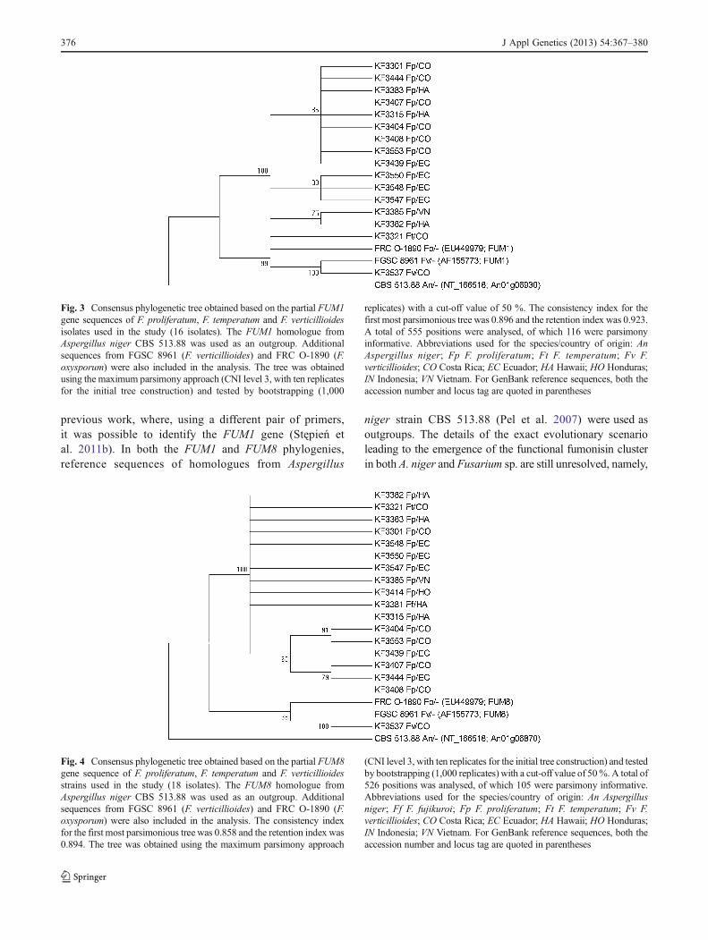
previous work, where, using a different pair of primers,it was possible to identify the FUM1 gene (Stępień etal. 2011b). In both the FUM1 and FUM8 phylogenies,reference sequences of homologues from Aspergillus
niger strain CBS 513.88 (Pel et al. 2007) were used asoutgroups. The details of the exact evolutionary scenarioleading to the emergence of the functional fumonisin clusterin both A. niger and Fusarium sp. are still unresolved, namely,
Fig. 3 Consensus phylogenetic tree obtained based on the partial FUM1gene sequences of F. proliferatum, F. temperatum and F. verticillioidesisolates used in the study (16 isolates). The FUM1 homologue fromAspergillus niger CBS 513.88 was used as an outgroup. Additionalsequences from FGSC 8961 (F. verticillioides) and FRC O-1890 (F.oxysporum) were also included in the analysis. The tree was obtainedusing the maximum parsimony approach (CNI level 3, with ten replicatesfor the initial tree construction) and tested by bootstrapping (1,000
replicates) with a cut-off value of 50 %. The consistency index for thefirst most parsimonious tree was 0.896 and the retention index was 0.923.A total of 555 positions were analysed, of which 116 were parsimonyinformative. Abbreviations used for the species/country of origin: AnAspergillus niger; Fp F. proliferatum; Ft F. temperatum; Fv F.verticillioides; CO Costa Rica; EC Ecuador; HA Hawaii; HO Honduras;IN Indonesia; VN Vietnam. For GenBank reference sequences, both theaccession number and locus tag are quoted in parentheses
Fig. 4 Consensus phylogenetic tree obtained based on the partial FUM8gene sequence of F. proliferatum, F. temperatum and F. verticillioidesstrains used in the study (18 isolates). The FUM8 homologue fromAspergillus niger CBS 513.88 was used as an outgroup. Additionalsequences from FGSC 8961 (F. verticillioides) and FRC O-1890 (F.oxysporum) were also included in the analysis. The consistency indexfor the first most parsimonious tree was 0.858 and the retention index was0.894. The tree was obtained using the maximum parsimony approach
(CNI level 3, with ten replicates for the initial tree construction) and testedby bootstrapping (1,000 replicates) with a cut-off value of 50%.A total of526 positions was analysed, of which 105 were parsimony informative.Abbreviations used for the species/country of origin: An Aspergillusniger; Ff F. fujikuroi; Fp F. proliferatum; Ft F. temperatum; Fv F.verticillioides; CO Costa Rica; EC Ecuador; HA Hawaii; HO Honduras;IN Indonesia; VN Vietnam. For GenBank reference sequences, both theaccession number and locus tag are quoted in parentheses
376 J Appl Genetics (2013) 54:367–380

the direction of the ancestral horizontal transfer (Khaldi andWolfe 2011) and dating cluster formation in light of differentorganization within F. verticillioides and F. proliferatum(Waalwijk et al. 2004). However, the outgroup choice itselfis corroborated by both strong sequence divergence (ca. 55–60 % protein sequence identity between A. niger and Fusar-ium genes) and previously mentioned analysis by Khaldi andWolfe (2011).
To validate the potential risk of pineapple contaminationwith mycotoxins, BEA, MON and fumonisins contents weremeasured in fruit tissue samples (Table 4). Also, rice cul-tures of individual isolates were prepared in order to estab-lish the mycotoxigenic potential of individual genotypes incontrolled laboratory conditions (Table 3). Across all theanalyzed isolates, F. proliferatum appeared to be the speciesproducing the highest amounts of fumonisins (FBs) and
MON. Concerning FBs, only one isolate could be qualifiedas a low-efficiency producer (KF 3385) and only anothertwo yielded less than 1 mg g−1 of FB1 and the most efficientproducer (KF 3414) yielded about 3.3 mg g−1 of FB1. The F.fujikuroi KF 3381 produced FB1 in a comparable amount ofover 1.5 mg g−1 (Table 3). Both F. temperatum and F.verticillioides are known to produce FBs in significantamounts (Proctor et al. 1999; Scauflaire et al. 2011) andcan be described as medium-efficiency producers (between50 and 100 μg g−1 of FB1 produced) and the remainingspecies as low-efficiency producers. MON was also pro-duced in the highest amounts by F. proliferatum isolates,though, in this case, the yield was not very high, with thehighest value of 158 μg g−1. F. ananatum isolates did notproduce MON, but KF 3436 synthesized BEA in the highestamount (91 μg g−1) of all the isolates tested (Table 3).
Table 3 Means and standard deviations (SDs) of the amounts of fumonisins (FB1–FB3), moniliformin (MON) and beauvericin (BEA) concen-trations produced in vitro by 28 isolates of nine Fusarium species
Isolate Fusarium species FB1
(μg/g)FB2
(μg/g)FB3
(μg/g)MON(μg/g)
BEA(μg/g)
KF 3302 F. ananatum 0.16±0.05 0.09±0.02 0.04±0.01 ND 0 0.42±0.09
KF 3330 F. ananatum 9.80±2.21 1.02±0.53 0.32±0.02 0.04±0.01 0.96±0.07
KF 3410 F. ananatum nt nt nt ND 0.70±0.03
KF 3417 F. ananatum nt nt nt ND 4.70±0.56
KF 3425 F. ananatum nt nt nt- ND 5.85±0.87
KF 3438 F. ananatum nt nt nt ND ND
KF 3436 F. ananatum 3.29±0.45 0.95±0.05 0.32±0.02 ND 91.47±11.12
KF 3406 F. concentricum 10.41±2.11 ND 0.28±0.04 30.04±4.12 0.51±0.06
KF 3536 F. concentricum 9.38±0.97 ND 0.28±0.03 ND ND
KF 3381 F. fujikuroi 1,558.98±187.15 465.96±39.76 81.64±9.76 27.55±3.54 1.65±0.28
KF 3327 F. guttiforme nt nt nt ND 7.70±1.15
KF 3386 F. oxysporum ND ND ND 17.23±2.27 ND
KF 3504 F. oxysporum ND ND ND 2.22±0.18 3.93±0.55
KF 3405 F. polyphialidicum 2.91±0.28 0.41±0.03 0.11±0.02 ND 6.49±1.09
KF 3540 F. polyphialidicum 18.87±3.42 4.30±0.37 1.15±0.23 0.02±0.01 6.78±0.75
KF 3301a F. proliferatum 1,353.00±154.90 496.00±39.65 133.00±21.98 110.56±9.76 0.83±0.06
KF 3315a F. proliferatum 1,820.00±202.11 534.00±21.70 113.00±20.54 59.40±4.44 0.09±0.02
KF 3382 F. proliferatum 1,785.76±176.53 450.54±51.33 132.90±18.77 18.40±2.09 3.39±0.28
KF 3383a F. proliferatum 930.00±88.69 204.00±017.73 79.00±6.55 158.46±11.63 1.08±0.18
KF 3385a F. proliferatum 7.01±0.64 2.16±0.18 0.79±0.04 14.22±1.08 2.26±0.65
KF 3404 F. proliferatum 856.39±78.15 330.03±20.18 109.07±9.23 49.31±3.32 3.99±0.57
KF 3407 F. proliferatum 2,419.32±199.53 379.86±43.09 139.77±10.44 81.73±7.65 4.15±0.22
KF 3408 F. proliferatum 2,686.23±303.41 757.94±54.31 367.82±40.12 27.68±1.72 0.35±0.02
KF 3414 F. proliferatum 3,299.01±276.80 855.64±68.98 593.08±49.52 15.98±0.98 41.13±3.74
KF 3439 F. proliferatum 1,032.76±97.45 134.60±10.25 96.49±10.01 93.08±10.13 8.61±1.43
KF 3444 F. proliferatum 1,568.18±209.83 210.93±15.37 140.35±9.12 65.01±5.43 24.75±3.11
KF 3321 F. temperatum 2.25±0.32 0.35±0.04 0.08±0.02 7.45±0.61 12.48±0.89
KF 3537 F. verticillioides 59.65±6.16 19.37±0.99 5.86±0.76 0.07±0.01 0.05±0.01
ND not detected; nt not testeda Isolates previously described by Stępień et al. (2011a) and/or Stępień et al. (2011b)
J Appl Genetics (2013) 54:367–380 377

Again, F. proliferatum isolates recovered from the analysedplant samples were the most effective FBs, MON and BEAproducers. Interestingly, in the original pineapple samplesCos2 and Cos4—samples that did not contain F.proliferatum but only F. ananatum and F. oxysporum—moreFBs have been identified in planta than in the respectiveisolates cultured on rice. Such inconsistency might beexplained at least two-fold:
(i) An additional unknown FBs producer was present inthe infected fruit, but was not identified in cultures, or
(ii) Rice culture used for FBs biosynthesis in vitro was nota suitable medium for those strains causing low bio-synthesis level.
In conclusion, concerning a contribution of individualspecies to the contamination of pineapple with mycotoxins,the role of F. ananatum, F. oxysporum, F. guttiforme and F.polyphialidicum as mycotoxin producers can be regarded aslimited, while F. proliferatum emerges as a major, under-stated threat. The same conclusion can be drawn from theanalysis of in planta mycotoxin content results. Pineappleskin from infected fruit contained as much as 250 μg g−1 ofFB1 and the metabolite concentration in pineapple juice wasabout ten times lower. High FB content in the skin samplesis likely caused by the concentration of toxins achieved by
the application of a freeze-drying step. In both fractions,BEA and MON were of minor significance (Table 4).According to the US Food and Drug Administration(FDA) exposure guidelines, the total fumonisin content incorn-based food products may reach 2–4 ppm. This meansthat the naturally occurring FB levels in (stored) pineapplefruits are potentially hazardous to human health.
Taken as a whole, the obtained results demonstrate that,of all the species identified, F. ananatum and F. proliferatumare probably the most commonly occurring Fusaria andregarding mycotoxin produced, F. proliferatum can be po-tentially the most dangerous species found in plant tissues ofpineapple. Additionally, they serve as a proof of the species’metabolic activity in planta and suggest the need for FBscontamination control in pineapple and, likely, in othertropical crops most often consumed in unprocessed form.
Acknowledgements This research was partially supported by theNational Centre of Science (NCN) project NN 310 732440. GrzegorzKoczyk has been supported by the National Centre for Research andDevelopment (NCBiR) grant LIDER 19/113/L-1/09/NCBiR/2010(“Modelling, prediction and verification of toxigenic potential in fungi”).
Open Access This article is distributed under the terms of the CreativeCommons Attribution License which permits any use, distribution, andreproduction in any medium, provided the original author(s) and thesource are credited.
Table 4 Means and standard deviations (SD) of the amounts offumonisins (FB1–FB3), moniliformin (MON) and beauvericin (BEA)concentrations measured in five samples of pineapple juice and skin
fractions originating from Costa Rica (Cos) and Ecuador (Ecu), togeth-er with the mycotoxin yield for seven isolates of three Fusariumspecies purified from the respective samples
Sample ID/Isolate No. FB1 FB2 FB3 MON BEA
Pineapple juice samples (μg/ml)
Cos1_j 8.18±1.07 0.94±0.09 0.17±0.02 0.20±0.01 0.00
Cos2_j 14.85±5.12 1.46±0.32 0.23±0.03 0.04±0.01 0.00
Cos3_j 21.01±4.89 0.74±0.08 0.17±0.03 0.04±0.02 0.00
Cos4_j 23.73±5.22 1.21±0.24 0.18±0.03 0.03±0.01 0.00
Ecu1_j 22.18±3.38 0.95±0.07 0.23±0.04 0.01±0.01 0.00
Pineapple skin samples (μg/g)
Cos1_s 128.57±11.09 1.27±0.09 0.70±0.05 0.79±0.06 0.31±0.02
Cos2_s 33.23±2.65 0.64±0.05 0.48±0.03 0.02±0.01 0.23±0.02
Cos3_s 33.06±2.88 0.82±0.05 0.74±0.05 0.04±0.02 0.36±0.01
Cos4_s 25.65±1.83 0.59±0.06 0.56±0.04 0.06±0.02 1.60±0.09
Ecu1_s 247.74±20.76 1.48±0.11 0.11±0.02 0.08±0.03 0.79±0.05
Rice cultures of isolates obtainedfrom analyzed material (μg/g)
KF 3546 (F. ananatum/Cos1) 3.62±0.27 0.45±0.05 0.11±0.01 0.06±0.02 2.46±0.34
KF 3552 (F. ananatum/Cos2) 10.32±0.87 0.45±0.04 0.39±0.05 0.01±0.01 3.49±0.37
KF 3553 (F. proliferatum/Cos3) 2,125.42±154.78 368.79±54.41 189.48±35.09 23.90±6.18 10.54±2.35
KF 3554 (F. ananatum/Cos3) 26.05±1.34 5.09±0.72 1.13±0.15 0.03±0.01 1.31±0.11
KF 3555 (F. oxysporum/Cos4) 10.97±0.99 6.27±0.55 0.47±0.05 5.22±0.44 3.24±0.45
KF 3556 (F. ananatum/Cos4) 3.73±0.41 1.08±0.23 0.47±0.06 0.04±0.01 1.73±0.15
KF 3548 (F. proliferatum/Ecu1) 1,861.16±115.76 481.92±34.80 98.40±10.11 97.44±9.98 53.88±7.68
378 J Appl Genetics (2013) 54:367–380

References
Błaszczyk L, Goyeau H, Huang XQ, Röder M, Stępień Ł, ChełkowskiJ (2004) Identifying leaf rust resistance genes and mapping geneLr37 on the microsatellite map of wheat. Cell Mol Biol Lett9:869–878
Boutigny A-L, Ward TJ, Van Coller GJ, Flett B, Lamprecht SC,O’Donnell K, Viljoen A (2011) Analysis of the Fusariumgraminearum species complex from wheat, barley and maize inSouth Africa provides evidence of species-specific differences inhost preference. Fungal Genet Biol 48:914–920
Brown DW, Butchko RA, Busman M, Proctor RH (2012) Identifica-tion of gene clusters associated with fusaric acid, fusarin, andperithecial pigment production in Fusarium verticillioides. FungalGenet Biol 49:521–532
de Oliveira Rocha L, Reis GM, da Silva VN, Braghini R, TeixeiraMMG, Corrêa B (2011) Molecular characterization and fumonisinproduction by Fusarium verticillioides isolated from corn grainsof different geographic origins in Brazil. Int J Food Microbiol145:9–21
Desjardins AE (2006) Fusarium mycotoxins: chemistry, genetics, andbiology. American Phytopathological Society, St. Paul, MN
Edgar RC (2004) MUSCLE: a multiple sequence alignment methodwith reduced time and space complexity. BMC Bioinformatics5:113
Gouy M, Guindon S, Gascuel O (2010) SeaView version 4: amultiplatform graphical user interface for sequence alignmentand phylogenetic tree building. Mol Biol Evol 27:221–224
Haas BJ, Zeng Q, Pearson MD, Cuomo CA, Wortman JR (2011)Approaches to fungal genome annotation. Mycology 2:118–141
Hatsch D, Phalip V, Jeltsch J-M (2004) Use of genes encodingcellobiohydrolase-C and topoisomerase II as targets for phyloge-netic analysis and identification of Fusarium. Res Microbiol155:290–296
Jacobs A, Van Wyk PS, Marasas WFO, Wingfield BD, Wingfield MJ,Coutinho TA (2010) Fusarium ananatum sp. nov. in theGibberella fujikuroi species complex from pineapples with fruitrot in South Africa. Fungal Biol 114:515–527
Jurado M, Marín P, Callejas C, Moretti A, Vázquez C, González-JaénMT (2010) Genetic variability and fumonisin production by Fu-sarium proliferatum. Food Microbiol 27:50–57
Kenényi Z, Mulé G, Moretti A, Waalwijk C, Hornok L (2002) Fertilityand mating type assessment within Fusarium proliferatum iso-lates from different host plants. J Appl Genet 43A:55–68
Khaldi N, Wolfe KH (2011) Evolutionary origins of the fumonisinsecondary metabolite gene cluster in Fusarium verticillioides andAspergillus niger. Int J Evol Biol 2011:423821
Kostecki M, Wiśniewska H, Perrone G, Ritieni A, Goliński P,Chełkowski J, Logrieco A (1999) The effects of cereal substrateand temperature on production of beauvericin, moniliformin andfusaproliferin by Fusarium subglutinans ITEM-1434. Food AdditContam 16:361–365
Kristensen R, Torp M, Kosiak B, Holst-Jensen A (2005) Phylogenyand toxigenic potential is correlated in Fusarium species as re-vealed by partial translation elongation factor 1 alpha gene se-quences. Mycol Res 109:173–186
Kvas M, Marasas WFO, Wingfield BD, Wingfield MJ, Steenkamp ET(2009) Diversity and evolution of Fusarium species in theGibberella fujikuroi complex. Fungal Divers 34:1–21
Larkin MA, Blackshields G, Brown NP, Chenna R, McGettigan PA,McWilliam H, Valentin F, Wallace IM, Wilm A, Lopez R,Thompson JD, Gibson TJ, Higgins DG (2007) Clustal W andClustal X version 2.0. Bioinformatics 23:2947–2948
Leslie JF, Summerell BA (2006) The Fusarium laboratory manual.Wiley-Blackwell, Hoboken, NJ
Lievens B, Claes L, Vakalounakis DJ, Vanachter ACRC, ThommaBPHJ (2007) A robust identification and detection assay to dis-criminate the cucumber pathogens Fusarium oxysporum f. sp.cucumerinum and f. sp. radicis-cucumerinum. Environ Microbiol9:2145–2161
Marchler-Bauer A, Lu S, Anderson JB, Chitsaz F, Derbyshire MK,DeWeese-Scott C, Fong JH, Geer LY, Geer RC, Gonzales NR,Gwadz M, Hurwitz DI, Jackson JD, Ke Z, Lanczycki CJ, Lu F,Marchler GH, Mullokandov M, Omelchenko MV, Robertson CL,Song JS, Thanki N, Yamashita RA, Zhang D, Zhang N, Zheng C,Bryant SH (2011) CDD: a Conserved Domain Database for thefunctional annotation of proteins. Nucleic Acids Res 39:D225–D229
Moretti A, Mulé G, Ritieni A, Láday M, Stubnya V, Hornok L,Logrieco A (2008) Cryptic subspecies and beauvericin productionby Fusarium subglutinans from Europe. Int J Food Microbiol127:312–315
Nicholson P, Simpson DR, Wilson AH, Chandler E, Thomsett M(2004) Detection and differentiation of trichothecene andenniatin-producing Fusarium species on small-grain cereals. EurJ Plant Pathol 110:503–514
O’Donnell K, Ward TJ, Geiser DM, Corby Kistler H, Aoki T (2004)Genealogical concordance between the mating type locus andseven other nuclear genes supports formal recognition of ninephylogenetically distinct species within the Fusariumgraminearum clade. Fungal Genet Biol 41:600–623
Pel HJ, de Winde JH, Archer DB, Dyer PS, Hofmann G, SchaapPJ, Turner G, de Vries RP, Albang R, Albermann K, AndersenMR, Bendtsen JD, Benen JA, van den Berg M, Breestraat S,Caddick MX, Contreras R, Cornell M, Coutinho PM,Danchin EG, Debets AJ, Dekker P, van Dijck PW, van DijkA, Dijkhuizen L, Driessen AJ, d’Enfert C, Geysens S,Goosen C, Groot GS, de Groot PW, Guillemette T, HenrissatB, Herweijer M, van den Hombergh JP, van den Hondel CA,van der Heijden RT, van der Kaaij RM, Klis FM, Kools HJ,Kubicek CP, van Kuyk PA, Lauber J, Lu X, van der MaarelMJ, Meulenberg R, Menke H, Mortimer MA, Nielsen J,Oliver SG, Olsthoorn M, Pal K, van Peij NN, Ram AF,Rinas U, Roubos JA, Sagt CM, Schmoll M, Sun J, UsseryD, Varga J, Vervecken W, van de Vondervoort PJ, Wedler H,Wösten HA, Zeng AP, van Ooyen AJ, Visser J, Stam H(2007) Genome sequencing and analysis of the versatile cellfactory Aspergillus niger CBS 513.88. Nat Biotechnol25:221–231
Ploetz RC (2001) Significant diseases in the tropics that are caused byspecies of Fusarium. In: Fusarium: Paul E. Nelson MemorialSymposium. The American Phytopathological Society Press, St.Paul, MN, pp 295–309
Ploetz RC (2006) Fusarium-induced diseases of tropical, perennialcrops. Phytopathology 96:648–652
Proctor RH, Desjardins AE, Plattner RD, Hohn TM (1999) A polyke-tide synthase gene required for biosynthesis of fumonisin myco-toxins in Gibberella fujikuroi mating population A. Fungal GenetBiol 27:100–112
Proctor RH, Plattner RD, Desjardins AE, Busman M, Butchko RAE(2006) Fumonisin production in the maize pathogen Fusariumverticillioides: genetic basis of naturally occurring chemical var-iation. J Agric Food Chem 54:2424–2430
Proctor RH, Busman M, Seo J-A, Lee YW, Plattner RD (2008) Afumonisin biosynthetic gene cluster in Fusarium oxysporum strainO-1890 and the genetic basis for B versus C fumonisin produc-tion. Fungal Genet Biol 45:1016–1026
Punja ZK, Wan A, Rahman M, Goswami RS, Barasubiye T, SeifertKA, Lévesque CA (2008) Growth, population dynamics, anddiversity of Fusarium equiseti in ginseng fields. Eur J Plant Pathol121:173–184
J Appl Genetics (2013) 54:367–380 379
Rohrbach KG, Pfeiffer JB (1976) Susceptibility of pineapple cultivarsto fruit diseases incited by Penicillium funiculosum and Fusariummoniliforme. Phytopathology 66:1386–1390
Sayers EW, Barrett T, Benson DA, Bolton E, Bryant SH, Canese K,Chetvernin V, Church DM, Dicuccio M, Federhen S, Feolo M,Fingerman IM, Geer LY, Helmberg W, Kapustin Y, Krasnov S,Landsman D, Lipman DJ, Lu Z, Madden TL, Madej T, MaglottDR, Marchler-Bauer A, Miller V, Karsch-Mizrachi I, Ostell J,Panchenko A, Phan L, Pruitt KD, Schuler GD, Sequeira E, SherryST, Shumway M, Sirotkin K, Slotta D, Souvorov A, StarchenkoG, Tatusova TA, Wagner L, Wang Y, Wilbur WJ, Yaschenko E, YeJ (2012) Database resources of the National Center for Biotech-nology Information. Nucleic Acids Res 40:D13–D25
Scauflaire J, Gourgue M, Munaut F (2011) Fusarium temperatum sp.nov. from maize, an emergent species closely related to Fusariumsubglutinans. Mycologia 103:586–597
Stankovic S, Levic J, Petrovic T, Logrieco A, Moretti A (2007)Pathogenicity and mycotoxin production by Fusariumproliferatum isolated from onion and garlic in Serbia. Eur J PlantPathol 118:165–172
Steenkamp ET, Wingfield BD, Coutinho TA, Wingfield MJ, MarasasWFO (1999) Differentiation of Fusarium subglutinans f. sp. piniby histone gene sequence data. Appl Environ Microbiol 65:3401–3406
Stępień Ł (2013) The use of Fusarium secondary metabolite biosyn-thetic genes in chemotypic and phylogenetic studies. Crit RevMicrobiol. doi:10.3109/1040841X.2013.770387
Stępień Ł, Waśkiewicz A (2013) Sequence divergence of the enniatinsynthase gene in relation to production of beauvericin andenniatins in Fusarium species. Toxins (Basel) 5:537–555
Stępień Ł, Chełkowski J, Wenzel G, Mohler V (2004) Combined useof linked markers for genotyping the Pm1 locus in commonwheat. Cell Mol Biol Lett 9:819–827
Stępień Ł, Koczyk G, Waśkiewicz A (2011a) FUM cluster divergencein fumonisins-producing Fusarium species. Fungal Biol 115:112–123
Stępień Ł, Koczyk G, Waśkiewicz A (2011b) Genetic and phenotypicvariation of Fusarium proliferatum isolates from different hostspecies. J Appl Genet 52:487–496
Stępień Ł, Gromadzka K, Chełkowski J (2012) Polymorphism ofmycotoxin biosynthetic genes among Fusarium equiseti isolatesfrom Italy and Poland. J Appl Genet 53:227–236
Sutherland ML, Pegg GF (1992) The basis of host recognition inFusarium oxysporum f. sp. lycopersici. Physiol Mol Plant Pathol40:423–436
Tamura K, Dudley J, Nei M, Kumar S (2007) MEGA4: MolecularEvolutionary Genetics Analysis (MEGA) software version 4.0.Mol Biol Evol 24:1596–1599
Thiel PG, Marasas WFO, Sydenham EW, Shephard GS, GelderblomWC (1992) The implications of naturally occurring levels offumonisins in corn for human and animal health. Mycopathologia117:3–9
von Bargen S, Martinez O, Schadock I, Eisold A-M, Gossmann M,Büttner C (2009) Genetic variability of phytopathogenic Fusari-um proliferatum associated with crown rot in Asparagusofficinalis. J Phytopathol 157:446–456
Waalwijk C, van der Lee T, de Vries I, Hesselink T, Arts J, Kema GHJ(2004) Synteny in toxigenic Fusarium species: the fumonisingene cluster and the mating type region as examples. Eur J PlantPathol 110:533–544
Waśkiewicz A, Goliński P, Karolewski Z, Irzykowska L, BocianowskiJ, Kostecki M, Weber Z (2010) Formation of fumonisins andother secondary metabolites by Fusarium oxysporum and F.proliferatum: a comparative study. Food Addit Contam 27:608–615
Watanabe M, Yonezawa T, Lee K, Kumagai S, Sugita-Konishi Y, GotoK, Hara-Kudo Y (2011) Molecular phylogeny of the higher andlower taxonomy of the Fusarium genus and differences in theevolutionary histories of multiple genes. BMC Evol Biol 11:322
Weber Z, Kostecki M, von Bargen S, Gossmann M, Waskiewicz A,Bocianowski J, Knaflewski M, Büttner C, Golinski P (2006)Fusarium species colonizing spears and forming mycotoxins infield samples of Asparagus from Germany and Poland. JPhytopathol 154:209–216
Zhang T, Jia X, Zhuo Y, Liu M, Gao H, Liu J, Zhang L (2012) Cloningand characterization of a novel 2-ketoisovalerate reductase fromthe beauvericin producer Fusarium proliferatum LF061. BMCBiotechnol 12(55):1–9
380 J Appl Genetics (2013) 54:367–380